Tài liệu Practical Food Microbiology 3rd Edition - Part 5 pptx
Bạn đang xem bản rút gọn của tài liệu. Xem và tải ngay bản đầy đủ của tài liệu tại đây (150.92 KB, 25 trang )
Enumeration of microorganisms 105
Enumeration of microorganisms
5.1 Dip slide culture
5.2 Membrane filtration
5.3 Pour plate
5.4 Spiral plate
5.5 Surface drop
5.6 Surface spread plate
5.7 Multiple tube (most probable number) methods
5.8 Surface contact methods
5.9 Surface swabs
5.10 Membrane slide cultures
5.11 Rinse method for watercress, other leaf vegetables and acidic berry fruits
5.12 Bottle rinse and plate count
Choice of method
A range of methods is available for the enumeration of microorganisms in food.
The choice of method will depend on a number of factors.
• Type of sample.
• Characteristics, including the physiological state, of specific organisms
sought.
• Characteristics of specific media.
• Lower limit of enumeration required.
• Purpose of the examination.
• Time available.
Legislation sometimes prescribes a specific counting method for the enu-
meration of microorganisms in a particular product, for example the pour plate
method is specified in European Union (EU) milk legislation. For environmental
samples such as surfaces, utensils and equipment a surface contact technique
may be the most useful method to choose.
Any of a number of methods given in this section may be selected for enu-
meration of microorganisms in food. Whilst the pour plate method using plate
count agar is regarded as the standard international method of enumeration
for a total aerobic colony count, it is common for laboratories to use surface
methods such as the surface drop and spiral plate. Apart from the obvious con-
venience of using pre-poured plates, these surface methods have the advantages
that they eliminate possible heat stress to the organisms from the molten agar,
provide fully aerobic conditions of growth and facilitate identification of the
organism types present.
Pour plate methods require the use of a clear growth medium to allow count-
ing of colonies that have grown below the surface of the medium. This also
applies to counts performed by automated colony counters using transmitted
light.
5
In most instances surface methods are preferable when selective media
are used for enumeration of specific groups of organisms because they allow
full manifestation of colonial properties such as morphology, pigmentation,
haemolysis, haloes of precipitation around the colonies or changes in colour
around the surrounding medium. However, some organisms with particular
atmospheric requirements, such as anaerobes, may be best enumerated by a
pour plate method where the depth of medium helps maintain an anaerobic
environment.
The use of a liquid method such as a multiple tube method for enumeration
of organisms that are highly stressed, due to drying or high salt content for
example, may allow better recovery and growth of the target organism and thus
result in a more accurate assessment of the level of the target organism in the
food sample. Multiple tube methods are also useful for enumeration of low
numbers of organisms (below 100/g) but are less suitable when high numbers
are expected.
If an enumeration is performed in order to determine compliance with limits
set in microbiological standards, guidelines or specifications the choice of enu-
meration method may also be affected by the required lower limit of detec-
tion. Pour plate methods, membrane filtration and multiple tube methods are
capable of detecting lower counts than surface methods of enumeration because
a larger quantity of the sample can be examined.
Where large numbers of similar samples are to be checked for a microbial load
within a defined range, such as in production runs within a factory, increasing
use is being made of sophisticated equipment that detects bacterial growth elec-
tronically by impedance or conductance within the growth medium. For any
given product it is first necessary to produce a calibration curve for growth in a
defined medium under carefully controlled test conditions. The advantage of
such methods is that batch rejection can be triggered as soon as a predefined
point on the calibration curve is reached and means that the samples with the
highest bacterial count will be detected in the minimum period of time, some-
times within 6 h. These methods are not included in this manual because of
the diversity of foods which most non-industrial laboratories are required to
examine.
Factors affecting the results [1]
The successful performance of the pour plate technique depends heavily on
adequate and appropriate tempering of the molten agar. Bottles of molten agar
should be placed in a water bath set at 44–47°C. The length of time required for
tempering to that temperature will depend on the volume of agar in each bottle
and should be determined on an individual basis. The number of bottles placed
in the water bath will also affect the rate of cooling. Extended storage of the
molten agar will reduce the gelling properties. Molten agar should be used with-
in 8 h of melting and preferably within 3 h, and should not be remelted once it
has set. For some particularly sensitive media such as agars containing bile, the
106 Section five
duration of holding in the molten state should not exceed 3 h. Even if adequate
tempering of the molten agar has been ensured, heat stress of organisms may
still occur, particularly in chilled and frozen foods.
Many of the organisms found in foods are obligate aerobes, for example some
species of Pseudomonas and Bacillus. The relatively anaerobic conditions found
in the depths of the agar in a pour plate may result in under-recovery of these or-
ganisms. Use of surface methods utilizing pre-poured plates will remove these
variables and may result in a more accurate determination of the levels of these
organisms. Pre-poured plates usually require some drying before use, so that the
inoculum used in the test is absorbed within 15 min of application. Over-drying
must be avoided as this can result in concentration of inhibitory components at
the surface of the plate with subsequent inhibition of growth.
Inoculated plates should be placed in the incubator as soon as possible after
the agar has set or the inoculum absorbed. International standards recommend
that plates should be stacked no more than three high to ensure good heat
penetration. This may be difficult to achieve in practice and studies have shown
that plates stacked six high are not subject to significant variation in heat
penetration [1].
At the end of the incubation period it is not always possible to perform the
colony counting, for example due to lack of time or work of a higher priority. In
most cases it is acceptable to refrigerate the plates until counting can be per-
formed. ISO 7218 [2] permits refrigerated storage of plates for up to 24h after the
incubation period unless otherwise specified in the method. For media contain-
ing pH indicators such as violet red bile agars the plates must be allowed to
regain ambient temperature before attempting to count the colonies to ensure
accurate identification of suspect colonies.
It is good practice to monitor the microbial contamination of the laboratory
environment, and this should be performed at regular intervals determined by
the level of activity in the laboratory. Settle plates may be used to monitor the
level of aerial environmental contamination in areas of sample processing by
exposing the agar surface for a defined length of time, e.g. 15 min. The number
of organisms are then counted after incubation. An action level should be estab-
lished above which remedial action should be taken, for example thorough
cleaning of the laboratory. Surface swabs may also be taken to monitor general
levels of hygiene and to ensure the absence of pathogens.
Preparation of dilutions [3]
In order to enumerate fully the number of organisms in a food sample it may be
necessary to prepare dilutions of the food homogenate. Commonly serial deci-
mal dilutions in peptone saline solution (maximum recovery diluent, MRD) are
prepared from the sample homogenate by adding 1 mL of sample homogenate
to 9 mL of diluent etc. to the required endpoint. The accuracy of the volumes of
diluent used should be ±2% and the accuracy of the sample volume dispensed
should be ±5%. The use of automatic pipettors and associated sterile tips is advo-
Enumeration of microorganisms 107
cated to help ensure accuracy when preparing dilutions. Precision of ±1% is
achievable with automatic pipettors compared with ±5% with volumetric
graduated pipettes. All automatic pipettors should be checked regularly to
ensure that the desired volume is being delivered. For dispensing volumes of
0.1 mL or more, the pipettor should be used in total delivery mode, that is the
plunger is depressed only to the first stop when drawing up the liquid, but fully
depressed when discharging the liquid. If the volume to be dispensed is less than
0.1 mL, the reverse pipetting technique should be used whereby the plunger is
fully depressed when aspirating the liquid but only depressed to the first stop
when discharging. In all cases care must be taken to prevent jump back of the
liquid inoculum that may result in contamination of the pipettor, as this may
also result in contamination of the sample inocula; regular sanitizing of the
pipettor is recommended.
If total delivery volumetric pipettes are used, correct delivery is ensured
by touching the tip of the pipette on an inside wall of the container when
emptying.
Quality control of media
Solid and liquid media used for the enumeration of microorganisms in foods
should be subjected to quality control tests using reference cultures. Details of
cultures for use in relation to media specific for particular organisms or groups of
organisms are given in Section 6. The organisms listed in Table 5.1 are recom-
mended for testing media used for enumeration of ‘total’ microbial content and
other non-selective procedures.
108 Section five
Table 5.1 Control organisms for testing enumeration and non-selective media.
Control strain Media for control
NCTC 6571 Staphylococcus aureus
}
Blood agar base, tryptone soya
NCTC 662 Lactococcus lactis agar, tryptone soya broth
NCTC 662 Lactococcus lactis
Plate count agar, yeast extract
NCTC 775 Enterococcus faecalis
}
agar, milk plate count agar
NCTC 10418/9001 Escherichia coli
NCTC 662 Lactococcus lactis
}
Nutrient agar
NCTC 10418/9001 Escherichia coli
NCTC 10418/9001 Escherichia coli
NCTC 11994 Listeria monocytogenes
}
Dilution fluid, e.g. MRD
NCTC 662 Lactococcus lactis
NCTC 4840 Salmonella poona
}
Buffered peptone water
NCTC 11994 Listeria monocytogenes
MRD, maximum recovery diluent; NCTC, National Collection of Type Cultures.
Uncertainty of measurement [4]
Uncertainty of measurement is a quantity associated with the result of a test
measurement that characterizes the dispersion of values that could reasonably
be attributed to that measurement (such as a count per g). Each laboratory
should evaluate the uncertainty associated with test methods used by that
laboratory.
• The standard uncertainty of a test method is defined as one standard deviation.
• The combined standard uncertainty is the result of the combination of all the
standard uncertainty components associated with that test method.
• The expanded uncertainty is obtained by multiplying the combined standard
uncertainty by a coverage factor (see below).
• Type A evaluations of uncertainty are done by calculations from a series of re-
peated observations, using statistical methods.
• Type B evaluations of uncertainty are derived from other sources, e.g. calibration
data.
Likely sources of uncertainty are shown in Table 5.2.
In microbiological testing the greatest sources of uncertainty arise from
sampling and the non-homogeneous distribution of microorganisms in the
sample. In order to evaluate uncertainty it has to be assumed that the organisms
are distributed randomly. When performing a microbiological test, type B un-
certainties usually form part of a type A evaluation and so may not need to be
considered separately. In addition, they usually represent such a small contribu-
Enumeration of microorganisms 109
Table 5.2 Factors contributing to uncertainty of measurement in microbiology.
Sample stability
Representative nature of subsampling in the laboratory
Uncertainty associated with weighing balance
Uncertainty associated with diluting equipment (dispensers, pipettors)
Uncertainty associated with inoculum volume (pipettes, pipettors)
Integrity of filtration membrane (quality, pore size)
Uncertainty of temperature measurement (thermometers)
Stability of incubation conditions
Penetration of heat during incubation
Achievement of designated incubation duration
Performance of the isolation medium (yield)
Uncertainty associated with counting:
particle statistical variation
crowding effect
between operator variation
accuracy of colony counter
personal interpretation of the target
Uncertainty associated with confirmatory tests:
number of colonies selected
tion to the combined standard uncertainty that they do not make a significant
contribution. Thus for microbiological testing purposes, the type A evaluation is
the dominant component and is not significantly different from the standard
uncertainty. Generally, the type B components can therefore be ignored for
microbiological tests.
Duplicate results from tests performed by different operators as part of inter-
nal or external quality control samples can be used to calculate uncertainty of
measurement using the analysis of variance to obtain the repeatability standard
deviation. This is equivalent to the standard uncertainty. In order to obtain a
level of confidence of approximately 95% the standard uncertainty (standard
deviation) is multiplied by a coverage factor of two. The value obtained is known
as the expanded uncertainty of the test.
This analysis should be repeated on a regular basis to maintain an estimate
that is relevant to the laboratory in its current situation. Results from all staff
should be included, to provide a result for the laboratory as a whole.
Interpretation of counts [4]
If a numerical limit is specified in a standard, guideline or specification and a
statement of compliance is required but no reference is made to taking uncer-
tainty into account, the following approach is recommended [4].
• Expand the count obtained in the test by the uncertainty interval at a level
of confidence of 95% before comparison with the numerical standard. For
microbiological tests, maximum values are usually specified.
• Compliance is achieved if the standard lies above the upper limit of the uncer-
tainty interval.
• If the standard is exceeded even when the measured count is decreased by half
the uncertainty interval, a statement of non-compliance can be made.
• If the lower limit of the uncertainty interval does not exceed the standard it is
not possible to confirm compliance or non-compliance. The test result and
expanded uncertainty should be reported together with a statement that
compliance was not demonstrated.
110 Section five
EXAMPLE
The uncertainty for a test at a 95% confidence level is ±0.21 (expressed as a logarithmic
value).
The standard to be met is 1.0 ¥10
5
/g (or log
10
5.0000).
The measured count for the test is 1.3 ¥10
5
/g (or log
10
5.1139).
The measured count expanded by the uncertainty is:
Log
10
4.9039 - log
10
5.3239 or 8.0 ¥10
4
- 2.1¥10
5
.
Because the measured count lowered by half the uncertainty interval (8.0 ¥ 10
4
) is less than
the standard it is not possible to confirm compliance or non-compliance.
ENUMERATION METHODS
Dip slide culture
Dip slides may be used for estimating numbers of bacteria in liquid food prod-
ucts and in food homogenates prepared as described in Section 4.2. The use of
dip slides for surface contact methods is described in method 3 of Section 5.8.
There is a wide choice of dip slides available and the selection of a particular type
will depend on the following:
• The organism or group of organisms sought (and therefore the agar medium
used).
• The potential use of the dip slide (the same medium or different media can be
used to coat the two sides of the slide).
• The surface area of the slide.
• The availability and storage life.
• Cost.
5.1
Enumeration of microorganisms 111
Procedure
(a) Remove the dip slide from its container and immerse the agar-covered area in the
sample.
(b) Remove the dip slide and drain.
(c) Replace the dip slide in its container and incubate as appropriate for the organ-
isms sought (see Section 6 for guidance).
After incubation
Estimate the number of microorganisms/mL of sample from diagrams supplied by
the manufacturer of the slide or count the number of colonies on each side of the
slide.
Calculation
For watery liquids only:
Calibration is necessary for other types of liquids, e.g. oil–water emulsions, milk or
milk products.
Total colonies on slide
Agar surface area cm
colony forming units cfu mL.
2
()
¥=
()
1000
Membrane filtration [5]
This method is suitable for water, beverages and liquid food products. Any meas-
ured volume of sample that is compatible with the equipment available may be
used, so this method is particularly useful for examining larger sample sizes such
as 100 mL or 1 L. If the sample is likely to contain high numbers of organisms,
5.2
the use of a small volume or preparation of serial decimal dilutions is
recommended.
112 Section five
Procedure
(a) Filter a measured volume of the sample or dilution using sterile membrane filtra-
tion equipment and a membrane with pore size 0.45 µm. For sample volumes
of less than 10 mL, aseptically pour 20 mL of sterile diluent into the filtration
funnel before addition of the measured volume of sample. Vacuum filtration is
recommended.
(b) After filtration, remove the filter membrane with sterile forceps and place it on a
culture pad previously soaked in appropriate culture medium or on the surface of
a suitable agar medium (see Section 6 for guidance).
(c) Incubate the culture pad or agar plus filter membrane as appropriate for the
organisms sought (see Section 6 for guidance).
After incubation
Count the number of colonies on the membrane and relate the number of colonies to
the volume (and dilution) of sample filtered to obtain a count per mL.
Pour plate [6]
This method is suitable for liquid food products or food homogenates. Serial
decimal dilutions of the sample should be made using peptone saline solution
(MRD) as diluent. As a guide, with ‘clean’ products dilution to 10
-3
may be suffi-
cient whereas heavily contaminated products may require dilution to 10
-6
.
5.3
Procedure
(a) Place 1 mL of the dilution into each of two sterile Petri dishes.
(b) Add about 15 mL of molten clear agar, tempered to 44–47°C, to each plate (e.g.
plate count agar for a total colony count).
(c) Mix each plate well by moving it five times in a vertical, clockwise, horizontal and
anticlockwise direction as shown, then allow the plates to set.
(d) Incubate all plates as appropriate for the organisms sought (see Section 6 for
guidance). For a total mesophilic aerobic colony count using plate count agar,
incubate for 72 ±3 h at 30°C.
continued
5 times
Allow to set
5 times5 times5 times
Enumeration of microorganisms 113
Calculation
Use the plates containing fewer than 300 colonies at two consecutive dilutions to cal-
culate the results from a weighted mean. The number (N) of cfu/g or mL of test sample
is calculated as follows:
N =C/v (n
1
+0.1 n
2
) d
where: C is the sum of colonies on all plates counted
v is the volume applied to each plate
n
1
is the number of plates counted at the first dilution
n
2
is the number of plates counted at the second dilution
d is the dilution from which the first count was obtained.
Round the result to two significant figures and express it as a number between 1.0 and
9.9 multiplied by 10
x
where x is the appropriate power of 10.
If a differential or selective medium (such as violet red bile glucose agar [VRBGA]) is
used for the pour plate method, plates containing no more than 150 colonies should
be selected for counting.
If plates at only one dilution contain countable colonies, calculate the count using
the formula N =C/2 vd.
If only one plate contains countable colonies, calculate the count using the formula
N =C/vd.
Confidence intervals
In certain circumstances, plates with colony counts falling within the count limits
expanded by the 95% confidence interval (CI) may be used for counting (Tables 5.3
and 5.4).
continued
EXAMPLE
Number of colonies at first dilution (10
-3
) =171 and 194.
Number of colonies at second dilution (10
-4
) =14 and 20.
Volume added to each plate = 1 mL.
N =(171 +194 +14 +20)/1 ¥(2 +[0.1 ¥2]) ¥10
-3
=399/0.0022 =181 363.
When rounded to two significant figures this becomes 180 000 or 1.8 ¥10
5
cfu/g
or mL.
Note: all counts from plates of the selected dilutions should be used, including any
plate with no colonies if the corresponding plate at that dilution contains colonies,
unless the count exceeds 300 or is overgrown.
114 Section five
Table 5.3 Enumeration of total colony count (e.g. aerobic plate count) using non-
selective medium.
Count
First dilution (d
1
) Second dilution (d
2
) Expression
n ≥15 and £300 Any Weighted mean
n ≥15 and £300 None Arithmetic mean
n <15 None Arithmetic mean
n =0 None Less than 1/d
1
n >300 and £324 ~<15 Weighted mean
n >324 n ≥10 Arithmetic mean d
2
n >324 n <10 Not acceptable
n >300 n >300 More than 300¥1/d
2
n >300 n <300 Arithmetic mean d
2
d, dilution (10
-1
, 10
-2
, etc.); n, total number of colonies.
Table 5.4 Enumeration of characteristic colonies on selective media.
Count
First dilution (d
1
) Second dilution (d
2
) Expression
n ≥15 and £150 with cc Any with cc Weighted mean
n ≥15 and £150 with cc No cc Arithmetic mean d
1
n <15 with cc No cc Arithmetic mean d
1
n =0 No cc Less than 1/d
1
n ≥150 with cc n £150 no cc Less than 1/d
2
and more than 1/d
1
n ≥150 no cc n £150 no cc Less than 1/d
1
n >150 and £167 with cc n <15 with cc Weighted mean
n >167 with cc n <15 with cc Arithmetic mean d
2
n >150 with cc n >150 with cc More than 150¥1/d
2
n >150 with cc n £150 with cc Arithmetic mean d
2
cc, characteristic colonies; d, dilution (10
-1
, 10
-2
, etc.); n, total number of colonies.
For a 95% probability, the CI can be calculated from the following equation:
CI =[C/B + 1.92/B ± 1.96 ÷C/B]1/d
where B = v(n
1
+ 0.1n
2
).
The limits of the CI can then be expressed as a percentage. An example of this is shown
below for the extreme counts of 15 and 300.
continued
Spiral plate
This method is suitable for liquid food products or food homogenates, but it is
necessary to allow all food particles to settle before proceeding with the test. The
spiral plater is a dispenser which distributes a set volume of liquid on to the sur-
face of a rotating agar plate. The dispensing arm moves from near the centre of
the plate towards the outside edge, depositing the sample in an Archimedes’
spiral. A cam-activated syringe dispenses a continually decreasing volume of
sample, resulting in a concentration range of up to 10 000 : 1 on a single plate.
The volume of sample on any particular segment of a plate is known and is
constant.
5.4
Enumeration of microorganisms 115
EXAMPLE
No. colonies counted
Dilution d Dilution d +1 Weighted mean CI
300 30 278–324
300 30 300 From -7% to +8%
15 1 10–20
15 1 14 From -29% to + 43%
The CI for counts derived from plates containing less than 15 colonies at the lowest
dilution are wide. Values may be found in tables contained in ISO 7218 [2]. For exam-
ple, for a microorganism count of 10 derived from two Petri dishes, for a 95%CI the
range is 6–15, the per cent error for the lower limit is -39% and for the upper limit is
+54%. If only one plate is used, the range is 5–18 with per cent errors of -52% and
+84%. Plates containing less than 10 colonies that are not part of a weighted mean
count should only be used if they contain the lowest dilution used.
Full details of rules for counts by the pour plate method outlined above can be found
in ISO 4833 [6].
Procedure
(a) Prepare a clear agar plate with a flat surface (e.g. plate count agar, lysed blood
agar). Always use plates with the same volume of agar.
(b) If required dilute the sample or food homogenate prepared as described in Section
4.2 and allow particles to settle to avoid blockage of the stylus.
continued
Surface drop [7]
This method is suitable for liquid food products or food homogenates. Serial
decimal dilutions should be made using MRD as diluent. As a guide, with clean
products dilutions to 10
-3
may be sufficient whereas heavily contaminated
products may require dilution to 10
-6
or higher.
5.5
116 Section five
(c) Disinfect the dispensing stylus of the spiral plater with 2–5% sodium hypochlo-
rite solution and rinse with sterile distilled water.
(d) Take up the liquid in the dispensing stylus and distribute the set volume (usually
0.05 mL but may be up to 0.4mL) on to the agar plate located on the rotating table
of the apparatus.
(e) Incubate the agar plate as appropriate for the organisms sought (see Section 6 for
guidance). For a mesophilic aerobic colony count, incubation is normally carried
out at 30°C for 48 ±2h.
(f) Repeat step (c) after each sample or between each dilution if higher than the pre-
vious dilution.
After incubation
Count the colonies on the agar plate. This can be done manually with a viewing
grid or with a laser colony counter or other automated counter/image analyser. For
manual counting, select any segment and count the colonies from the outer edge into
the centre until 20 colonies have been counted; continue to count the remaining
colonies in the subdivision of the segment containing the 20th colony. Record this
count together with the number assigned to the subdivision of the segment. Count
the colonies in the same area on the opposite side of the plate and record the count.
Add the counts together to obtain the number of colonies in that designated subdivi-
sion. If the whole plate contains fewer than 160 colonies count the colonies on the
whole plate.
Calculation
To calculate the count, divide the total count obtained by the volume constant for the
subdivision counted, then multiply by the appropriate dilution factor. Alternatively,
use the tables supplied by the manufacturer. If a 0.05-mL volume has been used the
countable range of cfu/mL of test dilution is 20–10
5
.
Procedure
(a) Start with the highest dilution of the sample (e.g. 10
-6
).
(b) Mix well, preferably using a vortex mixer.
(c) Using the reverse pipetting technique, draw up a known volume of the liquid, e.g.
20 µL using an automatic pipettor and sterile tip.
(d) Dispense the aspirated volume as a drop onto one sector of at least two agar plates
(e.g. plate count agar).
continued
Enumeration of microorganisms 117
Surface spread plate [7]
This method is suitable for liquid food products or food homogenates. Serial
decimal dilutions should be made using MRD as diluent. As a guide, with ‘clean’
products dilutions to 10
-3
may be sufficient whereas heavily contaminated
products may require dilution to 10
-6
.
5.6
(e) Repeat steps (b)–(d) for the remaining dilutions to produce two or more sectored
plates. The number of sectors will equal the number of dilutions, to a maximum
of six sectors per plate.
(f) Incubate the plates as appropriate to the organisms sought (see Section 6 for
guidance).
After incubation
Count the colonies on all sectors containing 30 or fewer colonies per drop.
Calculation
Determine the mean number of colonies per drop for the dilutions counted, C/x. Cal-
culate the number of cfu/g or mL of sample (N) as follows:
N =C/xvd
where: C is the sum of the colonies
d is the sample dilution used
x is the number of drops used at that dilution
v is the volume of sample dilution used per drop.
If there are countable colonies at more than one dilution, use a weighted mean, i.e. N
=C/x (1.1v) d, where d is the dilution from which the first counts were obtained.
Procedure
(a) Prepare at least two agar plates (e.g. plate count agar) for each dilution to be
tested.
(b) Starting with the highest dilution, use an automatic pipettor and sterile dispos-
able tip to transfer 0.1 mL of each test dilution to the surface of the appropriately
labelled dried agar plates.
(c) Spread the inoculum evenly over the entire surface of the plates using a sterile
bent spreader (glass or plastic). Avoid touching the sides of the plate.
continued
If greater sensitivity is required, a volume of up to 0.5 mL of the 10
-1
homogenate may
be applied to the agar plates, giving a lower detection limit of 10 cfu/g or mL.
118 Section five
Multiple tube (most probable number)
methods
Multiple tube methods, also known as most probable number (MPN) methods,
are suitable for liquid food products or food homogenates. The methods are
based on the probability of finding bacterial growth after culture of successive
dilutions of the food sample in a liquid medium. They are used for enumeration
of specific organisms or groups of organisms such as Escherichia coli and col-
iforms, and not for ‘total’ microbial counts on a product. The most commonly
used procedures are based on the use of three serial decimal dilutions of the sam-
ple (methods 3 and 4) using three or five aliquots of each dilution; further dilu-
tions can be made if necessary. The degree of dilution necessary and the number
of tubes at each dilution used depends on the initial bacterial load. For a sample
that is unlikely to contain many organisms to be counted, culture of 6¥18mL
aliquots of a single dilution (method 1) would give a counting range of 1–10
organisms/100 mL of the dilution used. For a wider count range, 10¥1mL
aliquots of a single dilution will allow counting of 10–230 organisms/100mL of
the dilution used (method 2). Higher number of organisms can be counted and
greater accuracy achieved with the methods utilizing three serial dilutions.
5.7
(d) Incubate all plates as appropriate for the organisms sought (see Section 6 for guid-
ance). For a total mesophilic aerobic colony count, incubation is normally per-
formed at 30 ±1°C for 48 ±2 h or 72 ±3h.
After incubation
Count all colonies on plates containing 300 or fewer colonies per plate or, if the
medium is selective, 150 colonies or fewer per plate.
Calculation
Use the plates containing not more than 300 colonies (or 150 for selective media) at
two consecutive dilutions to calculate the results. Calculate the number (N) of cfu/g
or mL of test sample as follows:
N =C/v (n
1
+0.1n
2
) d.
For the key to symbols and an example of calculation see Section 5.3. In this instance,
the volume added per plate, v, is 0.1 mL (or 0.5 mL).
Method 1 Six tube (six by 18mL or 18 g) test
Procedure
(a) If the sample is liquid, add 18 mL volumes to each of six 180 mL volumes of the
chosen liquid medium (e.g. buffered peptone water (BPW) or selective enrich-
ment medium). If the sample is solid, prepare six homogenates using 18 g of
sample with 180 mL of chosen liquid medium.
continued
Enumeration of microorganisms 119
(b) Incubate overnight at the temperature appropriate for the organism sought (see
Section 6 for guidance).
After incubation
Test all six tubes for the characteristic reactions of the organisms sought by subculture
from each tube to a suitable selective agar or liquid medium. If the medium used has
no characteristic reactions, subculture all the tubes. Incubate the plates or broths and
record the number of tubes that contain the target bacteria. Obtain the MPN of bacte-
ria/g or mL of the food sample from Table 5.5, taking into account any dilution factor
of the original sample.
Method 2 Ten tube (10 by 1mL or 1 g) test
Procedure
(a) For liquid samples add 1 mL volumes to 10 tubes of chosen liquid medium
(e.g. buffered peptone water or selective enrichment medium). For solid samples
add 10 mL of the 10
-1
homogenate (equivalent to 1 g) to either 10 mL of double
strength medium or 90 mL of single strength medium.
(b) Incubate overnight at the temperature appropriate for the organism sought (see
Section 6 for guidance).
After incubation
Test all 10 tubes for the characteristic reactions of the organisms sought by subculture
from each tube to a suitable selective agar or liquid medium. If the medium has no
characteristic reactions subculture all tubes. Incubate the plates or broths and record
the number of original tubes that contain the target bacteria. Obtain the MPN of
bacteria/g or mL of the food sample from Table 5.6.
Table 5.5 Most probable number (MPN)/100mL or 100g
using six 18 mL or 18g aliquots.
Number positive MPN/100 mL or 100g
11
22
34
46
510
6 >10
120 Section five
Method 3 Nine tube (3,3,3 tube) test [8,9]
Procedure
(a) Make serial decimal dilutions of the liquid food sample, or 10
-1
food homogenate
prepared as described in Section 4.3, using MRD as diluent.
(b) Prepare nine tubes, each containing 10 mL of single strength growth medium
appropriate for the organisms sought (e.g. BPW or a selective enrichment broth).
(c) Add 1 mL of the liquid food sample or food homogenate to each of three tubes
containing growth medium.
(d) Repeat step (c) for each of two subsequent decimal dilutions.
(e) Incubate all nine tubes as appropriate for the organisms sought (see Section 6 for
guidance).
After incubation
Test those tubes showing the characteristic reactions of the organisms sought by
subculture from each tube to a suitable confirmatory agar or liquid medium. If the
medium has no characteristic reactions subculture all the tubes. Incubate the plates
or broths and record the number of original tubes at each dilution that contain the
target bacteria.
Selection of dilutions [2,8,9]
Select three consecutive dilutions to obtain the MPN of bacteria/g or mL of the food
sample from Table 5.7. Select the highest dilution (i.e. that having the lowest sample
continued on p. 122
If greater sensitivity is required, also prepare three tubes containing 10 mL of double
strength medium and add 10 mL of liquid sample or 10
-1
food homogenate to each
tube. If high numbers of target organisms are expected, prepare three tubes of single
strength growth medium for each additional decimal dilution to be used and inocu-
late each of them with 1 mL of the additional dilution.
Table 5.6 Most probable number (MPN)/100 mL or 100 g using 10¥1mL or 1 g aliquots.
Number positive MPN/100 mL or 100 g 95% confidence limits
00<0–37
1 10 0.25–59
2 22 3–81
3 36 7–106
4 51 13–134
5 69 21–168
6 92 30–211
7 120 43–270
8 160 59–368
9 230 81–600
10 >230 118–>600
Enumeration of microorganisms 121
Table 5.7 Most probable number (MPN): three tubes at each dilution.
Number of positive
results
Category when the
10
-1
10
-2
10
-3
MPN/g or mL number of tests is 1 95%CI
000 <3 0.0–9.4
0 0 1 3 3 0.1–9.5
0 1 0 3 2 0.1–10.0
0 1 1 6 0 1.2–17.0
0 2 0 6 3 1.2–17.0
0 3 0 9 0 3.5–35.0
1 0 0 4 1 0.2–17.0
1 0 1 7 2 1.2–17.0
1 0 2 11 0 4.0–35.0
1 1 0 7 1 1.2–20.0
1 1 1 11 3 4.0–35.0
1 2 0 11 2 4.0–35.0
1 2 1 15 3 5.0–38.0
1 3 0 16 3 5.0–38.0
2 0 0 9 1 1.5–35.0
2 0 1 14 2 4.0–35.0
2 0 2 20 0 5.0–38.0
2 1 0 15 1 4.0–38.0
2 1 1 20 2 5.0–38.0
2 1 2 27 0 9.0–94.0
2 2 0 21 1 5.0–40.0
2 2 1 28 3 9.0–94.0
2 2 2 35 0 9.0–94.0
2 3 0 29 3 9.0–94.0
2 3 1 36 0 9.0–94.0
3 0 0 23 1 5.0–94.0
3 0 1 38 1 9.0–104.0
3 0 2 64 3 16.0–181.0
3 1 0 43 1 9.0–181.0
3 1 1 75 1 17.0–199.0
3 1 2 120 3 30.0–360.0
3 1 3 160 0 30.0–380.0
3 2 0 93 1 18.0–360.0
3 2 1 150 1 30.0–380.0
3 2 2 210 2 30.0–400.0
3 2 3 290 3 90.0–990.0
3 3 0 240 1 40.0–990.0
3 3 1 460 1 90.0–1980.0
3 3 2 1100 1 200.0–4000.0
333>1100
Adapted from ISO 7218 [2]. 95%CI, 95% confidence interval.
122 Section five
EXAMPLES
(a) If the reading is 0,3,1 tubes positive, it is reasonable to assume that the first set of
tubes should have been positive.
Adjusted reading = 3,3,1
From Table 5.7, MPN/g = 460
(b) If the reading is 0,3,1 tubes positive, the dilution series might be theoretically
extended so that the tubes may be read 0,3,1,0. Use the last three figures to
obtain the MPN value, then multiply by 10 to compensate for the further decimal
dilution.
Reading = 3,1,0
From Table 5.7, MPN value =43
MPN/g (MPN value ¥10) =430.
Note: as with other counts, MPN counts should be reported to two significant figures
with one figure before and one figure after the decimal point multiplied by the
appropriate power of 10.
concentration) yielding three positive tubes together with the next two dilutions. If
this is not possible because insufficient dilutions were made beyond the highest dilu-
tion yielding three positive tubes, select instead the three highest dilutions. If no
dilution contains three positive tubes, select the three highest dilutions in the series
amongst which at least one positive result was obtained.
Table 5.7 shows the MPN counts/g or mL obtained when the sample homogenate and
two further decimal dilutions are used to inoculate the three sets of tubes. If a liquid
sample and its 10
-1
and 10
-2
dilutions have been used the MPN value must be multi-
plied by 10 to obtain the MPN count/mL. This must also be done to obtain the MPN
count/g if 10 mL volumes of the sample homogenate are inoculated into double
strength medium in the first set of tubes. The MPN/g or mL can be obtained with any
set of three dilutions by using the formula:
MPN/g =MPN from Table 5.7¥j/100
where j is the dilution of the middle set of tubes.
The presence of inhibitory substances in the sample may prevent typical reactions
taking place in tubes containing the lowest dilution of food or the greatest volume of
liquid sample. If this is anticipated the dilution series should be extended. If, how-
ever, this is not done, the following examples indicate how to derive the MPN.
Interpretation of the probability tables
Wide variations in the results may occur with the MPN technique. Readings
at each dilution may vary by one or two tubes, even in tests made separately on
the same well-mixed sample. In addition to the MPN value, Table 5.7 shows the
category of result and the range defined by 95% confidence limits (95%CI).
The category indicates the acceptability of the combination of positive results.
Category 1 results are those that have the highest probability of being correct. If
Enumeration of microorganisms 123
combinations belonging to category 1 are obtained they should be used in pref-
erence to category 2, and so on. If more than one combination of the same cate-
gory is obtained the combination with the highest number of positive tubes
should be used. When the decision to be taken on the basis of the result is of
great importance, only category 1 or at most category 1 and 2 results should be
accepted. Category 0 results should be viewed with great suspicion as there is
only a 0.1% chance of obtaining a result in this category without anything
being wrong. For further information, consult the appropriate ISO standards.
Method 4 Fifteen tube (5,5,5 tube) test [8]
For greater accuracy, a 3¥5 tube method can be used, giving a total of 15 tubes incu-
bated. The procedure is the same as described for method 3. The MPN/100 g can be
derived from Table 9.2–9.4, pp. 233–7, which gives category 1 and category 2 values
for a 15 tube test.
Surface contact methods
The procedures available in this category are:
• Agar sausage.
• Surface contact plate.
• Dip slide.
The type of medium depends on the organisms sought.
5.8
Method 1 Agar sausage
Procedure
(a) Cut one end of an agar sausage and squeeze the agar a little way out of its protec-
tive wrapping.
(b) Press the open cut end of the sausage evenly against the test surface.
(c) Cut off a thin slice of the agar sausage and place this, test side face upwards, in a
Petri dish.
(d) Replace the lid of the Petri dish and incubate the test slice as appropriate for the
organisms sought.
(e) Proceed as for method 3 (p. 124) after incubation.
124 Section five
Method 3 Dip slide
Procedure
(a) Remove the dip slide from its container and, if necessary, detach the slide from
the cap of the container.
(b) Press the agar surface evenly against the test surface.
(c) Reattach the slide to the cap of the container if necessary; return the slide to its
container.
(d) Incubate as appropriate for the organisms sought.
After incubation
Count the number of colonies on the agar sausage slice, the central 4 cm
2
of the con-
tact plate, or the dip slide.
Calculation
Calculate the number of cfu/cm
2
of surface (N) from the formula:
N =C/A
where: C is the colony count
A is the area samples (in cm
2
).
For the surface contact plate method A =4cm
2
.
Template
The aluminium template to delineate the test area (e.g. 25 cm
2
) is made from thin
aluminium sheeting (Fig. 5.1a). Wrap and sterilize the template before use. Alterna-
tively, irradiated wrapped plastic templates are available commercially.
continued
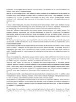
Surface swabs
This is a procedure for surface swabbing of a known area delineated by a tem-
plate. The organisms picked up by the swab are recovered in a known volume of
fluid and a bacterial count made on the fluid. From this the number of organisms
present on the area swabbed can be computed and the count per cm
2
of swabbed
area then calculated. If it is likely that sanitizers have been used on the surface,
the suspending fluid should contain appropriate neutralizers.
5.9
Method 2 Surface contact plate
Procedure
(a) Remove the lid from a surface contact agar plate and press the surface of the agar
evenly against the test surface.
(b) Replace the lid on the plate and incubate as appropriate for the organisms sought.
(c) Proceed as for method 3 (below) after incubation.
Enumeration of microorganisms 125
5 cm
5 cm
(a)
(b)
(c)
(d)
(e)
Fig. 5.1 Surface swabs.
Diluent
Neutralizer solution
—
MRD containing lecithin 3 g/L, polysorbate 80 30 g/L, sodium
thiosulphate 5 g/L, L-histidine 1 g/L [10].
Procedure
(a) Hold a sterile template firmly over the surface to delineate the area to be sampled
(Fig. 5.1b).
(b) Dip a cotton wool swab into a 10-mL volume of neutralizer solution contained in
a small screw-capped bottle with five small glass beads (e.g. 3 mm diameter).
Squeeze out the excess fluid against an inner surface of the bottle, and rub the
moistened swab thoroughly over the entire test area, turning the swab in order to
maximize its ability to pick up organisms (Fig. 5.1c).
(c) Break off the cotton wool end of this swab into the 10 mL neutralizer solution.
(d) Take a second dry cotton wool swab and rub it thoroughly over the entire test area
turning as before (Fig. 5.1d).
(e) Break off the end of this swab into the same 10 mL neutralizer solution.
(f) Shake the diluent bottle containing the swabs and beads until the cotton wool has
been broken down into fibres (Fig. 5.1e).
(g) Use one of the methods in Sections 5.1–5.7 to count the organisms in suspension.
continued
126 Section five
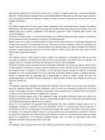
Membrane slide cultures
This is a procedure for surface testing using a commercially available kit consist-
ing of a swab in sterile buffer and a sampler consisting of a plastic tab supporting
a gridded filter membrane bonded to an absorbent pad containing dehydrated
culture medium (Fig. 5.2).*
5.10
Calculation
If the test suspension is found to contain N organisms/mL, there are 10N/25 organ-
isms/cm
2
of test surface.
If larger areas than 25 cm
2
are to be swabbed, commercially available sponges impreg-
nated with neutralizing fluid can be used in place of the cotton wool swabs. MRD may
then be used as diluent.
*This type of kit can be obtained from Millipore (UK) Ltd, Millipore House, Abbey Road,
London NW10 7SP. .
(a) (b) (c)
(d) (e) (f)
(g)
Fig. 5.2 Membrane slide cultures.
Enumeration of microorganisms 127
Rinse method for watercress, other leaf
vegetables and acidic berry fruits
[11,12]
The use of a rinse method is suitable for leaf vegetables and may enhance recov-
ery of target organisms from acidic fruits such as berries.
5.11
Procedure
(a) Remove the swab from its container and roll the tip against the inner surface of
the container to remove excess buffer solution (Fig. 5.2a).
(b) Swab the test surface by rotating the swab while moving it over a defined area (e.g.
10 ¥10 cm) (Fig. 5.2b).
(c) Replace the swab into its container, which contains 18 mL buffer solution (Fig.
5.2c). Seal the container and shake well (at least 30 times). Remove and discard
the swab.
(d) Take the sampler from its container and insert it in the test buffer solution from
which the swab was removed. Leave for 30 s (Fig. 5.2d,e).
(e) Remove the sampler, allow it to drain and replace it in its original container
(Fig. 5.2f).
(f) Incubate the sampler in its container as appropriate for the organisms sought.
After incubation
Count the colonies on the gridded membrane of the sampler (Fig. 5.2g), or compare
with the chart supplied with the kit. The sampler absorbs 1 mL of buffer solution;
therefore multiply the colony count by 18 to obtain the number of cfu/area sampled.
Procedure
(a) Place 100 g of watercress, other leaf vegetables or berry fruit in a large sterile con-
tainer with a screw cap or other airtight closure and add 200 mL of MRD. Secure
the lid.
(b) Shake the container well for 15–30 s so that the diluent wets all the vegetable/fruit
surfaces. Leave to stand for 30 min.
(c) Shake well again for 10–15 s.
(d) Examine the rinse fluid for the target organism or group of organisms using an ap-
propriate medium (see Section 6). If counts are expected to be low use membrane
filtration or a multiple tube method for enumeration. If counts are likely to be
high (>10
3
/g) a surface plating or pour plating technique can be used.
Calculation
Compute the count per mL of rinse fluid. Multiply this value by two to obtain the
count/g of sample.
Bottle rinse and plate count
This method is most commonly used for determining the effectiveness of milk
bottle washing [13]. It may also be used for single use bottles, in which case a
neutralizer is not needed in the diluent.
5.12
128 Section five
References
1 Peterz MEG. Temperature in agar plates and its influence on the results of quantitative
microbiological food analyses. Int J Food Microbiol 1991; 14: 59–66.
2 ISO 7218 (BS 5763 Part 0). Microbiology of Food and Animal Feeding Stuffs
—
General Rules
for Microbiological Examinations. Geneva: International Organization for Standardiza-
tion (ISO), 1996; incorporating amendment 1, 2001.
3 ISO 6887-1. Microbiology of Food and Animal Feeding Stuffs
—
Preparation of Test Samples,
Initial Suspension and Decimal Dilutions for Microbiological Examination. Part 1: General
Rules for the Preparation of the Initial Suspension and Decimal Dilutions. Geneva: Inter-
national Organization for Standardization (ISO), 1999.
4 LAB 12. The Expression of Uncertainty in Testing. Feltham, Middlesex: United Kingdom
Accreditation Service, 2000.
5 Standing Committee of Analysts. Methods for the Examination of Waters and Associated
Materials. The Microbiology of Drinking Water (2002)
—
Part I
—
Water Quality and Public
Health. Environment Agency, 2002.
6 ISO 4833 (BS 5763 Part 1). Microbiology
—
General Guidance for the Enumeration of
Microorganisms
—
Colony Count Technique at 30°C. Geneva: International Organiza-
tion for Standardization (ISO), 1991.
5.13
Procedure
(a) Set aside six washed and capped (or stoppered) bottles for testing.
(b) Add 20 mL of MRD (or quarter strength Ringer’s solution) containing 0.05%
sodium thiosulphate to each bottle and recap.
(c) Thoroughly wet the surfaces of all bottles by rotating gently 12 times in one
direction. Allow the bottles to stand for 15–30 min.
(d) Again rotate each bottle gently 12 times in the same direction so that the whole
internal bottle surface is thoroughly wetted.
(e) Place 5 mL of the rinse fluid from each bottle into a separate Petri dish.
(f) To each dish add 20 mL of molten milk plate count agar tempered to 44–47°C.
Include one plate containing agar only and another plate containing rinsing
fluid and agar to act as media sterility controls.
(g) Mix each plate five times in a vertical, clockwise, horizontal and anticlockwise
direction (as shown on p. 112), then allow to set.
(h) Incubate the plates at 30°C for 72±3h.
Calculation
Multiply the colony count in each plate by four to obtain the total colony count per
bottle. Add the counts together and divide by six to obtain a mean count per bottle.
If any individual bottle count is more than 25 times greater than the next highest
count in the series, it should be omitted from the mean bottle count and a note made
to that effect.
As a general rule, the colony count per bottle should not exceed 1 cfu/mL capacity of
the bottle. For 1 pint milk bottles, counts below 200 are considered satisfactory and
counts above 600 are considered unsatisfactory.
7 International Commission on Microbiological Specifications for Foods. Microorgan-
isms in Foods. 1 Their Significance and Methods of Enumeration, 2nd edn. London:
University of Toronto Press, 1978.
8 ISO 4831 (BS 5763 Part 3). Microbiology
—
General Guidance for the Enumeration of
Coliforms
—
Most Probable Number Technique. Geneva: International Organization for
Standardization (ISO), 1991.
9 ISO 7251 (BS 5763 Part 8). Microbiology
—
General Guidance for the Enumeration of
Presumptive Escherichia coli
—
Most Probable Number Technique. Geneva: International
Organization for Standardization (ISO), 1993.
10 CCFRA. Guideline No. 20. Effective Microbiological Sampling of Food Processing Environ-
ments. Chipping Campden, Gloucestershire: Campden and Chorleywood Food
Research Association, 1999.
11 Tee GH. Bacteriological examination of watercress. Mon Bull Min Hlth PHLS 1962; 21:
73–8.
12 Public Health Laboratory Service. The hygienic production of watercress: summary of
a report by the Watercress Working Party of the Public Health Laboratory Service. Mon
Bull Min Hlth PHLS 1966; 25: 146–51.
13 British Standards Institution (BSI). BS 4285. Microbiological Examination for Dairy Pur-
poses. Part 4. Methods for Assessment of Hygienic Conditions. London: BSI, 1991.
Enumeration of microorganisms 129