Tài liệu Practical Food Microbiology 3rd Edition - Part 6 docx
Bạn đang xem bản rút gọn của tài liệu. Xem và tải ngay bản đầy đủ của tài liệu tại đây (260.01 KB, 61 trang )
Isolation and enrichment of microorganisms 131
Isolation and enrichment
of microorganisms
6.1 Aeromonas spp.
6.2 Bacillus cereus and other Bacillus spp.
6.3 Brucella spp.
6.4 Campylobacter jejuni, C. coli, C. lari
6.5 Clostridium perfringens and other sulphite-reducing clostridia
6.6 Coliforms, thermotolerant (faecal) coliforms and Escherichia coli
6.7 Enterobacteriaceae
6.8 Enterococci
6.9 Lactobacilli and the lactic acid bacteria
6.10 Listeria monocytogenes and other Listeria spp.
6.11 Pseudomonas aeruginosa and other pseudomonads
6.12 Salmonella spp.
6.13 Shigella spp.
6.14 Staphylococcus aureus and other coagulase positive staphylococci
6.15 Vibrio spp.
6.16 Viruses
6.17 Yeasts and moulds
6.18 Yersinia spp.
The procedure used for isolation of a microorganism from a food sample will
depend upon a number of factors. If the organism is expected to be found in
large numbers, or its presence is only significant when there are large numbers,
direct enumeration on a suitable selective solid medium will be sufficient.
If, however, only small numbers of that organism are anticipated, or if their
presence is significant regardless of the number of cells (e.g. salmonellae)
then enrichment culture will be required. This may need to incorporate a
pre-enrichment or resuscitation stage if the organism is likely to have suffered
injury through freezing, drying, heating, etc. Isolation media and procedures
are often a matter of personal choice, but due regard should be given to their
suitability for recovery of stressed organisms, which are easily inhibited by
many selective agents and also by elevated incubation temperatures. In addition
the recovery of spoilage organisms may require adjustments to the isolation
medium, such as an increase in the levels of salt or glucose, in order to mimic the
nature of the spoiled commodity and thus to allow recovery of the organism.
The quantity of food examined is important; in general for pre-enrichment
or direct selective enrichment a 25 g portion should be cultured and the ratio of
sample to broth should be 1 :9 (or 1/10). For secondary enrichment a 1 :10 ratio
of inoculum to broth is usually maintained but this may vary depending on the
selective broth; for example, the ratio of pre-enrichment broth to Rappaport
Vassiliadis broth for isolation of salmonellae should be 1: 100.
6
It is also important to perform internal quality control tests on both the
media used for food examination and the whole test procedure. Reference
strains derived from a recognized culture collection, such as the National Col-
lection of Type Cultures (NCTC; see Appendix C), are used to compare their abil-
ity to grow and the degree of growth on or in the agar or liquid medium under
test with results from a non-selective medium. The reference strains can also be
used to assess recovery from artificially inoculated foods of different types by the
methods used.
Quality control cultures
A wide range of reference cultures is required to test the entire range of liquid and
solid culture and test media encountered in the microbiological examination of
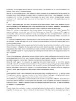
food. Reference cultures should be obtained on an annual basis in freeze dried
form from the appropriate culture collection and developed into reference stock
cultures on beads and working cultures according to the suggested procedure
shown in Fig. 6.1 [1].
132 Section six
Reference culture
(vial of freeze dried organisms from culture collection)
Subculture according to culture collection instructions on appropriate
non-selective medium (discard reference culture)
Prepare multiple beads in cryovials — minimum 20 beads
Reference stock cultures
(beads prepared from reference culture)
Every four weeks subculture from reference stock culture
Working culture
(slopes or liquid cultures)
Working cultures should not be used to prepare further stocks.
Where viability of cultures on slopes or liquid media is poor, a fresh
bead from a cryovial may be used as a working culture.
Documentation and detailed records on the handling of reference
strains from receipt in the laboratory is essential.
A new reference culture should be obtained annually.
Most working cultures can be maintained at 4°C after incubation to
establish sufficient growth for up to four weeks without loss of
viability or contamination.
The key considerations are the preparation of the reference bead
stocks and the life of the working cultures prior to replacement.
1
2
3
4
5
6
Fig. 6.1 Preparation and maintenance of quality control cultures.
Quality control testing of solid and liquid media
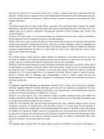
A standard procedure for testing solid media is the plating out, in a standard,
reproducible manner, of the test organism and the recording of the degree of
growth. An example of this type of procedure is the ‘ecometric’ method [2]
in which a loopful (1mL or 5 mL) of an overnight broth culture is spread on to
the surface of pre-dried plates in the manner illustrated in Fig. 6.2(a), the loop
moving through sections one to five without reloading.
After appropriate incubation the highest rate of dilution that still leads to
growth can be assessed and the results expressed as an absolute growth index
(AGI). For example growth in all five sectors would give an AGI of 5.0, whereas
growth on sections one and two and on only two inoculum lines of section
three would give an AGI of 2.4. The relative growth index (RGI), the proportion
of the AGI on the test medium compared with that on a control medium, can be
used to describe the productive and selective properties of a particular medium.
An alternative method is shown in Fig. 6.2(b). The culture is spread from
A1–B1–C1–D1–A2–B2, and so on, finishing at D5 without sterilizing the loop.
The AGI can be calculated from the last segment and line at which growth
occurs, the figure for each line increasing by five from A1 (5) through to D5
(100). Thus if the last line of growth is B4 then the AGI is 70. The RGI can be cal-
culated by comparing the AGI of the test medium with that of a control medium
as described above.
Alternatively a number of consecutive dilutions of the appropriate reference
organism can be enumerated on the test medium, for example using the Miles
and Misra surface drop method for testing solid media (see Section 5.5), and
compared with the results obtained with a control medium.
There are a number of other methods which can be used in the quality
assurance of culture media such as dilution to extinction (liquid media), mixed
cultures of wanted and unwanted organisms (liquid media) and assessment
of growth rate (liquid media). A summary of the available methods has been
published [3].
The appropriate positive and negative quality control cultures are listed
under each specific method or organism in the different sections of this manual
where appropriate.
Isolation and enrichment of microorganisms 133
(a) (b)
12
43
5
A
B
D
C
1
2
3
4
5
1 2 3 4 5
5 4 3 2 1
5
4
3
2
1
Fig. 6.2 Inoculation of plates using the ecometric technique: (a) method of Mossel et al.
[2]; (b) modified method.
Quality control of test procedures
The whole test procedure should also be challenged by the use of reference ma-
terials or foods known to contain the required target organism. The latter can be
achieved by preparing spiked samples or by the re-examination of samples pre-
viously found to be positive. Reference materials [4] are available that contain
small numbers of the target organism (e.g. Salmonella spp., Listeria monocyto-
genes) in an inert substrate (spray-dried milk powder) contained within a gelatin
capsule. These reference materials can be used alone to test the efficiency of the
medium or in the presence of the relevant food material, with its associated
competitive flora, to test the whole procedure.
Quality assurance
This is defined as the total process whereby the quality of laboratory reports
can be achieved and is a combination of internal quality control and external
quality assessment. Guidelines on the implementation of quality assurance pro-
grammes in laboratories involved in food, water and environmental laborato-
ries have been published by an European Union (EU) Working Group [5] with
the aim of making available, simply but accurately, procedures that have been
developed and applied successfully by the Working Group members.
Internal quality control
This comprises the continual monitoring of working practices, equipment,
media and reagents including performance of laboratory personnel. Procedures
for the quality control of media are as described earlier in this section. Equip-
ment should be regularly checked to ensure maintenance of optimum perfor-
mance. The operational techniques and activities used to fulfil the requirements
for quality are also referred to as analytical quality control [5], and can be dif-
ferentiated into three lines of checking as outlined in Table 6.1.
The first line of checking is a means of self-control by the analyst, but it
should be supervised by the direct superior responsible for setting criteria and
134 Section six
Table 6.1 Analytical control in microbiology.
Line of
checking Responsibility Frequency Purpose
First Analyst High All aspects of the analysis under control
and consistent over time
Second Person independent of Less frequent Different analysts or equipment produce
the analyst similar results. Individual results not
biased
Third Laboratory management Regular intervals To ensure interlaboratory standardization
defining action plans and should be included with every series of examinations.
First-line checks should cover equipment and procedures to be undertaken: (a)
before the examination (samples, equipment, media, filters and reagents); (b)
during the analysis (noting all the information that becomes available such as
temperature, anaerobic conditions, confirmation rates, colonial appearance,
background flora, etc.); and (c) in addition to the examination. The latter would
include internal quality control (IQC) procedures such as examination of addi-
tional samples, parallel plating, procedural blanks, positive and negative con-
trol samples, colony counts on different volumes/dilutions, use of control
charts and use of sufficient colonies for confirmatory tests.
Second-line checks are implemented to assure reproducibility between dif-
ferent analysts or equipment, during training of new workers and evaluation of
established staff in order to maintain standards of subjective interpretation.
Such checks would include: (a) duplicate counting by the same person to pro-
vide the counting error under repeatability conditions, and by different persons,
thus including both random and systematic components to the variation; (b)
duplicate analytical procedures to test the whole quantitative procedure, by
using duplicate samples and plotting control charts; and (c) intensified quality
control tests as listed for first-line checks.
Third-line checks should be supervised by the quality assurance officer and
include participation in an external quality assurance (EQA) scheme, also
known as proficiency testing, and the use of certified reference materials
(CRMs). In EQA schemes the samples are examined by different laboratories,
the results interpreted retrospectively by the central organization and the per-
formance compared with other participants. It is a flexible approach whereby
participants apply their own methods. With CRMs, all laboratories follow
a strict protocol and the certified value is valid only for the applied method.
Results obtained with other methods can be compared with the certified values.
External quality assessment
Quality assessment acts as a check on the efficiency of the quality control proce-
dures by the introduction of samples of known but undisclosed content for
examination by the normal routine methods of the laboratory. This external
challenge can be undertaken by participation in a proficiency testing scheme in
which such samples, containing a range of food-associated organisms, are
distributed on a regular basis. Such a system is offered by the Public Health
Laboratory Service (PHLS) Food Microbiology External Quality Assessment
Schemes (see Section 4.9 and Appendix C).
Temperature ranges
Incubators and water baths should be capable of maintaining the temperature
to within 1°C of the desired temperature. Where more accurate temperature
control is required, e.g. to within 0.5°C or 0.2°C, special fan-assisted incubators,
Isolation and enrichment of microorganisms 135
or water baths, will be needed. Temperatures should be checked and recorded at
least every working day, using thermometers or electronic temperature record-
ing equipment calibrated by techniques traceable to national standards, and
records kept for reference. Details of general laboratory practices can be found in
ISO 7218 (BS 5763 Part 0) [6].
For tests designated ‘recommended’ and ‘supplementary’ in Section 3, the
incubation temperatures given in this manual should be maintained to within
1°C and incubation times should not deviate from those stated by more than
2 h. For statutory tests, the temperature and time ranges permitted are quoted
in the relevant legislation.
Confirmatory tests
Procedures for the tests most frequently used in confirmation of the identity of
the microorganisms included in this section are given in Section 10. Details of
other confirmatory tests may be found in standard texts such as Cowan and
Steel’s Manual for the Identification of Medical Bacteria ([1] in Section 10).
In this manual the tests described for the identification of microorganisms
are based on traditional methods. However, multi-test micro-methods involv-
ing manual biochemical systems using dehydrated substrates (e.g. API
®
,
Minitek
®
, MicroID
®
) or agar bases (e.g. Enterotube
®
) have become established in
microbiological practice. These are simple and rapid to use and produce
reproducible results. Databases are often provided with computer back-up and a
telephone assistance service. Use of such methods is acceptable provided they
are fully validated against the traditional tests. Although the standards cited in
this manual describe traditional methods, the use of commercially produced
biochemical galleries is increasingly permitted.
Aeromonas spp.
Members of the genus Aeromonas are Gram negative, facultatively anaerobic,
non-sporing rod-shaped bacteria in the family Vibrionaceae. The genus can
be divided into two groups of species. One group contains only one species,
the psychrophilic fish pathogen A. salmonicida. The other group consists of the
psychrotrophic, ‘motile aeromonads’ that includes A. hydrophila, A. caviae and
A. sobria. The oxidase reaction is positive; motility can be variable as can gas
production.
The motile aeromonads of the hydrophila group [7,8] have been associated
with human disease and are regarded as potential human food-borne
pathogens. Illness can range from a mild diarrhoea to a life-threatening cholera-
like disease. A. hydrophila is the species most frequently implicated but, as there
are no simple tests to distinguish between the different strains, they are often
referred to as one species. These organisms are ubiquitous and are commonly
found in water, sewage, seafood, meat, vegetables and dairy produce, but their
significance in the epidemiology of food-borne disease is unclear.
6.1
136 Section six
Control cultures
NCTC 8049 Aeromonas hydrophila Positive, growth quantitative
NCTC 9001 Escherichia coli Negative, growth inhibited
Isolation and enrichment of microorganisms 137
Method 1 Direct enumeration
Media
A selective agar: e.g. bile salts irgasan brilliant green agar, Ryan’s modification of
xylose lysine desoxycholate agar (XLD) agar or ampicillin blood agar (contains
ampicillin 10 mg/L).
Procedure
(a) Prepare a 10
-1
homogenate using 25 g of food sample and 225 mL of maximum re-
covery diluent (MRD) and further decimal dilutions as described in Section 4.3.
(b) Using a surface counting method selected from Section 5 (eg: 5.4–5.6), enumerate
Aeromonas spp. on a suitable selective agar.
(c) Incubate at 30°C for 18–24 h.
(d) Examine the plates and count typical colonies; these appear translucent on bile
salts irgasan brilliant green agar, dark green, opaque colonies with a darker centre
on Ryan’s medium and large, colourless, usually haemolytic colonies on ampi-
cillin blood agar.
(e) Subculture five typical colonies (or all if fewer than five) to a non-selective
medium such as nutrient agar, then incubate at 30°C for 18–24 h.
(f) Perform an oxidase test (see Section 10.14). Retain oxidase-positive strains and
identify by biochemical tests (strains remain viable for up to 20 min after the
addition of oxidase reagent).
(g) Calculate the count per g from the proportion of colonies that are identified as
Aeromonas spp.
Identification
Oxidase-positive strains isolated in this way may be considered to be members of the
genus Aeromonas if they are fermentative and resistant to vibriostatic agent 0129 (2,4-
diamino-6,7-diisopropylpteridine), and capable of growth in 0% but not 6% sodium
chloride. Identification of the species can be obtained using the characteristics listed
in Table 6.2.
continued
Table 6.2 Identification of Aeromonas spp.
Test A. hydrophila A. sobria A. caviae
Voges–Proskauer test ++-
Growth at 42°C -+-
Aesculin hydrolysis +-+
Gas from glucose V +-
Acid from arabinose +-+
Lysine decarboxylase ++-
V, variable.
138 Section six
The ‘suicide’ test [9] for the speciation of Aeromonas based on the fermentation of glu-
cose, with or without gas production, and pelleting of bacteria (suicide phenomenon)
has been shown to be both accurate and simple to perform. This test, in combination
with a short series of other biochemical tests (Table 6.3), is also recommended for
identification of Aeromonas spp.
Table 6.3 Short scheme for identification of Aeromonas spp.
Test A. hydrophila A. sobria A. caviae
Suicide test* - V +
Gas from glucose V +-
Aesculin hydrolysis +-+
Hydrogen sulphide production ++-
*Aeromonas suicide phenomenon medium [9]: nutrient broth containing 0.5% (w/v)
glucose and 0.0015% (w/v) bromocresol purple, dispensed in 5mL volumes in 125 mm¥
16mm tubes containing inverted Durham tubes.
V, variable.
Method 2 Enrichment culture
Media
Enrichment medium. Alkaline peptone water with electrolyte supplement (contains
tryptone peptone 10 g, sodium chloride 10g, magnesium chloride hexahydrate 4 g,
potassium chloride 4 g/L), pH8.6.
Selective agar: e.g. bile salts irgasan brilliant green agar, Ryan’s aeromonas medium or
ampicillin blood agar.
Procedure
(a) Prepare a homogenate using 25 g of food sample and 225 mL of enrichment
medium.
(b) Incubate at 30°C for 18–24 h.
(c) Subculture to a suitable selective agar and proceed as described from step (c) of
method 1.
Specialized reference facilities are available in certain circumstances for identifi-
cation and serotyping of Aeromonas strains (see Appendix C).
Bacillus cereus and other Bacillus spp.
The Bacillus group includes a large number of Gram positive rod-shaped spore-
forming species with a wide variety of properties. The genus is taxonomically
non-homogeneous and many characters used for identification are variable
including the Gram reaction, motility, ability to grow under anaerobic condi-
tions, the oxidase reaction and method of breakdown of carbohydrates. The best
6.2
arrangement for subdividing the genus appears to be that of Smith et al. [10],
which divides the species into three groups based on traditional biochemical
tests, spore position and morphology. The main species involved in food-borne
illness include B. cereus (Group I) and the B. subtilis/licheniformis group (Group
III), although a number of other species have been incriminated.
Members of the Bacillus group are ubiquitous, being found widely in the dust
and soil, and are freqently isolated in varying numbers from a wide range of
foods especially those containing cereals. The spores may survive many heat
processes, and as high numbers are normally required to cause illness low num-
bers present in foods are not considered significant. Enrichment methods are
not normally required. Bacillus spp. will grow readily on non-selective media,
but for purposes of identification a selective medium should be used [11–14].
The media specified below do not recover all species of Bacillus, but do recover
the species that are recognized as capable of causing gastrointestinal symptoms.
An incubation temperature of 30°C is recommended to ensure the detection of
psychrophilic strains of B. cereus.
Control cultures
NCTC 7464 Bacillus cereus Positive, growth quantitative
NCTC 10400 Bacillus subtilis Positive, growth qualitative
NCTC 9001 Escherichia coli Negative, growth inhibited
Isolation and enrichment of microorganisms 139
Media
Polymyxin pyruvate egg yolk mannitol bromothymol blue agar (PEMBA)
or
Phenol red egg yolk polymyxin agar (MYP or PREP agar).
Both media contain 1% mannitol, 5% egg yolk emulsion and 100 IU polymyxin/mL.
The appropriate ISO method (EN ISO 7932; BS 5763 Part 11) [14] uses MYP agar inoc-
ulated by the surface plating method. However international studies have failed to
show a significant difference between the performance of the two media [15] and
many dairy microbiologists favour the use of PEMBA.
Procedure
(a) Prepare a 10
-1
homogenate and serial decimal dilutions of the food sample as
described in Sections 4.2 and 4.3.
(b) Select a surface counting method from Section 5 (eg: 5.4–5.6), and enumerate
using PEMBA or MYP agar.
(c) Incubate aerobically at 30°C for 24 h; if colonies are not clearly visible incubate at
30°C for a further 24 h. If PEMBA is used and a spore stain (see Section 10.4) will be
required after incubation the medium should be incubated at 37°C for the first
24 h followed by a further 24 h at room temperature.
(d) Examine plates for characteristic colonies, which will be large (3–7 mm diameter)
and dull. Colonies of B. cereus appear turquoise/peacock blue on PEMBA agar and
continued
140 Section six
pink on MYP agar due to absence of mannitol fermentation, and are usually sur-
rounded by a zone of opacity due to precipitation of hydrolysed lecithin (see Plate
Ia,b, facing p. 150). Most other members of the Bacillus group are mannitol
positive, appear as green or yellow colonies and do not produce lecithinase (see
Plate Ic,d, facing p. 150).
(e) Select plates containing up to 150 colonies for counting. Count and record the
number of colonies with morphology resembling Bacillus species to give the pre-
sumptive count. If B. cereus is also sought count and record blue (PEMBA) or pink
(MYP) colonies with and without lecithinase zones.
Note: Some members of the Enterobacteriaceae, such as Proteus, and many strains of
Staphylococcus aureus are able to grow on these selective media. However, they are
easily distinguished by colonial morphology and overall appearance, and by egg-yolk
clearing, in contrast to egg-yolk precipitation.
Identification
(f) Perform a Gram stain if necessary to confirm cell morphology (large Gram posi-
tive bacilli, with or without visible spores). Subculture at least five colonies of each
colonial type onto blood agar and incubate for 18–24 h at 30°C. Colonies of B.
cereus are b-haemolytic, that is they produce complete clearing of the red blood
cells around the colony growth.
Confirm the identity of presumptive B. cereus and characterize other Bacillus strains of
different morphology with appropriate biochemical tests The short scheme in Table
6.4 allows distinction of some of the most common strains of Bacillus of importance
in food poisoning. Details of the biochemical tests can be found in Section 10. To test
for anaerobic growth inoculate two blood agar plates; incubate one plate aerobically
and the other plate anaerobically at 30°C for 22 ±2 h, then examine both plates for the
presence of growth.
(g) Calculate the total Bacillus spp. or B. cereus count per g of food.
If the food under test is acidic or if the plate contains many colonies that ferment
mannitol the characteristic blue (PEMBA) or pink (MYP) colour due to absence of
mannitol fermentation may be masked. Further subculture of suspect colonies to
PEMBA or MYP will overcome this problem and aid identification.
Table 6.4 Identification of common food poisoning strains of Bacillus spp.
B. cereus B. pumilus B. subtilis B. licheniformis
Glucose (ASS) ++ + +
Arabinose (ASS) -+ + +
Mannitol (ASS) -+ + +
Xylose (ASS) -+ + +
Nitrate reduction +- + +
Anaerobic growth +- - +
ASS, ammonium salt sugars. For preparation see [1] in Section 10.
Specialized biochemical, serological and toxin production tests are available
(see Appendix C).
Brucella spp.
Brucella spp. are short Gram negative, aerobic or capnophilic, non-motile rods
belonging to the Moraxella-Acinetobacter Group. The genus comprises a single
genospecies B. melitensis but the old specific names are still generally used
—
B. abortus, B. melitensis and B. suis being the three classical species, all of which
cause infections in humans. They are catalase positive, usually oxidase positive
and do not show acid production from sugars in peptone-containing media
[16–19].
Brucella spp. are Hazard Group 3 pathogens, and samples and cultures must
be handled accordingly. Count methods are not normally applicable, the aim
being simply to detect the presence of brucellae. The methods described are for
the detection of brucellae in milk, but can be adapted for cream, soft cheese and
other milk products.
6.3
Isolation and enrichment of microorganisms 141
Method 1 Direct culture
Media
A selective agar: e.g. brucella agar base, which contains dextrose; or blood agar or
Columbia agar base plus 1% (w/v) sterile dextrose. These media are suitable for use
with the addition of 5% inactivated horse serum (i.e. serum held at 56°C for 30 min)
and an antibiotic cocktail containing polymyxin 5000 IU, bacitracin 25 000IU,
cycloheximide 100 mg, nalidixic acid 5mg, nystatin 100 000IU and vancomycin
20 mg/L.
Procedure
(a) Transfer the milk sample to sterile test tubes (180 mm¥25 mm) and store
overnight at 4°C.
(b) Dip a swab into the cream layer and inoculate the surface of a selective agar.
(c) Incubate the plates at 37°C in an atmosphere of air containing 10% carbon
dioxide.
(d) Examine the plates every 2 days for up to 10 days. Colonies are usually visible after
4 to 5 days’ incubation, and are 1–2 mm in diameter, convex, with round entire
edges.
Identification
Brucella spp. can be further identified using antibodies for slide agglutination.
Differentiation can also be achieved by the dyes-strip method [18] as follows:
1 Impregnate filter paper strips with 1 :200 basic fuchsin or 1 :600 thionin and
dry.
2 Place a strip of each dye parallel on the surface of a plate of serum dextrose agar
and cover with a thin layer of the same medium. Allow the medium to solidify.
continued
Facilities are available for the identification and serotyping of Brucella spp. (see
Appendix C).
Campylobacter jejuni, C. coli and C. lari
Thermotolerant, microaerobic campylobacters have only been recognized as
important causes of human enteritis since the early 1970s. Campylobacter jejuni
is responsible for most illness, with C. coli causing a small proportion of
cases and other species being isolated occasionally. Campylobacters are
microaerophilic, Gram negative, small vibrioid or spiral-shaped cells with rapid,
darting, reciprocating motility. They reduce nitrate, are unable to oxidize or fer-
6.4
142 Section six
3 Make streak inoculations of the Brucella strains at right angles to the strips.
4 Incubate in 10% carbon dioxide for 2 to 3 days at 37°C.
5 Examine for growth. Resistant strains grow right across the strip, but sensitive
strains show inhibition of growth up to 10 mm from the strip. Typical growth pat-
terns are given in Table 6.5.
Table 6.5 Typical patterns of Brucella spp. in the dye-strip tests.
Basic fuchsin 1 : 200 Thionin 1 : 600
B. abortus Growth No growth
B. melitensis Growth Growth
B. suis No growth Growth
Method 2 Enrichment culture
Media
Broth bases: e.g. brucella broth or media suitable for the culture of fastidious organ-
isms such as brain heart infusion broth or tryptone soya broth. Supplement the
medium with 5% sterile horse serum and antibiotics as described in method 1. The
use of amphotericin B (4 mg/L) and cycloserine (12.5mg/L) in addition to the antibi-
otics previously listed has also been recommended.
Procedure
(a) Centrifuge 100 mL of the milk for 30min at 1500 rev/min.
(b) Transfer the cream layer and deposit from the centrifuged milk to sufficient
enrichment broth in a screw-capped container to give an inoculation ratio of
1 :10.
(c) Incubate the broth, with screwcap loose, in air containing 10% carbon dioxide at
37°C for 5 days.
(d) Subculture the broth to selective agar and proceed as described from step (c) of
method 1.
ment carbohydrates and mostly reduce nitrite. C. jejuni, C. coli, C. upsaliensis and
C. lari are thermotolerant, growing at 42°C but not at 25°C. Campylobacters
may infect humans after direct contact with animals or indirectly via contami-
nated water, milk or meat [20].
Many food samples to be examined for the presence of Campylobacter spp.
[21–26] will have received treatments such as heating, freezing or chilling. These
treatments can cause sublethal injury to the organism resulting in increased sen-
sitivity to some antibiotics and lowered resistance to elevated incubation tem-
peratures. The enrichment culture method described below allows resuscitation
and recovery of injured organisms. Direct culture of fresh raw foods especially
poultry may also be productive. Enumeration of campylobacters is not normal-
ly attempted, as the aim of examination is to establish the presence of the or-
ganism.
Control cultures
NCTC 11322 Campylobacter jejuni Positive, growth quantitative
NCTC 9001 Escherichia coli Negative, growth inhibited
Isolation and enrichment of microorganisms 143
Method 1 Direct culture
This procedure is likely to be of most value with samples such as chicken skin.
Media
A selective agar: e.g. blood-free modified cefoperazone charcoal deoxycholate agar
(CCDA) [22], Exeter [23], Preston [21] or Skirrow [24].
Procedure
(a) Take a swab of the food sample and inoculate on to the surface of a suitable selec-
tive agar.
(b) Incubate the plates at 37°C for 4 h and then at 41.5°C for a further 44–68 h
in an atmosphere of nitrogen containing 5–15% carbon dioxide and 5–10%
oxygen.
(c) Examine the plates for typical colonies, which have the following characteristics
[20]:
C. jejuni (and C. lari)
—
flat, glossy, effuse colonies, with a tendency to spread along the
inoculation track. Well-spaced colonies resemble droplets of fluid. On moist agar a
thin, spreading film may be seen. With continued incubation colonies become low
and convex with a dull surface. A metallic sheen will eventually develop (see Plate II,
facing p. 150).
C. coli
—
less effuse, often umbonate colonies with the surface usually remaining
shiny.
continued
144 Section six
Identification
(d) Identification to genus level can be made by the following tests:
1 Oxidase test: positive (see Section 10.14).
2 Growth on blood agar incubated at 41.5°C for 24–48 h under microaerobic
conditions described in step (b) but no growth following incubation under
aerobic conditions.
3 Microscopy showing Gram negative, highly motile rods with S-shaped or
spiral morphology. This rapidly degenerates to a coccal form with exposure to
oxygen.
(e) C. jejuni, C. coli and C. lari can be differentiated by the biochemical tests shown in
Table 6.6.
Table 6.6 Differentiation of Campylobacter spp.
Hippurate hydrolysis Nalidixic acid sensitivity
C. jejuni + S
C. coli - S
C. lari - R
R, resistant; S, sensitive.
Method 2 Enrichment culture
Suitable enrichment broths contain FBP supplement (ferrous sulphate, sodium
metabisulphite and sodium pyruvate, each at 0.025% concentration) to improve
aerotolerance and allow aerobic incubation. A mixture of antibiotics is also required
to prevent overgrowth by competing organisms and are included in the formulation
of Preston [21], Exeter [23] and Bolton [26] broths. Preston broth is based on the for-
mulation of Preston agar. Exeter broth is similar but also includes cefoperazone for
greater selectivity. Exeter broth has been shown to produce superior isolation rates
to that of Preston broth. Sensitivity to some of the ingredients demonstrated by
sublethally injured campylobacters can be overcome by incubating the broths
at 37°C [25]. Bolton broth has been elaborated to optimize recovery of injured cells
(see method 3).
The method described below is similar to that described in one part of ISO 10272 (BS
5763 Part 17) [27].
Media
Exeter campylobacter-selective medium [23] of the following composition:
Nutrient broth (Oxoid No. 2) 1000mL
Lysed blood 50 mL
Trimethoprim 10 mg
Rifampicin 10mg
Cefoperazone 15mg
continued
Isolation and enrichment of microorganisms 145
Polymyxin 4 mg
Amphotericin 2 mg
Sodium pyruvate 250 mg
Sodium metabisulphite
}
FBP 250 mg
Ferrous sulphate 250mg
For plates add 15 g of agar.
FBP can be made as a combined 2.5% solution of each additive in water. Ten millilitres
of this can then be added to 1 L of medium. Discard stock solution after 7 days.
Antibiotics have to be made as separate solutions.
Selective agars: e.g. blood-free modified CCDA [22], Preston [21], Exeter [23] or Skirrow
[24].
Procedure
(a) Homogenize 25 g of the food sample in 225mL of Exeter enrichment broth. The
broth should be at room temperature on inoculation. Transfer the homogenate to
a screw-topped jar leaving very little headspace, and close the top tightly.
(b) Incubate at 37°C for 18–48 h preferably in a fan-assisted incubator to obtain rapid
heat transfer. Adjust the incubation period according to the expected degree of
contamination of the sample: for samples such as chicken skin, incubate at 37°C
for 18 h; for water samples, where cells will be severely damaged, incubate for 48 h.
(c) Subculture onto a suitable selective agar.
(d) Incubate the plates at 41.5°C for 24–48 h in a microaerobic atmosphere (see step
(b) of method 1).
(e) Proceed as described in steps (c)–(e) of method 1.
Specialized tests for biotyping and serotyping of campylobacters are available
(see Appendix C).
Method 3 Enrichment culture for isolation of
injured cells
A number of changes have been proposed to the current version of ISO 10272. The
new version (in preparation) contains a more convenient method for the recovery of
stressed Campylobacter cells, such as those that might be found in frozen foods. The
new method is oulined below.
Media
Enrichment broth: Bolton broth [26]
Selective agars: blood-free modified CCDA and a second selective agar of choice.
Procedure
(a) Homogenize 25 g of sample in 225mL of Bolton broth. Transfer the homogenate
to a screw-topped jar leaving very little headspace, and close the top tightly.
(b) Incubate at 37°C for 4 h; transfer to 41.5°C for a further 42–44h.
(c) Subculture onto modified CCDA agar and one other agar of choice.
(d) Incubate the plates at 41.5°C for 40–48 h.
(e) Proceeed as described in steps (c)–(e) of method 1.
Clostridium perfringens and other sulphite-
reducing clostridia
[28–32]
Clostridium perfringens is commonly found in human and animal faeces
and is widespread in the environment in soil, dust, flies and vegetation. Because
of current slaughtering practices it is difficult to obtain animal carcasses free
of gut contamination; the organism is therefore a common contaminant of
meat and poultry. It was associated with diarrhoea as early as 1895 and first
reports of its role in food poisoning date from 1943. It is a Gram positive,
square ended, anaerobic (but relatively oxygen tolerant) non-motile member
of the genus Clostridium. It forms oval, central spores rarely seen in culture
unless specially formulated media are used. The spores are readily formed in the
intestine; an enterotoxin is produced on sporulation in the gut. C. perfringens
produces a capsule, it reduces sulphite and nitrate and produces a lecithinase
(b-toxin activity). Sugar reactions may be irregular but lactose fermentation
can help differentiate the organisms from C. sordelli and C. novyi, while the
lack of motility and inability to sporulate freely can be used to separate
C. perfringens from C. bifermentans and also C. sordelli, to which it is antigenically
related [31].
Foods contaminated with large numbers of vegetative cells of C. perfringens
can give rise to illness characterized by diarrhoea and abdominal pain. The veg-
etative cells are very sensitive to chilling and freezing, and only the spore form
may survive in chilled and frozen foods. Other sulphite-reducing clostridia are
implicated in food spoilage, especially of poorly processed canned food. The
first method described for direct enumeration will detect almost all sulphite-
reducing clostridia and is capable of good recovery of both vegetative cells and
spores. The second method is useful for investigating food poisoning outbreaks,
but may not recover some strains.
Control cultures
NCTC 8237 Clostridium perfringens Positive, growth quantitative
NCTC 9001 Escherichia coli Negative, growth inhibited
(tryptose sulphite
cycloserine: TSC)
NCTC 10975 Proteus mirabilis Negative, growth inhibited
(neomycin blood agar)
NCTC 532 Clostridium sporogenes Positive, growth quantitative
6.5
146 Section six
Isolation and enrichment of microorganisms 147
Method 1 Direct enumeration
This method is based on BS EN 13401 and ISO 7937 [31]. The difference between
these two international methods lies in the confirmation technique. The revision
of ISO 7937 will allow either method to be used instead of only lactose sulphite
medium.
Media
Tryptose sulphite cycloserine agar [28,29,32] (TSC): perfringens agar base plus
D-cycloserine (400 mg/L); for spoilage clostridia sensitive to cycloserine, use per-
fringens agar base containing kanamycin sulphate (12 mg/L) and polymyxin B
(30 000IU/L).
Reagents
Nitrite reagents: equal volumes of 5-amino-2-naphthalene sulphonic acid (0.1% solu-
tion in 15% by volume acetic acid solution) and sulfanilic acid solution (0.4% in 15%
by volume acetic acid solution) mixed just before use.
Procedure
(a) Prepare a 10
-1
homogenate and serial decimal dilutions of the food as described in
Sections 4.2 and 4.3.
(b) Place 1 mL of the 10
-1
homogenate and each dilution into separate sterile Petri
dishes. Add 10–15 mL of molten, cooled agar. Rotate gently to mix the agar and
the inoculum and allow to solidify. (Modification of method is described in
Section 5.3.)
(c) Overlay the solidified agar with a further 10 mL of molten, cooled agar and allow
to set.
(d) Incubate the plates anaerobically at 37°C for 20 ±2h.
(e) Count the black colonies on plates containing up to 150 such colonies. These are
presumptive sulphite-reducing clostridia (see Plate IIIa, facing p. 150).
(f) Subculture at least five black colonies to two blood agar plates; incubate one plate
aerobically and the other anaerobically at 37°C for 18–24 h to ensure absence of
aerobic growth. Colonies which fail to grow aerobically are confirmed as sulphite-
reducing clostridia.
(g) Confirm the identity of black colonies that have grown anaerobically either by
the nitrate motility/lactose gelatin method (g)–(i) or by use of lactose sulphite (LS)
medium at 46°C (j)–(m).
Nitrate motility/lactose gelatin method
(h) Stab-inoculate the colonies into nitrate-motility and lactose-gelatin media in
screw-capped bottles that have been steamed and cooled just prior to use. Incu-
bate anaerobically with the bottle tops loose at 37°C for 24 h. If C. perfringens is
specifically sought and the headspace in the bottles is small, aerobic incubation
with the bottle tops tightly closed will help select for this relatively aerotolerant
species.
(i) Examine the nitrate-motility bottle for motility. C. perfringens is non-motile and
produces a distinct line of growth along the stab (as opposed to diffuse growth
continued
148 Section six
through the medium). Add the nitrite reagents to the nitrate-motility bottle;
C. perfringens usually reduces nitrate to nitrite with formation of a red colour
on the agar surface after addition of the reagents. If no red colour is produced after
addition of the nitrite reagent add a small amount of powdered zinc. Continued
absence of a red colour indicates that the nitrate in the original medium has been
reduced completely by the organism, and denotes a positive result. If a red colour
is detected, the nitrate in the medium has been reduced by the zinc rather than by
the organism.
(j) Examine the lactose-gelatin medium for the presence of acid and gas, then refrig-
erate the bottle for 30 min. If no liquefaction is noted after 24 h, reincubate the
lactose-gelatin medium for a further 24 h and re-examine. C. perfringens is lactose-
positive and liquefies gelatin.
Lactose sulphite method
(h) Inoculate each selected colony into fluid thioglycollate medium and incubate
anaerobically at 37°C for 18–24 h.
(i) Immediately after incubation use a sterile pipette to transfer five drops of the
thioglycollate culture to lactose sulphite medium containing an inverted
Durham tube, that has been steamed and cooled just prior to use.
(j) Incubate at 46°C for 18–24 h in a water bath.
(k) Tubes of LS medium containing a black precipitate and with Durham tubes more
than a quarter full of gas are considered positive. If the Durham tube, in a black-
ened medium, is less than one-quarter full of gas, transfer five drops of the
growth from this tube to a further tube of LS medium and incubate at 46°C.
Read as described above. Colonies giving the typical appearance in the TSC
medium and positive confirmation with the LS medium are considered to be
C. perfringens.
Colonies may be confirmed as C. perfringens type A by the Nagler reaction, i.e. by
demonstrating the ability of C. perfringens type A antitoxin to inhibit lecithinase pro-
duction using an egg yolk agar. A few strains do not produce lecithinase. However,
care must be taken not to confuse the reaction with that produced by other closely
related species of clostridia such as C. bifermentans and C. sordelli.
Bacteria that produce black colonies in the TSC medium, are non-motile, reduce
nitrate to nitrite, produce acid and gas from lactose and liquefy gelatin in 48 h
are considered to be C. perfringens. However, the confirmatory tests described
above will not distinguish between C. perfringens and other closely related but
less commonly encountered Clostridium spp. such as C. paraperfringens and
C. absonum.
Specialist tests for identification of clostridia and C. perfringens serotyping and
toxin testing are available (see Appendix C).
Coliforms, thermotolerant (faecal) coliforms
and Escherichia coli
Coliforms, thermotolerant (faecal) coliforms and Escherichia coli have long been
used as marker (index and indicator) organisms in the examination of a variety
of foods. These organisms are very sensitive to heat and so their presence in heat
processed foods indicates post-processing contamination. The coliform (coli-
aerogenes) group, defined as lactose-positive members of the Enterobacteri-
aceae, is frequently used by the dairy industry as an indicator of hygiene.
However, it is an ill-defined group and tests to demonstrate Gram negative bac-
teria growing on media containing bile salts and which produce acid from lac-
tose would also include all sorts of entirely different bacteria depending on the
medium and incubation conditions and the criteria used for reading results.
They would also sometimes erroneously exclude organisms on the basis of aber-
rant biochemical behaviour or unusual colonial type [33]. The term faecal col-
iform is used to denote a coliform of faecal origin and those that can grow at
44°C have been referred to as thermotolerant faecal coliforms. However, not all
thermotolerant coliforms are of faecal origin and not all faecal coliforms are
thermotolerant. Thus tests which determine the presence of well defined groups
or species are much more useful. For foods processed for safety a test for the
whole of the Enterobacteriaceae group is the test of choice, but there is limited
scope in the examination of fresh foods such as salad ingredients.
Escherichia coli originates from the intestinal tract of humans and animals. It
6.6
Isolation and enrichment of microorganisms 149
Method 2 Enrichment culture
This method can be used to determine the presence or absence of clostridia when the
number of cells is likely to be small or when only spores are present.
Procedure
(a) Weigh two 1g samples of the food into separate screw-capped bottles containing
25 mL volumes of cooked meat medium or reinforced clostridial medium that has
been boiled to expel oxygen and cooled immediately before use.
(b) Heat one bottle to 60–65°C for 15 min to heat shock the spores. Do not heat the
other bottle.
(c) Incubate both bottles at 37°C for 20–24 h.
(d) Subculture both bottles to a suitable selective agar to confirm the presence of
clostridia as described in steps (a)–(i) of method 1. (Bottles of reinforced clostri-
dial medium that have grown clostridia will have blackened.)
Cooked meat medium and reinforced clostridial medium may be used to enumerate
clostridia by a multiple tube (most probable number) method (see Section 5.7).
is a clear-cut taxonomic entity and can be used as a marker to demonstrate that
faecal pollution may have occurred at some stage during the production of a
food. Tests have traditionally been based on the detection of organisms that pro-
duce indole and gas from lactose at 44°C. However, most strains of E. coli are also
glucuronidase positive, and methods have latterly been introduced which de-
tect the presence of b-glucuronidase producing organisms by the cleavage
of fluorogenic or chromogenic substrates such as methylumbelliferyl b-
D-
glucuronide (MUG) and 5-bromo-4-chloro-3-indolyl b-
D-glucuronide (BCIG)
media (see method 7). The pathogenic strains of E. coli such as verocytotoxin
producing O157 are not usually sought routinely but only in instances of food
poisoning and in high-risk foods. Tests for this organism are dealt with in
method 10 of this section.
Control cultures
NCTC 9001 Escherichia coli Positive, growth quantitative
b -glucuronidase positive
NCTC 12900 Escherichia coli O157 Sorbitol negative
(non-toxigenic)
NCTC 13216 Escherichia coli b-glucuronidase weak
positive
Negative controls will vary with test media and conditions:
NCTC 6571 Staphylococcus aureus Brilliant green bile broth,
MacConkey agar,
MacConkey broth, lauryl
sulphate tryptose broth,
violet red bile agar
NCTC 9528 Klebsiella aerogenes Brilliant green bile broth at
44°C, peptone/tryptone
water (indole), MUG
media, BCIG media
NCTC 11047 Staphylococcus epidermidis Membrane enriched broths
NCTC 9001 Escherichia coli Sorbitol positive
150 Section six
Method 1 Coliforms
—
pour plate
This method is based on ISO 4832 (BS 5763 Part 2) [34]. Coliforms detected by this
method are defined as lactose fermenting Gram negative bacilli capable of growth in
the presence of bile. It can be used for liquid samples and food homogenates, and a
modification of the method is widely used by the dairy industry (see Section 7,
method 2). For dairy products and hygiene investigations incubation at 30°C is rec-
ommended; for other foods and public health investigations an incubation tempera-
ture of 37°C is preferable.
continued
Isolation and enrichment of microorganisms 151
Media
Violet red bile agar (VRBA).
Procedure
(a) Place 1 mL of liquid sample or 10
-1
homogenate into each of two Petri dishes; re-
peat with each dilution prepared.
(b) To each plate add 15 mL of molten VRBA cooled to 44–47°C. Mix carefully and
allow to set. Overlay each plate with a further 4–5 mL of molten, cooled VRBA and
allow to set. Incubate the plates at 30°C or 37°C for 24 ± 2h.
(c) Select dishes that contain not more than 150 colonies and count purplish red
colonies that have a diameter of 0.5 mm or greater, usually surrounded by a red-
dish zone.
(d) Calculate the count per g or mL as described in Section 5.3.
Method 2 Coliforms, thermotolerant (faecal)
coliforms and Escherichia coli
—
surface plate
This method is convenient in that it uses pre-poured plates. It will only detect aero-
genic coliforms, thermotolerant coliforms and E. coli. If the ratio of E. coli to other
organisms in the sample is low, the method may not detect E. coli.
Media
Violet red bile agar (VRBA)
Brilliant green bile (lactose) broth (BGBB)
1% tryptone water.
Procedure
(a) Prepare a 10
-1
homogenate and serial decimal dilutions of the food as described in
Sections 4.2 and 4.3.
(b) Select a surface counting method from Section 5 (eg: 5.4–5.6) and enumerate
using pre-poured VRBA plates. Incubate the plates at 37°C for 24 ±2h.
(c) Count the purplish-red colonies. This will give the presumptive coliform count.
(d) Confirm the identity of at least five of the purplish-red colonies by subculturing
into two tubes of BGBB containing an inverted Durham fermentation tube, and
into 1% tryptone water. Incubate one tube of BGBB at 37°C for 48h, and the sec-
ond tube of BGBB and the tryptone water at 44 ±0.5°C for 24 h.
(e) After incubation, add 0.2–0.3 mL of Kovac’s reagent to the tryptone water to
detect indole production, shown by a red surface layer, and examine the tubes
of BGBB for gas production (Table 6.7).
continued
152 Section six
Full identification of the organisms can be made if required after subculture of the
BGBB broths to an agar medium. Coliforms, thermotolerant coliforms and E. coli are
oxidase negative.
Table 6.7 Differentiation of coliforms, thermotolerant coliforms and Escherichia coli
type 1.
Gas in BGBB Gas in BGBB
37°C (48 h) 44°C (24 h) Indole production
Coliforms +-+or -
Thermotolerant (faecal) +++or -*
coliforms
E. coli (type 1) +++
*Escherichia coli are thermotolerant (faecal) coliforms. If thermotolerant (faecal) coliforms
are sought, colonies identified as E.coli should be included.
Method 3 Coliforms, thermotolerant (faecal)
coliforms and Escherichia coli
—
most probable
number
[35–37]
Although this method will only detect aerogenic strains, it will allow the enumera-
tion of low levels of E. coli in the presence of high levels of other coliforms. Some
liquid media also allow the growth of other organisms such as Bacillus species
that may give rise to false positive results. ISO 4831 [36] allows incubation of the
primary liquid medium at either 30°C or 37°C, depending on the reason for seeking
coliforms.
Media
Suitable liquid enrichment media containing Durham tubes for gas detection: e.g. lauryl
sulphate tryptose broth; minerals modified glutamate broth (MMGB) [37].
Selective confirmatory medium: e.g. brilliant green bile broth or Eserichia coli (EC) broth.
1% tryptone water.
Both ISO 48317 and ISO 7251 [36] specify the use of lauryl sulphate tryptose broth as
the enrichment medium and ISO 7251 specifies confirmation in EC broth.
Procedure
(a) Prepare a 10
-1
food homogenate and further serial decimal dilutions as described
in Sections 4.2 and 4.3.
(b) Using Section 5.7, method 3 or 4, inoculate the tubes of media with suitable dilu-
tions of the food sample. Incubate the tubes at 30°C or 37°C for 48 h.
(c) Examine the tubes after 24 h and 48 h for gas production (acid and gas production
in MMGB). Tubes showing gas production may be considered presumptively pos-
itive for coliforms.
continued
Isolation and enrichment of microorganisms 153
(d) Confirm the presence of coliforms, faecal coliforms and E. coli type 1 by subcul-
turing tubes showing the presence of gas (or acid and gas) to EC broth or BGBB as
described in steps (d) and (e) of method 2.
(e) Use the number of positive tubes at each dilution to compute the number of col-
iforms, thermotolerant coliforms and E. coli type 1 using Table 5.7 (pp. 121–2) for
three tubes per dilution and Table 9.2 (pp. 233–4) for five tubes per dilution.
Method 4 Coliforms, thermotolerant (faecal)
coliforms and Escherichia coli
—
presence/absence
If only information on presence or absence of the organisms is required, the following
method can be used.
Procedure
(a) Inoculate 10 mL of the sample if liquid or 10
-1
food homogenate if solid to 10 mL
of double strength liquid medium containing an inverted Durham fermentation
tube, as described in method 3.
(b) Proceed as described in steps (b)–(e) of method 3.
Method 5 Escherichia coli
—
direct enumeration
using membranes
The use of membranes and solid media allows rapid enumeration of E. coli and incor-
porates a resuscitation stage to permit recovery of injured E. coli cells [38]. The method
described is based on ISO 6391 (BS 5763 Part 13) [39] and BS ISO 11866-3 [36,40].
Media
Non-selective agar: e.g. minerals modified glutamate agar (MMGB solidified with agar)
or tryptone soya agar.
Selective agar: tryptone bile agar.
Procedure
(a) Prepare a 10
-1
food homogenate and serial decimal dilutions as described in
Sections 4.2 and 4.3.
(b) Using sterile forceps place cellulose ester membranes, 85mm diameter and
0.45–1.2 mm pore size with working surface (dull side) uppermost, onto the
surface of plates of a non-selective agar taking care to avoid trapping air bubbles
beneath the membrane. Smooth over the membrane surfaces with a sterile spread-
er. Use sufficient plates for the range of decimal dilutions selected for testing.
(c) Inoculate 1 mL of the 10
-1
food homogenate or dilution on to the centre of the
membrane. Spread this inoculum over the whole membrane surface, using a ster-
ile spreader, taking care not to spill over the membrane edge. Allow the inoculum
to soak in by leaving at room temperature for 15 min
(d) Incubate plates with the membrane/agar surface uppermost at 37°C for 4 h.
continued
154 Section six
(e) Transfer the membranes aseptically to plates of tryptone bile agar (do NOT
smooth over the membrane surface).
(f ) Incubate at 44± 1°C for 18–24h. Do not invert the plates.
(g) Remove the Petri dish lid, and place 2mL of Vracko and Sherris [41] indole reagent
(5% p-dimethylaminobenzaldehyde in 1
M hydrochloric acid) in the lid.
(h) Remove the membrane from the agar surface and lower it on to the indole reagent
so that the whole of the lower surface of the membrane is wetted. After 5 min,
pipette off the excess indole reagent.
(i) Develop the indole reaction by exposing the treated membrane to strong sunlight
or ultraviolet light (366 nm) for 30min.
(j) Count the number of pink-red (indole positive) colonies, selecting plates contain-
ing up to 150 pink colonies, and calculate the level of E. coli per g of food sample.
Method 6 b-glucuronidase positive Escherichia coli
Most strains of E. coli express the enzyme b-glucuronidase, the activity of which can be
demonstrated by the cleavage of fluorogenic or chromogenic substrates. Fluorogenic
methods use the substrate 4-methylumbelliferyl b-
D-glucuronide (MUG), which is
cleaved to form 4-methylumbelliferone with the production of blue/white fluores-
cence under ultraviolet light at 366 nm (see Plate IV, facing p. 150). The addition of
MUG to conventional media for the detection of E. coli at a concentration of 50 mg/L for
liquid media and 100 mg/L for agar media can be used to provide presumptive evidence
of the presence of E. coli which should be confirmed by further biochemical tests.
An example of the use of MUG is described in Section 7.4, method 1. Chromogenic
methods use the substrate 5-bromo-4-chloro-3-indolyl b-
D-glucuronide (BCIG or X-b-
D-glucuronide) which when cleaved forms insoluble coloured hydrolysis products and
glucuronic acid. E. coli absorbs the substrate and strains producing b-glucuronidase
form coloured colonies on agar media containing the substrate (see Plate IVb). Incuba-
tion at 44°C in the presence of bile salts provides highly specific conditions.
Method 7 Detection of b-glucuronidase positive
Escherichia coli
—
membrane method
The procedure in Part 1 of BS ISO 16649 [42] is identical to that in ISO 6391 [39] and
BS ISO 11866-3 [40] except that the trypone bile agar is supplemented with BCIG. If
glucuronidase positive E. coli is present, blue colonies are formed. No confirmation is
required.
Media
As for method 5, and in addition:
Tryptone bile agar containing 144 mmol BCIG (e.g. 0.075 g/L of cyclohexammonium
salt) (TBX/TBG agar).
Procedure
Follow method 5 from step (a) to step (f). Count the number of blue or blue-green
colonies in plates containing up to 300 colonies in total (blue and colourless). Calcu-
late the count per g of b-glucuronidase positive E. coli.
Isolation and enrichment of microorganisms 155
Method 8 Detection of b-glucuronidase positive
Escherichia coli
—
pour plate method
Part 2 of BS ISO 16649 [43] describes a pour plate method using TBX agar for detection
of b-glucuronidase positive E. coli. Incubation is performed throughout at 44°C,
although the option is given of initial incubation at 37°C for 4 h if stressed organisms
are likely to be present. Because of this the method may not recover stressed organ-
isms; for example, those present in frozen foods and dried foods.
Media
TBX agar.
Procedure
(a) Prepare a 10
-1
food homogenate and serial decimal dilutions as described in
Sections 4.2 and 4.3.
(b) Transfer 1mL volumes of each dilution to Petri dishes. To each plate, add
15–20 mL of molten TBX agar cooled to 44–47°C. Mix carefully and allow to set.
(c) Incubate at 44°C for 20–24 h (or at 37°C for 4 h followed by incubation at 44°C for
16–20 h).
(d) Count the number of blue or blue-green colonies in plates containing up to
300 colonies in total.
(e) Calculate the count per g as described in Section 5.3.
Method 9 Enumeration of b-glucuronidase
positive Escherichia coli
—
surface plate method
For routine purposes, pre-poured plates of TBX agar may be used in conjunction with
a surface method of enumeration [44].
Media
TBX agar.
Procedure
(a) Prepare a 10
-1
food homogenate and serial decimal dilutions if required as
described in Sections 4.2 and 4.3.
(b) Select a surface counting method from Section 5 (eg: 5.4–5.6) and enumerate
using pre-poured TBX plates.
(c) Incubate the plates at 30°C for 4 h, followed by incubation at 44°C for 16–20h.
(d) Count the number of blue or blue-green colonies in plates containing up to 300
colonies in total.
(e) Calculate the count per g as described in Section 5.
If it is not possible to transfer plates between the two incubation temperatures the
plates may be incubated at 37°C throughout. However any blue colonies that are
formed should be subjected to confirmation by indole testing (see Section 10.10).