Tài liệu Color Atlas of Pharmacology (Part 9): Systems Pharmacology pptx
Bạn đang xem bản rút gọn của tài liệu. Xem và tải ngay bản đầy đủ của tài liệu tại đây (1.45 MB, 29 trang )
Systems Pharmacology
Lüllmann, Color Atlas of Pharmacology © 2000 Thieme
All rights reserved. Usage subject to terms and conditions of license.
Sympathetic Nervous System
In the course of phylogeny an efficient
control system evolved that enabled the
functions of individual organs to be or-
chestrated in increasingly complex life
forms and permitted rapid adaptation
to changing environmental conditions.
This regulatory system consists of the
CNS (brain plus spinal cord) and two
separate pathways for two-way com-
munication with peripheral organs, viz.,
the somatic and the autonomic nervous
systems. The somatic nervous system
comprising extero- and interoceptive
afferents, special sense organs, and mo-
tor efferents, serves to perceive external
states and to target appropriate body
movement (sensory perception: threat
Ǟ response: flight or attack). The auto-
nomic (vegetative) nervous system
(ANS), together with the endocrine
system, controls the milieu interieur. It
adjusts internal organ functions to the
changing needs of the organism. Neural
control permits very quick adaptation,
whereas the endocrine system provides
for a long-term regulation of functional
states. The ANS operates largely beyond
voluntary control; it functions autono-
mously. Its central components reside
in the hypothalamus, brain stem, and
spinal cord. The ANS also participates in
the regulation of endocrine functions.
The ANS has sympathetic and
parasympathetic branches. Both are
made up of centrifugal (efferent) and
centripetal (afferent) nerves. In many
organs innervated by both branches, re-
spective activation of the sympathetic
and parasympathetic input evokes op-
posing responses.
In various disease states (organ
malfunctions), drugs are employed with
the intention of normalizing susceptible
organ functions. To understand the bio-
logical effects of substances capable of
inhibiting or exciting sympathetic or
parasympathetic nerves, one must first
envisage the functions subserved by the
sympathetic and parasympathetic divi-
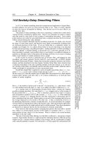
sions (A, Responses to sympathetic ac-
tivation). In simplistic terms, activation
of the sympathetic division can be con-
sidered a means by which the body
achieves a state of maximal work capac-
ity as required in fight or flight situa-
tions.
In both cases, there is a need for
vigorous activity of skeletal muscula-
ture. To ensure adequate supply of oxy-
gen and nutrients, blood flow in skeletal
muscle is increased; cardiac rate and
contractility are enhanced, resulting in a
larger blood volume being pumped into
the circulation. Narrowing of splanchnic
blood vessels diverts blood into vascular
beds in muscle.
Because digestion of food in the in-
testinal tract is dispensable and only
counterproductive, the propulsion of in-
testinal contents is slowed to the extent
that peristalsis diminishes and sphinc-
teric tonus increases. However, in order
to increase nutrient supply to heart and
musculature, glucose from the liver and
free fatty acid from adipose tissue must
be released into the blood. The bronchi
are dilated, enabling tidal volume and
alveolar oxygen uptake to be increased.
Sweat glands are also innervated by
sympathetic fibers (wet palms due to
excitement); however, these are excep-
tional as regards their neurotransmitter
(ACh, p. 106).
Although the life styles of modern
humans are different from those of
hominid ancestors, biological functions
have remained the same.
80 Drugs Acting on the Sympathetic Nervous System
Lüllmann, Color Atlas of Pharmacology © 2000 Thieme
All rights reserved. Usage subject to terms and conditions of license.
Drugs Acting on the Sympathetic Nervous System 81
Eyes:
pupillary dilation
CNS:
drive
alertness
Bronchi:
dilation
Saliva:
little, viscous
Heart:
rate
force
blood pressure
Fat tissue:
lipolysis
fatty acid
liberation
Bladder:
Sphincter
tone
detrusor muscle
Skeletal muscle:
blood flow
glycogenolysis
A. Responses to sympathetic activation
GI-tract:
peristalsis
sphincter tone
blood flow
Liver:
glycogenolysis
glucose release
Skin:
perspiration
(cholinergic)
Lüllmann, Color Atlas of Pharmacology © 2000 Thieme
All rights reserved. Usage subject to terms and conditions of license.
Structure of the Sympathetic Nervous
System
The sympathetic preganglionic neurons
(first neurons) project from the inter-
mediolateral column of the spinal gray
matter to the paired paravertebral gan-
glionic chain lying alongside the verte-
bral column and to unpaired preverte-
bral ganglia. These ganglia represent
sites of synaptic contact between pre-
ganglionic axons (1
st
neurons) and
nerve cells (2
nd
neurons or sympathocy-
tes) that emit postganglionic axons
terminating on cells in various end or-
gans. In addition, there are preganglion-
ic neurons that project either to periph-
eral ganglia in end organs or to the ad-
renal medulla.
Sympathetic Transmitter Substances
Whereas acetylcholine (see p. 98)
serves as the chemical transmitter at
ganglionic synapses between first and
second neurons, norepinephrine
(= noradrenaline) is the mediator at
synapses of the second neuron (B). This
second neuron does not synapse with
only a single cell in the effector organ;
rather, it branches out, each branch
making en passant contacts with several
cells. At these junctions the nerve axons
form enlargements (varicosities) re-
sembling beads on a string. Thus, excita-
tion of the neuron leads to activation of
a larger aggregate of effector cells, al-
though the action of released norepi-
nephrine may be confined to the region
of each junction. Excitation of pregan-
glionic neurons innervating the adrenal
medulla causes a liberation of acetyl-
choline. This, in turn, elicits a secretion
of epinephrine (= adrenaline) into the
blood, by which it is distributed to body
tissues as a hormone (A).
Adrenergic Synapse
Within the varicosities, norepinephrine
is stored in small membrane-enclosed
vesicles (granules, 0.05 to 0.2 µm in dia-
meter). In the axoplasm, L-tyrosine is
converted via two intermediate steps to
dopamine, which is taken up into the
vesicles and there converted to norepi-
nephrine by dopamine-!-hydroxylase.
When stimulated electrically, the sym-
pathetic nerve discharges the contents
of part of its vesicles, including norepi-
nephrine, into the extracellular space.
Liberated norepinephrine reacts with
adrenoceptors located postjunctionally
on the membrane of effector cells or
prejunctionally on the membrane of
varicosities. Activation of presynaptic
"
2
-receptors inhibits norepinephrine
release. By this negative feedback, re-
lease can be regulated.
The effect of released norepineph-
rine wanes quickly, because approx.
90 % is actively transported back into
the axoplasm, then into storage vesicles
(neuronal re-uptake). Small portions of
norepinephrine are inactivated by the
enzyme catechol-O-methyltransferase
(COMT, present in the cytoplasm of
postjunctional cells, to yield normeta-
nephrine), and monoamine oxidase
(MAO, present in mitochondria of nerve
cells and postjunctional cells, to yield
3,4-dihydroxymandelic acid).
The liver is richly endowed with
COMT and MAO; it therefore contrib-
utes significantly to the degradation of
circulating norepinephrine and epi-
nephrine. The end product of the com-
bined actions of MAO and COMT is van-
illylmandelic acid.
82 Drugs Acting on the Sympathetic Nervous System
Lüllmann, Color Atlas of Pharmacology © 2000 Thieme
All rights reserved. Usage subject to terms and conditions of license.
Drugs Acting on the Sympathetic Nervous System 83
B. Second neuron of sympathetic system, varicosity, norepinephrine release
A. Epinephrine as hormone, norepinephrine as transmitter
Psychic
stress
or physical
stress
First neuron
Second
neuron
Adrenal
medulla
NorepinephrineEpinephrine
M
A
O
Receptors
Receptors
COMT
Norepinephrine
Presynaptic
"
2
-receptors
"
!
2
!
1
3.4-Dihydroxy-
mandelic acid
Normeta-
nephrine
First neuron
Lüllmann, Color Atlas of Pharmacology © 2000 Thieme
All rights reserved. Usage subject to terms and conditions of license.
Adrenoceptor Subtypes and
Catecholamine Actions
Adrenoceptors fall into three major
groups, designated !
1
, !
2
, and ", within
each of which further subtypes can be
distinguished pharmacologically. The
different adrenoceptors are differential-
ly distributed according to region and
tissue. Agonists at adrenoceptors (di-
rect sympathomimetics) mimic the ac-
tions of the naturally occurring cate-
cholamines, norepinephrine and epi-
nephrine, and are used for various ther-
apeutic effects.
Smooth muscle effects. The op-
posing effects on smooth muscle (A) of
!-and "-adrenoceptor activation are
due to differences in signal transduction
(p. 66). This is exemplified by vascular
smooth muscle (A). !
1
-Receptor stimu-
lation leads to intracellular release of
Ca
2+
via activation of the inositol tris-
phosphate (IP
3
) pathway. In concert
with the protein calmodulin, Ca
2+
can
activate myosin kinase, leading to a rise
in tonus via phosphorylation of the con-
tractile protein myosin. cAMP inhibits
activation of myosin kinase. Via the for-
mer effector pathway, stimulation of !-
receptors results in vasoconstriction;
via the latter, "
2
-receptors mediate va-
sodilation, particularly in skeletal mus-
cle — an effect that has little therapeutic
use.
Vasoconstriction. Local application of
!-sympathomimetics can be employed
in infiltration anesthesia (p. 204) or for
nasal decongestion (naphazoline, tetra-
hydrozoline, xylometazoline; pp. 90,
324). Systemically administered epi-
nephrine is important in the treatment
of anaphylactic shock for combating hy-
potension.
Bronchodilation. "
2
-Adrenocep-
tor-mediated bronchodilation (e.g., with
terbutaline, fenoterol, or salbutamol)
plays an essential part in the treatment
of bronchial asthma (p. 328).
Tocolysis. The uterine relaxant ef-
fect of "
2
-adrenoceptor agonists, such as
terbutaline or fenoterol, can be used to
prevent premature labor. Vasodilation
with a resultant drop in systemic blood
pressure results in reflex tachycardia,
which is also due in part to the "
1
-stim-
ulant action of these drugs.
Cardiostimulation. By stimulating
"
1
-receptors, hence activation of ade-
nylatcyclase (Ad-cyclase) and cAMP
production, catecholamines augment all
heart functions, including systolic force
(positive inotropism), velocity of short-
ening (p. clinotropism), sinoatrial rate
(p. chronotropism), conduction velocity
(p. dromotropism), and excitability (p.
bathmotropism). In pacemaker fibers,
diastolic depolarization is hastened, so
that the firing threshold for the action
potential is reached sooner (positive
chronotropic effect, B). The cardiostim-
ulant effect of "-sympathomimetics
such as epinephrine is exploited in the
treatment of cardiac arrest. Use of "-
sympathomimetics in heart failure car-
ries the risk of cardiac arrhythmias.
Metabolic effects. "-Receptors me-
diate increased conversion of glycogen to
glucose (glycogenolysis) in both liver
and skeletal muscle. From the liver, glu-
cose is released into the blood, In adi-
pose tissue, triglycerides are hydrolyzed
to fatty acids (lipolysis, mediated by "
3
-
receptors), which then enter the blood
(C). The metabolic effects of catechola-
mines are not amenable to therapeutic
use.
84 Drugs Acting on the Sympathetic Nervous System
Lüllmann, Color Atlas of Pharmacology © 2000 Thieme
All rights reserved. Usage subject to terms and conditions of license.
Drugs Acting on the Sympathetic Nervous System 85
Membrane potential (mV)
Time
B. Cardiac effects of catecholamines
A. Vasomotor effects of catecholamines
!
1
G
i
!
2
Ad-cyclase
Phospholipase C
Ad-cyclase
Ca
2+
IP
3
cAMP
+ -
Calmodulin
Myosin
kinase
Myosin
Myosin-P
"
2
"
1
G
s
Ad-cyclase
+
cAMP
Force (mN)
Time
C. Metabolic effects of catecholamines
"
G
s
Ad-cyclase
+
Glucose
Glycogenolysis
cAMP
Glucose
Lipolysis
Fatty acids
Glycogenolysis
G
i
G
s
Lüllmann, Color Atlas of Pharmacology © 2000 Thieme
All rights reserved. Usage subject to terms and conditions of license.
Structure – Activity Relationships of
Sympathomimetics
Due to its equally high affinity for all !-
and "-receptors, epinephrine does not
permit selective activation of a particu-
lar receptor subtype. Like most cate-
cholamines, it is also unsuitable for oral
administration (catechol is a trivial
name for o-hydroxyphenol). Norepi-
nephrine differs from epinephrine by its
high affinity for !-receptors and low af-
finity for "
2
-receptors. In contrast, iso-
proterenol has high affinity for "-recep-
tors, but virtually none for !-receptors
(A).
norepinephrine Ǟ !, "
1
epinephrine Ǟ !, "
1
, "
2
isoproterenol Ǟ "
1
, "
2
Knowledge of structure–activity
relationships has permitted the syn-
thesis of sympathomimetics that dis-
play a high degree of selectivity at
adrenoceptor subtypes.
Direct-acting sympathomimetics
(i.e., adrenoceptor agonists) typically
share a phenylethylamine structure. The
side chain "-hydroxyl group confers af-
finity for !- and "-receptors. Substitu-
tion on the amino group reduces affinity
for !-receptors, but increases it for "-re-
ceptors (exception: !-agonist phenyl-
ephrine), with optimal affinity being
seen after the introduction of only one
isopropyl group. Increasing the bulk of
the amino substituent favors affinity for
"
2
-receptors (e.g., fenoterol, salbuta-
mol). Both hydroxyl groups on the aro-
matic nucleus contribute to affinity;
high activity at !-receptors is associated
with hydroxyl groups at the 3 and 4 po-
sitions. Affinity for "-receptors is pre-
served in congeners bearing hydroxyl
groups at positions 3 and 5 (orciprena-
line, terbutaline, fenoterol).
The hydroxyl groups of catechol-
amines are responsible for the very low
lipophilicity of these substances. Pola-
rity is increased at physiological pH due
to protonation of the amino group. De-
letion of one or all hydroxyl groups im-
proves membrane penetrability at the
intestinal mucosa-blood and the blood-
brain barriers. Accordingly, these non-
catecholamine congeners can be given
orally and can exert CNS actions; how-
ever, this structural change entails a loss
in affinity.
Absence of one or both aromatic
hydroxyl groups is associated with an
increase in indirect sympathomimetic
activity, denoting the ability of a sub-
stance to release norepinephrine from
its neuronal stores without exerting an
agonist action at the adrenoceptor (p.
88).
An altered position of aromatic hy-
droxyl groups (e.g., in orciprenaline, fe-
noterol, or terbutaline) or their substi-
tution (e.g., salbutamol) protects
against inactivation by COMT (p. 82). In-
droduction of a small alkyl residue at
the carbon atom adjacent to the amino
group (ephedrine, methamphetamine)
confers resistance to degradation by
MAO (p. 80), as does replacement on the
amino groups of the methyl residue
with larger substituents (e.g., ethyl in
etilefrine). Accordingly, the congeners
are less subject to presystemic inactiva-
tion.
Since structural requirements for
high affinity, on the one hand, and oral
applicability, on the other, do not
match, choosing a sympathomimetic is
a matter of compromise. If the high af-
finity of epinephrine is to be exploited,
absorbability from the intestine must be
foregone (epinephrine, isoprenaline). If
good bioavailability with oral adminis-
tration is desired, losses in receptor af-
finity must be accepted (etilefrine).
86 Drugs Acting on the Sympathetic Nervous System
Lüllmann, Color Atlas of Pharmacology © 2000 Thieme
All rights reserved. Usage subject to terms and conditions of license.
Drugs Acting on the Sympathetic Nervous System 87
B. Structure-activity relationship of epinephrine derivatives
A. Chemical structure of catecholamines and affinity for
!- and "-receptors
EpinephrineNorepinephrine Isoproterenol
Receptor affinity
Catecholamine-
O-methyltransferase
Monoamine oxidase
(Enteral absorbability
CNS permeability)
Metabolic
stability
Etilefrine Ephedrine Methamphetamine
Epinephrine Orciprenaline Fenoterol
Affinity for !-receptors
Affinity for "-receptors
Resistance to degradation
Absorbability
Indirect
action
Penetrability
through
membrane
barriers
Lüllmann, Color Atlas of Pharmacology © 2000 Thieme
All rights reserved. Usage subject to terms and conditions of license.
Indirect Sympathomimetics
Apart from receptors, adrenergic neu-
rotransmission involves mechanisms
for the active re-uptake and re-storage
of released amine, as well as enzymatic
breakdown by monoamine oxidase
(MAO). Norepinephrine (NE) displays
affinity for receptors, transport systems,
and degradative enzymes. Chemical al-
terations of the catecholamine differen-
tially affect these properties and result
in substances with selective actions.
Inhibitors of MAO (A). The enzyme
is located predominantly on mitochon-
dria, and serves to scavenge axoplasmic
free NE. Inhibition of the enzyme causes
free NE concentrations to rise. Likewise,
dopamine catabolism is impaired, mak-
ing more of it available for NE synthesis.
Consequently, the amount of NE stored
in granular vesicles will increase, and
with it the amount of amine released
per nerve impulse.
In the CNS, inhibition of MAO af-
fects neuronal storage not only of NE
but also of dopamine and serotonin.
These mediators probably play signifi-
cant roles in CNS functions consistent
with the stimulant effects of MAO inhib-
itors on mood and psychomotor drive
and their use as antidepressants in the
treatment of depression (A). Tranylcy-
promine is used to treat particular forms
of depressive illness; as a covalently
bound suicide substrate, it causes long-
lasting inhibition of both MAO iso-
zymes, (MAO
A
, MAO
B
). Moclobemide re-
versibly inhibits MAO
A
and is also used
as an antidepressant. The MAO
B
inhibi-
tor selegiline (deprenyl) retards the cat-
obolism of dopamine, an effect used in
the treatment of parkinsonism (p. 188).
Indirect sympathomimetics (B)
are agents that elevate the concentra-
tion of NE at neuroeffector junctions,
because they either inhibit re-uptake
(cocaine), facilitate release, or slow
breakdown by MAO, or exert all three of
these effects (amphetamine, metham-
phetamine). The effectiveness of such
indirect sympathomimetics diminishes
or disappears (tachyphylaxis) when ve-
sicular stores of NE close to the axolem-
ma are depleted.
Indirect sympathomimetics can
penetrate the blood-brain barrier and
evoke such CNS effects as a feeling of
well-being, enhanced physical activity
and mood (euphoria), and decreased
sense of hunger or fatigue. Subsequent-
ly, the user may feel tired and de-
pressed. These after effects are partly
responsible for the urge to re-adminis-
ter the drug (high abuse potential). To
prevent their misuse, these substances
are subject to governmental regulations
(e.g., Food and Drugs Act: Canada; Con-
trolled Drugs Act: USA) restricting their
prescription and distribution.
When amphetamine-like substanc-
es are misused to enhance athletic per-
formance (doping), there is a risk of dan-
gerous physical overexertion. Because
of the absence of a sense of fatigue, a
drugged athlete may be able to mobilize
ultimate energy reserves. In extreme
situations, cardiovascular failure may
result (B).
Closely related chemically to am-
phetamine are the so-called appetite
suppressants or anorexiants, such as
fenfluramine, mazindole, and sibutra-
mine. These may also cause dependence
and their therapeutic value and safety
are questionable.
88 Drugs Acting on the Sympathetic Nervous System
Lüllmann, Color Atlas of Pharmacology © 2000 Thieme
All rights reserved. Usage subject to terms and conditions of license.
Drugs Acting on the Sympathetic Nervous System 89
Controlled
Substances
Act regulates
use of
cocaine and
amphetamine
MAO
MAO
MAO
MAO
B. Indirect sympathomimetics with central stimulant activity and abuse potential
A. Monoamine oxidase inhibitor
Nor-
epinephrine
Norepinephrine
transport system
Effector organ
"Doping"
Runner-up
Pain stimulus Local
anesthetic
effect
Amphetamine Cocaine
§
§
Inhibitor: Moclobemide MAO-A
Selegiline MAO-B
Lüllmann, Color Atlas of Pharmacology © 2000 Thieme
All rights reserved. Usage subject to terms and conditions of license.
!-Sympathomimetics,
!-Sympatholytics
!-Sympathomimetics can be used
systemically in certain types of hypoten-
sion (p. 314) and locally for nasal or con-
junctival decongestion (pp. 324, 326) or
as adjuncts in infiltration anesthesia (p.
206) for the purpose of delaying the re-
moval of local anesthetic. With local
use, underperfusion of the vasocon-
stricted area results in a lack of oxygen
(A). In the extreme case, local hypoxia
can lead to tissue necrosis. The append-
ages (e.g., digits, toes, ears) are particu-
larly vulnerable in this regard, thus pre-
cluding vasoconstrictor adjuncts in in-
filtration anesthesia at these sites.
Vasoconstriction induced by an !-
sympathomimetic is followed by a
phase of enhanced blood flow (reactive
hyperemia, A). This reaction can be ob-
served after the application of !-sympa-
thomimetics (naphazoline, tetrahydro-
zoline, xylometazoline) to the nasal mu-
cosa. Initially, vasoconstriction reduces
mucosal blood flow and, hence, capil-
lary pressure. Fluid exuded into the
interstitial space is drained through the
veins, thus shrinking the nasal mucosa.
Due to the reduced supply of fluid, se-
cretion of nasal mucus decreases. In co-
ryza, nasal patency is restored. Howev-
er, after vasoconstriction subsides, reac-
tive hyperemia causes renewed exuda-
tion of plasma fluid into the interstitial
space, the nose is “stuffy” again, and the
patient feels a need to reapply decon-
gestant. In this way, a vicious cycle
threatens. Besides rebound congestion,
persistent use of a decongestant entails
the risk of atrophic damage caused by
prolonged hypoxia of the nasal mucosa.
!-Sympatholytics (B). The interac-
tion of norepinephrine with !-adreno-
ceptors can be inhibited by !-sympath-
olytics ( !-adrenoceptor antagonists, !-
blockers). This inhibition can be put to
therapeutic use in antihypertensive
treatment (vasodilation Ǟ peripheral
resistance ", blood pressure ", p. 118).
The first !-sympatholytics blocked the
action of norepinephrine at both post-
and prejunctional !-adrenoceptors
(non-selective !-blockers, e.g., phen-
oxybenzamine, phentolamine).
Presynaptic !
2
-adrenoceptors func-
tion like sensors that enable norepi-
nephrine concentration outside the
axolemma to be monitored, thus regu-
lating its release via a local feedback
mechanism. When presynaptic !
2
-re-
ceptors are stimulated, further release
of norepinephrine is inhibited. Con-
versely, their blockade leads to uncon-
trolled release of norepinephrine with
an overt enhancement of sympathetic
effects at #
1
-adrenoceptor-mediated
myocardial neuroeffector junctions, re-
sulting in tachycardia and tachyar-
rhythmia.
Selective !-Sympatholytics
!-Blockers, such as prazosin, or the
longer-acting terazosin and doxazosin,
lack affinity for prejunctional !
2
-adren-
oceptors. They suppress activation of
!
1
-receptors without a concomitant en-
hancement of norepinephrine release.
!
1
-Blockers may be used in hyper-
tension (p. 312). Because they prevent
reflex vasoconstriction, they are likely
to cause postural hypotension with
pooling of blood in lower limb capaci-
tance veins during change from the su-
pine to the erect position (orthostatic
collapse: " venous return, " cardiac out-
put, fall in systemic pressure, " blood
supply to CNS, syncope, p. 314).
In benign hyperplasia of the pros-
tate, !-blockers (terazosin, alfuzosin)
may serve to lower tonus of smooth
musculature in the prostatic region and
thereby facilitate micturition (p. 252).
90 Drugs Acting on the Sympathetic Nervous System
Lüllmann, Color Atlas of Pharmacology © 2000 Thieme
All rights reserved. Usage subject to terms and conditions of license.
Drugs Acting on the Sympathetic Nervous System 91
C. Indications for !
1
-sympatholytics
A. Reactive hyperemia due to !-sympathomimetics, e.g., following decongestion
of nasal mucosa
B. Autoinhibition of norepinephrine release and !-sympatholytics
!-Agonist
O
2
supply < O
2
demand
O
2
supply = O
2
demand
After
Before
O
2
supply = O
2
demand
NE
!
2
!
2
!
2
nonselective
!-blocker
!
1
!
1
!
1
#
1
#
1
#
1
!
1
-blocker
!
1
-blocker
e.g., terazosin
H
3
CO
O
O
H
3
CO
NH
2
N
N
N
N
High blood pressure
Benign
prostatic hyperplasia
Inhibition of
!
1
-adrenergic
stimulation of
smooth muscle
Neck of bladder,
prostate
Resistance
arteries
Lüllmann, Color Atlas of Pharmacology © 2000 Thieme
All rights reserved. Usage subject to terms and conditions of license.
!-Sympatholytics (!-Blockers)
!-Sympatholytics are antagonists of
norepiphephrine and epinephrine at !-
adrenoceptors; they lack affinity for "-
receptors.
Therapeutic effects. !-Blockers
protect the heart from the oxygen-
wasting effect of sympathetic inotrop-
ism (p. 306) by blocking cardiac !-re-
ceptors; thus, cardiac work can no long-
er be augmented above basal levels (the
heart is “coasting”). This effect is uti-
lized prophylactically in angina pectoris
to prevent myocardial stress that could
trigger an ischemic attack (p. 308, 310).
!-Blockers also serve to lower cardiac
rate (sinus tachycardia, p. 134) and ele-
vated blood pressure due to high cardiac
output (p. 312). The mechanism under-
lying their antihypertensive action via
reduction of peripheral resistance is un-
clear.
Applied topically to the eye, !-
blockers are used in the management of
glaucoma; they lower production of
aqueous humor without affecting its
drainage.
Undesired effects. The hazards of
treatment with !-blockers become ap-
parent particularly when continuous
activation of !-receptors is needed in
order to maintain the function of an or-
gan.
Congestive heart failure: In myocar-
dial insufficiency, the heart depends on
a tonic sympathetic drive to maintain
adequate cardiac output. Sympathetic
activation gives rise to an increase in
heart rate and systolic muscle tension,
enabling cardiac output to be restored
to a level comparable to that in a
healthy subject. When sympathetic
drive is eliminated during !-receptor
blockade, stroke volume and cardiac
rate decline, a latent myocardial insuffi-
ciency is unmasked, and overt insuffi-
ciency is exacerbated (A).
On the other hand, clinical evidence
suggests that !-blockers produce favor-
able effects in certain forms of conges-
tive heart failure (idiopathic dilated car-
diomyopathy).
Bradycardia, A-V block: Elimination
of sympathetic drive can lead to a
marked fall in cardiac rate as well as to
disorders of impulse conduction from
the atria to the ventricles.
Bronchial asthma: Increased sym-
pathetic activity prevents broncho-
spasm in patients disposed to paroxys-
mal constriction of the bronchial tree
(bronchial asthma, bronchitis in smok-
ers). In this condition, !
2
-receptor
blockade will precipitate acute respira-
tory distress (B).
Hypoglycemia in diabetes mellitus:
When treatment with insulin or oral hy-
poglycemics in the diabetic patient low-
ers blood glucose below a critical level,
epinephrine is released, which then
stimulates hepatic glucose release via
activation of !
2
-receptors. !-Blockers
suppress this counter-regulation; in ad-
dition, they mask other epinephrine-
mediated warning signs of imminent
hypoglycemia, such as tachycardia and
anxiety, thereby enhancing the risk of
hypoglycemic shock.
Altered vascular responses: When
!
2
-receptors are blocked, the vasodilat-
ing effect of epinephrine is abolished,
leaving the "-receptor-mediated vaso-
constriction unaffected: peripheral
blood flow # – “cold hands and feet”.
!-Blockers exert an “anxiolytic“
action that may be due to the suppres-
sion of somatic responses (palpitations,
trembling) to epinephrine release that
is induced by emotional stress; in turn,
these would exacerbate “anxiety” or
“stage fright”. Because alertness is not
impaired by !-blockers, these agents are
occasionally taken by orators and musi-
cians before a major performance (C).
Stage fright, however, is not a disease
requiring drug therapy.
92 Drugs Acting on the Sympathetic Nervous System
Lüllmann, Color Atlas of Pharmacology © 2000 Thieme
All rights reserved. Usage subject to terms and conditions of license.
Drugs Acting on the Sympathetic Nervous System 93
C. “Anxiolytic” effect of !-sympatholytics
A.
!-Sympatholytics: effect on cardiac function
B.
!-Sympatholytics: effect on bronchial and vascular tone
Stroke
volume
100 ml
!-Receptor!-Blocker
blocks
receptor
Heart failure Healthy
1 sec
!
1
-Blockade !
1
-Stimulation
!
2
-Blockade !
2
-Stimulation
HealthyAsthmatic
!
2
-Blockade !
2
-Stimulation
" !
2
" !
2
"
1 sec
!-Blockade
Lüllmann, Color Atlas of Pharmacology © 2000 Thieme
All rights reserved. Usage subject to terms and conditions of license.
Types of !-Blockers
The basic structure shared by most !-
sympatholytics is the side chain of !-
sympathomimetics (cf. isoproterenol
with the !-blockers propranolol, pindo-
lol, atenolol). As a rule, this basic struc-
ture is linked to an aromatic nucleus by
a methylene and oxygen bridge. The
side chain C-atom bearing the hydroxyl
group forms the chiral center. With
some exceptions (e.g., timolol, penbuto-
lol), all !-sympatholytics are brought as
racemates into the market (p. 62).
Compared with the dextrorotatory
form, the levorotatory enantiomer pos-
sesses a greater than 100-fold higher af-
finity for the !-receptor and is, there-
fore, practically alone in contributing to
the !-blocking effect of the racemate.
The side chain and substituents on the
amino group critically affect affinity for
!-receptors, whereas the aromatic nu-
cleus determines whether the com-
pound possess intrinsic sympathomi-
metic activity (ISA), that is, acts as a
partial agonist (p. 60) or partial antago-
nist. In the presence of a partial agonist
(e.g., pindolol), the ability of a full ago-
nist (e.g., isoprenaline) to elicit a maxi-
mal effect would be attenuated, because
binding of the full agonist is impeded.
However, the !-receptor at which such
partial agonism can be shown appears
to be atypical (!
3
or !
4
subtype). Wheth-
er ISA confers a therapeutic advantage
on a !-blocker remains an open ques-
tion.
As cationic amphiphilic drugs, !-
blockers can exert a membrane-stabi-
lizing effect, as evidenced by the ability
of the more lipophilic congeners to in-
hibit Na
+
-channel function and impulse
conduction in cardiac tissues. At the
usual therapeutic dosage, the high con-
centration required for these effects will
not be reached.
Some !-sympatholytics possess
higher affinity for cardiac !
1
-receptors
than for !
2
-receptors and thus display
cardioselectivity (e.g., metoprolol, ace-
butolol, bisoprolol). None of these
blockers is sufficiently selective to per-
mit its use in patients with bronchial
asthma or diabetes mellitus (p. 92).
The chemical structure of !-block-
ers also determines their pharmacoki-
netic properties. Except for hydrophilic
representatives (atenolol), !-sympatho-
lytics are completely absorbed from the
intestines and subsequently undergo
presystemic elimination to a major ex-
tent (A).
All the above differences are of
little clinical importance. The abundance
of commercially available congeners
would thus appear all the more curious
(B). Propranolol was the first !-blocker
to be introduced into therapy in 1965.
Thirty-five years later, about 20 different
congeners are being marketed in differ-
ent countries. This questionable devel-
opment unfortunately is typical of any
drug group that has major therapeutic
relevance, in addition to a relatively
fixed active structure. Variation of the
molecule will create a new patentable
chemical, not necessarily a drug with a
novel action. Moreover, a drug no longer
protected by patent is offered as a gener-
ic by different manufacturers under doz-
ens of different proprietary names.
Propranolol alone has been marketed by
13 manufacturers under 11 different
names.
94 Drugs Acting on the Sympathetic Nervous System
Lüllmann, Color Atlas of Pharmacology © 2000 Thieme
All rights reserved. Usage subject to terms and conditions of license.
Drugs Acting on the Sympathetic Nervous System 95
Talinolol
Sotalol
!
1
!
2
B. Avalanche-like increase in commercially available !-sympatholytics
Isoproterenol Pindolol Propranolol Atenolol
Agonist partial
Agonist
Antagonist
Effect No effect
selectivity
Presystemic elimination
100%
50%
A. Types of !-sympatholytics
Betaxolol
Carteolol
Mepindolol
Penbutolol
Carazolol
Nadolol
Acebutolol
Bunitrolol
Atenolol
Metipranol
Metoprolol
Timolol
Oxprenolol
Pindolol
Bupranolol
Alprenolol
Propranolol
1965 1970
1975 1980 1985 1990
Celiprolol
Bisoprolol
Bopindolol
Esmolol
Tertatolol
!
1
!
2
Cardio-
!
1
!
2
!
1
!
2
!
1
!
2
!-Receptor !-Receptor !-Receptor
Carvedilol
Befunolol
Year introduced
Antagonist
Lüllmann, Color Atlas of Pharmacology © 2000 Thieme
All rights reserved. Usage subject to terms and conditions of license.
Antiadrenergics
Antiadrenergics are drugs capable of
lowering transmitter output from sym-
pathetic neurons, i.e., “sympathetic
tone”. Their action is hypotensive (indi-
cation: hypertension, p. 312); however,
being poorly tolerated, they enjoy only
limited therapeutic use.
Clonidine is an !
2
-agonist whose
high lipophilicity (dichlorophenyl ring)
permits rapid penetration through the
blood-brain barrier. The activation of
postsynaptic !
2
-receptors dampens the
activity of vasomotor neurons in the
medulla oblongata, resulting in a reset-
ting of systemic arterial pressure at a
lower level. In addition, activation of
presynaptic !
2
-receptors in the periph-
ery (pp. 82, 90) leads to a decreased re-
lease of both norepinephrine (NE) and
acetylcholine.
Side effects. Lassitude, dry mouth;
rebound hypertension after abrupt ces-
sation of clonidine therapy.
Methyldopa (dopa = dihydroxy-
phenylalanine), as an amino acid, is
transported across the blood-brain bar-
rier, decarboxylated in the brain to !-
methyldopamine, and then hydroxylat-
ed to !-methyl-NE. The decarboxylation
of methyldopa competes for a portion of
the available enzymatic activity, so that
the rate of conversion of L-dopa to NE
(via dopamine) is decreased. The false
transmitter !-methyl-NE can be stored;
however, unlike the endogenous media-
tor, it has a higher affinity for !
2
- than
for !
1
-receptors and therefore produces
effects similar to those of clonidine. The
same events take place in peripheral ad-
renergic neurons.
Adverse effects. Fatigue, orthostatic
hypotension, extrapyramidal Parkin-
son-like symptoms (p. 88), cutaneous
reactions, hepatic damage, immune-he-
molytic anemia.
Reserpine, an alkaloid from the
Rauwolfia plant, abolishes the vesicular
storage of biogenic amines (NE, dopa-
mine = DA, serotonin = 5-HT) by inhibit-
ing an ATPase required for the vesicular
amine pump. The amount of NE re-
leased per nerve impulse is decreased.
To a lesser degree, release of epineph-
rine from the adrenal medulla is also
impaired. At higher doses, there is irre-
versible damage to storage vesicles
(“pharmacological sympathectomy”),
days to weeks being required for their
resynthesis. Reserpine readily enters
the brain, where it also impairs vesicu-
lar storage of biogenic amines.
Adverse effects. Disorders of extra-
pyramidal motor function with devel-
opment of pseudo-Parkinsonism (p. 88),
sedation, depression, stuffy nose, im-
paired libido, and impotence; increased
appetite. These adverse effects have
rendered the drug practically obsolete.
Guanethidine possesses high affin-
ity for the axolemmal and vesicular
amine transporters. It is stored instead
of NE, but is unable to mimic the func-
tions of the latter. In addition, it stabiliz-
es the axonal membrane, thereby im-
peding the propagation of impulses into
the sympathetic nerve terminals. Stor-
age and release of epinephrine from the
adrenal medulla are not affected, owing
to the absence of a re-uptake process.
The drug does not cross the blood-brain
barrier.
Adverse effects. Cardiovascular cri-
ses are a possible risk: emotional stress
of the patient may cause sympatho-
adrenal activation with epinephrine re-
lease. The resulting rise in blood pres-
sure can be all the more marked be-
cause persistent depression of sympa-
thetic nerve activity induces supersen-
sitivity of effector organs to circulating
catecholamines.
96 Drugs Acting on the Sympathetic Nervous System
Lüllmann, Color Atlas of Pharmacology © 2000 Thieme
All rights reserved. Usage subject to terms and conditions of license.
Drugs Acting on the Sympathetic Nervous System 97
Suppression of
sympathetic
impulses in
vasomotor
center
Release from adrenal medulla
unaffected
CNS
A. Inhibitors of sympathetic tone
No epinephrine from adrenal medulla
due to central sedative effect
Stimulation of central !
2
-receptors
!-Methyl-NE
False transmitter
Tyrosine
Dopa
Dopamine
NE
Clonidine
!-Methyldopa
Peripheral
sympathetic activity
Inhibition of
biogenic amine
storage
NE
DA
5HT
Varicosity
Reserpine
Inhibition of
peripheral
sympathetic activity
Active uptake and
storage instead of
norepinephrine;
not a transmitter
Guanethidine
Varicosity
Inhibition of
Dopa-decarb-
oxylase
!-Methyl-NE
in brain
Lüllmann, Color Atlas of Pharmacology © 2000 Thieme
All rights reserved. Usage subject to terms and conditions of license.
Parasympathetic Nervous System
Responses to activation of the para-
sympathetic system. Parasympathetic
nerves regulate processes connected
with energy assimilation (food intake,
digestion, absorption) and storage.
These processes operate when the body
is at rest, allowing a decreased tidal vol-
ume (increased bronchomotor tone)
and decreased cardiac activity. Secre-
tion of saliva and intestinal fluids pro-
motes the digestion of foodstuffs; trans-
port of intestinal contents is speeded up
because of enhanced peristaltic activity
and lowered tone of sphincteric mus-
cles. To empty the urinary bladder (mic-
turition), wall tension is increased by
detrusor activation with a concurrent
relaxation of sphincter tonus.
Activation of ocular parasympa-
thetic fibers (see below) results in nar-
rowing of the pupil and increased curva-
ture of the lens, enabling near objects to
be brought into focus (accommodation).
Anatomy of the parasympathetic
system. The cell bodies of parasympa-
thetic preganglionic neurons are located
in the brainstem and the sacral spinal
cord. Parasympathetic outflow is chan-
nelled from the brainstem (1) through
the third cranial nerve (oculomotor n.)
via the ciliary ganglion to the eye; (2)
through the seventh cranial nerve (fa-
cial n.) via the pterygopalatine and sub-
maxillary ganglia to lacrimal glands and
salivary glands (sublingual, submandib-
ular), respectively; (3) through the
ninth cranial nerve (glossopharyngeal
n.) via the otic ganglion to the parotid
gland; and (4) via the tenth cranial
nerve (vagus n.) to thoracic and abdom-
inal viscera. Approximately 75 % of all
parasympathetic fibers are contained
within the vagus nerve. The neurons of
the sacral division innervate the distal
colon, rectum, bladder, the distal ure-
ters, and the external genitalia.
Acetylcholine (ACh) as a transmit-
ter. ACh serves as mediator at terminals
of all postganglionic parasympathetic
fibers, in addition to fulfilling its trans-
mitter role at ganglionic synapses with-
in both the sympathetic and parasym-
pathetic divisions and the motor end-
plates on striated muscle. However, dif-
ferent types of receptors are present at
these synaptic junctions:
98 Drugs Acting on the Parasympathetic Nervous System
Localization Agonist Antagonist Receptor Type
Target tissues of 2
nd
ACh Atropine Muscarinic (M)
parasympathetic Muscarine cholinoceptor;
neurons G-protein-coupled-
receptor protein with
7 transmembrane
domains
Sympathetic & ACh Trimethaphan Ganglionic type
parasympathetic Nicotine (!3 "4)
ganglia
Nicotinic (N)
cholinoceptor ligand-
gated cation channel
formed by five trans-
membrane subunits
Motor endplate ACh d-Tubocurarine muscular type
Nicotine (!1
2
"1#$)
The existence of distinct cholino-
ceptors at different cholinergic synap-
ses allows selective pharmacological
interventions.
Lüllmann, Color Atlas of Pharmacology © 2000 Thieme
All rights reserved. Usage subject to terms and conditions of license.
Drugs Acting on the Parasympathetic Nervous System 99
Eyes:
Accommodation
for near vision,
miosis
Bronchi:
constriction
secretion
Saliva:
copious, liquid
GI tract:
secretion
peristalsis
sphincter tone
Heart:
rate
blood pressure
Bladder:
sphincter tone
detrusor
A. Responses to parasympathetic activation
Lüllmann, Color Atlas of Pharmacology © 2000 Thieme
All rights reserved. Usage subject to terms and conditions of license.
Cholinergic Synapse
Acetylcholine (ACh) is the transmitter
at postganglionic synapses of parasym-
pathetic nerve endings. It is highly con-
centrated in synaptic storage vesicles
densely present in the axoplasm of the
terminal. ACh is formed from choline
and activated acetate (acetylcoenzyme
A), a reaction catalyzed by the enzyme
choline acetyltransferase. The highly
polar choline is actively transported into
the axoplasm. The specific choline trans-
porter is localized exclusively to mem-
branes of cholinergic axons and termi-
nals. The mechanism of transmitter re-
lease is not known in full detail. The vesi-
cles are anchored via the protein synap-
sin to the cytoskeletal network. This ar-
rangement permits clustering of vesicles
near the presynaptic membrane, while
preventing fusion with it. During activa-
tion of the nerve membrane, Ca
2+
is
thought to enter the axoplasm through
voltage-gated channels and to activate
protein kinases that phosphorylate syn-
apsin. As a result, vesicles close to the
membrane are detached from their an-
choring and allowed to fuse with the
presynaptic membrane. During fusion,
vesicles discharge their contents into the
synaptic gap. ACh quickly diffuses
through the synaptic gap (the acetylcho-
line molecule is a little longer than
0.5 nm; the synaptic gap is as narrow as
30–40 nm). At the postsynaptic effector
cell membrane, ACh reacts with its re-
ceptors. Because these receptors can al-
so be activated by the alkaloid musca-
rine, they are referred to as muscarinic
(M-)cholinoceptors. In contrast, at gan-
glionic (p. 108) and motor endplate (p.
184) cholinoceptors, the action of ACh is
mimicked by nicotine and they are,
therefore, said to be nicotinic cholino-
ceptors.
Released ACh is rapidly hydrolyzed
and inactivated by a specific acetylchol-
inesterase, present on pre- and post-
junctional membranes, or by a less spe-
cific serum cholinesterase (butyryl chol-
inesterase), a soluble enzyme present in
serum and interstitial fluid.
M-cholinoceptors can be classified
into subtypes according to their molec-
ular structure, signal transduction, and
ligand affinity. Here, the M
1
, M
2
, and M
3
subtypes are considered. M
1
receptors
are present on nerve cells, e.g., in gan-
glia, where they mediate a facilitation of
impulse transmission from pregan-
glionic axon terminals to ganglion cells.
M
2
receptors mediate acetylcholine ef-
fects on the heart: opening of K
+
chan-
nels leads to slowing of diastolic depola-
rization in sinoatrial pacemaker cells
and a decrease in heart rate. M
3
recep-
tors play a role in the regulation of
smooth muscle tone, e.g., in the gut and
bronchi, where their activation causes
stimulation of phospholipase C, mem-
brane depolarization, and increase in
muscle tone. M
3
receptors are also
found in glandular epithelia, which sim-
ilarly respond with activation of phos-
pholipase C and increased secretory ac-
tivity. In the CNS, where all subtypes are
present, cholinoceptors serve diverse
functions, including regulation of corti-
cal excitability, memory, learning, pain
processing, and brain stem motor con-
trol. The assignment of specific receptor
subtypes to these functions has yet to be
achieved.
In blood vessels, the relaxant action
of ACh on muscle tone is indirect, be-
cause it involves stimulation of M
3
-cho-
linoceptors on endothelial cells that re-
spond by liberating NO (= endothelium-
derived relaxing factor). The latter dif-
fuses into the subjacent smooth muscu-
lature, where it causes a relaxation of
active tonus (p. 121).
100 Drugs Acting on the Parasympathetic Nervous System
Lüllmann, Color Atlas of Pharmacology © 2000 Thieme
All rights reserved. Usage subject to terms and conditions of license.
Drugs Acting on the Parasympathetic Nervous System 101
Acetyl coenzyme A + choline
Choline acetyltransferase
Acetylcholine
Serum-
cholinesterase
Smooth muscle cell
M
3
-receptor
Heart pacemaker cell
M
2
-receptor
Secretory cell
M
3
-receptor
Phospholipase C K
+
-channel activation Phospholipase C
Ca
2
+
in Cytosol
Slowing of
diastolic
depolarization
Ca
2
+
in Cytosol
Tone Rate Secretion
-30
-70
Time
0
-45
-90
ACh
effect
Control
condition
Time
A. Acetylcholine: release, effects, and degradation
mV
Ca
2
+
influx
Protein
kinase
Vesicle
release
Exocytosis
Receptor
occupation
esteric
cleavage
Action potential
Ca
2
+
mV
mN
active
reuptake of
choline
Acetylcholine
esterase:
membrane-
associated
Storage of
acetylcholine
in vesicles
Lüllmann, Color Atlas of Pharmacology © 2000 Thieme
All rights reserved. Usage subject to terms and conditions of license.
Parasympathomimetics
Acetylcholine (ACh) is too rapidly hy-
drolyzed and inactivated by acetylcholi-
nesterase (AChE) to be of any therapeu-
tic use; however, its action can be mim-
icked by other substances, namely di-
rect or indirect parasympathomimetics.
Direct Parasympathomimetics.
The choline ester, carbachol, activates
M-cholinoceptors, but is not hydrolyzed
by AChE. Carbachol can thus be effec-
tively employed for local application to
the eye (glaucoma) and systemic ad-
ministration (bowel atonia, bladder ato-
nia). The alkaloids, pilocarpine (from Pil-
ocarpus jaborandi) and arecoline (from
Areca catechu; betel nut) also act as di-
rect parasympathomimetics. As tertiary
amines, they moreover exert central ef-
fects. The central effect of muscarine-
like substances consists of an enliven-
ing, mild stimulation that is probably
the effect desired in betel chewing, a
widespread habit in South Asia. Of this
group, only pilocarpine enjoys thera-
peutic use, which is limited to local ap-
plication to the eye in glaucoma.
Indirect Parasympathomimetics.
AChE can be inhibited selectively, with
the result that ACh released by nerve
impulses will accumulate at cholinergic
synapses and cause prolonged stimula-
tion of cholinoceptors. Inhibitors of
AChE are, therefore, indirect parasym-
pathomimetics. Their action is evident
at all cholinergic synapses. Chemically,
these agents include esters of carbamic
acid (carbamates such as physostig-
mine, neostigmine) and of phosphoric
acid (organophosphates such as para-
oxon = E600 and nitrostigmine = para-
thion = E605, its prodrug).
Members of both groups react like
ACh with AChE and can be considered
false substrates. The esters are hydro-
lyzed upon formation of a complex with
the enzyme. The rate-limiting step in
ACh hydrolysis is deacetylation of the
enzyme, which takes only milliseconds,
thus permitting a high turnover rate
and activity of AChE. Decarbaminoyla-
tion following hydrolysis of a carba-
mate takes hours to days, the enzyme
remaining inhibited as long as it is car-
baminoylated. Cleavage of the phos-
phate residue, i.e. dephosphorylation,
is practically impossible; enzyme inhi-
bition is irreversible.
Uses. The quaternary carbamate
neostigmine is employed as an indirect
parasympathomimetic in postoperative
atonia of the bowel or bladder. Further-
more, it is needed to overcome the rela-
tive ACh-deficiency at the motor end-
plate in myasthenia gravis or to reverse
the neuromuscular blockade (p. 184)
caused by nondepolarizing muscle re-
laxants (decurarization before discon-
tinuation of anesthesia). The tertiary
carbamate physostigmine can be used
as an antidote in poisoning with para-
sympatholytic drugs, because it has ac-
cess to AChE in the brain. Carbamates
(neostigmine, pyridostigmine, physos-
tigmine) and organophosphates (para-
oxon, ecothiopate) can also be applied
locally to the eye in the treatment of
glaucoma; however, their long-term use
leads to cataract formation. Agents from
both classes also serve as insecticides.
Although they possess high acute toxic-
ity in humans, they are more rapidly de-
graded than is DDT following their
emission into the environment.
Tacrine is not an ester and interferes
only with the choline-binding site of
AChE. It is effective in alleviating symp-
toms of dementia in some subtypes of
Alzheimer’s disease.
102 Drugs Acting on the Parasympathetic Nervous System
Lüllmann, Color Atlas of Pharmacology © 2000 Thieme
All rights reserved. Usage subject to terms and conditions of license.
Drugs Acting on the Parasympathetic Nervous System 103
Effector organ
A. Direct and indirect parasympathomimetics
Arecoline =
ingredient of
betel nut:
betel
chewing
AChE
Direct parasympatho-
mimetics
AChE
Inhibitors of
acetylcholinesterase
(AChE)
Indirect
parasympathomimetics
Carbachol
Acetylcholine
Arecoline
ACh
Neostigmine Paraoxon (E 600)
Physostigmine
AChE
Phosphoryl
Dephosphorylation impossible
Paraoxon
+
AChE
Carbaminoyl
Hours to days
Decarbaminoylation
Neostigmine
+
AChE
Acetyl
ms
Deacetylation
Acetylcholine
+
Nitrostigmine =
Parathion =
E 605
Choline
Lüllmann, Color Atlas of Pharmacology © 2000 Thieme
All rights reserved. Usage subject to terms and conditions of license.