Tài liệu TRADITIONAL AND NOVEL RISK FACTORS IN ATHEROTHROMBOSIS pptx
Bạn đang xem bản rút gọn của tài liệu. Xem và tải ngay bản đầy đủ của tài liệu tại đây (7.02 MB, 150 trang )
TRADITIONAL AND
NOVEL RISK FACTORS IN
ATHEROTHROMBOSIS
Edited by Efraín Gaxiola
TRADITIONAL AND
NOVEL RISK FACTORS IN
ATHEROTHROMBOSIS
Edited by Efraín Gaxiola
Traditional and Novel Risk Factors in Atherothrombosis
Edited by Efraín Gaxiola
Published by InTech
Janeza Trdine 9, 51000 Rijeka, Croatia
Copyright © 2012 InTech
All chapters are Open Access distributed under the Creative Commons Attribution 3.0
license, which allows users to download, copy and build upon published articles even for
commercial purposes, as long as the author and publisher are properly credited, which
ensures maximum dissemination and a wider impact of our publications. After this work
has been published by InTech, authors have the right to republish it, in whole or part, in
any publication of which they are the author, and to make other personal use of the
work. Any republication, referencing or personal use of the work must explicitly identify
the original source.
As for readers, this license allows users to download, copy and build upon published
chapters even for commercial purposes, as long as the author and publisher are properly
credited, which ensures maximum dissemination and a wider impact of our publications.
Notice
Statements and opinions expressed in the chapters are these of the individual contributors
and not necessarily those of the editors or publisher. No responsibility is accepted for the
accuracy of information contained in the published chapters. The publisher assumes no
responsibility for any damage or injury to persons or property arising out of the use of any
materials, instructions, methods or ideas contained in the book.
Publishing Process Manager Anja Filipovic
Technical Editor Teodora Smiljanic
Cover Designer InTech Design Team
First published April, 2012
Printed in Croatia
A free online edition of this book is available at www.intechopen.com
Additional hard copies can be obtained from
Traditional and Novel Risk Factors in Atherothrombosis, Edited by Efraín Gaxiola
p. cm.
ISBN 978-953-51-0561-9
Contents
Preface IX
Chapter 1 Pathology and Pathophysiology of
Atherothrombosis: Virchow’s Triad Revisited 1
Atsushi Yamashita and Yujiro Asada
Chapter 2 Biomarkers of Atherosclerosis and Acute
Coronary Syndromes – A Clinical Perspective 21
Richard Body, Mark Slevin and Garry McDowell
Chapter 3 Roles of Serotonin in
Atherothrombosis and Related Diseases 57
Takuya Watanabe
and Shinji Koba
Chapter 4 Endothelial Progenitor Cell in Cardiovascular Diseases 71
Po-Hsun Huang
Chapter 5 CD40 Ligand and Its Receptors in Atherothrombosis 79
Daniel Yacoub, Ghada S. Hassan, Nada Alaadine,
Yahye Merhi and Walid Mourad
Chapter 6 In Search for Novel Biomarkers
of Acute Coronary Syndrome 97
Kavita K. Shalia and Vinod K. Shah
Chapter 7 Lower Extremity Peripheral Arterial Disease 119
Aditya M. Sharma and Herbert D. Aronow
Preface
Atherothrombosis has reached pandemic proportions worldwide. It is the underlying
condition that results in events leading to myocardial infarction, ischemic stroke and
vascular death. As such, it is the leading cause of death worldwide manifested mainly
as cardiovascular/cerebrovascular death.
As the population of many countries becomes more aged, so the burden of
atherothrombosis increases. The burden of atherothrombosis is felt in numerous ways:
shortened life expectancy, increased morbidity and mortality and future risk of
consequences in multiple systems.
Although therapeutic improvements and public health policies for risk factors control
have brought about a reduction in atherothrombosis among the general population,
this success has not been extended to some group populations as diabetics.
The complex and intimate relationship between atherothrombosis and traditional and
novel risk factors is discussed in the following chapters of Traditional and Novel Risk
Factors in Atherothrombosis – from basic science to clinical and therapeutic concerns.
Beginning with pathology and pathophysiology of atherothrombosis, plaque
rupture/disruption, this book continues with molecular, biochemical, inflammatory,
cellular aspects and finally analyzes several aspects of clinical pharmacology.
This book is made up of seven chapters. In the first, Yamashita and Asada delineate
the pathophysiologic mechanisms of plaque disruption and thrombus formation as
critical steps for the onset of cardiovascular events, and that simultaneous activation of
coagulation cascade and platelets play an important role in thrombus formation after
plaque disruption. Next, Body, Slevin and McDowell discuss current methods for
assessment of the presence, degree of severity and ‘plaque composition’ in patients
with atherosclerosis, incuding current and novel imaging technology and
measurement of circulating biomarkers of atherosclerosis. Subsequently, Watanabe
and Koba clarify the roles of Serotonin in atherothrombosis and its related diseases,
and how serotonin plays a crucial role in the formation of thrombosis and
atherosclerotic lesions through 5-HT
2A receptors. Po-Hsun Huang analyzes the
therapeutic use of endothelial progenitor cell in cardiovascular diseases. Yacoub,
Hassan, Alaadine, Merhi, and Mourad discuss the role of CD40 Ligand and its
X Preface
receptors in atherothrombosis. They show that besides its pivotal role in humoral
immunity, CD40L is now regarded as a key player to all major phases of
atherothrombosis, a concept supported in part by the strong relationship between its
circulating soluble levels and the occurrence of cardiovascular diseases. The last two
chapters are dedicated to diagnostic and therapeutic issues. Shalia and Shah describe
the current use of diagnostic biomarkers in ACS, as well as novel cardiac biomarkers
of ACS. Sharma and Aronow talk about the optimal diagnosis and management of
lower extremity peripheral arterial disease, detailing both the classical and modern
therapeutic options.
I would like to pay tribute and express our appreciation to the distinguished and
internationally renowned collaborators of this book for their outstanding contribution.
Despite their many commitments and busy time schedules, all of them enthusiastically
stated their acquiescence to cooperate. This book could not have become a reality were
it not for their dedicated efforts.
Efraín Gaxiola, MD, FACC
Cardiology Chief
Jardínes Hospital de Especialidades
Guadalajara,
México
1
Pathology and Pathophysiology of
Atherothrombosis: Virchow’s Triad Revisited
Atsushi Yamashita and Yujiro Asada
University of Miyazaki,
Japan
1. Introduction
In 1856, Rudolf Virchow published “Cellular pathology” based on macroscopic and
microscopic observation of diseases, and described a triad of factors on thrombosis. The
three components were vascular change, blood flow alteration, and abnormalities of blood
constituents. Although Virchow originally referred to venous thrombosis, the theory can
also be applied to arterial thrombosis, and it is considered that atherothrombus formation is
regulated by the thrombogenicity of exposed plaque contents, local hemorheology, and
blood factors. Thrombus formation on a disrupted atherosclerotic plaque is a critical event
that leads to atherothrombosis. However, it does not always result in complete thrombotic
occlusion with subsequent acute symptomatic events (Sato et al., 2009). Therefore, thrombus
growth is also critical to the onset of clinical events. In spite of intensive investigation on the
mechanisms of thrombus formation, little is known about the mechanisms involved in
thrombogenesis or thrombus growth after plaque disruption, because thrombus is assessed
with chemical or physical injury of “normal” arteries in most animal models of thrombosis.
Vascular change is an essential factor of atherothrombosis. Atherothrombosis is initiated by
disruption of atherosclerotic plaque. The plaque disruption is morphologically
characterized, however, the triggers of plaque disruption have not been completely
understood. Tissue factor (TF) is an initiator of the coagulation cascade, is normally
expressed in adventitia and variably in the media of normal artery (Drake et al., 1989).
Because the atherosclerotic lesion expresses active TF, it is considered that TF in
atherosclerotic lesion is a major determinant of vascular wall thrombogenicity (Owens &
Mackman, 2010). Therefore, atherosclerotic lesions with TF expression are indispensable for
studying atherothrombosis. To examine thrombus formation on TF-expressing
atherosclerotic lesions, we established a rabbit model of atherothrombosis (Yamashita et al.,
2003, 2009). This allowed us to investigate the “Virchow’s triad” on atherothrombosis.
Blood flow is a key modulator of the development of atherosclerosis and thrombus
formation. The areas of disturbed flow or low shear stress are susceptible for atherogenesis,
whereas areas under steady flow and physiologically high shear stress are resistant to
atherogenesis (Malek et al., 1999). The transcription of thrombogenic or anti-thrombogenic
genes is also regulated by shear stress (Cunningham & Gotlieb, 2005). The blood flow can be
altered by vascular stenosis, acute luminal change after plaque disruption, and micovascular
constriction induced by distal embolism (Topol & Yadav, 2003). The blood flow alteration
after plaque disruption may affect thrombus formation.
Traditional and Novel Risk Factors in Atherothrombosis
2
Blood circulates in the vessel as a liquid. This property suddenly changes after plaque
disruption. The exposure of matrix proteins and TF induce platelet adhesion, aggregation
and activation of coagulation cascade, resulted in platelet-fibrin thrombus formation.
Clinical studies revealed increased platelet reactivity, coagulation factors, and reduced
fibrinolytic activity in patients with atherothrombosis (Feinbloom & Bauer, 2005), and that
risk factors for atherothrombosis can affect these blood factors (Lemkes et al., 2010, Rosito et
al., 2004). In addition, recent evidences suggest that white blood cells can influence arterial
thrombus formation. It seems that abnormalities on blood factors affect thrombus growth
rather than initiation of thrombus formation.
This article focuses on pathology and pathophysiology of coronary atherothrombosis.
Because mechanisms of atherothrombus formation are highly complicated, we separately
discuss the “Virchow’s triad” on atherothrombogenesis and thrombus growth.
2. Pathology of atherothrombosis
Traditionally, it is considered that arterial thrombi are mainly composed of aggregated
platelets because of rapid blood flow condition, and the development of platelet-rich
thrombi has been regarded as a cause of atherothrombosis. However, recent evidences
indicate that atherothrombi are composed of aggregated platelets and fibrin, along
erythrocytes and white blood cells, and constitutively immunopositive for GPIIb/IIIa (a
platelet integrin), fibrin, glycophorin A (a membrane protein expressed on erythrocytes),
von Willbrand factor (VWF, a blood adhesion molecule). And neutrophils are major white
blood cells in coronary atherothrombus (Nishihira et al., 2010, Yamashita et al., 2006a).
GPIIb/IIIa colocalized with VWF. TF was closely associated with fibrin (Yamashita et al.,
2006a). The findings suggest that VWF and/or TF contribute thrombus growth and
obstructive thrombus formation on atherosclerotic lesions, and that the enhanced platelet
aggregation and fibrin formation indicate excess thrombin generation mediated by TF.
Overexpression of TF and its procoagulant activity have been found in human
atherosclerotic plaque, and TF-expressing cells are identified as macrophages and smooth
muscle cells (SMC) in the intima (Wilcox et al., 1989). The TF activity is more prominent in
fatty streaks and atheromatous plaque than in the diffuse intimal thickening in aorta
(Hatakeyama et al., 1997). Thus, atherosclerotic plaque has a potential to initiate coagulation
cascade after plaque disruption, and TF in the plaque is thought to play an important role in
thrombus formation after plaque disruption. Interestingly, TF pathway inhibitor (TFPI), a
major down regulator of TF-factor VIIa (FVIIa) complex, is also upregulated in
atherosclerotic lesions (Crawley et al., 2000). In addition to endothelial cells, macrophages,
medial and intimal SMCs express TFPI. These evidence suggest that imbalance between TF
and TFPI contribute to vascular wall thrombogenicity.
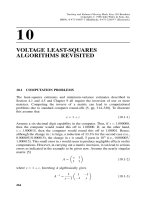
Two major patterns of plaque disruption are plaque rupture and plaque erosion (Figure 1).
Plaque rupture is caused by fibrous cap disruption, allowing blood to come in contact with
the thrombogenic necrotized core, resulting in thrombus formation. Ruptured plaque is
characterized by disruption of thin fibrous caps, usually less than 65 μm in thickness, rich in
macrophages and lymphocytes, and poor in SMCs (Virmani et al., 2000). Thus, the thinning
of the fibrous cap is though to be a state vulnerable to rupture, the so-called thin-cap
fibroatheroma (Kolodgie et al., 2001). However, the thin-cap fibroatheroma is not taken into
Pathology and Pathophysiology of Atherothrombosis: Virchow’s Triad Revisited
3
account in the current American Heart Association classification of atherosclerosis (Stary et
al., 1995). Plaque erosion is characterized by a denuded plaque surface and thrombus
formation, and defined by the lack of surface disruption of the fibrous cap. Compared with
plaque rupture, patients with plaque erosion are younger, no male predominance.
Angiographycally, there is less narrowing and irregularity of the luminal surface in erosion.
The morphologic characteristics include an abundance of SMCs and proteoglycan matrix,
expecially versican and hyaluronan, and disruption of surface endothelium. Necrotic core is
often absent. Plaque erosion contains relatively few macrophages and T cells compared with
plaque rupture (Virmani et al., 2000). Thrombotic occlusion is less common with plaque
erosion than plaque rupture, whereas microembolization in distal small vessels is more
common with plaque erosion than plaque rupture (Schwartz et al., 2009). The proportions of
fibrin and platelets differ in coronary thrombi on ruptured and eroded plaques. Thrombi on
ruptured plaque are fibrin-rich, but those on eroded plaque are platelet-rich. TF and C
reactive protein (CRP) are abundantly present in ruptured plaque, compared with eroded
plaques (Sato et al., 2005). These distinct morphologic features suggest the different
mechanisms in plaque rupture and erosion.
500μm
500μm
100μm
100μm
100μm
100μm
G
PI
I
b/IIIa Fibrin
rupture
erosion
HE
Fig. 1. Human coronary plaque rupture and erosion in patients with acute myocardial
infarction.
Large necrotic core and disrupted thin fibrous cap is accompanied by thrombus formation
in ruptured plaque. Eroded plaque has superficial injury of SMC-rich atherosclerotic lesion
with thrombus formation. Both thrombi comprise platelets and fibrin. HE, Hematoxylin
eosin stain (from Sato et al. 2005, with permission).
3. Pathology of asymptomatic atherothrombus
On the other hands, the disruption of atherosclerotic plaque does not always result in
complete thrombotic occlusion with subsequent acute symptomatic events. The clinical
studies using angioscopy have revealed that multiple plaque rupture is a frequent
complication in patients with coronary atherothrombosis (Okada et al., 2011). Healed stages
Traditional and Novel Risk Factors in Atherothrombosis
4
of plaque disruption are also occasionally observed in autopsy cases with or without
coronary atherothrombosis (Burke et al., 2001). To evaluate the incidence and morphological
characteristics of thrombi and plaque disruption in patients with non-cardiac death, Sato et
al. (2009) examined 102 hearts from non-cardiac death autopsy cases and 19 from those who
died of acute myocardical infarction (AMI). They found coronary thrombi in 16% of cases
with non-cardiac death, and most of them developed on plaque erosion, and the thrombi
were too small to affect coronary lumen (Figure 2, Table 1). The disrupted plaques in non-
cardiac death case had smaller lipid areas, thicker fibrous caps, and more modest luminal
narrowing than those in cases with AMI. A few autopsy studies have examined the
incidence of coronary thrombus in non-cardiac death. Davies et al. (1989) and Arbustini et
al. (1993) found 3 (4%) mural thrombi in 69, and 10 (7%) thrombi in 132 autopsy cases with
non-cardiac death. The all coronary thrombi from non-cardiac death were associated with
plaque erosion (Arbustini et al., 1993). Although the precise mechanisms of plaque erosion
remain unknown, it is possible that the superficial erosive injury is a common mechanism of
coronary thrombus formation. The results suggest that plaque disruption does not always
result in complete thrombotic occlusion with subsequent acute symptomatic events, that
thrombus growth is critical step for the onset of clinical events, and that at least the regional
factors influence the size of coronary thrombus after plaque disruption.
Fig. 2. Human coronary plaque erosion in patient with non-cardiac death.
No
n
-cardiac death
(n=102)
Acute m
y
ocardial infarctio
n
(n=19)
P value
Fresh thrombus 10 (10%) 14 (74%) <0.001
erosio
n
7 (7%) 4 (21%) 0.07
rupture 3 (3%) 10 (53%) <0.001
Old thrombus 6 (6%) 5 (26%) <0.05
(From Sato et al. 2009, with permission)
Table 1. Incidence of thrombosis in non-cardiac death and acute myocardial infarction.
Pathology and Pathophysiology of Atherothrombosis: Virchow’s Triad Revisited
5
The atherosclerotic lesion shows superficial erosive injury with mural thrombus (arrows).
The thrombus is too small to obstruct coronary lumen and induce symptomatic event
(hematoxyline eosin stain, from Sato et al. 2009, with permission).
4. Pathophysiology of atherothrombosis
4.1 Triggers on plaque disruption
As described above, atherothrombosis is initiated by plaque rupture or plaque erosion. The
plaque disruption is probably affected by vascular wall change and local blood flow. Our
recent study revealed that disturbed blood flow could trigger plaque erosion in rabbit
femoral artery with SMC-rich plaque. We separately discuss possible factors that affect
plaque rupture or plaque erosion in atherosclerotic vessels.
4.1.1 Vascular change in plaque rupture
The thinning and disruption of fibrous cap by metalloproteases together with local rheological
forces and emotional status is likely to be involved in plaque rupture. Accumulating evidence
supports a key role for inflammation in the pathogenesis of plaque rupture. The inflammatory
cells that appear quite numerous in rupture-prone atherosclerotic plaques can produce
enzymes degrading the extracellular matrix of the fibrous cap. Macrophages in human
atheroma overexpress interstitial collagenases and gelatinases, and elastolytic enzymes.
Activated T lymphocytes and macrophages can secrete interferon γ (INF-γ), which inhibits
collagen synthesis and induces apoptotic death of SMC (Shah, 2003). Moreover, INF-γ can
induce interleukine (IL)-18, an accelerator of inflammation. IL-18 is colocalized with INF-γ in
macrophage located at shoulder region, but not at necrotic core, and is associated with
coronary thrombus formation in patients with ischemic heart disease (Nishihira et al., 2007).
IL-10, an important anti-inflammatory cytokine, also is upregulated in macrophage in
atherosclerotic lesion from patients with unstable angina compared with stable angina
(Nishihira et al., 2006b). Heterogeneity of macrophages in atherosclerotic plaque could explain
the paradoxical findings (Waldo et al., 2008). These evidences indicate that the imbalance of
inflammatory pathway appear to participate in the destabilization of the plaque that triggers
thrombosis in fibrous cap rupture.
Other possible trigger of plaque rupture is intraplaque hemorrhage. The frequency of
previous hemorrhages is greater in coronary atherosclerotic lesions with late necrosis and
thin fibrous cap than those lesions with early necrosis or intimal thickening (Kolodgie et al.,
2003). Plaque hemorrhage is present in majority (>75%) of acute ruptures, and in 40% of
fibrous cap and thin-fibrous cap atheromas. In addition, intraplaque hemorrhage is more
frequently seen in patients with AMI compared to patients with healed myocardial
infarction or non-cardiac death (Virmani et al., 2003). In coronary culprit lesions obtained by
directional coronary atherectomy, intraplaque hemorrhage and iron deposition were more
prominent in patients with unstable angina pectoris than with stable angina pectoris. The
iron deposition correlated with oxidized low density lipoprotein and thioredoxin, an anti-
oxidant protein, and was also associated with thrombus formation (Nishihira et al., 2008b).
The pathological findings imply a possible relationship among intraplaque hemorrhage,
oxidative stress, and plaque instability. However, the direct evidence that links intraplaque
hemorrhage to plaque instability is still lacking.
Traditional and Novel Risk Factors in Atherothrombosis
6
4.1.2 Blood flow-induced mechanical stress on plaque rupture
Blood flow-induced mechanical stress is an essential factor of development of
atherosclerosis and atherothrombosis. The low shear stress and oscillatory shear stress are
both important stimuli for induction of atherosclerosis. Using a perivascular shear stress
modifier in mice, Cheng et al. (2006) revealed that low shear stress induces larger lesions
with vulnerable plaque phenotype (more lipids, more proteolytic enzymes, less SMCs, and
less collagen) whereas vortices with oscillatory shear stress induce stable lesions. Chatzizisis
et al. (2011) reported development of thin fibrous cap atheroma in coronary artery with low
shear stress in pigs. In addition, the shear stress-induced changes in atherosclerotic plaque
composition are modulated by chemokines. Inhibition of fractalkine, which is exclusively
expressed in the low shear stress-induced atherosclerotic plaque, was reduced lipid and
macrophage accumulation in the brachiocephalic arteries in mice (Cheng et al., 2007).
Therefore, lower shear stress can induce atherosclerotic lesion prone to plaque rupture.
Although it is well recognized that a mechanical stress triggers the disruption of fibrous cap,
it remains unclear which factor is mainly responsible for the disruption of the thin fibrous
cap. A variety of mechanical factors have been postulated to play a role in plaque rupture,
including hemodynamic shear stress, turbulent pressure fluctuation (Loree et al., 1991),
sudden increases in intraluminal pressure (Muller et al., 1989), and tensile stress
concentration within the wall of the lesion. To investigate the relationship between shear
stress distribution and coronary plaque rupture, Fukumoto et al. (2008) analyzed 3-
dimmensional intravascular ultrasound images in patients with acute coronary thrombosis
by a program for calculating the fluid dynamics. The ruptured sites were located in the
proximal or top portion of the plaques, and the localized high shear stress is frequently
correlated with the rupture sites. This finding is inconsistent with role of low shear stress on
atherogenesis. It is possible that the process of initiating plaque rupture is quite different
form that of atherogenesis. On the other hand, an excessive concentration of tensile stress
within the plaque may be one of the triggers of plaque rupture. When the tensile stress
becomes greater than the fragility of the fibrous cap, a plaque disruption may be initiated.
The tensile stress is increased by development of a lipid core, thinning of the fibrous cap
(Loree et al., 1992). Cheng et al. (1993) analyzed the distribution of circumferential stress in
human coronary arteries. The maximum circumferential stress in ruptured plaques was
significantly higher than that in stable plaques, although plaque rupture does not always
occur at the region of highest stress. These results suggest that a mechanical factor that
triggers plaque rupture differ in each case and lesion.
4.1.3 Disturbed blood flow on plaque erosion
Although it has been postulated that erosions result from coronary vasospasm of SMC-rich
plaque, the mechanisms of plaque erosion are poorly understood. Approximately 80%
thrombi of plaque erosion are nonocclusive in spite of sudden coronary death (Virmani et
al., 2000). Platelet rich emboli are found in 74% of patients dying suddenly with plaque
erosion compared with plaque rupture (40%). Because activated platelets release
vasoconstrictive agents, such as 5-hydroxytriptamine (5-HT, serotonin) and thromboxane
A2, these emboli might increase peripheral resistance leading to alteration of coronary blood
flow. 5-HT can induce vasoconstriction and reduce coronary blood flow in human
atherosclerotic vessels but not in normal arteries (Golino et al., 1991).
Pathology and Pathophysiology of Atherothrombosis: Virchow’s Triad Revisited
7
Experimental aortic stenosis can induce acute endothelial change or damage of the normal
aorta (Fry, 1968). Therefore, hemodynamic force, particularly disturbed blood flow induced
by stenosis or vasoconstriction, could be a crucial factor in generating surface vascular
damage and thrombosis. To address the relation between disturbed blood flow and plaque
erosion, we investigated the pathological change after acute luminal narrowing in SMC-rich
plaque in rabbit. The SMC-rich plaque was induced by a balloon injury of rabbit femoral
artery, and expressed TF as human atherosclerotic plaques. Actually, the disturbed blood by
acute vascular narrowing induced superficial erosive injury to the SMC-rich plaque at post
stenotic regions in rabbit femoral arteries. Figure 3 shows microscopic images of the
longitudinal section of the neointima at the post- stenotic region 15 min after vascular
narrowing. The endothelial cells and SMCs at this region were broadly detached with time,
and associated with platelet adhesion to the sub-endothelium. Apoptosis of endothelial cells
Fig. 3. Representative images of superficial erosive injury of SMC-rich plaque and thrombus
formation at the post-stenotic region.
SMC-rich plaque 15 min after vascular narrowing shows endothelial detachement (small
arrows) accompanies platelet adhesion (arrow heads) at 1mm form vascular narrowing (A,
hematoxyline eosin stain). Detachment of endothelial cells and exposure of subendothelial
matrix is accompanied by platelet aggregation on the left side, and residual endothelial cell
layer is present on right side (inset, high magnification of aggregated platelets) (B. scanning
electron microscopy). Immunohistochemistry for VWF (C, a marker of endothelium) or
smooth muscle actin (D, a marker of SMC) confirm detachment of endothelial cells and
SMCs at post stenotic region (from Sumi et al. 2010, with permission).
Traditional and Novel Risk Factors in Atherothrombosis
8
and superficial SMCs was also observed at the post- stenotic region within 15 minutes (Sumi
et al., 2010). The vascular narrowing induced large mural thrombi which composed of
platelets and fibrin, as human plaque erosion. Thus, disturbed blood flow can induce
superficial erosive injury to SMC-rich plaque and thrombus formation at post stenotic
region. Computational fluid simulation analysis indicated that oscillatory shear stress
contributes to the development of the erosive damage to the plaque (Sumi et al., 2010).
Although direct clinical evidence has not yet supported the notion that coronary artery
vasospasm plays a role in plaque erosion, the superficial erosive injury of SMC-rich plaque
by disturbed blood flow is similar to those of human plaque erosion (Sato et al., 2005). And,
platelet and blood coagulation in coronary circulation are activated after vasospastic angina
(Miyamoto et al. 2001, Oshima et al., 1990). Therefore, these evidence suggest that an acute-
onset disturbed blood flow due to vasoconstriction could trigger plaque erosion.
Hemodynamic factors could play an important role in development of plaque erosion.
4.2 Virchow’s triads on thrombus growth
As described above, plaque disruption does not always result in complete thrombotic
occlusion. Thrombus growth is considered critical to the onset of clinical events. Although
thrombus formation is regulated by the vascular wall thrombogenicity, local blood flow,
and blood contents, their contribution to thrombus growth has not been clearly defined. We
separately discuss three factors that affect thrombus growth in atherosclerotic vessels.
4.2.1 Vascular factors on thrombus growth
Most fundamental difference between normal artery and atherosclerotic artery is presence
of abundant active TF in atherosclerotic lesions (Hatakeyama et al., 1997, Wilcox et al., 1989).
It seems that vascular wall TF contribute to thrombus size/propagation on atherosclerotic
lesions. However, recent studies indicate that a small amount of TF is detectable in the blood
and is capable of supporting clot formation in vitro. Plasma TF levels are elevated in
patients with unstable angina and AMI and correlate with adverse outcomes (Mackman,
2004). Therefore, it is still controversial whether vascular wall and/or blood-derived TF
support thrombus propagation. Hematopoietic cell-derived, TF-positive microparticles
contributed to laser injury-induced thrombosis in the microvasculature of mouse cremaster
muscle (Chou et al. 2004). In contrast, vascular smooth muscle-derived TF contributed to
FeCl
3
induced thrombosis in mouse carotid artery (Wang et al., 2009). We investigated
whether plaque and/or blood TF contribute to thrombus formation in rabbit femoral artery
with or without atherosclerotic lesions. The atherosclerotic lesions in rabbit femoral arteries
were induced by a 0.5% cholesterol diet and balloon injury, and showed TF expression and
increased procoagulant activity compared with normal femoral arteries (Figure 4). Balloon
injury of the atherosclerotic plaque induced thrombin-dependent large platelet-fibrin
thrombi. In contrast, balloon injury of normal femoral artery induced thrombin-independent
small platelet thrombi (Figure 5). Moreover, whole blood coagulation in the rabbits was not
affected by blood TF inhibition with a TF antibody even in hyperlipidemic condition
(Yamashita et al., 2009). Therefore, at least, atherosclerotic plaque-derived TF might
contribute to activation of intravascular coagulation cascade and thrombus
size/propagation on atherosclerotic lesions.
Pathology and Pathophysiology of Atherothrombosis: Virchow’s Triad Revisited
9
HE/VB
SMC
Macrophage
TF
100μm
100μm
A
B
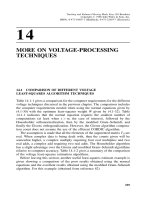
Fig. 4. Histological images of rabbit femoral arteries.
Representative immunohistochemical microphotographs of normal (A) and balloon-injured
femoral artery at 3 weeks after injury under 0.5% cholesterol diet (B). Atherosclerotic lesion
composed of SMCs and macrophages develops in injured artery. TF expression is present in
the lesion and adventitia of both arteries. HE/VB, hematoxyline eosin/Victoria blue stain
(From Yamashita et al. 2009, with permission).
A
B
M
I
I
IEL
Fig. 5. Immunofluorescence images of thrombus on rabbit femoral artery.
Representative immunofluorescent microphotographs of thrombi 15 minutes after balloon
injury of normal femoral artery and of atherosclerotic plaque under 0.5% cholesterol diet.
Rows show differential interference contrast images, images stained with fluorescein
isothiocyanate-labeled GPIIb/IIIa (green), Cy3-labeled fibrin (red), and merged
immunofluorescent images. Areas with colocalized factors are stained yellow. The thrombi
on normal intima is composed of small aggregated platelet (A), while the thrombi on
atherosclerotic plaque is large, and composed of platelet and fibrin (B). I, intima; M, media;
IEL, internal elastic lamina. (From Yamashita et al. 2009, with permission).
Traditional and Novel Risk Factors in Atherothrombosis
10
Several factors can influence TF expression in plaques and atherothrombus formation after
plaque disruption. CRP is an inflammatory acute-phase reactant that has emerged as a
powerful predictor of cardiovascular disease (Ridker, 2007). CRP is localized in
atherosclerotic plaques and is more in thrombotic plaques than non-thrombotic ones
(Ishikawa et al., 2003, Sun et al., 2005). The findings imply that CRP is implicated in
atherothrombogenesis. To address this issue, CRP-transgenic rabbits were generated,
because as human CRP, CRP in rabbits but not in mice works as an acute-phase reactant
during inflammation (Koike et al., 2009). In the rabbits, CRP was overexpressed in livers and
circulated in blood and deposited in the both SMC-rich and macrophage-rich atherosclerotic
lesions. The thrombus size on SMC-rich plaque or macrophage-rich plaque after balloon
injury was significantly increased in CRP-transgenic rabbits as compared with wild non-
transgenic rabbits (Figure 6). TF expression and its acivity in the plaques were significantly
increased in CRP-transgenic rabbits. The degree of CRP deposition correlated with TF
expression in plaques and thrombus size on injured plaques (Matsuda et al., 2011). On the
100μm
HE
GP IIb/IIIa
Fibrin
100μm
Non-trans
g
enic Rb
CRP-trans
g
enic Rb
Fig. 6. Thrombus formation on SMC-rich plaque in CRP-transgenic or non-transgenic rabbit
femoral artery.
The images show thrombus formation on SMC-rich plaque (arrows) 15 min after balloon
injury of rabbit femoral arteries. The thrombus size is increased in CRP-transgenic rabbits as
compared with non-transgenic rabbit. Immunopositive areas for GPIIb/IIIa and fibrin also
increase in CRP-transgenic rabbit (from Matsuda et al. 2011, with permission).
Pathology and Pathophysiology of Atherothrombosis: Virchow’s Triad Revisited
11
other hand, the CRP overexpression did not enhance atherosclerosis induced by
hyperchoresterol diets (Koike et al., 2009). CRP localized in atherosclerotic plaques might
enhance vascular wall thrombogenicity and thrombus formation after plaque disruption
rather than atherogenesis.
4.2.2 Altered blood flow on thrombus growth
Blood flow is a key modulator of thrombus growth. Clinical studies revealed an alteration of
coronary blood flow in patients with ischemic heart diseases. Marzilli et al. (2000) reported
an approximate 80% reduction in coronary blood flow during ischemia in patients with
unstable angina. An autopsy study reported that intramyocardial microemboli were
frequently present in sudden coronary death patients (Schwartz et al. 2009). Distal
microvascular embolism and/or vasoconstriction could affect blood flow alteration and
thrombus formation and growth at the culprit lesions (Erbel & Heusch, 2000). To assess the
issue, we examined the effects of the blood flow reduction to thrombus formation in our
animal model. Blood flow reduction (>75%) promoted the growth of thrombus, a mixture of
platelets and fibrin, on atherosclerotic lesion, which grew to occlusive one. The flow
reduction also induced thrombus formation on normal arteries, but the thrombi were very
small and composed only of platelets (Yamashita et al. 2004). Therefore, blood flow
reduction associated with increased vascular wall thrombogenecity is considered to
contribute thrombus growth. We also demonstrated an important role of 5-HT
2A
receptor on
platelets and SMCs in this process via platelet aggregation and thrombogenic
vasoconstriction (Nishihira et al., 2006a, 2008a).
In addition to distal vascular resistance, disturbed blood flow by acute vascular narrowing
promotes thrombus growth at post stenotic regions. As described above, vascular narrowing
of rabbit femoral artery induced superficial erosive injury to SMC-rich plaque at post
stenotic regions. The thrombi consisted of a mixture of aggregated platelets and a
considerable amount of fibrin. The whole blood hemostatic parameters in the rabbits was
not changed after vascular narrowing or anti-rabbit TF antibody treatment, which evidence
indicates that TF derived from eroded plaque rather than circulating TF plays an important
role in fibrin generation and thrombus growth (Sumi et al. 2010).
The rheological effect on thrombus growth may be partly explained by a shear gradient-
dependent platelet aggregation mechanism. Using in vitro and in vivo stenotic microvessels
and imaging systems, Nesbitt et al. (2009) revealed a shear gradient-dependent platelet
aggregation process which is preceded by soluble agonist-dependent aggregation. Shear
microgradient at post stenosis region or down stream face of thrombi induced stable
platelets aggregates, and the shear microgradients directly influenced the platelet
aggregation size. This process required ligand binding to integrin αIIbβ3, transient Ca
2+
flux,
but did not required global platelet shape change or soluble agonists. The findings suggest
that platelets principally use a biomechanical platelet aggregation mechanism in early phase
of platelet adhesion and aggregation. Vessel and/or thrombus geometry itself may promote
thrombus formation.
4.2.3 Blood factors on thrombus growth
As described above, platelet is a major cellular component in coronary thrombus, and
platelets play an important role in growing phase of thrombus formation, as well as initial
Traditional and Novel Risk Factors in Atherothrombosis
12
phase of thrombus formation. Adhesion molecules and its receptors on platelets are
essential for thrombus formation, because these molecules support platelet tethering, firm
adhesion, aggregation and platelet recruitment to thrombus surface. VWF is a large,
multimeric, plasma protein that undergoes a conformational change when bound to matrix
under permit its binding to GPIbα. Recent studies in vitro and in vivo showed that platelet
recruitment on thrombus surface was primary mediated by VWF and GPIbα on flowing
platelets (Bergmeier et al. 2006, Kulkuni et al. 2000). We demonstrated that a large amount
of VWF was localized in coronary thrombi in patients with AMI (Nishihira et al., 2010,
Yamashita et al., 2006a), and that monoclonal antibody against VWF A1 domain, which
interacts platelet GPIbα, significantly suppressed formation of platelet-fibrin thrombi and
completely inhibited occlusive thrombus formation in rabbit atherosclerotic lesions
(Yamashita et al., 2003, 2004). These findings indicated a crucial role of VWF in thrombus
growth via platelet recruitment. The multimer size of VWF can affect thrombus size and is
regulated by a plasma protease, a disintegrin and metalloprotease with a thrombospondin
type 1 motif 13 (ADAMTS-13). A deficiency of ADAMTS-13 activity causes an increased
level of circulating ultralarge VWF multimers, and correlates with the onset of the general
thrombotic disease, thrombotic thrombocytopenic purpura (TTP). A clinical evidence
suggested dysregulation of VWF multimer size in AMI patient. The ratio of
VWF/ADAMTS-13 antigen was higher in patients with AMI than in those with stable
angina pectris, and there was a inverse correlation between plasma VWF antigen and
ADAMTS-13 activity in AMI patients (Kaikita et al. 2006). The ADAMTS-13 closely localized
with VWF in fresh coronary thrombi from AMI patients (Moriguchi-Goto et al., 2009). A
reducing ADAMTS-13 activity by monoclonal antibody against distintegrin-like domain
enhanced platelet thrombus growth on immobilized type I collagen at a high shear rate
(1500S
-1
) and platelet-fibrin thrombus formation on injured atherosclerotic lesion of rabbit
femoral arteries (Moriguchi-Goto et al., 2009). The study also showed cleavage of large sized
VWF multimer during platelet thrombus formation under a high shear rate. The VWF-
cleaving site by ADAMTS-13 localized on the surface of platelet thrombus, and the
ADAMTS-13 activity was shear dependent manner (Shida et al. 2008). Thus, ADAMTS-13
may work at the site of ongoing thrombus generation and limit thrombus growth.
The recent studies in vitro showed various blood cells, not only monocytes but also
neutrophils, eosinophils, and even if platelets, can synthesize TF. Although there is much on
debate on the TF expression in blood cells, it is likely that monocytes are the only blood cells
that synthesize and express TF (Østerud, 2010). A related topic is contribution of
microparticles (MPs) to thrombus formation. MPs are small fragments of membrane-bound
cytoplasm that are shed from the surface of an activated or apoptotic cells (Blann et al. 2009).
The procoagulant activity of MPs is increased with the exposure of phosphatidylserine and
the presence of TF. In fact, MPs have significantly elevated in acute coronary syndrome and
ischemic strokes (Geiser et al. 1998, Singh et al. 1995). However, it is still unclear whether the
elevated levels of MPs are a cause or consequence of atherothrombosis. Moreover, our
animal studies did not support the role of blood-derived TF in atherothrombus formation as
described above. Future studies are required to clarify contribution of blood derived TF
and/or MPs to thrombus propagation on atherosclerotic lesions.
Among the white blood cells, neutrophils are mostly found in coronary thrombus in
patients with AMI, and CD34 positive leukocytes are also found in the thrombus (Nishihira
et al., 2010). Recent evidences revealed neutrophils and endothelial progenitor cells
influence thrombus growth. Neutrophils can positively or negatively affect thrombus
Pathology and Pathophysiology of Atherothrombosis: Virchow’s Triad Revisited
13
formation by degradation of coagulation or fibrinolysis factors and promoting platelet
function (Kornecki et al., 1988, Moir et al., 2002). Inhibition of interaction between p-selectin
and p-selectin glycoprotein ligand 1 reduced fibrin formation in vivo (Palabrica et al., 1992).
These adhesion molecules have been implicated in recruitment of leukocytes and leukocyte
MPs to thrombi (Vandendries et al., 2004). To reveal the neutrophil-mediated procoagulant
mechanisms, Massberg et al. (2010) investigated thrombus formation using neutrophil
elastase and cathepsin G deficient mice. Proteolysis of TFPI by these proteases enhanced
fibrin and thrombus formation after FeCl
3
-induced vessel injury. In addition, activated
platelets by collagen accelerated nucleosome externalization by neutrophils. The neutrophil-
derived externalized nucleosomes can form neutrophil extracellular traps that provide a
scaffold for platelets and red blood cells and histone 3/4 can induce platelet aggregation
(Fuchs et al., 2010). On the other hands, neutrophil elastase has fibriolytic potential, and
there is significant correlation between neutrophil elastase-digested fibrin and leukocyte
content in human atherothrombi (Rábai et al., 2010). Zeng et al. (2002) investigated
contribution of polymorphonuclear leukocytes (PMNs) to fibrinolysis in vivo using
plasminogen deficient mice. The PMNs accumulated within the thrombi by 6 hours after
FeCl
3
-induced vessel injury and peaked at 24 hours. There were no significant differences
between the PMNs from plasminogen deficient mice and wild type mice within the 6 hour
after thrombus formation, whereas there was significant greater retention of PMNs within
the thrombus over 24 hours after thrombus formation. PMNs from both mice showed
fibrinolytic activity, but the degradation products were a distinct pattern. Therefore, it is
possible that neutrophils works as positive or negative regulator of early or late phase of
thrombus formation, respectively.
Endothelial progenitor cells (EPC) contributes to angiogenesis and wound healing (Asahara
et al., 1997), and the number of EPCs in blood is associated with cardiovascular risk (Hill et
al., 2003). The mechanisms that regulate mobilization, migration, and differentiation of EPCs
and their homing to sites of vascular injury are complex and involve several mediators and
receptors, such as P-selectin glycoprotein ligand-1, CXC chemokine, and integrins (Chavakis
et al., 2005, Massberg et al., 2006). Interaction of thrombus contents and EPCs influences
their mobilization and differentiation to mature endothelial cells during vascular injury (de
Boer HC et al., 2006). Abou-Saleh et al. (2009) reported that human peripheral blood
mononuclear cell derived EPCs bound platelets via p-selectin and inhibit platelet activation,
aggregation, and adhesion to collagen in vitro, and that injection of these EPCs reduced
thrombus formation after FeCl
3
-induced vessel injury of mouse carotid arteries.
Other possible mechanism contributing thrombus propagation in vivo is intrinsic
coagulation pathway. The intrinsic coagulation pathway is initiated when coagulation factor
XII (FXII) comes into contact with negatively charged surfaces in a reaction involving the
plasma proteins, high molecular mass kininogen and plasma kallikrein. Factor XI (FXI) is
activated by activated FXII, thrombin, and activated XI. Feedback activation of FXI by
thrombin promotes further thrombin generation in vitro (Gailani & Broze, 1991). FXI was
present in platelet-fibrin thrombus induced balloon injury of atherosclerotic lesion in
rabbits, and anti-FXI antibody reduced thrombus growth without prolonging bleeding
(Yamashita et al., 2006b). FXI plays an important role in thrombus growth via further
thrombin generation. On the other hand, there are conflicts of evidence that FXII supports
arterial thrombus growth. FXII deficient mice were resistant to thrombotic occlusion after
FeCl
3
induced vessel injury of carotid arteries (Cheng Q et al., 2010). However, a clinical
study demonstrated an inverse relationship between FXII level and risk of myocardial
Traditional and Novel Risk Factors in Atherothrombosis
14
infarction (Doggen et al., 2006). Moreover, inhibition of FXII did not change platelet
aggregation and fibrin formation on atherosclerotic plaque surface under flow in vitro. The
effect of FXII on coagulation became obvious only absence of TF (Reininger et al., 2010).
5. Conclusion
More than 150 years ago, Virchow described the mechanims of thrombus formation. It has
still remained as a fundamental theory of thrombus formation. To date, pathological and
experimental studies have clarified the mechanisms of atherothrombus formation. The
thrombus formation is initiated by plaque rupture and plaque erosion. Among the
Virchow’s triad, vascular and rheological factors are responsible for plaque rupture.
Disruption of thin fibrous cap atheroma triggers plaque rupture. On the other hand,
disturbed blood by acute luminal change can trigger plaque erosion to SMC-rich plaque.
Pathological findings of human atherothrombosis suggest that thrombus growth rather than
plaque disruption is a critical step for the onset of cardiovascular events, and that
simultaneous activation of coagulation cascade and platelets play an important role in
thrombus formation after plaque disruption. All three factors contribute to atherothrombus
growth. Our rabbit model of atherothrombosis revealed that excess thrombin generation
mediated by plaque TF contribute to large plate-fibrin thrombus formation on
atherosclerotic lesion, and that disturbed flow condition after plaque disruption promote
thrombus growth. Recent evidence suggests that leukocytes influence arterial thrombus
formation as well as platelet and coagulation/fibrinolysis factors. Differences between
hemostasis and thrombus growth may shed light on a novel anti-atherothrombogic drug
with a wide safety margin.
6. Acknowledgement
The work is supported in part by Grants-in-Aid for Scientific Research in Japan
(No.23790410), Mitsubishi Pharma Research Foundation, and Integrated Research Project for
Human and Veterinary Medicine.
7. References
Abou-Saleh, H., Yacoub, D., Théorêt, JF., Gillis, MA., Neagoe, PE., Labarthe, B., Théroux, P.,
Sirois, MG., Tabrizian, M., Thorin, E., Merhi, Y. (2009). Endothelial progenitor
cells bind and inhibit platelet function and thrombus formation. Circulation,
Vol.120, No.22, pp.2230-2239.
Arbustini, E., Grasso, M., Diegoli, M., Morbini, P., Aguzzi, A., Fasani, R., Specchia, G.
(1993). Coronary thrombosis in non-cardiac death. Coron Artery Dis, Vol.4, No.9,
pp.751-759.
Asahara, T., Murohara, T., Sullivan, A., Silver, M., van der Zee, R., Li, T., Witzenbichler, B.,
Schatteman, G., Isner, JM. (1997). Isolation of putative progenitor endothelial
cells for angiogenesis. Science. Vol.275, No.5302, pp.964-967.
Bergmeier, W., Piffath, CL., Goerge, T., Cifuni, SM., Ruggeri, ZM., Ware, J., Wagner, DD.
(2006). The role of platelet adhesion receptor GPIbalpha far exceeds that of its main
ligand, von Willebrand factor, in arterial thrombosis. Proc Natl Acad Sci U S A.
Vol.103, No.45, pp.16900-16905.
Pathology and Pathophysiology of Atherothrombosis: Virchow’s Triad Revisited
15
Blann, A., Shantsila, E., Shantsila, A. (2009). Microparticles and arterial disease. Semin
Thromb Hemost. Vol.35, No.5, pp.488-496.
Burke, AP., Kolodgie, FD., Farb, A., Weber, DK., Malcom, GT., Smialek J, Virmani R.
(2001). Healed plaque ruptures and sudden coronary death: evidence that
subclinical rupture has a role in plaque progression. Circulation, Vol.103, No.7,
pp.934-940.
Chatzizisis, YS., Baker, AB., Sukhova, GK., Koskinas, KC., Papafaklis, MI., Beigel, R., Jonas,
M., Coskun, AU., Stone, BV., Maynard, C., Shi, GP., Libby, P., Feldman, CL.,
Edelman, ER., Stone, PH. (2011). Augmented expression and activity of
extracellular matrix-degrading enzymes in regions of low endothelial shear stress
colocalize with coronary atheromata with thin fibrous caps in pigs. Circulation,
Vol.123, No.6, pp.621-630.
Chavakis, E., Aicher, A., Heeschen, C., Sasaki, K., Kaiser, R., El Makhfi, N., Urbich, C.,
Peters, T., Scharffetter-Kochanek, K., Zeiher, AM., Chavakis, T., Dimmeler, S.
(2005). Role of beta2-integrins for homing and neovascularization capacity of
endothelial progenitor cells. J Exp Med, Vol.201, No.1, pp.63-72Chou, J., Mackman,
N., Merrill-Skoloff, G., Pedersen, B., Furie, BC., Furie, B. (2004). Hematopoietic
cell-derived microparticle tissue factor contributes to fibrin formation during
thrombus propagation. Blood, Vol.104, No.10, pp.3190-3197.
Cheng, C., Tempel, D., van Haperen, R., van der Baan, A., Grosveld, F., Daemen, MJ., Krams,
R., de Crom, R. (2006). Atherosclerotic lesion size and vulnerability are determined
by patterns of fluid shear stress. Circulation, Vol.113, No.23, pp.2744-2753.
Cheng, C., Tempel, D., van Haperen, R., de Boer, HC., Segers, D., Huisman, M., van
Zonneveld, AJ., Leenen, PJ., van der Steen, A., Serruys, PW., de Crom, R., Krams,
R. (2007). Shear stress-induced changes in atherosclerotic plaque composition are
modulated by chemokines. J Clin Invest, Vol.117, No.3, pp.616-626.
Cheng, GC., Loree, HM., Kamm, RD., Fishbein, MC., Lee, RT. (1993). Distribution of
circumferential stress in ruptured and stable atherosclerotic lesions. A structural
analysis with histopathological correlation. Circulation, Vol.87, No.4, pp.1179-1187.
Cheng, Q., Tucker, EI., Pine, MS., Sisler, I., Matafonov, A., Sun, MF., White-Adams, TC.,
Smith, SA., Hanson, SR., McCarty, OJ., Renné, T., Gruber, A., Gailani, D. (2010).
A role for factor XIIa-mediated factor XI activation in thrombus formation in vivo.
Blood. Vol.116, No.19, pp.3981-3989.
Chou, J., Mackman, N., Merrill-Skoloff, G., Pedersen, B., Furie, BC., Furie, B. (2004).
Hematopoietic cell-derived microparticle tissue factor contributes to fibrin
formation during thrombus propagation. Blood, Vol.104, No.10, pp.3190-3197.
Crawley, J., Lupu, F., Westmuckett, AD., Severs, NJ., Kakkar, VV., Lupu, C. (2000).
Expression, localization, and activity of tissue factor pathway inhibitor in normal
and atherosclerotic human vessels. Arterioscler Thromb Vasc Biol, Vol.20, No.5,
pp.1362-1373.
Cunningham, KS., Gotlieb, AI. (2005). The role of shear stress in the pathogenesis of
atherosclerosis. Lab Invest
, Vol.85, No.1, pp.9-23.
Davies, MJ., Bland, JM., Hangartner, JR., Angelini, A., Thomas, AC. (1989). Factors
influencing the presence or absence of acute coronary artery thrombi in sudden
ischaemic death. Eur Heart J, Vol.10, No.3, pp.203-208.
Doggen, CJ., Rosendaal, FR., Meijers, JC. (2006). Levels of intrinsic coagulation factors and
the risk of myocardial infarction among men: Opposite and synergistic effects of
factors XI and XII. Blood, Vol.108, No.13, pp.4045-4051.