Tài liệu Magnetic resonance imaging and gynecological devices doc
Bạn đang xem bản rút gọn của tài liệu. Xem và tải ngay bản đầy đủ của tài liệu tại đây (232.89 KB, 6 trang )
Review article
Magnetic resonance imaging and gynecological devices
☆,☆☆
Lúcia Correia
a,
⁎
, Ana Beatriz Ramos
b
, Ana Isabel Machado
a
, Duarte Rosa
a
, Carlos Marques
a
a
Maternidade Dr. Alfredo da Costa, Lisbon, Portugal
b
Centro Hospitalar do Porto-Hospital Geral de Santo António, Oporto, Portugal
Received 22 November 2010; revised 16 October 2011; accepted 19 October 2011
Abstract
Background: Performing magnetic resonance imaging (MRI) on women with gynecological devices is a completely accepted practice. The
goal of our review is to assess how safe it is to perform MRI on women using contraceptive implants or devices.
Study Design: Literature review, searching in PubMed-Medline/Ovid for the following keywords: magnetic resonance imaging, intrauterine
devices, Implanon® and Essure®.
Results: Though plastic devices do not represent a contraindication to the use of the technique, those including metallic components have
been submitted to several tests, after which they were classified as MR Conditional (devices presenting no risks in MR-specific
environments) by the Food and Drug Administration. Thus, the use of MRI can be safely advised to women with this type of device as long
as the magnetic resonance equipment is ≤3.0 T.
Conclusions: Presently, there is no scientific evidence that contraindicates performing MRI on women with any kind of gynecological
device. Therefore, this procedure is safe as long as it is performed under previously tested conditions.
© 2012 Elsevier Inc. All rights reserved.
Keywords: Magnetic resonance imaging; Intrauterine devices; Implanon®; Essure®
1. Introduction
Magnetic resonance imaging (MRI) is an increasingly
popular imaging technique, having become one of the
preferred techniques due to its several advantages over other
methods, namely, (a) its multiplane capability, allowing for
the capture of cuts or layers in all directions in space, (b) its
high contr ast resolution and (c) the absence of known
harmful effects since ionizing radiation is not employed.
The differentiation of pelvic organs through contrast, an
exclusive feature of MRI, has rendered it the technique of
choice as far as exploring the pelvic cavity is concerned. The
correct interpretation of MRI images requires an under-
standing of the basic mechanisms necessary for image
formation. In its more basic form, MRI can be analyzed in
terms of energy transference [1]. Magnetic resonance is the
physical feature shown by the nuclei of some elements
which, when submitted to a strong magnetic field and excited
by radio waves of a particular frequency, will broadcast a
radio signal, which can then be captured by an antenna
and converted into an image [2,3].
Hydrogen atoms are the most common and the simplest in
the human body since their nuclei consist of a single proton.
This proton exhibits a feature called spin, which is essentially
a rotational motion, similar to how the earth rotates around its
own axis [2]. Thus, the magnetic field is the result of an
electric charge in mot ion. Because of this behavior, the
hydrogen proton is the most suitable for extracting MRI
images due to its abundant presence in the human body and
the capability to broadcast the strongest radio signal of all
stable nuclei.
Under normal conditions, the protons in the body exhibit
random orientation; however, if submitted to the influence of
an external magnetic field, the spins become aligned in either
the direction of the magnetic field or the opposite direction.
In fact, the number of protons which become aligned with
the magnetic field is a little higher than the rest. This fact
results in a small magnetization, powerful enough to
Contraception 85 (2012) 538 – 543
☆
No funding was provided for this study.
☆☆
Declaration of interest: The authors report no conflicts of interest.
The authors alone are responsible for the content and writing of the paper.
The authors stated no financial relationship to disclose.
⁎
Corresponding author. Praça das Flores N°3 2°Dto, 2526-419 Forte
da Casa, Portugal. Tel.: +35 1934236134.
E-mail address: (L. Correia).
0010-7824/$ – see front matter © 2012 Elsevier Inc. All rights reserved.
doi:10.1016/j.contraception.2011.10.011
broadcast an MRI signa l [3]. When the radio frequency is
turned off, the nuclei release themselves from the energy
they absorbed. Each body tissue reemits radiation at a
different rate, according to its own chemical composition and
physical status. This radiation is then captured by an antenna,
which converts it to electric current. Finally, the current is
used to create the desired images. The variation in the several
tissues' reemission times enables the creation of an image
which shows the contrast between them.
It is important to point out the strength of the magnetic
field created by the huge magnets present in MRI devices.
Magnetic fiel ds are usually measured in tesla units (T);
another measurement unit normally used is the gauss (1
T=10.000 gauss) [3]. The magnets in use today in MR are in
the 0.5–3.0-T range, or 5000 to 30,000 gauss. MRI uses
gigantic magnets, which are capable of creating magnetic
fields with an intensity varying from 0.2 to 9.4 T [4].
Nowadays, MRI for medical diagnosis employs devices
ranging from 0.5 to 3.0 T. For comparison purposes, the
earth's magnetic field is approximately 0.00005 T, exhibit-
ing slight variations around the equator and the poles.
As is the case with other imaging methods, MRI is subject
to several types of artifacts, which can compromise image
quality and interfere in its interpretation. Thus, it is necessary
to know the different types of artifacts, distinguishing them
from anatomical variations and pathological processes.
Artifacts can result from either data acquisition and treatment
or the patient's own features [5]. One of the most relevant
patient-related artifacts is the so-called magnetic suscepti-
bility arti fact. The magnetic susceptibilit y of a tissue
showcases i ts capability to acquire self-magnetization
when submitted to a magnetic field. Such acquired
magnetization may be concordant (parallel) or discordant
(antiparallel). In the presence of the former, it is said that a
substance exhibits positive magnetic susceptibility, thus
strengthening the resulting magnetic field. Such a substance
is called paramagnetic (substances which show strong
positive magnetic susceptibility are called superparamag-
netic or ferromagnetic). In the presence of discordant
(antiparallel) magnetization, the substances are classified as
exhibiting negative magnetic suscep tibility and are called
diamagnetic. This magnetic susceptibility artifact is com-
monly found in the presence of air, metal, calcium or a
concentrated gadolinic contrast medium. As far as the image
is concerned, it can be identified as a hypointensity of focal
signal, surrounded by a hyperintense halo, which can be
associated to several levels of distortion in the surrounding
tissue [5]. The size and shape of the artifact depend on the
size, shape, orientation and nature of the metal, as well as the
sequences used in the exam [6]. The artifact created by a
ferromagnetic object is larger than that originated by a
nonferromagnetic object [7] — the stronger the ferromag-
netic nature of the object, the more intense the registered
signal will be. Technically speaking, magnetic susceptibility
artifacts are more prominent in gradient-echo and echo-
planar sequences, and we may try to reduce them using a
smaller voxel, shorter echo time, or larger bandwidth or
even performing the exam with equipment featuring a
lower-intensity magnetic field [6].
Due to the evolution and increasingly common usage of
medical devices, some of which incorporate metallic
components, the use of MRI in patients with such devices
can lead to concern. Safety in the application of MRI on
women using gynecological devices depends, essentially,
on the device's structure.
For women using intrauterine devices (IUDs), pondered
images in T2 allow us to observe three different areas of
the uterus: the endometrium, with a high intensity signal; the
junction area (junction between the endometrial mucosa
and the myometrium), if we employ a low-frequency signal;
and the myometrium, using a medium- intensity signal.
IUDs must be correctly placed inside the uterine cavity and
are visualized in MRI images as areas without signal — their
shape depends on their orientation relative to the magnetic
field. Being external to the patient's tissue, they are
considered external devices. The description of the different
materials present in IUDs will be approached more
thoroughly throughout this paper, though we find it relevant,
due to the aforementioned facts, to discuss the safety of
MRIs in the presence of such devices, particularly those
whose compositions include copper.
Since we favor the patient's well-being over image
degradation, it is necessary to know the type of material
being studied. In fact, in addition to the artifacts generated
by metallic objects, which can decrease the diagnostic
capability of MRI, when submitting a patient to an external
magnetic field, we may cause dislocation, torsion or over-
heating of these devices, which in turn can provoke
injuries [8].
Any complaint of pelvic pain should result in the
immediate suspension of the exam. The most common,
though seldom described, consequences include the burning
of adjacent tissues, incorrect placement of contraceptive
devices or, more seri ously, uterine perfor ation in women
using IUDs.
Recent studies reveal that gynecological devices generate
minimal artifacts and that MRI can be safely performed since
the majority of these devices do n ot exhibit relevant
ferromagnetic proprieties.
Thus, this paper intends to review the literature on the
topic of safety regarding the use of MRI on women with
contraceptive devices or implants.
2. Material and methods
The authors researched in PubMed-Medline/Ovid using
the following keywords: Magnetic Resonance Imaging,
Intrauterine Devices, Implanon® and Essure®. This article
presents the review of data published between 1985 and
2010 on the application of magnetic resonance on women
using gynecological devices.
539L. Correia et al. / Contraception 85 (2012) 538–543
3. Results
3.1. Medi cal devices
The increasing use of MRI implied a greater demand for
information regarding the safety of its application to patients
with implants or other medical devices. As such, the Food
and Drug Administration (FDA) has acknowledged the need
for conduct studies in order to clarify these issues. Over the
last few years, testing methods were developed by several
organizations, e.g., the American Society for Testing and
Materials (ASTM), in order to assess the presence of
movement/deflection, torsion, a rise in the device's temper-
ature due to radiofrequency or the creation of image artifact s.
In 1997, the FDA's Center for Devices and Radiological
Health (CDRH) proposed the first classification method,
which splits devices into two groups: MR Safe (devices
which have been shown not to increase the risk for patients,
though potentially compromising the quality of diagnostic
information) and MR Compatible (devices which do not
increase the risk for patients and also do not compromise the
quality of the diagnostic information collected). According
to this terminology, testing “MRI safety” required in vitro
tests in order to assess static magnetic field interactions, MR-
related heating and, in some cases, induced electrical
currents. In order to test “MR compatibility,” the assessment
and description of artifacts are also required, in addition to
the above-mentioned tests [4,9].
After a while, the CDRH's classification method proved
confusing, which could potentially lead to accidents and
injured patients; therefore, as of August 2005, the ASTM
published the classification method currently in use, which
defines three groups [4,9]:
1. MR Safe: devices presenting no risks in all MR
environments. This group includes devices made of
nonconductive and nonmagnetic elements, e.g., plas-
tic, silicone or glass devices.
2. MR Conditional: devices presenting no risks in MR-
specific environments, under specific use conditions.
Field conditions that define the MR environment
characterization include static magnetic field
strength, spatial gradient, time rate of change of the
magnetic field, radiofrequency fields and specific
absorption rate.
3. MR Unsafe: devices presenting risks in all MR
environments. P erforming MR in these cases is
contraindicated. This group includes all electromag-
netic devices.
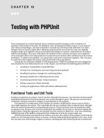
Each group was represented graphically (Fig. 1).
The “MR Safe” icon can be represented by the acronym
“MR” in green lettering inside a white square with green
borders or by the acronym “MR” written in white over a
green square. The “MR Conditional” icon consists of the
acronym “MR” in black lettering inside a yellow triangle
with black borders. The “MR Unsafe” icon consi sts of the
acronym “MR” in black lettering over a white background
inside a red circle with a red diagonal bar.
3.2. Gynecological devices
3.2.1. IUDs
IUDs are an effective contraceptive method used
worldwide.
These devices can consist of nonmetallic elements, like
the levonorgestrel-releasing intr auterine system (LNG-IUS)
(Bayer HealthCare Pharmaceuticals Inc.), or a combination
of metallic and nonmetallic elements. In the latter case,
copper is the most commonly used metal.
Due to its polyethylene structure, the LNG-IUS is
included in the group of MR Safe devices, according to the
ASTM. Therefore, its use does not pose any risk to women
in case MRI is performed.
Even though copper is not ferromagnetic, some
concerns regarding the application of MRI on women
using a copper IUD (Cu-IUD) have emerged since the
presence of a device including a metal component in a
patient submitted to MRI can lead to injuries deriving from
its movement/deflection or from an increase in the device's
temperature; in the Cu-IUD's case, this could result in
Fig. 1. Icons recommended by the ASTM. Legend: A, MR Safe; B, MR
Conditional; C, MR Unsafe.
540 L. Correia et al. / Contraception 85 (2012) 538–543
injuries to the endometrium, in addition to the possibility
of generating image artifacts which would compromise the
MRI's diagnostic capability [8].
Until the 1980s, healthcare providers' concerns, associ-
ated to a lack of guidelines and of previous studies, led many
medical centers to contraindicate the performance of MRI
in the presence of a Cu-IUD. However, due to the increasing
use of MRI, women with Cu-IUD started to be submitted to
this imaging technique when there was suspicion of pelvic
disease or, more frequently, extrapelvic disease.
In 1987, Mark and Hricak [10] carried out in vitro and in
vivo studies using RMI 0.35 T and 1.5 T on women with a
nonmetallic IUD (Lippes Loop Intrauterine Double-S; Ortho
Pharmaceutical, Raritan, NJ, USA) and a Cu-IUD (Cu-7;
Searle Pharmaceuticals, Chic ago, IL, USA) in order to assess
the possible occurrences of movement, IUD temperature
increase and generation of image artifacts during the MRI
performance. They have concluded that performing MRI on
those women was a safe procedure since there was no
rotation or deflection of either metallic or nonmetallic IUDs,
nor were there statistically significant differences in
temperature, when compa red to the placebo. Furthermor e,
no changes in image quality were recorded. Removing the
IUD for the single purpose of performing a safe MRI was no
longer justified. According to the same authors, and even
though ultrasonography is still the main imaging metho-
dology for the detection of a dislocated or extrauterine
IUD, the MRI can be considered an alternative in cases
where an ultrasonography is technically contraindicated or
suboptimal, such as for overweight women or for those with
a retroverted uterus.
Research in PubMed-Medline/Ovid using the keywords
“Magnetic Resonance” and “Intra-Uterine Device” allowed
us to find three more articles related to in vitro studies carried
out in order to assess the safety of performing MRI on
women with four different Cu-IUDs [11–13].
Using a 1.5-T MRI, Hess et al. [11] assessed the safety
of three different IUDs — Multiload® CU375 (Noury-
pharma, Oberschleissheim, Germany), Nova-T® (Schering
AG, Berlin, Germany) and Gyne-T® (Cilag, Sulzbach,
Germany) — while Pasquale et al. [12] tested the ParaGard®
CuT380A (Ortho-McNeil Pharmaceutical Corp., Raritan,
NJ, USA). More recently, Zieman and Kanal [13] reassessed
the usage safety of the ParaGard® CuT380A IUD on a 3.0-T
MRI system.
The results obtained by the different authors corroborated
and supported those of Mark and Hricak's studies [10],
showing that the application of MRI on women using Cu-
IUDs did not produce relevant effects (movement/deflection,
torsion, temperat ure rise, image artifacts) which would
contraindicate the use of MRI up to 3.0 T in women using
this kind of device (Table 1). Hess et al. [11] recorded a
maximum temperat ure increase in the IUD location that
varied between 0.3 and 0.4°C.
Even though the presence of a metallic component may
influence the image, due to the slight magnetic susceptibility
of copper and the small size of the devices, the artifacts
produced are not particularly relevant and do not interfere
with the interpretation of the collected images [8,10].
IUDs are captured in MR images as no-signal zones
inside the uterine cavity, assuming different structures
according to the type of analyzed IUD and the cutting
plane [10].
3.2.2. Essure®
The Essure® system (Conceptus Inc., San Carlo, CA,
USA) consists of two metallic microimplan ts and is an
increasingly popular female sterilization method. Each
implant includes an internal stainless steel section, wrapped
in polyethylene fibers, as well as an external section made of
26 expandable nickel and titanium spirals, with an initial
diameter of 0.8 mm, reaching 1.5 to 2 mm in order to fit the
Table 1
Effects of MRI on women using Cu-IUD
Article Year Tests Cu-IUD MRI Variables tested Conclusion
Intrauterine contraceptive devices:
MR imaging
Mark and Hricak [10]
1987 In vitro Lippes Loop Intrauterine
Double-S
Cu-7
0.35 T
and
1.5 T
Movement
Heat
Artifacts
IUD does not move under the influence
of the magnetic field, does not heat and
does not produce artifacts in vitro or in
vivo.In vivo Retrospective review of six MR
images of women with Cu-IUD
Safety of intrauterine contraceptive
devices during MR imaging
Hess et al [11]
1996 In vitro Multiload Cu 375
NovaT
GyneT
1.5 T Deflection
Heat
Maximum temperature rise of 0.4°C.
Lack of interaction between MRI
and the copper T380A IUD
Pasquale et al [12]
1997 In vitro CuT380A 1.5 T Movement
Torque
Heat
There was no deflection, turning motion
(torque) or temperature change.
There appears to be no reason to exclude
women with IUDs of the type examined
from an MRI system or its environs.
Copper T380A IUD and MRI
Zieman and Kanal [13]
2007 In vitro Cu T380A 3.0 T Deflection
Torque
Heating
Artifacts
No significant deflection, torque,
heating or artifact was found.
No safety concerns regarding the use of
the CuT380A IUD at 3.0 T, under the
conditions of testing.
541L. Correia et al. / Contraception 85 (2012) 538–543
tubarian lumen. These microimplants are placed, via
hysteroscopy, in the isthmic part of the tube, where they
generate a foreign body reaction, resulting in the growth of
fibrous tissue around the microimplants. Simultaneously,
after a 3-month period, this leads to the fastening of implants
and to an occlusion of the tubarian lumen, thus enabling
definitive contraception [8].
The security of performing MRI on a woman using
Essure® has raised some concerns due to its metallic cons-
titution. After some ex vivo studies where several issues
were tested, such as interactions with the magnetic field
(torsion and deflection), temperature changes and the
presence of image artifacts, Shellock [8] concluded that the
use of Essure® did not increase the risk for the patients
submitted to a 1.5-T MRI. Essure® was considered MR Safe
in those conditions since no interactions with the magnetic
field were identified and the maximum temperature change
recorded was ≤0.6°C. Likewise, the use of these device s
does not generate significant image artifacts, except when
the studied region is exactly the same or is very close to the
device's position — in this case, it may compromise the
quality of the MR images. To overcome this problem, it
might be necessary to optimize the MRI parameters in order
to suit this particular device [4].
In 2002, while using a 3.0-T MR under the same
circumstances, Shellock [14] concluded that the performance
of MRI on a woman using Essure® was a safe procedure in
vivo, though a 3° deflection angle was identified in vitro, as
well as a slight torsion. However, there was no alignment
towards the magnetic field. These changes are still within
the limits defined by the ASTM in order to classify a device
as MR Safe.
In MR images, the Essure® microimplants may be visible
as linear losses of signal in the initial part of the tubes and
in the uterine cornu region [15].
3.2.3. Contraceptive implant
The Implanon® (Organon USA Inc.) is a contraceptive
implant approved by the FDA in July 2006. It is a small,
40-mm-long, 2-mm-thick rod made of soft and flexible
plastic, with no metallic components, located inside an
application system, which allows for the subcutaneous
placement of the implant in the internal surface of the
nondominant upper limb. It contains 68 mg of etonogestrel,
spread through an ethylene–vinyl acetate core, which is
surrounded by a thin, 0.6-mm membrane made of the same
material. The prolonged release of etonogestrel allows for
contraceptive effectiveness of up to 3 years [16].
The absence of metallic elements and its plastic
composition both justify the classification of Implanon as
an MR Safe device; that is, under no circumstances will
its use pose a risk for women submitted to an MRI.
In the rare instances when it is not possible to clinically
identify the location of the implant, the MRI is considered
a second-line diagnostic examination after soft tissue
ultrasound [17,18].
4. Discussion
MRI is an increasingly popular imaging technique,
enabling the differentiation of organs and tissues by contrast
through the use of radiofrequency waves. The generation of a
magnetic field may have harmful effects, particularly when
foreign metallic bodies are present inside the patients'
bodies. The assessed effects consist of device movement/de-
flection, torsion, temperature rise and the creation of artifacts
in the images obtained.
Even though most devices (e.g., prostheses, surgical
clips) do not interfere with magnetic fields, some others are
of an electromagnetic nature (e.g., electrodes for electrocar-
diography), and patients using the latter should not be
submitted to MRI.
Improvement in the family planning field led to the
introduction of several contraceptive methods, some of
which include metallic components. In this review, we
assessed the available information regarding the application
of MRI on women who use some of the contraceptive
methods available in our national market, both those devoid
of metallic components (Implanon®, LNG-IUS) and those
which include them (Cu-IUD and Essure®).
Nonmetallic contraceptive methods are considered MR
Safe. Therefore, women using those devices can be safely
submitted to any MRI environment.
Published studies on the application of MRI on women
using Cu-IUD and Essure® have all concluded that it is a safe
procedure, provided that the MRI is of a maximum of 3.0 T;
thus, Cu-IUD and Essure® are classified as MR Conditional.
The only identified effect was a slight increase in the
temperature of the device and its surroundings — in vivo,
these effects were shown to be nonsignificant. The use of
specific MRI sequences allows for the capture of images with
no artifacts that may compromise the diagnostic assessment.
Presently, there is no scientific evidence contraindicating
the application of MRI on women who use the assessed
gynecological devices.
References
[1] Lee JKT, Sagel SS, Standley RJ, Heikein JP. Computed body
tomography with MRI correlation, 4th ed, Vol. 20. Philadelphia:
Lippincott Williams & Wilkins; 2006, pp. 1341–78.
[2] Nunes MCF, Soares H, Masao I. Imagem por ressonância
magnética: princípios básicos. Ciência Rural 2009;39:1287–95.
[3] Pooley RA. Fundamental physics of MR imaging. RadioGraphics
2005;25:1087–99.
[4] Shellock FG, Woods TO, Crues III JV. MR labeling information
for implants and devices: explanation of terminology. Radiology
2009;253:26–30.
[5] Palácio GAS, Francisco VV, Abbehusen CL, Tiferes DA, D'Ippolito
G, Szejnfeld J. Artefactos em ressonância magnética do abdómen:
ensaio iconográfico. Radiol Bras 2002;35:371–6.
[6] Suh JS, Jeong EK, Shin KH, et al. Minimizing artifacts caused by
metallic implants at MR imaging: experimental and clinical studies.
AJR Am J Roentgenol 1998;171:1207–13.
542 L. Correia et al. / Contraception 85 (2012) 538–543
[7] Laakman RW, Kaufman B, Han JS, Nelson AD, Clampitt M, O'Block
AM, et al. MR imaging in patients with metallic implants. Radiology
1985;157:711–4.
[8] Shellock FG. New metallic implant used for permanent contraception
in women: evaluation of MR saf ety. AJR Am J Roe ntge nol
2002;178:1513–6.
[9] Woods TO. Standards for medical devices in MRI: present and future.
J Magn Reson Imaging 2007;26:1186–9.
[10] Mark AS. Hricak H. Intrauterine contraceptive devices: MR imaging.
Radiology 1987;162:311–4.
[11] Hess T, Stepanow B, Knopp MV. Magnetic resonance imaging.
Safety of intrauterine contraceptive devices during MR imaging. Eur
Radiol 1996;6:66–8.
[12] Pasquale SA, Russer TJ, Foldesy R, Mezrich RS. Lack of interaction
between magnetic resonance imaging and the copper-T380A IUD.
Contraception 1997;55:169–73.
[13] Zieman M, Kanal E. Copper T 380A IUD and magnetic resonance
imaging. Contraception 2007;75:93–5.
[14] Shellock FG. Biomedical implant s and devices: assessment of
magnetic field interactions with a 3.0-tesla MR system. J Magn
Reson Imaging 2002;16:721–32.
[15] Wittmer MH, Brown DL, Hartman RP, Famuyide AO, Kawashima A,
King BF. Sonography, CT, and MRI appearance of the Essure microinsert
permanent birth control device. AJR A m J Ro entgenol 2006;187:959–64.
[16] Speroff L, Darney PD. Implant contraception: a clinical guide for
contraception. 4th ed. Philadelphia: Lippincott Williams & Wilkins;
2005, pp. 165–200.
[17] Westerway SC, Picker R, Christie J. Implanon implant detection
with ultrasound and magnetic resonance imaging. Aust Z J Obstet
Gynaecol 2003;43:346–50.
[18] Shulman LP, Gabriel H. Management and localization strategies for
the nonpalpable Implanon rod. Contraception 2006;73:325–30.
543L. Correia et al. / Contraception 85 (2012) 538–543