Tài liệu Visuospatial Memory in Healthy Elderly, AD and MCI: A Review doc
Bạn đang xem bản rút gọn của tài liệu. Xem và tải ngay bản đầy đủ của tài liệu tại đây (386.11 KB, 17 trang )
Current Aging Science, 2009, 2, 43-59 43
1874-6098/09 $55.00+.00 © 2009 Bentham Science Publishers Ltd.
Visuospatial Memory in Healthy Elderly, AD and MCI: A Review
Tina Iachini
*,1
, Alessandro Iavarone
2
, Vincenzo Paolo Senese
1
, Francesco Ruotolo
1
and
Gennaro Ruggiero
1
1
Department of Psychology, Second University of Naples, Italy
2
Unit of Neurology ASL Naples 1 CTO, Italy
Abstract: In the literature it is commonly reported that several spatial abilities decline with normal aging, even though
such a decline is not uniform. So far, it is not yet clear which spatial components present a normal age-related decline,
which ones are preserved and at what point the deficit is so severe to represent an index of mild cognitive impairment
(MCI) or a symptom of potential degenerative progression as in the early-stage Alzheimer’s disease (AD). In particular,
AD (from early onset) is characterised by impairments in constructive abilities, visuospatial intelligence, spatial short-
term memory deficits, and disorders of spatial orientation (topographical disorientation). MCI indicates a condition,
generally affecting older individuals, characterized by cognitive deficits including memory and/or non memory
impairments and at high risk of progression to dementia. Three MCI subgroups have been distinguished and a very high
risk of developing AD is associated to the amnestic MCI subtypes. Further, recent studies have suggested that the
allocentric component of spatial memory might be taken as predictor of AD from MCI. Given the frequency of
visuospatial deficits in early-stage AD, evaluation of visuospatial processes is a promising approach to find predictive
markers of AD. Here we report a review of the literature exploring specific visuospatial components in normal aging,
MCI, and AD. In this way we could shed some light on the role of these components in the progression from MCI to AD
and pave the way for future studies.
Keywords: Normal aging, MCI, AD, visuospatial abilities, visuospatial memory, predictors.
INTRODUCTION
Over the past decades, interest has been growing in
determining the predictors of Alzheimer’s disease (AD).
Accordingly, research efforts have been devoted to early pre-
dementia stages of AD when subjects typically present with
memory complaints and show deficits on neuropsychological
tests, but do not fulfil the clinical criteria for dementia
because of the isolated nature of the cognitive deficits and
the preservation of everyday abilities. Although a number of
different labels have been applied to patients in this
prodromal state [1], there is now wide acceptance of the term
MCI, i.e. Mild Cognitive Impairment. Many patients with
MCI may progress to AD in few years [2]. Typically,
performance of MCI patients on standard psychometric tests
is in between that of healthy elderly and AD patients. MCI
patients can be difficult to differentiate from individuals with
normal age-related cognitive decline or mild memory loss
associated with depression. The possibility of framing a
memory deficit as normal or pathological for age and
education standards is therefore crucial to differ the natural
course of aged cognition from MCI and, possibly, to predict
future onset of AD.
A long-standing literature has addressed the question of
which deficits can be taken as early predictors of AD. So far,
the greatest attention has been paid to verbally-mediated
memory disorders, specifically episodic and semantic
memory that are traditionally considered the earliest and
deepest deficits [3]. Visuospatial deficits, even in early
*Address correspondence to this author at the Department of Psychology,
Second University of Naples, Via Vivaldi, 43, 81100, Caserta, Italy; Fax:
+39 0823 323000; Tel: +39 0823 274789; E-mail:
stages of AD, have long been recognized but have been
studied much less closely [4,5]. Disorders of spatial
orientation (topographical disorientation) are considered an
early symptom of dementia [6], and often attributed to the
hippocampal damage [7]. Some authors have suggested that
visuospatial deficits can precede typical memory
impairments in very prodromal phases [8,9]. Therefore,
consensus is still lacking on the staging of the cognitive
deficits that follow, precede, or coexist with memory
impairments during the progression of the disease,
particularly early in its course. Here we discuss some studies
about visuospatial memory in AD and MCI patients.
Definition and taxonomy of MCI patients and data about
rates of conversion to AD are also provided. We do not focus
on Topographical Disorientation (extensive reviews are
already available [10]). It is not our aim to provide a
comprehensive review of all studies dealing with spatial
processes in MCI and AD (if ever possible) but to analyze
critically the theoretical constructs measured and the
psychometric tasks used in comparison with models and
paradigms of cognitive psychology. In particular, we will try
to clarify what is “spatial” in visuospatial processes and to
analyze the cognitive processing components of frequently
poorly specified tasks. In doing so, the hypothesis that
spatial memory deficits may represent an early sign of
degenerative dementia will be discussed and findings
suggesting this possibility will be presented. Further, a brief
overview of visuospatial abilities in healthy elderly people
will be sketched in order to provide a baseline of normal
functioning of spatial cognition with aging. We wish to
emphasize that the research efforts to find out early
predictors of AD would benefit from a closer cross-talk
between clinical approaches and cognitive psychology.
44 Current Aging Science, 2009, Vol. 2, No. 1 Iachini et al.
1. METHOD AND MATERIALS
The review of the literature was conducted using a
systematic method. The search was carried out in PubMed, a
free digital archive of biomedical and life sciences journal
literature, and CSA Illumina, a digital archive of literature
comprising social science, technology, and medicine
databases.
Relevant articles were identified through searches using
the terms Alzheimer and Mild Cognitive Impairment with no
restriction as to year. This produced 2385 articles and 4418
articles, respectively from PubMed and CSA Illumina. In
order to refine the research, articles were further narrowed to
those that contained the word visuospatial. The final result
was 709 articles. Starting from the abstracts, we selected
articles tapping specifically visuospatial abilities and
considering humans. This led to a selection of about 40
articles. Additional information from relevant publications
were used for the background information about definitions
and taxonomies of MCI and spatial memory in normal aging.
2. MCI BETWEEN NORMAL AGING AND AD
Healthy elderly people between 60 and 80 years should
reveal a decline in the efficiency of cognitive functions of
10%, and this change should be mainly concerned with
reasoning, learning, recalling events and experiences [11].
The detection of a predementia state from normal aging is
burdened by the fact that MCI lies subtly between normal
aging and AD [12-17]. Indeed, the typical prodromal sign of
onset of dementia, i.e. memory loss, is also associated with
other clinical conditions such as depression, anxiety,
learning disability, physical illness and so forth that should
be excluded from investigations to ascertain the risk of
developing dementia. As illustrated in Table 1 (adapted from
[13]), different subtypes of mild cognitive impairments can
be characterized by several damaged domains and by diverse
etiology.
Starting from the definition proposed by Kral [14] of
normal aging as “benign senescent forgetfulness” state, it
was later introduced a further distinction between “age-
associated memory impairment” which is benign
(corresponding to at least 1 SD below the scores of young
people) and a more severe decline (corresponding to at least
1 or 2 SDs below the scores of a normal sample) [15]. The
concept of MCI was initially introduced by Flicker and
colleagues [16] and the Mayo Clinic group [17] to fill the
gap between cognitive changes associated with normal aging
and those associated with dementia. Officially, the
classification of predementia states as MCI appeared in the
ICD-10 and DSM-IV manuals.
2.1. Taxonomy of Mild Cognitive Impairment and Rate
of Conversion in AD
The term MCI as reported by Petersen and colleagues
[18] indicates a condition, generally affecting older
individuals, characterized by isolate memory deficits.
According to the diagnostic criteria for MCI, memory
complaints referred by the patient have to be confirmed by a
relative and/or a General Practitioner. Cognitive decline has
to be greater than that expected for an individual’s age and
education level but such that does not interfere notably with
daily life activities. The memory impairment must be
documented by a performance falling below -1.5 standard
deviation at memory tests. Furthermore, a diagnosis of overt
dementia has to be excluded.
Petersen and colleagues [2] have classified MCI into
three subtypes: I, amnestic; II, multiple-domain slightly
impaired; and III, single non-memory domain impaired. The
criteria for amnestic-MCI are specified by Petersen [19] as:
memory complaints (preferably corroborated by an
informant); objective memory impairment on a delayed
recall test; relatively normal general cognitive functioning,
with the exception of memory (other cognitive domains may
be impaired but only to a minimal degree); and normal or
only minimally impaired daily activities. Non-amnestic MCI
can be further classified by the impairment in a single
domain (language, executive function, visuospatial relations)
or in multiple domains (combination of cognitive
dysfunctions).
Even if data from the literature report high variability in
the rate of conversion of MCI to AD [2,20,21], there is wide
consensus that MCI is a positive prodrome of subsequent
AD. The prevalence of dementia depends on the age group:
2.1/100 cases in 65-74 years, 6.9/100 cases in 75–84 years
and 27/100 cases in the group beyond 84 years [22].
Kivipelto and colleagues [23] recorded a rate of MCI of 6%
in people aged 65–79 years. According to Visser [12], the
prevalence of MCI should vary between 2 and 30% in the
general population and between 6 and 85% in clinical
settings. As suggested by Amieva and colleagues [20] the
Table 1. Subtypes of Mild Cognitive Impairment (MCI) classified on the Basis of Presumed Aetiology. Adaptation from Petersen
(2004)
[13]
Aetiology
Subtypes of Mild Cognitive Impairment
Degenerative Vascular Psychiatric
Amnestic AD - Depression
Multiple-domain with amnesia AD VaD Depression
Multiple-domain without amnesia DLB VaD -
Single non-memory domain FTD - DLB - -
AD = Alzheimer’s disease; DLB = Dementia with Lewy Bodies; FTD = Frontotemporal Dementia; VaD = Vascular dementia.
Visuospatial Memory in Healthy Elderly, AD and MCI Current Aging Science, 2009, Vol. 2, No. 1 45
rate of conversion to AD can rise up to 50% at 2-3 years
from the initial stage. After 6 years, 80% of 76 MCI patients
(mean age = 81 years) can convert to AD [2,24]. Several
factors may account for the discrepancies often found in
epidemiological studies and clinical statistics: the selected
population, the screening and neuropsychological tools to
assess memory functions and the criteria adopted to diagnose
the disorder. When clinical criteria have been strictly
applied, a prevalence of 3% in the elderly population has
been reported [2].
According to some authors MCI, particularly of type II,
is associated with higher risk in developing AD than pure
amnestic-MCI [25]. Instead, Petersen and colleagues [2]
point out that patients with amnestic-MCI are more likely to
develop AD than non-amnestic MCI patients. Two
longitudinal studies performed in a memory clinical setting
with a follow-up of 2 to 3.8 years found that all subjects with
multiple domain-MCI (md-MCI, divided in md-MCI with
memory impairment and without memory impairment) who
developed dementia at follow-up had AD [26]. Moreover,
71% to 80% of the cases with AD at follow-up had md-MCI
at baseline, and only 15% to 29% had amnestic-MCI.
Busse and co-workers [27], studying a sample of 1045
dementia-free individuals aged from 75 to 99 years, showed
that the positive predictive power for subsequent dementia
(after 2.6 years) was higher for the criteria of amnestic-MCI
(33%) and multiple domain-MCI (29%). Zanetti e colleagues
[28] found that subjects with amnestic-MCI who developed
dementia at follow-up all had Alzheimer-type dementia,
whereas subjects with multiple impaired cognitive domains
MCI (md-MCI) who developed dementia all had vascular
dementia.
Recent data from 269 Italian patients with amnestic-MCI
report a conversion rate to dementia of 21.4% a year [29];
among them, about 83% resulted affected by AD. It is
interesting to note that, in the same study, a large proportion
of patients (24.1%) was still affected by MCI at 24-month
follow-up, 13.3% had changed their neuropsychological
profile, and 17.2% resulted cognitively normalized.
In sum, it is not yet clear which MCI sub-type is more
likely to progress to AD and efforts to define more sensitive
assessment tools and more precise classification criteria are
necessary.
3. VISUOSPATIAL ABILITIES IN NORMAL AGING
MCI is typically defined as number of SDs from the
normal average for different age groups. The boundaries
between normal aging and dementia may comprise
conditions in which heterogeneous patterns of cognitive
impairment may be observed. Indeed, memory disorders
with no dementia in the elderly population are frequently
reported, and their prevalence varies from 22% to 56% [30].
Therefore, a clear picture of cognitive functioning and
normal decline in healthy elderly adults has yet to be
defined. Within the visuospatial domain, it is not clear which
spatial components present a normal age-related decline,
which ones are preserved and at what point the deficit is so
severe to represent a sign of MCI. One reason of this
variability is that spatial memory is not a unitary function but
includes a wide range of processes and components [31,32]
which could be selectively sensitive to aging effects.
Consequently, it is important to use tasks clearly defined as
regards the cognitive processing components and the spatial
concepts measured. In the subsequent paragraph, a definition
of what is “spatial” and basic models of spatial memory are
provided.
3.1. What is “Spatial”?
The term “spatial” is somewhat ambiguous as it has
assumed different meanings and has been considered in
various ways. For example, spatial competence is associated
with the processing of geometric (or metric) properties such
as distance and size, as well as dynamic properties such as
velocity and strength. Clearly, the ability to navigate in the
environment requires an understanding of all these
properties, thus linking the idea of an intuitive geometry with
that of an intuitive physics [33]. This ability is fundamental
to our survival and it is not surprising that spatial abilities are
often synonymous of navigational abilities. However,
characteristics of objects such as size, orientation and
location are also defined “spatial” [34]. Ungerleider and
Mishkin [35] proposed a distinction between spatial
information and object information in terms of “where” and
“what” systems. The visual system comprises two different
streams. According to the authors, the dorsal stream
processes spatial or “where” information for object
localization, whereas the ventral stream processes visual or
“what” features (such as shape, color, luminance) for object
recognition.
Potentially, all kinds of processes and information useful
to locate positions and directions in the environment can be
defined spatial. To encode the position of an object, a second
object is needed that acts as a point of reference. Humans
can use two fundamental classes of frames of reference to
encode and organize in memory spatial information:
egocentric and allocentric [36-38]. Egocentric frames of
reference specify spatial information in relation to one’s
body and therefore maintain the viewing perspective.
Egocentric spatial representations are often defined as
orientation-specific or orientation-dependent [39].
Allocentric frames of reference are independent of the
body’s position and are centred on external elements such as
objects and features of the environment [40,41, for a recent
review see 42]. Allocentric spatial representations are not
biased by the viewing perspective and are often called
orientation-independent or orientation-free [39].
Kosslyn [36] proposed a distinction between two kinds of
spatial information: one relies on categorical spatial
representations which preserve non metric spatial relations
between objects, such as object A is to the left of object B;
the other relies on coordinate spatial representations which
preserve locational information within a metric coordinate
system, such as object A is 2 m far from object B. Therefore,
this theoretical distinction specifies the grain of spatial
information that links a point of reference (object B in the
example) to other objects or locations, and is complementary
to the egocentric/allocentric distinction [43]. In short, spatial
relationships between the Self and external locations and
between locations in space can be defined in terms of
distances, directions and relative positions, and are
46 Current Aging Science, 2009, Vol. 2, No. 1 Iachini et al.
concerned with landmarks in the large-scale environment,
objects and internal parts of objects.
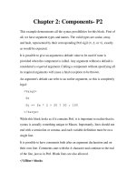
As illustrated in Fig. (1), these two fundamental distinc-
tions, i.e. egocentric/allocentric frames and categorical/
coordinate information, form the basic structure of spatial
memory and afford complex representations and behaviors.
We can represent our environment as an allocentric survey
map with embedded directions and distances or as a route
sequence with left-right turns from a first-person egocentric
perspective or we can simply focus on isolated landmarks
[44].
These three levels of spatial representation, landmark,
route and survey, form the developmental model proposed
by Siegel and White [44] to explain the acquisition of spatial
knowledge in the child. Then the model has been extended to
the development by adults of knowledge of the environment
and spatial strategies [45]. We can use diverse navigational
strategies to find out our way in the environment and to act
with objects. The fundamental role of spatial processes
between action and cognition is highlighted by Milner and
Goodale [46] in their re-interpretation of Ungerleider and
Mishkin’s model. They proposed that the ventral stream
generates object-centered, allocentric representations to the
purpose of building up long-term representations of objects,
whereas the dorsal stream generates egocentric
representations necessary to plan and execute reaching
movements under the guidance of vision. Finally,
visuospatial information and processes enable non verbal
cognitive abilities, such as mental imagery, that can be
defined as the capacity to represent and manipulate
information by relying on a spatial medium [36].
3.2. Models and Neurofunctional Bases of Spatial
Memory
The neural mechanisms underlying spatial memory have
yet to be fully understood, but it is agreed that the
hippocampus, together with its fundamental role in general
memory, is a key structure in supporting spatial memory.
Fig. (1). Fundamental features of spatial memory as sketched in the text.
Visuospatial Memory in Healthy Elderly, AD and MCI Current Aging Science, 2009, Vol. 2, No. 1 47
The experimental evidence is robust and encompasses
studies involving rodents, non-human primates and humans
[see 47,48]. According to one influential theory, spatial
information is maintained in the hippocampus in the form of
a cognitive map, which specifies the directions and relative
distances between locations in the environment [37,49].
Spatial information is integrated into an allocentric
representation that is maintained in long term memory. More
recently, it has been proposed that egocentric and allocentric
information is processed in parallel in the parietal lobe and
the hippocampal formation, with final transfer to the
hippocampus for long-term storage in allocentric coordinates
[50-52]. However, there is still debate on the status of long-
term spatial memory: according to one view egocentric
representations would be transient to the service of
perceptual control of movement in space whereas only stable
allocentric representations would be stored [53,54];
according to another view both egocentric and allocentric
representations would be maintained [41]. In any case, the
involvement of the hippocampus in allocentric spatial
memory is commonly accepted (for review see [55]).
Few studies have investigated directly the cerebral
networks subserving egocentric and allocentric processing.
A fMRI study showed that egocentric information activated
posterior parietal and lateral frontal premotor regions, more
extensively in the right hemisphere [56]. A succeeding study
confirmed the involvement of the fronto-parietal network in
the egocentric processing, while a subset of these regions
was also involved in the allocentric task [57]. Committeri
and co-workers [58] compared viewer-centered, object-
centered and landmark-centered spatial coding of visually
presented realistic 3D-information. Viewer-centered
egocentric coding activated mainly areas in the dorsal stream
and in frontal lobes, whereas allocentric coding recruited
both dorsal and ventral regions [58]. Zaehle and colleagues
[59] found that the processing of egocentric spatial relations
is mediated by medial superior-posterior areas with an
important role of the precuneus, whereas allocentric spatial
coding requires an additional involvement of the right
parietal cortex, the ventral visual stream and the
hippocampal formation.
With an ecological fMRI procedure, Rosenbaum and
collaborators [60] assessed participants familiar with the city
of Toronto in several navigational tasks: judgment of relative
distance, estimation of distance, correct order of sequences
of landmarks and spatial problem-solving. These tasks were
associated with cerebral activation of the medial temporal
lobe, in particular involving the right parahippocampal
gyrus, and of the following areas: retrosplenial cortex
(allocentric processing), medial and posterior parietal cortex
(egocentric processing), prefrontal cortex (spatial processing
requiring executive functions).
Maguire and colleagues [61] adapted a virtual reality
paradigm to a PET procedure. Normal subjects had to
mentally navigate to a goal, both directly and with detours.
Direct navigation strongly activated the right hippocampus
and the right inferior parietal cortex. Navigation with detour
also activated the left superior and middle frontal gyri. An
activation of the right caudate nucleus was also observed. In
a second fMRI study normal subjects had to learn a route in
a virtual environment and then to give judgements about
either the appearance (landmark processing) or the position
of particular locations (survey processing). Landmark
processing activated the lingual and fusiform gyri of the
occipital cortex, whereas survey processing activated the
posterior parietal and premotor areas. The overall data were
interpreted in terms of a specific mental navigation network
which included the right hippocampus, the left precuneus
and the insula [see also 62].
As regards coordinate and categorical spatial
representations, neuroimaging [63] and neurofunctional data
[64] in normal subjects performing spatial imagery tasks
have shown that the right hemisphere is particularly involved
in processing coordinate metric relations, while the left
hemisphere seems more specialized in computing categorical
spatial relations.
Recently, Iachini and colleagues [65] compared left- and
right-parietal brain lesioned patients on an egocentric and
allocentric spatial memory task. The results suggested that
the right hemisphere is specialized in processing metric
information according to egocentric frames of reference.
In conclusion, the heterogeneity of functions and
processes of spatial memory is reflected in the complexity of
the underlying cerebral networks, with a central role of
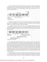
hippocampal and fronto-parietal circuits. Fig. (2) provides a
tentative description of the cerebral areas more involved in
spatial memory.
3.3. Spatial Memory and Normal Aging: General
Hypotheses
The reasons to study spatial memory and aging are
multiple. First, spatial ability plays a fundamental role in
everyday human activities, like way-finding, geographical
orientation, using a map of space for navigation, localizing
places or grasping objects. The assessment of visuospatial
abilities, which are the necessary pre-requisite of
independent mobility in the environment, is therefore crucial
to monitor elderly people's well-being. Second, episodic
memory is particularly vulnerable to decline with aging and
is among the firsts and most profound deficits of dementia.
Episodic memory has an inherently contextual nature, i.e.
previous experiences are embedded in a spatial and temporal
structure [66]. Spencer and Raz [67] reviewed the literature
about age differences in episodic memory by distinguishing
memory for content and context of a message. The results of
the meta-analysis showed that age differences in context
memory were reliably greater than those in content memory.
Third, spatial memory is a basic component of more general,
complex and non verbal cognitive processes such as mental
imagery.
Age-related changes in basic visuospatial abilities,
mental imagery and navigational abilities have been
investigated. Laboratory-based psychometric tasks, such as
mental rotation, and more ecological tasks, such as direction-
finding and map learning have been used [68]. The results
obtained are still controversial and it is not yet clear which
spatial processes decline with age and which ones are
preserved. Some data suggest that working memory is a very
important structure in understanding cognitive aging and it
has been hypothesized that a variation in its capacity is one
of the main variables associated with reduced mental
48 Current Aging Science, 2009, Vol. 2, No. 1 Iachini et al.
efficiency. Salthouse and Mitchell [69] suggested that in
working memory it is possible to distinguish between a
structural component, i.e. number of information units
that
can be memorized at the same time, and an operational
capacity component, i.e. number of processing operations
that can be performed. Mayr and collaborators [70] reported
pronounced age differences in active tasks requiring the
integration and coordination of information. In a series of
studies, Iachini and colleagues [32,71,72] compared two
general hypotheses about the cognitive decline associated
with healthy aging: the Slowing view and the Limited
Resources view. According to the first view, the speed of
cognitive processes is the main mediator of decrease with
age and would have global and uniform effects on cognitive
functioning [73,74]. According to the second view, age-
related decline is a consequence of reductions in basic
processing resources such as attention and working memory
[75,76]. This hypothesis predicts selective age-related effects
depending on the complexity of the task at hand. Iachini and
colleagues [71] compared young and elderly healthy adults
in a battery of psychometric tests assessing general cognitive
functions (Story Retell, immediate and delayed, Attentional
Matrices, Token, Verbal Fluency, Frontal Assessment
Battery (FAB) devised by Dubois, Raven’s matrices), and
visuospatial abilities: Line length perceptual judgement,
Mental rotation, Mental construction (all perceptually-
driven), visuospatial working memory span (Corsi), Line
length memory and Line Length inference. The results
showed selective effects of aging. Some abilities were well
preserved, such as memory for line length and perceptual
discrimination of line length. Some others were instead
impaired, such as the ability to infer new information from
memorized spatial information, the ability to manipulate the
spatial structure of mental images and to construct mental
images, and the ability of abstract spatial reasoning. Further,
basic processing resources such as attentional capacity and
visuospatial working memory showed a reduction in the
elderly. Two subsequent studies [32,72] confirmed that
aging has a detrimental effect on tasks that require active
manipulation and strategic control of spatial information (the
abilities to mentally rotate visual images, to retrieve spatio-
temporal sequences and to infer new spatial information).
Consistently, age had no detrimental effect on more passive
tasks requiring only perceptually-based comparisons or pure
maintenance of spatial information.
An interpretative framework similar to the Limited
Resources View is offered by the active/passive model
proposed by Cornoldi and Vecchi [77] within the Working-
Memory domain. The model is based on the level of activity
that cognitive processes require, that is the amount of
integration, modification or transformation of information.
Passive processes correspond to the simple maintenance of
information, whereas active processes imply simultaneous
maintenance and manipulation of information. Vecchi and
Cornoldi [78] compared young and elderly healthy adults on
passive and active visuospatial tasks. The battery included
the Corsi test, the Visual Pattern task, the Mental Pathway
task and the Jigsaw-Puzzle task. In the Jigsaw-Puzzle task,
Fig. (2). Graphic illustration of the relationships among neocortical regions, dorsal and ventral streams and hippocampal formation . The
arrows indicate the connections among cerebral structures that allow the processing of spatial information.
Visuospatial Memory in Healthy Elderly, AD and MCI Current Aging Science, 2009, Vol. 2, No. 1 49
participants are presented with numbered fragmented
pictures of everyday objects that must be assembled by
writing down in a blank grid the corresponding numbers.
The Visual Pattern task consists in the presentation of
pathways in matrices with increasing number of squares;
participants have to reproduce these pathways in a blank
matrix. In its Active version, the response matrix is
presented in a different orientation and hence mental rotation
of original pictures is needed. Overall, the results showed
marked differences due to active tasks and suggested that
age-related decline is due to a reduced capacity to
manipulate and transform visuospatial information (see also
[69]).
3.4. Basic Visuospatial Abilities in Normal Aging
As regards the egocentric/allocentric distinction, to the
best of our knowledge the literature on aging and spatial
cognition has not directly addressed this issue. In general,
several spatial tasks have been used, such as pointing tasks,
and the results are interpreted as consistent with the
allocentric or the egocentric organization of spatial
knowledge. Few attempts to compare directly these two
kinds of processing with young people have been made
[58,79] and it would be of theoretical and clinical relevance
to determine their developmental course. Parkin and
colleagues [80] used a spatial discrimination task that
involved egocentric spatial memory to compare elderly and
young people. They found no significant negative effect of
age on the spatial performance, but only a slight decline.
As regards the coordinate/categorical distinction, only
one study has addressed directly this issue. Meadmore and
co-workers [81] studied the hemispheric specialisation and
the effect of age on categorical and coordinate processing.
The results showed in all age groups a left hemisphere
advantage for the categorical task and a right hemisphere
advantage for the coordinate spatial task. However, older
adults were slower to process information and provide
spatial judgements. The results, therefore, did not clarify if
age exerted a selective negative impact on the two kinds of
processing. Again, this gap should be filled in future
research.
An important basic spatial ability is object location
memory. Sharps and Gollin [82] reported that memory for
objects and their spatial locations was more facilitated in
older than younger adults when items were studied in a
distinctive visual context. In Cherry and Park [83] younger
and older adults had to study and later recreate an
arrangement of small objects that were placed on a plain map
or a visually distinctive context. The objects were either
unrelated or categorically related. The results indicated that
the distinctive context enhanced spatial memory in all age
groups, whereas working memory resources accounted for
an important proportion of age-related variance in memory
for spatial location. Uttl and Graf [84] studied memory for
spatial locations within a museum and a secretarial office. In
Experiment 1 the subjects were 302 visitors (years from 15
to 74) to the museum; in Experiment 2 subjects were two
groups of young and older adults. The results showed an
age-related decline that appeared around the sixties. Cherry
and Jones [85] assessed the effects of structural and
organizational spatial context on memory for an arrangement
of dollhouse furniture pieces in younger and older adults. For
half of the participants, landmark objects and a floor plan
beneath the array served as structural context. Organizational
context was varied by grouping items either randomly or
prototypically. Landmark structural cues improved younger
adults' performance, whereas both groups benefited from the
floor plan. Connelly and Hasher [86] compared older and
younger adults on a composite object location task. They
found evidence that inhibition of identity and location may
function separately within the dorsal and ventral visual
streams. The findings are discussed in terms of reduced
inhibitory efficiency of irrelevant information in the elderly.
Overall, these studies tell us that contextual factors and
attentional/executive resources play a major role in the
spatial memory decline normally associated with healthy
aging. However, it is not clear which specific contextual
factors are particularly susceptible to age effects and how
they interact with executive factors.
3.5. Visuospatial Abilities and Mental Imagery
Mental imagery can be defined as a perceptual-like
representation of external objects or scenes that is able to
simulate a sensory-motor interaction with the environment in
absence of actual sensorial stimuli [36]. In this domain,
mental rotation and mental scanning of spatial images have
been among the firsts and most studied imagery processes,
possibly because they helped to clarify the spatial nature of
imagery [87,88].
Research on mental rotation has shown that this ability
declines with age [e.g. 89-92].
Craik and Dirkx [93] reported a negative impact of age
on visuospatial imagery using three different tasks: the
Brooks Letter Test (subjects have to imagine walking along
a block letter and describe the way), the East-West Test
(subjects have to state the direction they are facing after
changing direction), and the Clock Test (subjects have to
state whether the hands of an imagined clock subtend an
angle greater than 90°). Dror and Kosslyn [94] studied the
effects of aging on four components of mental imagery:
image generation, image maintenance, image scanning, and
image transformation. The authors found a progressive
impairment with age in image generation and rotation, but
not in image maintenance and scanning. Further studies
about generation and maintenance of mental images
confirmed this trend and showed a prevalence of self-related
images in the old [95,96].
Finally, some works addressed the topic of how metric
properties, such as distance, are processed by means of a
mental scanning paradigm [88]. Brown, Kosslyn and Dror
[97] found that as the scanning distance increased perceptual
and mental scanning of a small squared grid became harder
for the elderly than it did for the younger. Iachini, Poderico,
Ruggiero and Iavarone [71] adopted a mental scanning
procedure that was adapted to an ecological situation: young
and old participants had to study by vision and locomotion a
real 3-D pathway and then had to mentally explore it. The
results showed that aging had a negative impact on the
quality of metric information embedded in mental maps of
that environment. Elderly people retrieved the various
50 Current Aging Science, 2009, Vol. 2, No. 1 Iachini et al.
positions in their correct order, but were not able to depict
consistently in their mental map the different distances.
3.6. Visuospatial Abilities and Navigation
A review of the literature [98] shows a clear decline of
spatial abilities in the elderly when abstract laboratory tasks
are used, whereas the decrement seems to reduce with more
familiar tasks set in ecological contexts. For example, elderly
people can cope effectively with several everyday spatial
tasks [99]. Kirasic [100] found no negative effect of age
when elderly people had to perform their spatial tasks in a
familiar environment. Elderly participants can cope
effectively with tasks requiring self-orientation in familiar
environments and tend to judge their sense of direction more
positively than the younger [90].
However, even in more ecological tasks there is evidence
showing that age has a negative impact on various
navigational abilities: selecting and remembering landmarks
[101], learning unfamiliar routes [99,100], inferring
distances and directions among locations [102], and finding
the way [68]. A number of studies have found that older
adults tend to perform worse than young adults on many
measures of memory for routes [103]. Age differences
favoring young adults have also been reported in learning
how to navigate through real [104,105] or virtual [106]
environments. Typically, in a route learning task participants
have to explore a real path or a fictitious map and then to
answer various questions. Salthouse and Siedlecki [107]
investigated whether the age-related decline in navigational
abilities is due to reduced efficiency in route selection. The
results confirmed a moderate decline in measures of the
efficiency of route selection as age increased from 18 to 93
years. This finding is consistent with the results of similar
studies [108] and suggests that the age-related decline is due
to a deficit in the planning of the pathway rather than in its
execution.
Finally, a very popular task to assess navigational
abilities is the Morris Water Maze test (MWM). In its
standard version it is settled in a circular pool and the aim is
to reach an invisible platform, located under the water level.
As the target is not visible, it must be located with reference
to several cues. Several versions of MWM have been
designed to test human participants [109]. Moffat and
Resnick [106] adopted virtual reality to test healthy elderly
participants in MWM. They found that old participants, as
compared to young adults, covered a greater distance to
locate the hidden target, took shorter and showed greater
difficulty to set up a cognitive map of the environment.
Moffat and co-workers [110] also used the Virtual Water
Maze to assess possible relationships between navigational
abilities and structural integrity of hippocampal and
extrahippocampal brain regions. The results confirmed that
age-related deficits in navigational ability do not depend
solely on the hippocampus but are also associated with larger
regional volumes of multiple cortical and subcortical
structures.
4. VISUOSPATIAL ABILITIES IN AD AND MCI
At a first look, works measuring visuospatial abilities in
AD and MCI patients and reporting disturbances are huge,
about 709 articles. A closer reading led us to restrict our
interest to few articles and to exclude the remaining for two
main reasons: the terms visuospatial and visual were
sometimes used as synonymous in reference to tasks
requiring visual analysis of object properties; the assessment
of visuospatial abilities often relied on measures poorly
specified from a cognitive point of view. In our opinion, a
careful identification of the task demands is essential in
order to understand both the nature of the affected cognitive
processes and the sequence in which such effects may occur.
For example, many researchers use constructional tasks
that require participants to copy or to remember complex
figures such as the Rey-Osterrieth test [4,111-115], the most
used in the literature. Similarly, the Block Construction from
the Performance subtests of the Wechsler Adult Intelligence
Scale-Revised [116,117] requires to arrange painted wooden
blocks in order to copy a design formed by the examiner or
shown on a diagram. Both tests make demands on several
cognitive components, including planning and praxis, as well
as visuospatial abilities; this complexity does not allow to
separate the relative contribution of visuospatial and
executive components. Some works use the Raven’s Colored
Progressive Matrices [118] to assess visuospatial abilities
[111,113,114]. Although the Raven test implies visual and
geometric materials, assesses a complex and general ability
such as abstract reasoning. Finally, other researchers use the
Tower of London [119] and the Trail Making Test [120],
that can be better considered as executive function tests,
even if a visuospatial component may be implied.
Some tests clearly tap visuospatial abilities: Clock
Drawing [121], Benton Line Orientation [122] and Dot
Counting [123]. In all these cases, perceptual discrimination
of simplistic visual stimuli is measured. To measure
topographical orientation, it is often used the Money Road
Map test [124] in which subjects have to trace a route on a
map while identifying left and right turns [125,126]. Route-
description and map-drawing tests are usually adopted to
evaluate Topographical Disorientation in AD patients, but
they are ambiguous in their task demands [10]. As an
example, one could draw a map of a familiar environment by
recalling either the route usually covered or the mental
survey map of that environment: the final output would be
the same although resulting from different spatial strategies
(respectively egocentric/route and allocentric/survey). We
selected about 20 studies investigating visuospatial
disturbances in AD and MCI patients and using specific
perceptual spatial tasks.
As regards spatial memory, the Corsi test is usually used
to measure the short-term sequential memory span. It
consists of a set of nine identical blocks arranged irregularly
on a board. Participants have to reproduce the sequences of
blocks of increasing length as tapped by the experimenter in
forward-recall order and sometimes in backward-recall order
[127,128]. The final score corresponds to the span length,
that is the maximum level of block-tapping sequences
reproduced.
About ten studies, discussed below, devised tasks that
successfully removed the confounding elements of
constructional praxis and object identity processing, and
required memory for simple spatial arrangements or complex
routes/environments.
Visuospatial Memory in Healthy Elderly, AD and MCI Current Aging Science, 2009, Vol. 2, No. 1 51
4.1. Visuospatial Perceptual Abilities in AD and MCI
The staging of visuospatial deficits in AD has not been
investigated extensively and the few attempts to examine the
relationship between patterns of deficit and age of patients
are still inconclusive [4,123,129]. Initial interest in
visuospatial abilities was motivated by the heterogeneity of
deficits characterizing AD and the possibility to distinguish
different subgroups of patients [130]. In these studies
visuospatial abilities were assessed at perceptual level.
Martin and colleagues [4,131] identified two subgroups of
similar size (about 20% of their overall sample in each
domain): one showed impairment of word-finding ability
with preserved visuospatial and constructional skills,
whereas the other one showed the opposite profile. The
remaining group showed global cognitive decline. Complex
tasks were used to assess the visuospatial domain (Rey,
Block Design and Mosaic comparisons). Becker and
colleagues [129] identified similar groups with focal deficits,
although the percentage of visuospatial AD was only 5%.
Mendez and colleagues [5] used several visuoperceptual
tasks, including object, face and color recognition and form
discrimination, to examine visual disturbances in AD
patients. Deficits in spatial localization and object
recognition were present in half the sample, which ranged
from mild to severe stages of the disease. They concluded
that complex visual disturbances such as deficits in figure-
ground discrimination, visual object recognition and spatial
localization are common in AD.
Kaskie and Storandt [132] used a complex test, the
Visual Form Discrimination, to compare very mild and mild
AD patients with healthy controls and found visuospatial
deficits in several AD patients. Kurylo and colleagues [133]
found that scores on tests of visual processing did not
correlate with severity of dementia and suggested that visual
deficits may reflect the heterogeneity of neuropathological
changes rather than overall disease progression. Nordlund
and colleagues [134] examined attention, memory and
learning, visuospatial functions, language and executive
functions in MCI patients and matched controls. The results
showed impairments in all five cognitive domains.
The assessment of visuospatial abilities first
demonstrated the heterogeneity of degenerative deficits and
then led to the hypothesis that they could represent an early
predictor of AD [135]. For example, interest in possible
visual mechanisms underlying topographical disorientation
in AD patients led to hypothesize that early visual motion
perception deficits could precede navigational impairments
[136]. Mapstone and colleagues [125] compared young and
older healthy adults with MCI and AD patients in perception
of panoramic visual motion stimuli. One fifth of the older
adults, one third of the patients with MCI, and half of the
patients with AD showed pervasive impairments of visual
motion perception that correlated with poorer performance
on the Money Road Map test. In line with O’Brien and
colleagues [136], the authors suggested that visuospatial
deficits may develop as an early sign of neurodegenerative
disease.
Pursuing the visuospatial hypothesis, Rizzo and
colleagues [137] compared mild AD patients and healthy
controls on tests measuring visual perception and general
cognition. AD patients showed deficits in static spatial
contrast sensitivity, visual attention, shape-from-motion,
visuospatial construction and visual memory. The findings
are compatible with the hypothesis that neurodegenerative
processes involve multiple visual neural pathways and visual
dysfunctions may contribute to decrements in other cognitive
domains.
In a PET study, Fujimori and colleagues [138] assessed
spatial vision and object vision (based on the Milner and
Goodale’s model [46]) in 49 patients with mild-to-moderate
AD. Spatial vision was tested by means of the Visual
Counting test, whereas object vision by means of the
Overlapping Figure Identification and the Visual Form
Discrimination tests. The results showed that the visual
spatial disturbance was correlated to the metabolic rate of the
bilateral inferior parietal lobules, whereas the visual object
disturbance was correlated to the right middle temporal
gyrus and the right inferior temporo-parietal metabolism.
Caine and Hodges [123] examined the staging of
visuospatial and semantic deficits in 26 minimal/ mild AD
patients and healthy controls to determine whether
visuospatial deficits may occur prior to the presence of
semantic deficits. They emphasized that psychometric tests
must be highly specific as regards the underlying cognitive
requirements. Visuospatial abilities were assessed by tests
based on visual perception: Line Orientation, Object
Decision (where participants had to decide whether line
drawings depicted real or unreal items) and Object Matching
(where participants had to recognize a target object between
two distractors: same object from an unusual view or
different object but visually similar). In a second study the
Visual Object and Space Perception Battery was used
(VOSP) that included the Dot Counting test and two tests of
positional discrimination. A small group of early AD
patients showed visuospatial deficits and poor episodic
memory without coexisting semantic impairment, and this
suggested that damage can occur in occipito-parietal or
parietal regions at an earlier stage than currently recognized.
This study deserves some comments. First, Caine and
colleagues [123] have the merit of adopting tests of spatial
perception independent of executive, praxic or object-based
components, although these tests used quite abstract and
simplistic elements and did not assess more ecological
situations. Second, the association of visuospatial and
episodic memory deficits might imply that damage in
visuospatial cerebral areas is primary and is responsible for
memory losses, as discussed below.
In a fMRI study, Vannini and co-workers [139]
investigated the visuospatial cerebral networks in 18 MCI
patients. Along three years, they were periodically submitted
to an extensive battery of tests that included: WAIS-R, Rey-
Osterrieth Copy and Retention Test, Rey Auditory Verbal
Learning Test [140], and Trail Making Test A and B [141].
To assess visuospatial abilities an angle discrimination task
was adopted. The authors concluded that MCI patients who
progress to AD revealed a reduced neuronal efficacy during
execution of the angle-discrimination task. Furthermore, the
increased activation in the left hemisphere in MCI converters
suggested that compensatory mechanisms might be activated
before the onset of clinical symptoms of AD.
52 Current Aging Science, 2009, Vol. 2, No. 1 Iachini et al.
In conclusion, all these studies raised the possibility that
visuospatial abilities could represent an early predictor of
subsequent disease. However, as the testing was limited to
the perceptual level of spatial processing, the relative
contribution of the visuospatial modality to the well-known
memory deficits and its possible anticipatory role was not
assessed.
4.2. Visuospatial Memory Deficits in AD and MCI
There are few recent studies about the visuospatial
modality in the memory process of AD and MCI patients. In
the past years, it has been showed that memory for spatial
locations [142], spatial patterns [143] and object locations in
a grid [144] is impaired in AD patients as compared to
normal controls. Apart from some recent investigations,
there are no systematic data about AD and MCI patients.
Here we review those few studies assessing basic and
navigational visuospatial memory processes and adopting
clearly defined tasks (see Table 2 below).
Vecchi and colleagues [145] compared 16 early-stage
AD patients with a healthy elderly group in order to
determine the contribution of passive and active processes in
the limitations of working memory functions observed in
AD. There were four tasks: a verbal passive task, a verbal
active task, the Corsi test and a visuospatial active task
(Mental pathway). The results showed that AD patients
performed less accurately than the control group in all tasks,
but the deficit was maximized with active verbal and spatial
processes. Therefore, a clear impairment of executive
processes was confirmed while the staging of verbal and
spatial deficits remained unclear, presumably because of the
lack of MCI patients in the sample.
Lineweaver and colleagues [146] submitted AD patients
to a mental rotation task and found that accuracy decreased
as rotational angle increased. According to the authors, the
spatial manipulation deficit of AD patients may reflect
pathology in parietal and temporal lobes.
Some works have found an impairment in visuospatial
short-term memory as measured by the Corsi test in very
mild and mild AD patients [111] and in AD patients
followed for two years [113]. The authors suggested that
visuospatial deficits might constitute an early predictor of
AD and that cognitive decline may be better predicted by
deficits diffused in linguistic and visuospatial domains.
Toepper and co-workers [147] compared 13 AD patients
with elderly controls on several tests (Block Suppression,
clock drawing, digit-word transformation, verbal memory
span). Interestingly, the Corsi test was used in forward and
backward orders. The results showed that in AD patients the
active inhibition of irrelevant stimuli and the Corsi backward
span were significantly reduced, confirming the substantial
impairment in attentional and executive resources.
Kessels and colleagues [148] investigated object-location
memory in 18 AD patients and a matched control group by
using an ecologically valid computer task in which
participants had to remember the locations of objects in
common rooms. There were colored photographs of eight
domestic rooms and 80 everyday objects that were
semantically related to these rooms. Participants had to learn
the locations of various objects and next to relocate these
objects to their original locations. The results showed an
impairment of explicit but not implicit spatial memory in AD
patients. This suggests that the preservation of implicit
memory in AD extends to the spatial domain, and this could
have an important rehabilitative value.
Kavcic and colleagues [126] compared 15 AD patients
and matched controls to assess navigational impairments in
AD. They measured visual motion evoking potentials
responses to optic flow simulating observer self-movement
to verify how these potentials were linked to navigational
performance. Participants were submitted to a
neuropsychological battery that included visuospatial tests
such as the Money Road Map and the Judgement of Line
Orientation and to a real-world navigational task.
Participants were led with a wheelchair along a route and
then asked several questions that assessed their knowledge of
the route, of the landmarks and both. Afterwards, there were
three route learning tasks: re-trace the route by indicating
which turn was taken previously, point to several locations
from the starting/finishing positions and draw the route on a
map. There were three landmark tasks: name as many
landmarks as possible from the route, name features that
could help in finding the way along the route and recognize
views of the route depicted on photographs. Two tasks
assessed the integration of route and landmark knowledge:
identify which direction allowed to see the viewpoint shown
on photographs and indicate the direction and extent of
movements shown on video clips. The results showed that
the navigational impairment in AD patients was linked to a
disorder of extrastriate visual cortical motion processing that
was reflected in specific perceptual and memory measures of
spatial abilities.
deIpolyi and collaborators [114] compared 13 mild AD
and 21 MCI patients with matched controls on a route-
learning task and a neuropsychological battery. In the route-
learning task, subjects were led along a novel route through a
Care Center. Subsequently, they had to repeat the route by
giving themselves the proper directions and to draw the route
on a map. Next, subjects were shown with three sets of
photographs and had to recognize: photographs of objects
and places along the route (Landmark Recognition), the
position of places along the route (Landmark Location) and
the order in which several targets were encountered along the
route (Order Memory). Finally, subjects were asked to
traverse the route from the end to the start and were
submitted to a pointing task. A subsample also took part in a
neuroimaging study to determine the neural correlates of the
tested spatial abilities. The results showed that AD and MCI
patients recognized landmarks as effectively as controls, but
could not find their locations on maps or recall the order in
which they were encountered. Half of AD and one-quarter of
MCI patients got lost on the route, compared with less than
10% of controls. Patients who got lost had lower right
posterior hippocampal and parietal volumes than patients
and controls who did not get lost. The ability to identify
locations on a map correlated with right posterior
hippocampal and parietal volumes, whereas order memory
scores correlated with bilateral inferior frontal volumes. In
sum, the navigational disability in AD and MCI patients
involved a selective impairment of spatial cognition,
presumably concerning the capacity to represent
environmental information at route level. This deficit was
Visuospatial Memory in Healthy Elderly, AD and MCI Current Aging Science, 2009, Vol. 2, No. 1 53
Table 2. Relevant Studies Investigating Visuospatial Abilities in Healthy Elderly, AD and MCI
References Year Sample/s Main Visuospatial Task/s Results
[143] 1988
12 AD, 27 PD and 39
matched NC
Computerized tests of visuospatial memory
The AD patients were severely impaired in the
visuospatial memory task
[142] 1992
15 mild AD, 16 moderate
AD and 16 NC
Spatial order and spatial recognition memory
tasks
Mild AD patients were impaired in memory for
early serial positions, while moderate AD patients
on all serial positions for both spatial order and
spatial recognition memory
[144] 1997
19 AD, 12 VAD and 29
NC
Location Learning Test (LLT)
The AD and VAD patients were impaired in the
LLT
[145] 1998 16 AD and 16 NC
A verbal passive task, a verbal active task, Corsi
test, a visuospatial active task (Mental pathway)
AD had lower performances than NC. The deficit
was maximized in active processes
[148] 2005
18 AD and 18 matched
NC
Rooms Task
Impairment of explicit spatial memory in AD, but
no difference with the control group on implicit
spatial memory
[146] 2005
18 AD, 18 HD, 36
matched NC
A computer based mental rotation test
The accuracy of AD patients decreased with
increasing angle of orientation
[126] 2006 15 AD and 15 NC
Money Road Map test, Judgement of Line
Orientation test, a real-world navigational task
AD patients showed deficits of visual motion
processing and were not able to link navigational
information into an integrated cognitive map of the
environment
[111, 113]
2006,
2007
36 AD (18 Very mild
AD, 18 mild AD) and 17
NC
43 AD: 22 fast CD and
21 slow CD 43 in a
longitudinal study (24
months)
Corsi test
AD patients were impaired in Visuospatial short-
term memory
[8] 2007 8 AD, 8 MCI and 8 NC
Visual short-term memory (VSTM), and
visuospatial short term memory (VSSTM) tasks
VSTM and VSSTM deficits in MCI and AD
patients, VSSTM deficits were more severe in AD
[114] 2007
13 mild AD, 21 MCI and
24 matched NC
A route-learning task (RTL) comprising: RLT-
Forward, Landmark Recognition, Landmark
Location, Order Memory, RLT-Reverse and
Dead Reckoning sub-tasks
AD and MCI patients recognized as many
landmarks as controls, but could not find their
locations on maps or recall the order in which they
were encountered
[9] 2007
21 AD, 36 MCI, 8 SMC
and 26 NC
Adaptation of MWM
Impairment in the allocentric component of spatial
memory in aMCI, overall spatial impairment in AD
and multiple domain aMCI
[147] 2008 13 AD and 13 NC
Block Suppression, clock drawing, digit-word
transformation, verbal memory span, Corsi test
(backward and forward)
AD patients were impaired in active inhibition of
irrelevant stimuli and in backward span
[149] 2008 29 aMCI and 30 NC
Brief Visuospatial Memory Test-Revised
(BVMT-R), Digit Symbol incidental recall
Early neuroanatomical changes in the hippocampus
and entorhinal cortex in aMCI cause the
impairment of the ability to integrate associative
information in memory
Abbreviations: AD = Alzheimer’s disease patients; MCI = mild cognitive impairment patients; aMCI = mild cognitive impairment patients amnestic domain;
NC = normal control participants; SMC = elderly people with subjective memory complaints; PD = Parkinson’s disease; HD = Huntington’s disease patients;
VAD = vascular dementia patients; MWM= Morris Water Maze.
54 Current Aging Science, 2009, Vol. 2, No. 1 Iachini et al.
associated with atrophy of the right-lateralized navigation
network. Therefore, we can comment that by joining the
behavioral methods of cognitive psychology and the
neuroimaging techniques of neuroscience, this study was
able to detect parallel changes at behavioral and
neurofunctional level in the navigational abilities. Notably,
the authors adopted navigational tasks that required specific
processing components within the complex domain of spatial
memory. The extensive spatial impairments observed in MCI
patients suggest that navigation tests may help to find out
early markers of dementia.
Troyer and colleagues [149] compared 29 individuals
with amnestic MCI and 30 matched controls on standardized
tests of object–location recall and symbol–symbol recall.
The amnestic-MCI group showed marked deficits in the
ability to integrate associative information in memory, and
this was attributed to early neuroanatomical changes in the
hippocampus and the entorhinal cortex. According to the
authors, then, associative memory deficits may represent an
early cognitive sign of AD.
Finally, two recent studies suggest interesting hypotheses
about the predictive role of specific spatial memory
processes. Alescio-Lautier and colleagues [8] compared 8
MCI and 8 AD patients with healthy controls to determine
which modality, i.e. visual or visuospatial, is more
implicated in the early memory impairment typical of AD. In
the visual short term memory (VSTM) task, patients had to
encode a composite image comprising various concrete
objects and to recognize whether these images changed or
not. In the visuospatial short term memory (VSSTM) task,
patients had to encode the location of similar images and had
to recognize if the entire pattern changed or not its position.
A span control task was used to determine the number of
images with which patients could perform the recognition
task at their memory capacity level. After each presentation,
a target image was presented at three different intervals
(1sec, 10sec, 30sec) and the participants had to recognize if
images (VSTM) or locations (VSSTM) had changed. In
order to disentangle the relative contribution of attentional
resources in the memory impairment, for half trials a
distractor in the interval between the presentation and the
recognition was presented. Results showed VSTM and
VSSTM deficits in MCI and AD patients as compared to
elderly healthy controls, with the spatial performance being
worse than the visual one. MCI patients had an intermediate
performance between controls and AD patients. However,
cognitive memory profiles differed between MCI and AD
patients depending on the modality tested and this indicated
an alteration of different processes. Indeed, AD patients
presented a greater deficit in the visuospatial modality than
MCI patients and were differently affected by the
experimental manipulations. In the visual recognition task,
AD patients had more difficulty with the no change
condition (in which images were the same) than the change
condition, whereas this did not happen with MCI patients.
The incapacity to detect no change was explained by the
phenomenon of attentional blink: a temporary functional
blindness to the second of sequentially presented stimuli.
Further, the span measure could have affected the VSTM
task with more errors as the number of images increased. In
the VSSTM task the set of images can be considered as a
whole and this should have facilitated the performance,
although it did not. Consequently, the deficit in the VSTM
task might depend on the number of images, whereas in the
VSSTM task it should be due to the spatial component rather
than the visual one. When the distractor was presented in the
VSTM task, more errors appeared at 1sec interval than in
other intervals. Instead, the visuospatial task was not so
sensitive to the presence and timing of the distractor. The
visual recognition deficit, then, could derive from an
impairment in disengaging-engaging attention in MCI and
AD patients. The overall results, therefore, suggest that
deficits in visual recognition are secondary to impairments in
attentional and executive resources, whereas deficits in
spatial recognition are primary and reflect a genuine spatial
disorder. They might also imply that visuospatial short-term
deficits appear earlier than visual short-term ones in the
disease progression. Studies based on the complementary
assessment of attentional resources and visuospatial
memory, then, could help to identify the cognitive origin and
the neurofunctional bases of the deficits shown by MCI and
AD patients, and this is necessary to understand the staging
of the deficits and their predictive value.
Hort and colleagues [9] investigated navigation deficits
in AD and MCI patients in order to assess which spatial
components of navigational ability could represent a positive
marker of subsequent AD and in which sub-group of MCI
patients this marker is present. The sample included 26
normal controls, 21 AD patients, 8 elderly people with
subjective memory complaints (SMC) and 3 groups of MCI
patients sub-classified according to the Petersen’s criteria: 7
nonamnestic (naMCI), 11 amnestic single domain (aMCI),
18 amnestic multiple domain (aMCImd). They adopted the
MWM test in a version that allowed to discriminate the
allocentric and egocentric components of navigational
ability. Participants were required to locate an invisible goal
inside a circular arena, and to this purpose they could use
either egocentric cues (the relationship between the goal and
their starting position) or allocentric cues (external features).
The results showed strong differences in the patterns of
spatial navigation impairments among the subtypes of MCI.
The AD and aMCImd groups were impaired in all
conditions, whereas the naMCI and SMC groups were
similar to controls. Finally, the aMCI group showed a
specific impairment in the allocentric processing. The
similarity of spatial navigation impairments in the aMCImd
and AD groups confirmed that aMCImd could represent an
advanced prodromal stage of dementia, whereas aMCI could
represent an even earlier stage [25]. In sum, the authors
suggest a developmental course starting from aMCI to
aMCImd and finally to AD. The impairment in the
allocentric component of spatial memory could allow the
monitoring of the disease progression and could help in
detecting the early stages preceding AD.
4.3. Neurofunctional Evidence in AD and MCI
On the basis of histological and neuropathological
evidence, AD is characterized by degeneration of neurons
and their synapses and by the appearance of neurofibrillary
tangles and senile plaques that are considered generally
linked to the hippocampus atrophy [150]. Studies
investigating the changes in the levels of markers of tangle
and plaque formations in the cerebrospinal fluid (CFS) have
Visuospatial Memory in Healthy Elderly, AD and MCI Current Aging Science, 2009, Vol. 2, No. 1 55
shown a detectable potential index for diagnosis of
conversion to degenerative dementia [151]. In particular, it
seems that tangles and plaques, initially accumulated many
years before the clinical onset of the disease, could correlate
with the severity of the disease [152]. The degeneration
seems to prefer cerebral structures such as the
transentorhinal and the entorhinal cortexes, the hippocampus
and, then, the neocortical associative areas. This involvement
can explain the dysfunction of encoding and storing
information that reflects deficits at the level of consolidation
of information [6]. Furthermore, Apostolova and co-workers
[150] found that a high risk for conversion from MCI to AD
is associated with increased involvement of the hippocampal
subregion (CA1) and the subiculum. As pointed out by
Killiany and co-workers [153], the atrophy of some mesial
temporal lobe structures could represent a predictor for the
conversion from MCI to AD. Thompson and colleagues
[154] reported losses of grey matter being faster in the left
hemisphere than in the right one distinctively in AD with
respect to normal aging.
Still, by adopting single photon emission computed
tomography (SPECT) and positron emission tomography
(PET), many studies demonstrated reduced blood flow and
metabolic deficits in temporoparietal cortices in patients with
AD [155]. Furthermore, damage in parietal cortex could
indicate impairments in visuospatial processes that can be
recognized in the early clinical stages of AD [156].
Accordingly, evidence from functional magnetic resonance
imaging (fMRI) examining brain activation evoked by
visuospatial processing, showed decreased activation in the
dorsal visual pathway as well as compensatory recruitment
of remote brain areas in AD patients [157]. From this
perspective, Vannini and associates [139] argued that
compensatory mechanisms may mask the starting
degenerative process by determining functional changes. The
same authors hypothesized that an increased parietal
activation in MCI patients could reflect a reduced neuronal
efficacy due to accumulating AD pathology as proof of a
compensatory mechanism.
Given a predominance of temporal lobe damage,
especially in early stages of AD, Kurylo and colleagues
[133] suggested that it may be particularly useful to assess
the dorsal-ventral streams, especially in relation to visual
tasks. Visuoconstructional dysfunction in AD patients is
significantly correlated with a lower metabolism in the right
parietal cortex [158] or in the bilateral occipital and temporo-
parietal regions [159]. Pietrini and colleagues [160] showed
that patients with visuospatial symptoms had larger
metabolic deficits in the bilateral parietal and occipital
cortices than did patients without the symptoms.
CONCLUSIONS: SPATIAL MEMORY AND
ALZHEIMER'S DISEASE
A great effort has been devoted to the definition of
behavioral, biological and neurofunctional correlates which
could predict the conversion of MCI in AD, but most studies
have focused on verbally-mediated memory disorders.
Surprisingly, in contrast with the number of studies
addressed to disentangle the multiple cognitive processes
subtending (normal) spatial memory, both with methods of
cognitive psychology and neuroscience, there are relatively
few studies aimed at evaluating disorders of spatial memory
in AD.
We believe it is worth exploring this topic for the
following reasons. First, a progressive disorder primarily
involving memory (including spatial memory) could be
assumed as a theoretical paradigm to get insights into the
nature of normal spatial memory. Second, the AD is a
degenerative disease primarily involving brain structures
(hippocampus and medial temporal lobes) heavily implicated
in spatial memory processes. Consequently, studies on pre-
clinical stages of AD (namely, the MCI), or AD in its early
stages, could be assumed, with some limitations, as a
"lesional" paradigm to evaluate the role of these structures in
the complex organization of spatial memory. Studies on
patients with focal brain damage have the limitation given by
the wide heterogeneity of the site, extension and nature of
the lesion, which prevent to carry on studies on large cohorts
of subjects. Patients with AD or MCI, conversely, do not
undergo these limitations, given the putative pathogenetic
homogeneity of the disease and the relative simplicity to
match them according to general cognitive functioning.
Third, data from visuospatial functioning could be of great
aid to detect patients in early stages of AD, in such a way to
contribute to a timely diagnosis of dementia and to detect
subjects with MCI at higher risk to develop AD.
In the above paragraphs it has been shown that spatial
memory is heavily dependent on brain structures which
exhibit a particular vulnerability to both normal aging and
degenerative dementia. As shown in Fig. (2), the
hippocampus, the fronto-parietal network and the temporal
lobe are strongly involved in spatial memory. Researches
indicate that the neurodegeneration in AD primarily disrupts
hyppocampus, which accounts for the early appearance of
anterograde episodic amnesia. However, the hyppocampus is
also strongly implicated in visuospatial processes, like
topographic orientation and allocentric processing, and this
may account for the symptoms of spatial disorientation
which may precede, in some cases, disorders of episodic
memory. Furthermore, temporal-parietal areas, which are
related to spatial and visuo-constructive abilities, are early
involved in AD. The converging evidence about the neural
circuits subserving normal spatial memory and about the
evolution of the cerebral degenerative process suggests that
subtle disorders of spatial functions could be associated with
MCI and/or be considered a putative "cognitive marker" of
risk of conversion to AD. However, if we aim at identifying
very early pre-dementia states we have to adopt a research
strategy that is able to detect functional changes before
structural changes become evident. As suggested by Vannini
and colleagues [139], a reduced metabolic rate in specific
cerebral areas may precede future damages. To this purpose,
it is necessary to join neuroimaging techniques and
neuropsychological knowledge with experimental paradigms
derived from cognitive psychology. Indeed, there is need of
tasks clearly specified in their cognitive processing
components and in the concepts measured in order to put
forward hypotheses about the specific cerebral areas
involved and about the correlation between behavioral
changes and metabolic rate in those areas.
The results discussed above suggest that deficits in
spatial memory may play an important role in this research
56 Current Aging Science, 2009, Vol. 2, No. 1 Iachini et al.
strategy. The impairment of the allocentric component of
spatial memory found by Hort and colleagues [9] may
underlie navigational deficits and, more importantly, deficits
in broader cognitive processes such as object recognition
[35,46]. Alescio-Lautier and colleagues [8] attributed visual
recognition deficits in MCI and AD patients to attentional
factors. In normal aging several cognitive deficits are
mediated by a reduction in attentional and working memory
resources: from this point of view only a quantitative
difference between AD patients and healthy elderly would
appear. Instead, spatial deficits seem primary and not
secondary to attentional factors, consequently they could
represent a qualitative marker of departure from normal
aging. Further, if they were primary we could even speculate
that the well-known episodic memory deficits might be due
to spatial memory impairments or rather they may coexist.
Nowadays there is not enough experimental evidence to state
that spatial memory deficits occur earlier than other deficits
in the disease progression, but there is enough matter to
suggest deeper scientific investigation.
REFERENCES
[1] Hodges JR. Alzheimer's centennial legacy: origins, landmarks and
the current status of knowledge concerning cognitive aspects. Brain
129: 2811-2822 (2006).
[2] Petersen RC, Doody R, Kurz A, Mohs RC, Morris JC, Rabins PV,
et al. Current concepts in mild cognitive impairment. Arch Neurol
58: 1985-1992 (2001).
[3] Fox NC, Warrington EK, Seiffer AL, Agnew SK and Rossor MN.
Presymptomatic cognitive defitits in individual at risk of familiar
Alzheimer’s disease. A longitudinal prospective study. Brain 121:
1631-1639 (1998).
[4] Martin A, Brouwers P, Lalonde F, Cox C, Teleska P, Fedio P, et al.
Towards a behavioral typology of Alzheimer's patients. J Clin Exp
Neuropsychol 8(5): 594-610 (1986).
[5] Mendez MF, Mendez MA, Martin R, Smyth KA and Whitehouse
PJ. Complex visual disturbances in Alzheimer's disease. Neurology
40: 439-443 (1990).
[6] Braak H and Braak E. Neuropathological stageing of Alzheimer-
related changes. Acta Neuropathol 82: 239-259 (1991).
[7] Carli M, Luschi R and Samanin R. Dose-related impairment of
spatial learning by intrahippocampal scopolamine: antagonism by
ondansetron, a 5-HT3 receptor antagonist. Behav Brain Res 82(2):
185-194 (1997).
[8] Alescio-Lautier B, Michel BF, Herrera C, Elahmadi A, Chambon
C, Touzet C, et al. Visual and visuospatial short-term memory in
mild cognitive impairment and Alzheimer disease: role of attention.
Neuropsychologia 45(8): 1948-1960 (2007).
[9] Hort J, Laczò J, Vyhnàlek M, Bojar M, Bure J and Vlek K.
Spatial navigation deficit in amnestic mild cognitive impairment.
Proc Natl Acad Sci USA 104: 4042-4047 (2007).
[10] Aguirre GK and D'Esposito M. Topographical disorientation: a
synthesis and taxonomy. Brain 122: 1613-1628 (1999).
[11] Petersen RC, Stevens JC, Ganguli M, Tangalos EG, Cummings JL
and DeKosky ST. Practice parameter: early detection of dementia:
mild cognitive impairment (an evidence-based review). Report of
the Quality Standards Subcommittee of the American Academy of
Neurology. Neurology 56(9): 1133-1142 (2001).
[12] Visser PJ. Mild cognitive impairment subtypes and vascular
dementia. J Am Geriatr Soc 54(12): 1966-1967 (2006).
[13] Petersen RC. Challenges of Epidemiological Studies of Mild
Cognitive Impairment. Alzheimer Dis Assoc Disord 18(1): 3-10
(2004).
[14] Kral VA. Senescent forgetfulness: benign and malignant. Can Med
Assoc J 86: 257-260 (1962).
[15] Burns A and Zaudig M. Mild cognitive impairment in older people.
Lancet 361(9364): 1227-1228 (2002).
[16] Flicker C, Ferris SH and Reisberg B. Mild cognitive impairment in
the elderly: predictor of dementia. Neurology 41: 1006-1009
(1991).
[17] Petersen RC. Normal aging, mild cognitive impairment, and early
Alzheimer’s disease. Neurologist 1: 326-344 (1995).
[18] Petersen RC, Smith GE, Waring SC, Ivnik RJ, Kokmen E,
Tangelos EG. Aging, memory, and mild cognitive impairment. Int
Psychogeriatr 9: 65-69 (1997).
[19] Petersen RC. Mild cognitive impairment as a diagnostic entity. J
Intern Med 256(3): 183-194 (2004).
[20] Amieva H, Letenneur L, Dartigues JF, Rouch-Leroyer I, Sourgen
C, D'Alchée-Birée F, et al. Annual rate and predictors of
conversion to dementia in subjects presenting mild cognitive
impairment criteria defined according to a population-based study.
Dement Geriatr Cogn Disord 18(1): 87-93 (2004).
[21] Petersen RC and Negash S. Mild cognitive impairment: an
overview. CNS Spectr 13(1): 45-53 (2008).
[22] Jönsson L and Berr C. Cost of dementia in Europe. Eur J Neurol
12: 50-53 (2005).
[23] Kivipelto M, Helkala EL, Hanninen T, Laakso MP, Hallikainen M,
Alhainen K, et al. Midlife vascular risk factors and late-life mild
cognitive impairment: A population-based study. Neurology
56(12): 1683-1689 (2001).
[24] Morris J, Storandt M, Miller J, McKeel D, Price J, Rubin E, et al.
Mild cognitive impairment represents early-stage Alzheimer
disease. Arch Neurol 58: 397-405 (2001).
[25] Alexopoulos P, Grimmer T, Perneczky R, Domes G and Kurz A.
Do all patients with mild cognitive impairment progress to
dementia? Dement Geriatr Cogn Disord 22: 27-34 (2006).
[26] Rasquin SM, Lodder J, Visser PJ, Lousberg R and Verhey FR.
Predictive accuracy of MCI subtypes for Alzheimer's disease and
vascular dementia in subjects with mild cognitive impairment: a 2-
year follow-up study. Dement Geriatr Cogn Disord 19(2-3): 113-
119 (2005).
[27] Busse A, Bischkopf J, Riedel-Heller SG and Angermeyer MC.
Subclassifications for mild cognitive impairment: Prevalence and
predictive validity. Psychol Med 33: 1029-1038 (2003).
[28] Zanetti M, Ballabio C, Abbate C, Cutaia C, Vergani C and
Bergamaschini L. Mild cognitive impairment subtypes and vascular
dementia in community-dwelling elderly people: a 3-year follow-
up study. J Am Geriatr Soc 54(4): 580-586 (2006).
[29] Perri R, Serra L, Carlesimo GA and Caltagirone C. Preclinical
dementia: an Italian multicentre study on amnestic mild cognitive
impairment. Dement Geriatr Cogn Disord 23(5): 289-300 (2007).
[30] DeCarli C, Miller BL, Swan GE, Reed T, Wolf PA and Carmelli D.
Cerebrovascular and brain morphologic correlates of mild
cognitive impairment in the National Heart, Lung, and Blood
Institute Twin Study. Arch Neurol 58(4): 643-647 (2001).
[31] Iachini T, Sergi I, Ruggiero G and Gnisci A. Gender differences in
object location memory in a real three-dimensional environment.
Brain Cogn 59(1): 52-59 (2005).
[32] Ruggiero G, Sergi I and Iachini S. Gender differences in
remembering and inferring spatial distances. Memory 2008;
DOI: 10.1080/09658210802307695
[33] Eilan N. Molyneux's question and the idea of an external world. In:
Eilan N, McCarthy RA and Brewer B, Eds. Spatial representation:
Problems in philosophy and psychology. Blackwell Publishing,
Malden, MA, US, pp. 236-255 (1993).
[34] Pinker S. Visual cognition: an introduction. Cognition 18(1-3): 1-
63 (1984).
[35] Ungerleider LG and Mishkin M. Two cortical visual systems. In:
Ingle DJ, Goodale MA and Mansfield RJW, Eds. Analysis of visual
behavior. MIT Press, Cambridge, MA, pp. 549-586 (1982).
[36] Kosslyn SM, Ed. Image and brain: The resolution of the imagery
debate. Cambridge, MA, US, The MIT Press (1994).
[37] O'Keefe J and Nadel L. The hippocampus as a cognitive map.
Oxford, Oxford University press (1978).
[38] Paillard J, Ed. Brain and space. Oxford, Oxford University Press
(1991).
[39] Roskos-Ewoldsen B, McNamara TP, Shelton AL and Carr W.
Mental representations of large and small spatial layouts are
orientation dependent. J Exp Psychol Learn Mem Cogn 24(1): 215-
226 (1998).
[40] McNamara TP, Rump B and Werner S. Egocentric and geocentric
frames of reference in memory of large-scale space. Psychon Bull
Rev 10(3): 589-595 (2003).
[41] Mou W and McNamara TP. Intrinsic frames of reference in spatial
memory. J Exp Psychol Learn Mem Cogn 28(1): 162-170 (2002).
Visuospatial Memory in Healthy Elderly, AD and MCI Current Aging Science, 2009, Vol. 2, No. 1 57
[42] Avraamides MN and Kelly JW. Multiple systems of spatial
memory and action. Cogn Process 9(2): 93-106 (2008).
[43] Jager G and Postma A. On the hemispheric specialization for
categorical and coordinate spatial relations: a review of the current
evidence. Neuropsychologia 41(4): 504-515 (2003).
[44] Siegel AW and White SH. The development of spatial
representations of large-scale environments. In: Reese HW, Ed.
Advances in child development and behaviour. Academic Press,
New York, pp. 37-55 (1975).
[45] Iachini T, Ruotolo F and Ruggiero G. The effects of familiarity and
gender on spatial representation of a real environment. J Environ
Psychol 2008; DOI:10.1016/j.jenvp.2008.07.001
[46] Milner AD and Goodale MA, Eds. The visual brain in action.
Oxford, Oxford University Press (1995).
[47] Burgess N, Maguire EA and O'Keefe J. The human hippocampus
and spatial and episodic memory. Neuron 35(4): 625-641 (2002).
[48] Morris RG and Parslow D. Neurocognitive Components of Spatial
Memory. In: Allen G, Ed. Human Spatial Memory: Remembering
Where. Lawrence Erlbaum Associates, Mahwah, NJ, pp. 217-250
(2003).
[49] Burgess N. The hippocampus, space, and viewpoints in episodic
memory. Q J Exp Psychol A 55: 1057-1080 (2002).
[50] Moscovitch M, Rosenbaum S, Gilboa A, Addis DR, Westmacott R,
Grady C, et al. Functional neuroanatomy of remote, episodic,
semantic and spatial memory: a unified account based on multiple
trace theory. J Anat 207: 36-66 (2005).
[51] Feigenbaum JD and Morris RG. Allocentric versus egocentric
spatial memory after unilateral temporal lobectomy in humans.
Neuropsychology 18: 462-472 (2004).
[52] Kesner RP. Behavioral analysis of the contribution of the
hippocampus and parietal cortex to the processing of information:
Interactions and dissociations. Hippocampus 10: 483-490 (2000).
[53] Wang RF and Spelke ES. Human spatial representation: insights
from animals. Trends Cogn Sci 6(9): 376-382 (2002).
[54] Waller D and Hodgson E. Transient and enduring spatial
representations under disorientation and self-rotation. J Exp
Psychol Learn Mem Cogn 32(4): 867-882 (2006).
[55] Nadel L and Hardt O. The spatial brain. Neuropsychology 18: 473-
476 (2004).
[56] Vallar G, Lobel E, Galati G, Berthoz A, Pizzamiglio L and Le
Bihan D. A fronto-parietal system for computing the egocentric
spatial frame of reference in humans. Exp Brain Res 124(3): 281-
286 (1999).
[57] Galati G, Lobel E, Vallar G, Berthoz A, Pizzamiglio L and Le
Bihan D. The neural basis of egocentric and allocentric coding of
space in humans: a functional magnetic resonance study. Exp Brain
Res 133(2): 156-164 (2000).
[58] Committeri G, Galati G, Paradis AL, Pizzamiglio L, Berthoz A and
LeBihan D. Reference frames for spatial cognition: different brain
areas are involved in viewer-, object-, and landmark-centered
judgments about object location. J Cogn Neurosci 16(9): 1517-
1535 (2004).
[59] Zaehle T, Jordan K, Wüstenberg T, Baudewig J, Dechent P and
Mast FW. The neural basis of the egocentric and allocentric spatial
frame of reference. Brain Res 1137: 92-103 (2007).
[60] Rosenbaum RS, Ziegler M, Winocur G, Grady CL and Moscovitch
M. "I have often walked down this street before": fMRI studies on
the hippocampus and other structures during mental navigation of
an old environment. Hippocampus 14: 826-835 (2004).
[61] Maguire EA, Burgess N, Donnett JG, Frackowiak RSJ, Frith CD
and O´Keefe J. Knowing where and getting there: a human
navigation network. Science 280: 921-924 (1998).
[62] Gahem O, Mellet E, Crivello F, Tzourio N, Mazoyer B, Berthoz A,
et al. Mental navigation along memorized routes activates the
hippocampus, precuneus, and insula. Neureport 8: 739-744 (1997).
[63] Trojano L, Grossi D, Linden DE, Formisano E, Goebel R, Cirillo S,
et al. Coordinate and categorical judgements in spatial imagery. An
fMRI study. Neuropsychologia 40(10): 1666-1674 (2002).
[64] Trojano L, Conson M, Maffei R and Grossi D. Categorical and
coordinate spatial processing in the imagery domain investigated
by rTMS. Neuropsychologia 44(9): 1569-1574 (2006).
[65] Iachini T, Ruggero G, Conson M and Trojano L. Lateralization of
egocentric and allocentric spatial processing after parietal brain
lesions. Brain Cogn; in press; DOI: 10.1016/j.bandc.2008.11.001.
[66] Tulving E. Episodic and semantic memory. In: Tulving E and
Donaldson W, Eds. Organization of memory. Academy Press, New
York (1972).
[67] Spencer WD and Raz N. Differential effects of aging on memory
for content and context: a meta-analysis. Psychol Aging 10(4): 527-
539 (1995).
[68] Kirasic KC. Age difference in adults' spatial abilities, learning
environmental layout, and wayfinding behavior. Spat Cogn
Comput 2: 117-134 (2000).
[69] Salthouse TA and Mitchell DRD. Structural and operational
capacities in integrative spatial ability. Psychol Aging 4: 18-25
(1989).
[70] Mayr U, Kliegl R and Krampe RT. Sequential and coordinative
processing dynamics in figural transformations across the life span.
Cognition 59: 61-90 (1996).
[71] Iachini T, Poderico C, Ruggiero G and Iavarone A. Age differences
in mental scanning of locomotor maps. Disabil Rehabil 27: 741-
752 (2005).
[72] Iachini T, Ruggiero G, Ruotolo F and Pizza R. Age and gender
differences in some components of spatial cognition. In:
Benninghouse HT and Rosset AG, Eds. Women and Aging: New
Research. Nova Science Publishers, New York (2008).
[73] Cerella J. Age effects may be global, not local: Comment on Fisk
and Rogers. J Exp Psychol Gen 120: 215-223 (1991).
[74] Salthouse TA. General and specific speed mediation of adult age
differences in memory. J Gerontol B Psychol Sci Soc Sci 51B: 30-
42 (1996).
[75] Craik FIM. A functional account of age differences in memory. In:
Klix F and Hagendorf H, Eds. Human memory and cognitive
capabilities. North-Holland, Amsterdam, pp. 409-422 (1986).
[76] Light LL. Memory and aging: four hypotheses in search of data.
Annu Rev Psychol 42: 333-376 (1991).
[77] Cornoldi C and Vecchi T, Eds. Visuo-spatial working memory and
individual differences. Hove, Psychology Press (2003).
[78] Vecchi T and Cornoldi C. Passive storage and active manipulation
in visuo-spatial working memory: Further evidence from the study
of age differences. Eur J Cognit Psychol 11: 391-406 (1999).
[79] Iachini T and Ruggiero G. Egocentric and allocentric spatial frames
of reference: a direct measure. Cogn Proc 7: 126-127 (2006).
[80] Parkin AJ, Walter BM and Hunkin NM. Relationships between
normal aging, frontal lobe function, and memory for temporal and
spatial information. Neuropsychology 9: 304-312 (1995).
[81] Meadmore KL, Dror IE and Bucks RS. Lateralisation of spatial
processing and age. Laterality 24: 1-13 (2008).
[82] Sharps MJ and Gollin ES. Aging and free recall for objects located
in space. J Gerontol 43(1): 8-11 (1988).
[83] Cherry KE and Park DC. Individual difference and contextual
variables influence spatial memory in younger and older adults.
Psychol Aging 8(4): 517-526 (1993).
[84] Uttl B and Graf P. Episodic spatial memory in adulthood. Psychol
Aging 8(2): 257-273 (1993).
[85] Cherry KE and Jones MW. Age-related differences in spatial
memory: effects of structural and organizational context. J Gen
Psychol 126(1): 53-73 (1999).
[86] Connelly SL and Hasher L. Aging and the inhibition of spatial
location. J Exp Psychol Hum Percept Perform 19(6): 1238-1250
(1993).
[87] Shepard RN and Metzler J. Mental rotation of three-dimensional
objects. Science 171(972): 701-703 (1971).
[88] Kosslyn SM, Ball TM and Reiser BJ. Visual images preserve
metric spatial information: Evidence from studies of image
scanning. J Exp Psychol Hum Percept Perform 4(1): 47-60 (1978).
[89] Berg C, Hertzog C and Hunt E. Age differences in the speed. of
mental rotation. Dev Psychol 18: 95-107 (1982).
[90] De Beni R, Pazzaglia F and Gardini S. The role of mental rotation
and age in spatial perspective-taking tasks: when age does not
impair perspective-taking performance. Appl Cogn Psychol 20(6):
807-821 (2006).
[91] Gaylord SA and Marsh GR. Age differences in the speed of a
spatial cognitive process. J Gerontol 30(6): 674-678 (1975).
[92] Hertzog C, Vernon MC and Rypma B. Age differences in mental
rotation task performance: The influence of speed/accuracy trade-
offs. J Gerontol 48(3): 150-156 (1993).
[93] Craik FI and Dirkx E. Age-related differences in three tests of
visual imagery. Psychol Aging 7(4): 661-665 (1992).
58 Current Aging Science, 2009, Vol. 2, No. 1 Iachini et al.
[94] Dror IE and Kosslyn SM. Mental imagery and aging. Psychol
Aging 9(1): 90-102 (1994).
[95] De Beni R, Pazzaglia F and Gardini S. The generation and
maintenance of visual mental images: evidence from image type
and aging. Brain Cogn 63(3): 271-278 (2007).
[96] Palladino P and De Beni R. When mental images are very detailed:
image generation and memory performance as a function of age.
Acta Psychol 113(3): 297-314 (2003).
[97] Brown HD, Kosslyn SM and Dror IE. Aging and scanning of
imagined and perceived visual images. Exp Aging Res 24: 181-194
(1998).
[98] Devlin AS, Ed. Mind and maze: Spatial cognition and environ-
mental behavior. Westport, Connecticut, Praeger (2001).
[99] Willis SL. Cognition and Everyday Competence. Annu Rev
Gerontol Geriatr 11: 80-109 (1991).
[100] Kirasic KC. The effects of age and environmental familiarity on
adults' spatial problem-solving performance: evidence of a
hometown advantage. Exp Aging Res 15: 181-187 (1989).
[101] Kirasic KC, Allen GL and Haggerty D. Age-Related Differences in
Adults' Macrospatial Cognitive Processes. Exp Aging Res 18: 33-
39 (1992).
[102] Kirasic KC. Spatial cognition and behavior in young and elderly
adults: implications for learning new environments. Psychol Aging
6(1):10-18 (1991).
[103] Lipman PD and Caplan LJ. Adult age differences in memory for
routes: effects of instruction and spatial diagram. Psychol Aging
7(3): 435-442 (1992).
[104] Barrash J. Age-Related Decline in Route Learning Ability. Dev
Neuropsychol 10: 189-201 (1994).
[105] Wilkniss SM, Jones MG, Korol DL, Gold PE and Manning CA.
Age-related differences in an ecologically based study of route
learning. Psychol Aging 12(2): 372-375 (1997).
[106] Moffat SD and Resnick SM. Effects of age on virtual environment
place navigation and allocentric cognitive mapping. Behav
Neurosci 116(5): 851-859 (2002).
[107] Salthouse T and Siedlecki KL. Efficiency of route selection as a
function of adult age. Brain Cogn 63: 279-286 (2007).
[108] Allain P, Nicoleau S, Pinon K, Etcharry-Boux F, Barre J, Gerrut G,
et al. Executive functioning in normal aging: a study of action
planning using the Zoo Map Test. Brain Cogn 57: 4-7 (2005).
[109] Newman MC and Kaszniak AW. Spatial memory and aging:
performance on a human analog of the Morris water maze.
Neuropsychol Dev Cogn B Aging Neuropsychol Cogn 7: 86-93
(2000).
[110] Moffat SD, Kennedy KM, Rodrigue KM and Raz N.
Extrahippocampal Contributions to Age Differences in Human
Spatial Navigation. Cereb Cortex 17(6): 1274-1282 (2007).
[111] Baudic S, Barba GD, Thibaudet MC, Smagghe A, Remy P and
Traykov L. Executive function deficits in early Alzheimer's disease
and their relations with episodic memory. Arch Clin Neuropsychol
21:15-21 (2006).
[112] Beversdorf DQ, Ferguson JLW, Hillier A, Sharma UK, Nagaraja
HN, Bornstein RA, et al. Problem solving ability in patients with
Mild Cognitive Impairment. Cogn Behav Neurol 20(1): 44-47
(2007).
[113] Buccione I, Perri R, Carlesimo GA, Fadda L, Serra L, Scalmana S,
et al. Cognitive and behavioural predictors of progression rates in
Alzheimer’s disease. Eur J Neurol 14: 440-446 (2007).
[114] deIpolyi AR, Rankin KP, Mucke L, Miller BL and Gorno-Tempini
ML. Spatial cognition and the human navigation network in AD
and MCI. Neurology 69(10): 986-997 (2007).
[115] Kasai M, Meguro K, Hashimoto R, Ishizaki J, Yamadori A and
Mori E. Non-verbal learning is impaired in very mild Alzheimer's
disease (CDR 0.5): normative data from the learning version of the
Rey-Osterrieth Complex Figure Test. Psychiatry Clin Neurosci
60(2): 139-146 (2006).
[116] Wechsler D. WAIS-R manual. San Antonio, TX, The
Psychological Corporation (1981).
[117] Strite D, Massman PJ, Cooke N and Doody RS.
Neuropsychological asymmetry in Alzheimer's disease: Verbal
versus visuoconstructional deficits across stages of dementia. J Int
Neuropsychol Soc 3: 420-427 (1997).
[118] Raven JC. Guide to using the coloured progressive matrices.
London (1965).
[119] Franceschi M, Caffarra P, De Vreese L, Pelati O, Pradelli S, Savarè
R, et al. Visuospatial planning and problem solving in Alzheimer's
disease patients: a study with the Tower of London Test. Dement
Geriatr Cogn Disord 24(6): 424-428 (2007).
[120] Hashimoto R, Meguro K, Lee E, Kasai M, Ishii H and Yamaguchi
S. Effect of age and education on the Trail Making Test and
determination of normative data for Japanese elderly people: The
Tajiri Project. Psychiatr Clin Neurosci 60: 422-428 (2006).
[121] Saumier D, Murtha S, Bergman H, Phillips N, Whitehead V and
Chertkow H. Cognitive predictors of donepezil therapy response in
Alzheimer disease. Dement Geriatr Cogn Disord 24(1): 28-35
(2007).
[122] Benton AL, Hamsher K, Varney HR and Spreen O. Contributions
to neuropsychological assessment: A clinical manual. New York,
Oxford University Press (1983).
[123] Caine D and Hodges JR. Heterogeneity of semantic and
visuospatial deficits in early Alzheimer's disease. Neuropsychology
15(2): 155-164 (2001).
[124] Money J. A Standardized road map test of direction sense. San
Rafael, CA, Academic Therapy Publications (1976).
[125] Mapstone M, Steffenella TM and Duffy CJ. A visuospatial variant
of mild cognitive impairment: getting lost between aging and AD.
Neurology 60(5): 802-808 (2003).
[126] Kavcic V, Fernandez R, Logan D and Duffy CJ. Neurophysio-
logical and perceptual correlates of navigational impairment in
Alzheimer's disease. Brain 129: 736-746 (2006).
[127] Milner B. Interhemispheric differences in the localization of
psychological processes in man. Br Med Bull 27: 272-277 (1971).
[128] Della Sala S, Gray C, Baddeley A, Allamano N and Wilson L.
Pattern span: a tool for unwelding visuo-spatial memory.
Neuropsychologia 37(10): 1189-1199 (1999).
[129] Becker JT, Huff J, Nebes RD, Holland A and Boiler F.
Neuropsychological function in Alzheimer's disease: Pattern of
impairment and rates of progression. Arch Neurol 45: 263-268
(1988).
[130] Galton CJ, Patterson K, Xuereb JH and Hodges JR. Atypical and
typical presentations of Alzheimer's disease: A clinical
neuropsychological, neuroimaging and pathological study of 13
cases. Brain 123: 484-498 (2000).
[131] Martin A. Neuropsychology of Alzheimer's disease: The case for
subgroups. In: Schwartz MF, Ed. Modular deficits in Alzheimer-
type disease. The MIT Press, Cambridge, MA, pp. 143-175 (1990).
[132] Kaskie B and Storandt M. Visuospatial deficit in dementia of the
Alzheimer type. Arch Neurol 52: 422-425 (1995).
[133] Kurylo DD, Corkin S and Rizzo JF. Greater relative impairment of
object recognition than of visuospatial abilities in Alzheimer's
disease. Neuropsychology 10: 74-81 (1996).
[134] Nordlund A, Rolstad S, Hellström P, Sjögren M, Hansen S and
Wallin A. The Goteborg MCI study: mild cognitive impairment is a
heterogeneous condition. J Neurol Neurosurg Psychiatry 76(11):
1485-1490 (2005).
[135] Pai MC and Jacobs WJ. Topographical disorientation in
community-residing patients with Alzheimer's disease. Int J Geriatr
Psychiatry 19(3): 250-255 (2004).
[136] O'Brien HL, Tetewsky SJ, Avery LM, Cushman LA, Makous W
and Duffy CJ. Visual mechanisms of spatial disorientation in
Alzheimer's disease. Cereb Cortex 11(11): 1083-1092 (2001).
[137] Rizzo M, Anderson SW, Dawson J, Myers R and Ball K. Visual
attention impairments in Alzheimer’s disease. Neurology 54: 1954-
1959 (2000).
[138] Fujimori M, Imamura T, Hirono N, Ishii K, Sasaki M and Moria E.
Disturbances of spatial vision and object vision correlate differently
with regional cerebral glucose metabolism in Alzheimer's disease.
Neuropsychologia 38: 1356-1361 (2000).
[139] Vannini P, Almkvist O, Dierks T, Lehmann C and Wahlund LO.
Reduced neuronal efficacy in progressive mild cognitive
impairment: a prospective fMRI study on visuospatial processing.
Psychiatr Res 156(1): 43-57 (2007).
[140] Rey A. L'Examen Clinique en Psychologie. Paris, Presses
Universitaires de France (1959).
[141] Reitan RM. Validity of the trail-making tests as an indication of
organic brain damage. Percept Mot Skills 8: 271-276 (1958).
[142] Adelstein TB, Kesner KP, Strassberg DS. Spatial recognition and
spatial order memory in patients with dementia of the Alzheimer’s
type. Neuropsychologia 30: 59-67 (1992).
[143] Sahakian BJ, Morris RG, Evenden JL, Heald A, Levy R, Philpot
M, et al. A comparative study of visuospatial memory and learning
Visuospatial Memory in Healthy Elderly, AD and MCI Current Aging Science, 2009, Vol. 2, No. 1 59
in Alzheimer-type dementia and Parkinson’s disease. Brain 111:
695-718 (1988).
[144] Bucks RS and Willison JR. Development and validation of the
Location Learning Test (LLT): a test of visuo-spatial learning
designed for use with older adults and in dementia. Clin
Neuropsychologist 11: 273-286 (1997).
[145] Vecchi T, Saveriano V and Paciaroni L. Storage and processing
working memory functions in Alzheimer-type dementia. Behav
Neurol 11: 227-231 (1998).
[146] Lineweaver TT, Salmon DP, Bondi MW and Corey-Bloom J.
Differential effects of Alzheimer's disease and Huntington's disease
on the performance of mental rotation. J Int Neuropsychol Soc
11(1): 30-39 (2005).
[147] Toepper M, Beblo T, Thomas C and Driessen M. Early detection of
Alzheimer's disease: a new working memory paradigm. Int J
Geriatr Psychiatry 23(3): 272-278 (2008).
[148] Kessels RP, Feijen J and Postma A. Implicit and explicit memory
for spatial information in Alzheimer's disease. Dement Geriatr
Cogn Disord 20: 184-191 (2005).
[149] Troyer AK, Murphy KJ, Anderson ND, Hayman-Abello BA, Craik
FI and Moscovitch M. Item and associative memory in amnestic
mild cognitive impairment: performance on standardized memory
tests. Neuropsychology 22(1): 10-16 (2008).
[150] Apostolova LG and Thompson PM. Mapping progressive brain
structural changes in early Alzheimer's disease and mild cognitive
impairment. Neuropsychologia 46(6): 1597-1612 (2008).
[151] Blennow K. Cerebrospinal fluid protein biomarkers for Alzheimer's
disease. NeuroRx 1(2): 213-225 (2004).
[152] Delacourte A, David JP, Sergeant N, Buée L, Wattez A, Vermersch
P, et al. The biochemical pathway of neurofibrillary degeneration
in aging and Alzheimer's disease. Neurology 52(6): 1158-1165
(1999).
[153] Killiany RJ, Gomez-Isla T, Moss M, Kikinis R, Sandor T, Jolesz F,
et al. Use of structural magnetic resonance imaging to predict who
will get Alzheimer’s disease. Ann Neurol 47: 430-439 (2000).
[154] Thompson PM, Hayashi KM, de Zubicaray G, Janke AL, Rose SE,
Semple J, et al. Dynamics of gray matter loss in Alzheimer’s
disease. J Neurosci 23(3): 994-1005 (2003).
[155] Jelic V and Nordberg A. Early diagnosis of Alzheimer's disease
with positron emission tomography. Alzheimer Dis Assoc Disord
14: S109-S113 (2000).
[156] Arnaiz E, Blomberg M, Fernaeus SE, Wahlund LO, Winblad B and
Almkvist O. Psychometric discrimination of Alzheimer's disease
and mild cognitive impairment. Alzheimers Rep 3: 97-104 (2000).
[157] Prvulovic D, Hubl D, Sack AT, Melillo L, Maurer K, Frolich L, et
al. Functional imaging of visuospatial processing in Alzheimer's
disease. NeuroImage 17: 1403-1414 (2000).
[158] Foster NL, Chase TN, Fedio P, Patronas NJ, Brooks RA and Di
Chiro G. Alzheimer's disease: focal cortical changes shown by
positron emission tomography. Neurology 33: 961-965 (1983).
[159] Ober BA, Jagust WJ, Koss E, Delis DC and Friedland RP.
Visuoconstructive performance and regional cerebral glucose
metabolism in Alzheimer's disease. J Clin Exp Neuropsychol 13:
752-772 (1991).
[160] Pietrini P, Furey ML, Graff-Radford N, Freo U, Alexander GE,
Grady CL, et al. Preferential metabolic involvement of visual
cortical areas in a subtype of Alzheimer's disease: clinical
implications. Am J Psychiatry 153: 1261-1268 (1996).
Received: October 10, 2008 Revised: November 05, 2008 Accepted: November 13, 2008