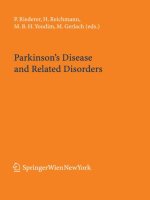
Tài liệu Parkinson’s Disease and Related Disorders docx
Bạn đang xem bản rút gọn của tài liệu. Xem và tải ngay bản đầy đủ của tài liệu tại đây (9.4 MB, 484 trang )
W
P. Riederer, H. Reichmann,
M. B. H. Youdim, M. Gerlach (eds.)
Parkinson’s Disease and Related Disorders
SpringerWienNewYork
Journal of Neural Transmission
Supplement 70
Prof. Dr. P. Riederer
Klinik und Poliklinik für Psychiatrie und Psychotherapie
Würzburg, Germany
Prof. Dr. H. Reichmann
Universitätsklinikum der TU Dresden
Dresden, Germany
Prof. Dr. M. B. H. Youdim
Israel Institute of Technology
Haifa, Israel
Prof. Dr. M. Gerlach
Klinik und Poliklinik für Psychiatrie und Psychotherapie
Würzburg, Germany
This work is subject to copyright.
$OOULJKWVDUHUHVHUYHGZKHWKHUWKHZKROHRUSDUWRIWKHPDWHULDOLVFRQFHUQHGVSHFL¿FDOO\
those of translation, reprinting, re-use of illustrations, broadcasting, reproduction by photo-
copying machines or similar means, and storage in data banks.
Product Liability: The publisher can give no guarantee for the information contained in this
book. This also refers to that on drug dosage and application thereof. In each individual case
the respective user must check the accuracy of the information given by consulting other
pharmaceutical literature. The use of registered names, trademarks, etc. in this publication
GRHVQRWLPSO\HYHQLQWKHDEVHQFHRIVSHFL¿FVWDWHPHQWWKDWVXFKQDPHVDUHH[HPSWIURP
the relevant protective laws and regulations and therefore free for general use.
© 2006 Springer-Verlag/Wien
Printed in Austria
SpringerWienNewYork is part of Springer Science+Business Media
springer.com
Typesetting: Thomson Press India Ltd., New Dehli
Printing: Druckerei Theiss GmbH, 9431 St. Stefan im Lavanttal, Austria
Printed on acid-free and chlorine-free bleached paper
SPIN: 11516439
Library of Congress Control Number: 2006928439
With 75 Figures
ISSN 0303-6995
ISBN-10 3-211-28927-5 SpringerWienNewYork
ISBN-13 978-3-211-28927-3 SpringerWienNewYork
J Neural Transm (2006) [Suppl] 70: V–VI
# Springer-Verlag 2006
Preface
It is our pleasure to present the Proceedings of the 16
th
International Congress on Parkinson’s Disease (PD) and
Related Disorders (16
th
ICPD) which took place in Berlin
from June 5 –9, 2005. This congress was the most succe ss-
ful congress ever with more than 3500 participants in the
roaring German capital, consisting of an innovative program
and with emphasis on bringing basic and clinical scientists
together. Special attention has was paid in inviting young
scientists. Therefore, the major aspect of scientific sessions
was to identify young and up coming individuals in the field,
with novel approaches to PD and novel models as a whole.
The congress gave us the opportunity to present Germany and
its capital after the burden of recent history in the new light
of a reunified and peaceful country. We have succeeded in
presenting the country as an important part of Europe and as
a country of arts, architecture and renewal. The Congress at-
tracted new friends from more than 75 countries worldwide.
For this reason, we are most thankful to the World Federa-
tion of Neurology (WFN), Research Group on Parkinsonism
and Related Disorders (RGPD), chaired by Professor Donald
Calne for bringing this congress to Germany!
The Congress had many highlights with lectures cover-
ing all the major fields in PD and Related Disorders. The
opening ceremony was highlighted by the inspiring presen-
tation of Nobel Laureate Paul Greengard who lectured on
dopamine-related signalling pathways in the brain, followed
by the welcome addresses by Professor Riederer, President of
the 16
th
ICPD, Professor Calne, President of the WFN-RGPD,
Dr. Slewett, President emeritus of the National Parkinson
Foundation, Miami, USA (NPF), Professor Kimura, President
WFN, Professor Reichmann, President German Parkinson
Society and Professor Einh
€
aaupl, Chairman of the Germ any
Science Council. The speeches were followed by a musical
interlude of the ‘‘Sunda y Night Orchestra’’ and the award
ceremony of the WFN Research Committee on Parkinson-
ism and Related Disorders. The welcome reception pre-
sented typical German dishes and drinks.
In total the congress included 4 plenary lectures, 20
symposia, 6 hot topics, 4 video sessions, 1 workshop with
demonstration, 29 educational seminars, more than 600
posters which were presented throughout the congress, 44
guided poster tours, 4 poster symposia, and 14 satellite
symposia.
There were many scientific highlights and this proceed-
ing intends to give a representative overview of congress
programme. In this preface we are only able to give a
glimpse of the outstanding lectures and scientific events
during the 5 days.
The congress started with a satellite symposium on the
significance of neuromelanin in the human brain. This sym-
posium was dedicated to Prof. Youdim on the occasion
of his 65
th
birthday. These contributions are published
separately in a Special Issue of the Journal of Neural
Transmission. Professor Carlsson, 2000 Nobel Laureate,
spent significant time at the congress site and was often
seen discussing topics of mutual interest with congress
participant’s. There was an interesting new study presented
by Professor Deuschl, Kiel, in which he demonstrates that
deep brain stimulation results in even better outcome of
motor function than regular medication. For this reason,
he advocated earlier use of deep brain stimulation in PD.
New medications were discussed in detail both in the ple-
nary lectures and satellites and new drugs such as rasagiline,
the new MAO-B-inhibitor and rotigotine, the new dermal
patch, were discussed in detail. There were satellite meet-
ings on apomorphine, COMT-inhibitors, levodopa, sphera-
mine (a new promising cell therapy for the treatment of PD
in the advanced stage), dopamine transporter scanning, do-
pamine agonists such as pramipexole, ropinirole and ca-
bergoline, adenosine antagonists, restless legs, deep brain
stimulation, botolinum toxine A, and the new lisuride der-
mal patch. All satellites were of highest quality and deliv-
ered valuable insights in present and new therapy of PD.
Special lectures addressed the advent of gene therapy and
stem cell therapy, although it is apparent that there is still
a long way to go until this therapy cab be safe and afford-
able for many PD patients longing for disease modifying
treatment.
Professors Schapira and Olanow gave an overview on the
ever contradictory aspects of neuroprotection. While neu-
roprotection is generally accepted in animal models and
cell culture, there is still discussion on whether SPECT-
and PET-analyses and the delayed start desi gn, as em-
ployed in the rasagiline study indicated neuroprotection
in man. For neuroprotection to be successful earlier diag-
nosis of PD is mandatory. For this reason, groups from
Amsterdam, Dresden, T
€
uubingen and W
€
uurzburg are working
on early diagnosis procedures such as olfactory tests, pa-
renchymal sonography, REM sleep analyses, and biochem-
ical markers.
There were lectures on treatment of PD and many on
genetic abnormalities causing PD, mitochondrial abnor-
malities and other disturbances of cell function which lead
to dopaminergic cell death.
The other major aspect of the scientific session was the
field of basic neuroscience to illuminate our current under-
standing of how neurons die in sporadic and familial PD.
This included symposia on development of midbrain dopa-
minergic neurons, the role of iron in neurodegeneration,
and the progress on genetics and proteomics and the con-
cept of developing novel multifunctional neuroprotective
drugs for such a complex disease.
Twenty nine educational seminars covered the most im-
portant topics and problems in clinical science bringing
theory to practice and treatment strategies.
The guided poster tours allowed exchange of scientific
ideas and shed light on new findings in etiology, diagnosis
and treatment of PD and related disorders.
A special highlight of the Congress was the Art Exhi-
bition, demonstrating the creativity of our patients with
movement disorders. This exhibition was organized by the
German Parkinson’s lay organisation as well as by the
Austrian lay organisation. Professor Maurer, Frankfurt, pre-
sented Art from an Alzheimer’s patient, the Carolus Horn
Exhibition, which impressively demonstrated change in the
way to paint during a dementive process.
Another highlight was the Medi cal Historical Exhibition
which was organised by Dr. Ch. Riederer, W
€
uurzburg, which
focused on the history of the treatment of PD and empha-
sized the Berlin contributions by H. Lewy, W. v. Humboldt,
R. Hassler and others.
A special tribute was paid to Melvin Yahr who sadly
passed away in early 2005 shortly before this Congress.
He was greatly missed.
Due to generous educational grants from the industry the
organizers were able to honour outstanding scient ists and
clinicians, Toshiharu Nagatsu, Yoshikuni Mizuno, Japan
(Award of the WFN Research Group on Parkinsonism
and Related Disorders), Saskia Biskup, Germany and
Andrew B. Singleton, USA (16
th
ICPD Junior Research
Award), Jonathan Brodie, Canada and Alan Crossman, UK
(Merck KGaA Dyskinesia Research Award). GE Health-
care sponsored the 16
th
ICPD Senior Resear cher Award
given to Silvia Mandel, Israel and Vincenzo Bonifati,
The Netherlands. Both companies gave educational grants
for the 12 Poste r Prizes while the Melvin Yahr Foundation
sponsored 26 Fellowships. In addition the congress made
it possible to bring numerous young scientists to the con-
gress by giving them financial support for travelling and
accommodation.
The Senator Dr. Franz Burda Award presented by
Helmut Lechner, Austria, and Franz Gerstenbrand, was
given to Laszlo Vecsei, Hungary and Tino Battistin, Italy.
We thank all the participants who gave us their creative
input to organize a World Congress on PD (as indicated in
the First Announcement) which fulfilled the criteria of excel-
lence and made the congress so successful. This was YOUR
congress and which many of you influenced by letting us
know your wishes and expectations. New concepts, formats
and innovations, the active and constructive cooperation by
the participating industry and the lay organisations made all
this possible. This can measured by the numerous compli-
mentary letters and emails we have received since then and
we hope it sets the standards for future meetings! By doing
all this we tried to come close to our milestone ‘‘Present
and Future Perspectives of Parkinson’s Syndrome’’.
Our special thanks go to CPO Hanser Congress Organi-
sation, the programme committee and the WFN Research
Group which all worked so hard to make this Congress so
successful.
Finally the congress proceedings are published and we
thank all those who contributed to this volume. Special
thanks go to Springer Verlag, Vienna, New York for their
efficient and splendid ability in being able to publish the
proceeding so rapidly.
Peter Franz Riederer, Heinz Reichmann, Moussa Youdim,
Manfred Gerlach
W
€
uurzburg, Dresden, Haifa, spring 2006
VI Preface
Contents
Powell, C.: Melvin Yahr (1917–2004). An appreciation 1
Kaufmann, H.: Melvin D. Yahr, 1917–2004. A personal recollection 5
1. Pathology
Hornykiewicz, O.: The discovery of dopamine deficiency
in the parkinsonian brain 9
Heimer, G., Rivlin, M., Israel, Z., Bergman, H.: Synchronizing activity
of basal ganglia and pathophysiology of Parkinson’s disease 17
Wichmann, T., DeLong, M. R.: Basal ganglia discharge abnormalities
in Parkinson’s disease . 21
Brown, P.: Bad oscillations in Parkinson’s disease 27
McKeown, M. J., Palmer, S. J., Au, W L., McCaig, R. G.,
Saab, R., Abu-Gharbieh, R.: Cortical muscle coupling in Parkinson’s disease
(PD) bradykinesia 31
Burke, R. E.: GDNF as a candidate striatal target-derived neurotrophic
factor for the development of substantia nigra dopamine neurons 41
Gherbassi, D., Simon, H. H.: The engrailed transcription factors
and the mesencephalic dopaminergic neurons 47
Smits, S. M., Smidt, M. P.: The role of Pitx3 in survival of midbrain
dopaminergic neurons . 57
Ryu, S., Holzschuh, J., Mahler, J., Driever, W.: Genetic analysis of dopaminergic
system development in zebrafish 61
Deutch, A. Y.: Striatal plasticity in parkinsonism: dystrophic changes
in medium spiny neurons and progression in Parkinson’s disease 67
Fuxe, K., Manger, P., Genedani, S., Agnati, L.: The nigrostriatal DA pathway
and Parkinson’s disease 71
Parent, M., Parent, A.: Relationship between axonal collateralization
and neuronal degeneration in basal ganglia 85
Braak, H., Mu
¨
ller, C. M., Ru
¨
b, U., Ackermann, H., Bratzke, H.,
de Vos, R. A. I., Del Tredici, K.: Pathology associated with sporadic
Parkinson’s disease – where does it end? . 89
Halliday, G. M., Del Tredici, K., Braak, H.: Critical appraisal of brain
pathology staging related to presymptomatic and symptomatic cases
of sporadic Parkinson’s disease 99
Giorgi, F. S., Bandettini di Poggio, A., Battaglia, G., Pellegrini, A.,
Murri, L., Ruggieri, S., Paparelli, A., Fornai, F.: A short overview
on the role of a-synuclein and proteasome in experimental models
of Parkinson’s disease . 105
Gispert-Sanchez, S., Auburger, G.: The role of protein aggregates
in neuronal pathology: guilty, innocent, or just trying to help? 111
2. Iron and neuromelanin
Double, K. L., Halliday, G. M.: New face of neuromelanin . 119
Maruyama, W., Shamoto-Nagai, M., Akao, Y., Riederer, P., Naoi, M.:
The effect of neuromelanin on the proteasome activity in human
dopaminergic SH-SY5Y cells 125
Gerlach, M., Double, K. L., Youdim, M. B. H., Riederer, P.:
Potential sources of increased iron in the substantia nigra
of parkinsonian patients 133
Pandolfo, M.: Iron and Friedreich ataxia. . . 143
3. Genetics
Chade, A. R., Kasten, M., Tanner, C. M.: Nongenetic causes
of Parkinson’s disease . 147
Lannuzel, A., Ho
¨
glinger, G. U., Champy, P., Michel, P. P.,
Hirsch, E. C., Ruberg, M.: Is atypical parkinsonism in the Caribbean
caused by the consumption of Annonacae? 153
Mellick, G. D.: CYP450, genetics and Parkinson’s disease:
geneÂenvironment interactions hold the key 159
Ravindranath, V., Kommaddi, R. P., Pai, H. V.: Unique cytochromes
P450 in human brain: implication in disease pathogenesis . 167
Viaggi, C., Pardini, C., Vaglini, F., Corsini, G. U.: Cytochrome P450
and Parkinson’s disease: protective role of neuronal CYP 2E1
from MPTP toxicity. . . 173
Miksys, S., Tyndale, R. F.: Nicotine induces brain CYP enzymes:
relevance to Parkinson’s disease 177
Riess, O., Kru
¨
ger, R., Hochstrasser, H., Soehn, A. S., Nuber, S.,
Franck, T., Berg, D.: Genetic causes of Parkinson’s disease:
extending the pathway . 181
Mizuno, Y., Hattori, N., Yoshino, H., Hatano, Y., Satoh, K.,
Tomiyama, H., Li, Y.: Progress in familial Parkinson’s disease 191
Hattori, N., Machida, Y., Sato, S., Noda, K., Iijima-Kitami, M.,
Kubo, S., Mizuno, Y.: Molecular mechanisms of nigral neurodegeneration
in Park2 and regulation of parkin protein by other proteins 205
Dawson, T. M.: Parkin and defective ubiquitination in Parkinson’s disease 209
Heutink, P.: PINK-1 and DJ-1 – new genes for autosomal recessive
Parkinson’s disease . . . 215
Whaley, N. R., Uitti, R. J., Dickson, D. W., Farrer, M. J., Wszolek, Z. K.:
Clinical and pathologic features of families with LRRK2-associated
Parkinson’s disease . . . 221
Gasser, T.: Molecular genetic findings in LRRK2 American, Canadian
and German families . . 231
4. Imaging
Lu, C S., Wu Chou, Y H., Weng, Y H., Chen, R S.: Genetic and DAT
imaging studies of familial parkinsonism in a Taiwanese cohort 235
Lok Au, W., Adams, J. R., Troiano, A., Stoessl, A. J.:
Neuroimaging in Parkinson’s disease 241
Berg, D.: Transcranial sonography in the early and differential diagnosis
of Parkinson’s disease . 249
VIII Contents
5. Models
Hirsch, E. C.: How to judge animal models of Parkinson’s disea se in terms
of neuroprotection 255
Falkenburger, B. H., Schulz, J. B.: Limitations of cellular models
in Parkinson’s disease research 261
Ho
¨
glinger, G. U., Oertel, W. H., Hirsch, E. C.: The Rotenone model
of Parkinsonism – the five years inspection 269
Schmidt, W. J., Alam, M.: Controversies on new animal models
of Parkinson’s disease Pro and Con: the rotenone model
of Parkinson’s disease (PD) 273
Kostrzewa, R. M., Kostrzewa, J. P., Brus, R., Kostrzewa, R. A., Nowak, P.:
Proposed animal model of severe Parkinson’s disease: neonatal
6-hydroxydopamine lesion of dopaminergic innervation of striatum 277
Mochizuki, H., Yamada, M., Mizuno, Y.: a-Synuclein overexpression model . 281
Ne
´
meth, H., Toldi, J., Ve
´
csei, L.: Kynurenines, Parkinson’s disease
and other neurodegenerative disorders: preclinical and clinical studies 285
6. Clinical approaches
Goetz, C. G.: What’s new? Clinical progression and staging of Parkinson’s disease 305
Wolters, E. Ch., Braak, H.: Parkinson’s disease: premotor
clinico-pathological correlations 309
Berendse, H. W., Ponsen, M. M.: Detection of preclinical Parkinson’s disease
along the olfactory trac(t) 321
Giladi, N., Balash, Y.: The clinical approach to gait disturbances
in Parkinson’s disease; maintaining independent mobility. . 327
Bodis-Wollne r, I., Jo, M Y.: Getting around and communicating
with the environment: visual cognition and language in Parkinson’s disease . . 333
Goldstein, D. S.: Cardiovascular aspects of Parkinson disease 339
Mathias, C. J.: Multiple system atrophy and autonomic failure 343
Comella, C. L.: Sleep disturbances and excessive daytime sleepiness
in Parkinson disease: an overview 349
Arnulf, I.: Sleep and wakefulness disturbances in Parkinson’s disease 357
7. Neuroinflammation
Burn, D. J.: Parkinson’s disease dementia: what’s in a Lewy body? 361
Qian, L., Hong, J S., Flood, P. M.: Role of microglia in inflammation-mediated
degeneration of dopaminergic neurons: neuroprotective effect of Interleukin 10 367
Sawada, M., Imamura, K., Nagatsu, T.: Role of cytokines in inflammatory
process in Parkinson’s disease 373
8. Neurosurgery
Benabid, A. L., Chabarde
`
s, S., Seigneuret, E., Fraix, V., Krack, P., Pollak, P.,
Xia, R., Wallace, B., Sauter, F.: Surgical therapy for Parkinson’s disease . . . 383
Hamani, C., Neimat, J., Lozano, A. M.: Deep brain stimulation for the treatment
of Parkinson’s disease . 393
Stefani, A., Fedele, E., Galati, S., Raiteri, M., Pepicelli, O., Brusa, L.,
Pierantozzi, M., Peppe, A., Pisani, A., Gattoni, G., Hainsworth, A. H.,
Bernardi, G., Stanzione, P., Mazzone, P.: Deep brain stimulation
in Parkinson’s disease patients: biochemical evidence 401
Contents IX
Agid, Y., Schu
¨
pbach, M., Gargiulo, M., Mallet, L., Houeto, J. L., Behar, C.,
Malte
ˆ
te, D., Mesnage, V., Welter, M. L.: Neurosurgery in Parkinson’s disease:
the doctor is happy, the patien t less so? . . 409
9. L-Dopa
de la Fuente-Ferna
´
ndez, R., Lidstone, S., Stoessl, A. J.: Pla cebo effect
and dopamine release. . 415
Fahn, S.: A new look at levodopa based on the ELLDOPA study 419
Chouza, C., Buzo
´
, R., Scaramelli, A., Romero, S., de Medina, O., Aljanati, R.,
Dieguez, E., Lisanti, N., Gomensoro, J.: Thirty five years of experience
in the treatment of Parkinson’s disease with levodopa and associations 427
10. Neuroprotection
Uitti, R. J., Wszolek, Z. K.: Concerning neuroprotective therapy
for Parkinson’s disease . 433
Zigmond, M. J.: Triggering endogenous neuroprotective mechanisms
in Parkinson’s disease: studies with a cellular model 439
Weinstock, M., Luques, L., Bejar, C., Shoham, S.: Ladostigil, a novel
multifunctional drug for the treatment of dementia co-morbid with depression 443
Gal, S., Fridkin, M., Amit, T., Zheng, H., Youdim, M. B. H.: M30, a novel
multifunctional neuroprotective drug with potent iron chelating
and brain selective monoamine oxidase-ab inhibitory activity
for Parkinson’s disease . 447
Weinreb, O., Amit, T., Bar-Am, O., Sagi, Y., Mandel, S., Youdim, M. B. H.:
Involvement of multiple survival signal transduction pathways
in the neuroprotective, neurorescue and APP processing activity
of rasagiline and its propargyl moiety . . . 457
11. Other treatment strategies
Schulz, J. B.: Anti-apoptotic gene therapy in Parkinson’s disease 467
Kaufmann, H.: The discovery of the pressor effect of DOPS and its blunting
by decarboxylase inhibitors 477
12. Dystonia
Hallett, M.: Pathophysiology of dystonia . . 485
Bressman, S.: Genetics of dystonia 489
Index 497
Listed in Current Contents/Life Sciences
X Contents
J Neural Transm (2006) [Suppl] 70: 1–4
# Springer-Verlag 2006
Melvin Yahr (1917–2004). An appreciation
C. Powell
Dalhousie University, Nova Scotia, Canada
Melvin Yahr (1917–2004)
I am honoured yet somewhat wary in being
invited to write an appreciation of Melvin
David Yahr. Can an outsider, a non-neurologist
and a non-American, really grasp his contri-
bution to movement disorder clinical prac-
tice, to the specialty of neurology, and to
the larger world of Medicine? In so far as
Melvin Yahr’s importance extended beyond
the borders of Neurology into all corners of
the world, the answer is, perhaps, yes. Given
such a long period of consistent and exten-
sive activity (first paper in Journal of Pedia-
trics in 1944, 357
th
in 2003), much of the
customary academic and professional rivalry
and anguish, well described by Hornykiewicz
(2004), will be unknown to an outsider and
perhaps is better left that way until some
future disinterested biographer intervenes.
Here follow brief comments on some of his
papers.
Duvoisin et al. (1963) give an insight into
some 1960s thinking. Yahr and his colleagues
studied a clinical sample from Columbia-
Presbyterian Medical Center (225 subjects
attending in 1962 of whom 195 had classical
paralysis agitans) and refuted authors who
asserted that, with the passing of the post-
encephalitic cohort, Parkinsonism would
largely disappear [by 1980] thereafter con-
stituting a numerically insignificant disease
entity.
Melvin Yahr (Yahr et al., 1969) was
important in those early years showing the
efficacy of L-dopa (from Birkmayer and
Hornykiewicz, 1961 onwards) and affirming
that enough L-dopa would produce and sus-
tain clinical response. (Hornykiewicz, 2004
engagingly and courageously records the
chronology and conflict of those papers and
their authors.) In a placebo controlled, double
blinded study, with careful evaluation (more
later about the Scale used for evaluation), 60
subjects, 56 with Parkinson’s Disease, aged
44–78 years of at least 3 years duration and
followed for 4 to 13 months, were given
750 mg to 1 gram of L-dopa 3 to 5 times
daily. All these patients had been hospital-
ized for the study – those were the days!
After initial symptomatic improvement, ob-
jectively there was ‘renewed ability to perform
simple movements which had been lost for
several years, such as turning over in bed or
rising from a chair’. They noted that some
subjects did not reach ultimate funct ional
improvement until treated for 3 to 4 months.
Abrupt cessation of the drug led to loss of
effect in the ensuing week and restoration
took at least a further week.
Younger clinicians will have no memory
of the excitement produced when L-dopa was
introduced (in today’s parlance, ‘‘Awaken-
ings’’ is a ‘must-read’). It is in the same
league as witnessing the original clinical
response to penicillin (Fletcher, 1984) or the
present writer’s joy as an intern at the effec-
tiveness of oral diuretics replacing parenteral
mercurials.
In his 1970 presidential address to the
American Neurological Association’s 95
th
Annual General Meeting, Melvin Yahr be-
gan: ‘‘I’m not a philosopher or historian,
much less a prophet’’ and then described
‘‘Neurology’s position in the present crisis
in American medicine’’. His analysis bears
repetition and response even today. He blunt-
ly asserted: ‘‘the public is disaffected with
the health care we are giving them’’ the
affluent complain about their waits to see
physicians, the indigent complain they have
no access to physicians. He warned that the
false dichotomy of ‘‘medical research’’ or
‘‘medical care’’ ignored research as the cata-
lyst for both clinical care and teaching. While
recognizing the complementary nature of
basic and applied research, he pleaded that
their funding should not be in direct compe-
tition. Where he differs from many presiden-
tial addresses, which focus on the clinician
and his (certainly his in 1970) preoccupa-
tions, is his dissection of the (American)
health care delivery system ‘‘which is about
as unhealthy, uncaring and unsystematic a
delivery system as one can imagine’’. He
emphasized the context: ‘‘the senseless war
in Vietnam, poverty, hunger, environmental
pollution, divisions between the races, alie-
nation of our young people. And somewhere
in that group inadequate medical care.’’
He challenged then and now: ‘‘the large sums
of money expended by our government on
misguided military adventures should, in-
stead, be serving the cause of human better-
ment and as physicians we have an obligation
to say so’’.
‘‘100 years ago we were unable to exist
with half slave half free, so we cannot now
continue to exist with half our people barred
from decent health care’’. He envisaged de-
veloping ‘‘a comprehensive health plan for
all to which ability to pay will cease to be
a barrier to participation’’. He then applied
these principles to neurological practice and
training. He perceptively commented on:
urban=rural practice (‘‘the irresistible ambi-
ence of West Coast living’’ – very pertinent
for a former Winnipeg physician when read
during a January sabbatical in Vancouver);
the needed continuum of care required
‘‘through the various phases of the many
long-term diseases with which we are in-
volved’’; and he made an impassioned plea
for ‘‘one class of care – first class’’. Other
topics included telehealth (not his term)
consultations, relevant CME in neurological
matters for primary care physicians, and the
relationship between the academic health
centre and its medical hinterland. To this
writer, this address was unexpectedly refresh-
ing, revealing, and still relevant.
The Hoehn and Yahr Scale (1967)
It is a truism that Parkinson’s Disease was
and is a clinical diagnosis: there are no lab-
oratory tests, no imaging techniques, no ge-
netic markers to confirm the diagnosis. It is
the clinician’s decision. This judgment nicely
combines the art and science of medicine
but the first attempt to supply a scientific
basis for this judgment appeared in Hoehn
and Yahr (1967). This is Melvin Yahr’s most
famous paper (at least 2886 citations by mid-
January, 2004) because it laid the foundation
for measuring Parkinsonism.
The Hoehn and Yahr Scale appeared be-
fore the obligatory application of psycho-
metric and clinimetric measures to clinical
scaling, before sensitivity and specificity,
before predictive values, before receiver oper-
ating curves and the rest of the scientific appa-
ratus ensuring those twin pots of gold: validity
and reliability (albeit tempered with simplic-
ity, acceptability, accuracy, cost – Cochrane
and Holland, 1971 – sensibility – Feinstein,
1987 – and responsiveness – Rockwood et al.,
2003). The main objective of the paper was to
determine the clinical variability, progression
2 C. Powell
and mortality of Parkinson’s Disease given the
then paucity of information about the natural
history of the condition. This would subse-
quently give the background upon which to
judge the effectiveness of the newly intro-
duced L-dopa therapy.
Hoehn and Yahr reported on 802 subjects
derived from a retrospective clinical sample
of 856 patients diagnosed with paralysis agi-
tans, Parkinson’s Disease and Parkinsonism
seen at the Columbia-Presbyterian Medical
Center from 1949 to 1964. Nearly 85% had
classic Parkinson’s Disease and 13% had
post-encephalitic associated Parkinsonism.
This was the largest clinical sample hitherto
studied. Two hundred and sixty three subjects
attending in 1963–4 were examined more
closely and it was from this subsample that
the famous clinical stratification was derived.
They found only 10% free of tremor at onset
and incidentally note the continuing occa-
sional clinical conundrum of Parkinsonism
and essential tremor; 14% exhibited ‘‘mild-
to-moderate organic mental syndrome usu-
ally characterized by recent memory defects
and some impairment of judgment and in-
sight’’; 4% were ‘‘moderately to severely
depressed’’ (no further details given).
They wisely point out that the presence of
the classical signs of tremor, rigidity and aki-
nesia varies with respect to disability – its
presence and progress; hence the need to
quantify this interaction of physical signs
and functional consequences into clinical
stages. Hoehn and Yahr recognized that these
stages may not correlate with pathology but
they claimed a clinimetric basis for ‘‘repro-
ducible assessments by independent exami-
ners of the general functional level of the
patient’’.
Five clinical stages, ‘‘based on the level
of clinical disability’’ were reported on 183
patients with ‘‘primary parkinsonism’’ (viz.
Parkinson’s Disease, paralysis agitans or
idiopathic Parkinsonism) – a subset of the
263 ‘more closely examined’. They dicho-
tomized these stages into: mildly affected
(Stages 1–III) and severely affected (Stages
IV–V).
Five clinica l stages: degrees of disability
Stage I: unilateral involvement only, usu-
ally with minimal or no functional
impairment.
Stage II: bilateral or midline involvement,
without impairment of balance.
Stage III: first sign of impaired righting
reflexes. This is evident by
unsteadiness as the patient turns
or is demonstrated when he is
pushed from standing equili-
brium with the feet together and
the eyes closed. Functionally the
patient is somewhat restricted in
his activities but may have some
work potential depending on the
type of employment. Patients are
usually capable of leading inde-
pendent lives and their disability
is mild to moderate.
Stage IV: fully developed, severely dis-
abling disease; the patient is still
able to walk and stand unassisted
but is markedly incapacitated.
Stage V: confinement to bed or wheelchair
unless aided.
It is unfair to criticize a 1967 paper in
terms of current epidemiological standards
(McDowell, 1996). Clinically derived scales
(e.g. Rankin, 1957 for stroke rehabilitation)
are still used inspite of academic strictures.
Ramaker et al. (2002) in their recent com-
prehensive review of measuring Parkinson’s
Disease, regret that the Hoehn and Yahr
Scale is frequently chosen ‘‘as the gold stan-
dard for testing other scales’’ because of its
lack of psychometric and clinimetric proper-
ties – but at the time it emerged it was
groundbreaking.
Melvin Yahr contributed to many major
textbooks of Medicine, Neurology and Move-
ment Disorders. In his 357 publications,
themes included: amino acid biology, the
Melvin Yahr (1917–2004). An appreciation 3
continuing relationship of central nervous
system infection and Parkinsonism, auto-
nomic nervous system failure with special
attention to orthostatic hypotension, and
every aspect of the drug management of
Parkinson’s Disease. His experience and
expertise (in others not necessarily the same
thing) were highly valued. No part of move-
ment disorder neurology was untouched by
his presence: as an explorer, quantifier, ana-
lyser, teacher and practitioner. An obituary
by former students (Di Rocco and Werner,
2004) expresses the richness of his contribu-
tion to neurology and neurologists. He was
an exemplar of successful ageing. Rejecting
the curse of mandatory retirement, he contin-
ued his clinical and academic work into the
last weeks of his life: he was the compleat
physician.
References
Birkmayer W, Hornykiewicz O (1961) The effect of
L-3, 4-dihydroxyphenyl alanine (¼DOPA) on the
Parkinsonian akinesia. Wien Klin Wochenschr 73:
787–788 (republished in English translation in
Parkinson’s Disease & Related Disorders (1998)
4: 59–60)
Cochrane AL, Holland WW (1971) Validation of
screening procedures. Br Med Bull 27: 3–8
Di Rocco A, Werner P (2004) In memoriam – Professor
Melvin David Yahr, 1917–2004. Parkinsonism Rel
Disord 10: 123–124
Duvoisin RC, Yahr MD, Schweitzer MD, Merritt HH
(1963) Parkinsonism before and since the epidemic
of Encephalitis Lethargica. Arch Neurol 9: 232–236
Feinstein AR (1987) Clinimetrics. Yale University
Press, New Haven, pp 141–166
Fletcher C (1984) First clinical use of penicillin. Br
Med J 289: 1721–1723
Hoehn MM, Yahr MD (1967) Parkinsonism: onset,
progression, and mortality. Neurology 17: 427–442
Hornykiewicz O (2004) Oleh Hornykiewicz. In: Squire
LR (ed) The history of neuroscience autobiogra-
phy, vol 4. Elsevier Academic Press, Amsterdam,
pp 243–281
Kaufmann H, Saadia D, Voustianiouk A, Goldstein DS,
Holmes C, Yahr MD, Nardin R, Freeman R (2003)
Norepinephrine precursor therapy in neurogenic
orthostatic hypotension. Circulation 108: 724–728
McDowell I (1996) Measuring health: a guide to
rating scales and questionnaires, 2nd edn. Oxford
University Press, New York
Ramaker C, Marinus J, Stiggelbout AM, van Hilten BJ
(2002) Systemic evaluation of rating scales for
impairment and disability in Parkinson’s disease.
Mov Disord 17: 867–876
Rankin J (1957) Cerebral vascular accidents in patients
over the age of 60. II. Prognosis. Scottish Med J 2:
200–215
Rockwood K,Howlett S, Stadnyk K, CarverD, PowellC,
Stolee P (2003) Responsiveness of goal attainment
scaling in a randomized trial of comprehensive
geriatric assessment. J Clin Epidemiol 56: 736–743
Sacks OW (1973) Awakenings. Duckworth, London
Yahr MD (1970) Retrospect and prospect in neurology.
Arch Neurol 23: 568–573
Yahr MD, Davis TK (1944) Myasthenia gravis – its
occurrence in a seven-year-old female child.
J Pediatr 25: 218–225 (a case report of a girl whose
symptoms probably began before her first birthday;
perhaps the youngest case then reported)
Yahr MD, Duvoisin RC, Scheer MJ, Barrett RE,
Hoehn MM (1969) Treatment of Parkinsonism with
Levodopa. Arch Neurol 21: 343–354
Author’s address: C. Powell, MB, FRCP, Professor
of Medicine, Dalhousie University, Nova Scotia,
Canada, e-mail:
4 C. Powell: Melvin Yahr (1917–2004). An appreciation
J Neural Transm (2006) [Suppl] 70: 5–7
# Springer-Verlag 2006
Melvin D. Yahr, 1917–2004. A personal recollection
H. Kaufmann
Mount Sinai School of Medicine, New York, NY, USA
Melvin D. Yahr, one of the giants of 20th
century neurology died on January 1st 2004,
aged 87, of lung cancer, at his home in
Scarsdale, New York. His was an intense and
long life of uninterrupted scientific produc-
tivity. His first paper, on myasthenia gravis,
was published in 1944 and his last one, of
course on Parkinson’s disease, appeared in
press in 2005, sixty one years later. Born in
1917 in New York City, Yahr was the young-
est of six children of immigrant parents. His
family lived in Brooklyn where his father
owned a bakery. He went to New York
University School of Medicine and completed
an internship and residency at Lenox Hill
Hospital and Montefiore Hospital in New
York City. As a student he played the clarinet
in a jazz combo to earn extra money, but in-
sisted that he was not a talented musician.
Later, when questioned about the origin of
the phenomenal musical talent of his daugh-
ters, he attributed all to his wife Felice, whom
he married when she was a 23-year-old writer
working at Fortune Magazine. Yahr served
in the US army from 1944 to 1947 and was
discharged with the rank of Major. Back
in NY, he joined the faculty at Columbia Uni-
versity College of Physicians and Surgeons
where he began his work as an academic
neurologist. He had wide clinical interests
but after a few years he began focusing on
Parkinson’s disease. Building on the work of
Carlsson, Hornykiewicz and Cotzias, in the
1960’s Yahr conducted the first double blind
randomized large clinical trials of Ldopa
in the treatment of Parkinson’s disease. The
success and impact of this treatment was
tremendous; patients were ‘‘unfrozen’’ from
statue-like rigidity and brought back to life.
In 1967, together with Peggy Hoehn, he de-
vised a 5-stage scale, simplicity itself, to de-
termine the severity of Parkinson disease. The
Hoehn and Yahr rating scale is still the gold
standard and levodopa remains the most
widely used medication for the treatment of
Parkinson’s disease.
Melvin Yahr became H Houston Merritt
professor of neurology at Columbia University
before moving downtown, as he used to say,
to Mount Sinai School of Medicine, where he
become professor of neurology and chairman
of the department. Yahr brought to Mount
Sinai the country’s first multidisciplinary cen-
ter for research in Parkinson’s disease and
related disorders, a pioneering example of
translational research. Under his leadership,
basic scientists and clinical investigators
working in close proximity, made significant
contributions to the understanding and treat-
ment of these disorders.
He chaired study sections for the National
Institute of Neurological Communicative
Disorders and Stroke, he was an adviser for
the National Research Council, the National
Academy of Sciences, and the New York City
Board of Education. He was president of the
American Board of Neurology and Psychiatry,
the American Neurological Association, and
the Association for Research in Nervous and
Mental Diseases. He received many prizes
and awards and was an honorary member of
the British, French, Belgium and Argentine
Neurological societies.
Melvin Yahr was an imposing presence. I
first met him in 1982 during my neurology
residency at Mount Sinai. He was 64, famous
and at the top of his game. He had a low
baritone voice and a very characteristic way
of speaking that we all used to imitate. He
was impeccably dressed and always wore a
crisp shirt and tie under his white lab coat.
And he smoked a pipe, an indispensable tool
for the neurologist-detective of his generation.
Yahr was first and foremost a clinician;
but believed strongly in basic research. He
loved neurology and he got great satisfaction
from his work. He was a superb teacher.
I remember vividly Morning Reports as a
senior neurology resident; every day of the
week at 9 in the morning, after rounding the
neurology ward, the senior residents went into
his office in the 14th floor of the Annenberg
Building, junior residents were not allowed.
The 5 or 6 seniors sat in couches and chairs
facing him who was sitting behind his desk,
reclined backwards, almost always smoking
a pipe. The curtains were usually lowered, so
the room was dark. Many times we couldn’t
see his face because it was covered by the
desk lamp and by a journal he was reading
and holding in front of him. One could only
see the smoke from his pipe coming up from
behind the journal. We felt we were in front
of the oracle. We presented each new patient
trying to be brief and to the point. At the end
of each patient’s presentation we heard his
voice saying: next! or some short comment.
But sometimes it was different. He would put
the journal down and ask a few more ques-
tions and then go through the differential
diagnosis or focus on one particular aspect of
the history and what it meant. For us it was
magic, it all made sense when he explained
it. He left us mesmerized and we walked out
of his office full of ideas and imagining that
we actually knew what we were all doing.
Clinical neurology was an exciting job with
Melvin Yahr.
Twice a week he also did ‘‘Chief of
Service Rounds.’’ With all the residents and
medical students sitting around him, he in-
terrogated and examined a patient from the
Neurology ward. With Melvin Yahr this was
high theatre. He was a master performer.
6 H. Kaufmann
Melvin Yahr was outspoken and blunt
and was used to be in charge. He was not
easily convinced ( – of anything), and his
most typical questions were – ‘‘What do
you want?’’ to his students and ‘‘What is it
that you cannot do?’’ to his patients. He was
frequently gruff and stern but had a fine sense
of humor and compassion.
Almost everything that is necessary for
a neurological diagnosis is in the history,
he used to say and he mostly stuck to that.
Of course he used radiology and electro-
physiology extensively, but he had a deep
distrust for all forms of testing. He asked
patients very clear questions and had the abil-
ity to make them talk and reveal information
that nobody else seemed to have been able to
obtain. He listened intently, rarely interrupt-
ing with his gaze locked on the patient. His
neurological examinations were very focused
brief and revealing: as residents, we enter-
tained the possibility that Yahr could actually
alter plantar responses in patients at will, and
we believed that he always knew what he was
going to find, as he never appeared surprised.
He kept the tradition of clinical neurology
training one on one, almost like an appren-
tice. Neurology was his passion. He was a
methodical thinker, disciplined, focused and
persistent.
Melvin Yahr did not believe in retire-
ment. When he stepped down as chairman
at Mount Sinai in 1992, his office was
demolished, literally. I guess the powers to
be thought he would have stayed there other-
wise. Undeterred, he got a new office, and
a new endowed chair remaining as active
as ever.
He appreciated beauty, loved red wine
and cognac. A favorite line of his was ‘‘It’s
a racket!’’ applied to a variety of senseless
medical or everyday life schemes. He was a
Democrat, which in the US, means liberal.
He is survived by Nancy his companion
after the death of his wife Felice in 1992,
and by 4 brilliant daughters, Carol an opera
singer, Barbara, an orchestra conductor, Laura
a pathologist and Nina a social worker, and
5 grandchildren.
Melvin Yahr died 200 years after James
Parkinson. He would have pointed that out.
Until the end Yahr remained intellectually
vibrant. He was writing and seeing patients
just a few weeks before his death. He will be
missed.
Author’s address: H. Kaufmann, MD, Mount Sinai
School of Medicine, Box 1052, New York, NY 10029,
USA, e-mail:
Melvin D. Yahr, 1917–2004. A personal recollection 7
J Neural Transm (2006) [Suppl] 70: 9–15
# Springer-Verlag 2006
The discovery of dopamine deficiency in the parkinsonian brain
O. Hornykiewicz
Center for Brain Research, Medical University of Vienna, Vienna, Austria
Summary. This article gives a short histori-
cal account of the events and circumstances
that led to the discovery of the occurrence of
dopamine (DA) in the brain and its deficiency
in Parkinson’s disease (PD). Some important
consequences, for both the basic science and
the patient, of the work on DA in the PD
brain are also highlighted.
Early opportunities
In 1951, Wilhelm Raab found a catecholamine
(CA)-like substance in animal and human
brain (Raab and Gigee, 1951). He knew that
this CA was neither noradrenaline (NA)
nor adrenaline; today, we know that it was,
at least in part, dopamine (DA). Raab exam-
ined its regional distribution in the brain of
humans, monkeys and some ‘‘larger animals’’,
and found highest levels in the caudate
nucleus. He found no changes of this CA in
the caudate in 11 ‘‘psychotic’’ patients. He
did not try to look for this compound in the
caudate nucleus of patients with Parkinson’s
disease (PD).
In 1952, G. Weber analyzed brains of
patients with PD, obtained postmortem, for
cholinesterase activity (Weber, 1952). He
found a reduction of the enzyme activity in
the putamen, and hypothesized about the
significance for PD. Had Weber known of
Raab’s study published the year before, he
might have measured Raab’s CA-like com-
pound in his PD postmortem material. In
his report, Weber does not refer to Raab’s
study.
In 1952–1954, Marthe Vogt performed
her landmark study of the regional distribu-
tion of NA and adrenal ine in the brain of
the dog (Vogt, 1952, 1954). She isolated the
amines from brain tissue extracts by paper
chromatography and eluted the corresponding
‘‘spots’’ for (biological) assays. Marthe Vogt
was well aware of Raab’s work. However, for
practical reasons, she did not stain the CA
(with ferricyanide) on the chromatograms of
regions that contained little NA, such as the
caudate; thus she let pass the opportunity of
detecting DA’s presence in the brain and its
striatal localisation.
Setting the stage for the DA===PD
studies
In August 1957, Kathleen Montagu reported
on the presence of DA, identified by paper
chromatography, in the brain of several
species, including a whole human brain
(Montagu, 1957). In November 1957, Hans
Weil-Malherbe confirmed this discovery and
examined DA’s intracellular distribution in
the rabbit brain stem (Weil-Malherbe and
Bone, 1957). Neither he, nor Montagu, offered
any speculations on the physiological role
of brain DA. At the same time as Weil-
Malherbe, in November 1957, Arvid Carlsson
observed that in na
€
ve and reserpine treated
animals ‘‘3,4-dihydroxyphenylalanine caused
central stimulation which was markedly
potentiated by iproniazid’’ (Carlsson et al.,
1957). He concluded that the study ‘‘supports
the assumption that the effect of 3,4-dihy-
droxyphenylalanine was due to an amine
formed from it’’ – leaving the question of
whether this amine was NA or DA, unconsid-
ered. In the Fall of 1957, a few weeks before
Carlsson’s report, Peter Holtz published
observations on, inter alia, L-dopa’s central
stimulant and ‘‘awakening’’ (from hexobarbi-
tal anesthesia) effects, and clearly suggested,
apparently for the first time, that this could be
due to the accumulation of ‘‘the dopamine
formed in the brain from L-dopa’’ (Holtz
et al., 1957). (Raab, in 1951, was the first to
observe increased brain levels of his CA-like
substance after i.p. L-dopa; but he does not
mention any behavioral L-dopa effects [Raab
and Gigge, 1951].)
Holtz’s conclusion was soon confirmed in
two biochemical studies. In February 1958,
Carlsson reported that reserpine depleted, in
addition to NA and serotonin, brain DA, and
L-dopa replenished it while causing central
excitation (Carlsson et al., 1958). In May
1958, Weil-Malherbe obtained, independently,
the same biochemical results in a well do-
cumented study (Weil-Malherbe and Bone,
1958). Neither Carlsson nor Weil-Malherbe
ventured any explicit statements about brain
DA’s possible physiological role or its involve-
ment in the reserpine syndrome.
More than a year before these first brain
DA studies, in the Fall of 1956, Blaschko
had already proposed that DA – until then
seen as being merely an intermediate in the
biosynthesis of CA – had ‘‘some regulating
functions of its own which are not yet
known’’ (Blaschko, 1957). In early 1957,
Hornykiewicz, in Blaschko’s Oxford labora-
tory, tested this idea experimentally. He ana-
lyzed DA’s vasodepressor action (in the
guinea pig) and proved that DA had actions
distinct from NA and adrenaline and thus
qualified as a biologically active substance in
its own right; L-dopa behaved exactly like DA
(Hornykiewicz, 1958). In 1958, Hornykiewicz
(now back in Vienna) examined (in the rat)
the central actions of several substances, in-
cluding the parkinsonism-inducing chlorpro-
mazine and bulbocapnine, as well as cocaine
and MAO inhibitors, and showed that only
the latter affected (increased) the levels of
brain DA (Holzer and Hornykiewicz, 1959).
Marthe Vogt, in her 1954 NA study in
the dog brain, inferred NA’s possible role
in brain function from the amine’s specific
distribution pattern. In January 1959, A
˚
ke
Bertler and Evald Rosengren, patterning them-
selves on Marthe Vogt’s NA study, published
a study, also in the dog, on the regional dis-
tribution of brain DA (Bertler and Rosengren,
1959a); a few weeks later, Isamu Sano re-
ported on DA’s regional distribution in the
human brain (Sano et al., 1959) (followed by
Bertler and Rosengren, 1959b). Both research
groups found that DA was mostly con-
centrated in the nuclei of the basal ganglia,
especially caudate and putamen. Bertler
and Rosengren (1959a) concluded that their
‘‘results favour [ed] the assumption that
dopamine is connected with the function of
the corpus striatum and thus with the control
of movement’’; and Sano ‘‘considered DA to
function in the extrapyramidal system which
regulates the central motoric function’’ (Sano
et al., 1959). Although Bertler and Rosengren
pointed out DA’s possible involvement in
reserpine parkinsonism, neither they nor Sano
suggested the possibility of striatal DA being
directly involved in diseases of the basal
ganglia.
DA is severely reduced
in PD striatum
Several eyewitness accounts have recently
been written about the historical events and
consequences of the discovery of the DA de-
ficiency in PD (Sourkes, 2000; Hornykiewicz,
2001a, b, 2002a, b).
Early in 1959, Hornykiewicz, aware of
DA’s localisation in the basal ganglia, started
10 O. Hornykiewicz
a study on DA in postmortem brain of patients
with PD and other basal ganglia disorders. He
and his collaborator Herbert Ehringer ana-
lyzed the brains of 17 adult non-neurological
controls, 6 brains of patients with basal gan-
glia disease of unknown etiology, 2 brains of
Huntington’s disease, and 6 Parkinson brains.
Of the 14 cases with basal ganglia disease,
only the 6 PD cases had a severe loss of
DA in the caudate and putamen (Ehringer
and Hornykiewicz, 1960). Ehringer and
Hornykiewicz concluded that their observa-
tions ‘‘could be regarded as comparable
in significance [for PD] to the histological
changes in substantia nigra’’ so that ‘‘a
particularly great importance would have to
be attributed to dopamine’s role in the patho-
physiology and symptomatology of idiopathic
Parkinson’s disease’’. This discovery was
published in December 1960. Ever since, it
has provided a solid, rational basis for all the
following research into the mechanisms, the
causes, and new treatments of PD.
It is interesting to note that in none of the
brain DA and=or L-dopa studies preceding
the Ehringer and Hornykiewicz 1960 paper,
is there any hint to be found that such a study
should be done. The first such suggestion was
made in an article from Montreal, submitted
for publication end of November 1960, report-
ing on reduced urinary DA in PD patients.
The authors concluded that future investiga-
tions should ‘‘include analysis of the cate-
cholamine content in the brains of patients
who have died with basal ganglia disorders’’,
so as to ‘‘help determine whether the concen-
tration of cerebral dopamine itself undergoes
major changes’’. The article was published in
May 1961; a ‘‘note added in proof’’ informed
the readers that the suggested study has, in the
meantime, been done (Barbeau et al., 1961).
The fact that the Montreal group quoted
the paper from Vienna so soon after it was
published on December 15, 1960, deserves a
comment. This article was written in German
and published in a German language journal.
Theodore Sourkes, the leading biochemist of
the Montreal group, must have read it almost
immediately after it came out. He contacted
Hornykiewicz about this article by letter
dated February 10, 1961. For the Vienna dis-
covery, there were, obviously, neither lan-
guage nor information transfer barriers.
This was opposite to what happened to a
(lecture) overview article of Sano, published
in Japanese in 1960. Independently from
Hornykiewicz, Sano had analyzed the brain
of a single PD patient, but was ‘‘reluctant to
speculate, from that single experience [low
putamen DA] about the pathogenesis of
Parkinson’s disease’’ (Sano, 1962). The publi-
cation remained unnoticed until it was recently
reprinted in English translation (Sano, 2000).
The question arises: Why did none of the
pioneers of the early brain DA research think
of studying the PD brain? It appears that the
main reason was their too exclusive preoccu-
pation with the central effects of reserpine.
This is surprizing because even then it was
obvious that reserpine, like most pharmaco-
logical animal models, was not a perfect cen-
trally acting drug; it depleted, to the same
degree as DA, also the brain NA and seroto-
nin, making a clear decision about the rela-
tive importance of these changes impossible.
The exclusive ‘‘fixation’’ on reserpine made
leading monoamine researchers of that period
overlook the most obvious, that is, PD as the
ultimate ‘‘brain DA experiment of Nature’’.
Two practical consequences
Inaugurating the nigrostriatal
DA pathway
When the DA deficiency in PD was discovered,
nothing was known about DA’s cellular loca-
lisation in the brain. In Huntington’s disease,
Ehringer and Hornykiewicz (1960) had found
normal striatal DA. Since in Huntington’s
disease there is a severe loss of striatal neu-
rons accompanied by marked gliosis, the nor-
mal striatal DA suggested that the amine was
probably contained in terminals of fibre tracts
originating outside the striatum. Rolf Hassler
The discovery of dopamine deficiency in the parkinsonian brain 11
had proved, back in 1938, that in PD, loss
of the substantia nigra compacta neurons
was the most consistent pathological change
(Hassler, 1938). Thus, in 1962, Hornykiewicz
started a study of the substantia nigra in 10
PD brains. The outcome of such a study was
by no means certain. Hassler himself rejected
the possibility of a nigro-striatal connection
(see page 869 in: Jung and Hassler, 1960);
and Derek Denny-Brown declared, in 1962,
that ‘‘we have presented reasons against the
common assumption that lesions of the sub-
stantia nigra are responsible fo parkinson-
ism’’ (Denny-Brown, 1962). In his study,
Hornykiewicz found markedly reduced nigral
DA, similar to the DA loss in the striatum. In
the report published in 1963, Hornykiewicz
concluded from his observation that ‘‘on the
other hand, cell loss in the [PD] substantia
nigra could well be the cause of the dopamine
deficit in the striatum’’ (Hornykiewicz, 1963).
At the time of Hornykiewicz’s DA=
substantia nigra study, two research groups
were already trying to tackle the question
of brain DA’s cellular localization. In
Montreal, Poirier and Sourkes were using
electrolytic brain lesions, in the primate; in
Sweden, Fuxe, Dahlstr
€
oom (and others) were
applying, in the rat, the just developed
CA histofluorescence method. A year after
Hornykiewicz published his study, each of
the two research groups was able to report
on the existence of a DA-containing nigros-
triatal connection. Both groups referred, in
their first publications, to Hornykiewicz’s
1963 nigral DA study (And
een et al., 1964;
Dahlstr
€
oom and Fuxe, 1964; Poirier and
Sourkes, 1965). This contribution to the dis-
covery of the nigrostriatal DA pathway had
for Hornykiewicz yet another consequence.
Several years later, Hassler wrote him a letter
in which he expressed his candid opinion on
the nigrostriatal DA pathway. He wrote:
‘‘I believe that your interpretation of your
observations does not agree with many
known facts, this being so because you accept
the American [?!] opinion about the direction
of the nigrostriatal connections. I believe that
all your observations can be equally well, or
even better, explained by the striatonigral
direction [of that pathway] ’’ (Hassler, 1967).
L-dopa for the PD patient
The discovery of the severe striatal DA defi-
ciency in PD had also a far-reaching clinical
consequence. Hornykiewicz immediately took
the step ‘‘from brain homogenate to treat-
ment’’ and asked the neurologist Walther
Birkmayer to do clinical trials with i.v.
L-dopa. After a delay of eight months, in July
1961, Birkmayer injected 50–150 mg L-dopa
i.v. in 20 PD patients, most of them pre-
treated with an MAO inhibitor. The first
report, published in November 1961, conveys,
even today, the excitement about what since
has been called ‘‘the dopamine miracle’’; it
reads as follows:
The effect of a single i.v. administration of
L-dopa was, in short, a complete abolition
or substantial relief of akinesia. Bed-ridden
patients who were unable to sit up; patients
who could not stand up when seated; and
patients who when standing could not start
walking, performed after L-dopa all these
activities with ease. They walked around
with normal associated movements and they
even could run and jump. The voiceless,
aphonic speech, blurred by pallilalia and
unclear articulation, became forceful and
clear as in a normal person. For short periods
of time the patients were able to perform
motor activities which could not be prompted
to any comparable degree by any other known
drug. (Birkmayer and Hornykiewicz, 1961).
Simultaneously with, and independently
from, the trials in Vienna, Sourkes and
Murphy, in Montreal, proposed to Barbeau
a trial of oral L-dopa. They observed, with
200 mg L-dopa, an amelioration of rigidity
that ‘‘was of the order of 50 percent’’
(Barbeau et al., 1962). Interestingly, Sano in
his overview in 1960 also mentioned that he
12 O. Hornykiewicz
had injected 200 mg L-dopa i.v. in two
patients; however, he did not evaluate the
effect clinically, being ‘‘more interested in
subjective complaints’’ (Sano, 1962). Sano
concluded that ‘‘treatment with dopa has no
practical value’’ (Sano, 2000).
Today, especially thanks to Cotzias’s in-
troduction of the high dose oral treatment
regimen (Cotzias et al., 1967), L-dopa is
recognized as the most powerful drug avail-
able for PD. As Sourkes very aptly expressed
it, the discovery of L-dopa ‘‘proved to be the
culmination of a century-and-a-half search
for a treatment of Parkinson’s disease’’
(Sourkes, 2000).
Despite the unprecedented success, doubts
were expressed about L-dopa’s ‘‘miraculous’’
antiparkinson effect. Many neurologists sus-
pected a placebo effect of the i.v. injected
L-dopa, ignoring the fact that Birkmayer
and Hornykiewicz (1962) had described,
already in 1962, the ineffectiveness of i.v.
injected compounds related to L-dopa, such
as: D-dopa, 3-O-methyldopa, DA, D, L-dops,
and also 5-HTP. This should have convinced
the doubters that the L-dopa effect could not
have been a placebo effect.
Especially counterproductive were various
statements by some rather prominent brain
scientists. Thus, some claimed that ‘‘the
actions of DOPA and DOPS [the direct pre-
cursor of NA] were similar’’, cautioning that
‘‘dopamine can activate not only its own
receptors [in the brain], but also those of nor-
adrenaline, and vice versa’’ (Carlsson, 1964,
1965); others felt that ‘‘the effect of L-dopa
was too complex to permit a conclusion
about disturbances of the dopamine system in
Parkinson’s disease’’ (Bertler and Rosengren,
1966), still others compressed all their doubts
in the terse phrase that L-dopa ‘‘was the right
therapy for the wrong reason’’ (Ward, 1970;
Jasper, 1970); and, finally, there was the
statement that ‘‘since L-dopa floods the brain
with dopamine, to relate its [antiparkinson]
effects to the natural function of dopamine
neurons may be erroneous’’ (Vogt, 1973).
These and similar critical statements dimin-
ished the status of L-dopa as a specific DA
replacing agent and put in doubt the very
concept of DA replacement in PD.
Viewed against the background of the
initial skepticism, today’s opinion has sub-
stantially changed, as reflected, for instance,
in a recent ‘‘Editorial’’:
The identification of the dopaminergic de-
ficit in Parkinson’s disease and the develop-
ment of dopamine replacement therapy by
Hornykiewicz and his contemporaries pro-
foundly influenced research into Parkinson’s
disease, and perhaps even all neurological dis-
orders. This is especially true for Alzheimer’s
disease, in which current cholinergic therapy
is the intellectual heir of dopamine replace-
ment therapy for Parkinson’s disease. (Hardy
and Langston, 2004).
Thus has theoretically based research led,
in an amazingly straight line, to very practi-
cal results. As Immanuel Kant, that eminent
philosopher of the Age of Enlightenment, put
it some 200 years ago: ‘‘There is nothing
more practical than a sound theory’’.
References
And
een NE, Carlsson A, Dahlstr
€
oom A, Fuxe K, Hillarp
NA, Larsson K (1964) Demonstration and mapping
out of nigro-striatal dopamine neurons. Life Sci 3:
523–530
Barbeau A, Murphy GF, Sourkes TL (1961) Excretion
of dopamine in diseases of basal ganglia. Science
133: 1706–1707
Barbeau A, Sourkes TL, Murphy GF (1962) Les
cat
eecholamines dans la maladie de Parkinson. In:
deAjuriaguerra (ed) Monoamines et syst
eeme ner-
veux centrale. Georg, Gen
eeve and Masson, Paris,
pp 247–262
Bertler A
˚
, Rosengren E (1959a) Occurrence and dis-
tribution of dopamine in brain and other tissues.
Experientia 15: 10–11
Bertler A
˚
, Rosengren E (1959b) On the distribution in
brain monoamines and of enzymes responsible for
their formation. Experientia 15: 382–383
Birkmayer W, Hornykiewicz O (1961) Der L-Dioxy-
phenylalanin (¼DOPA)-Effekt bei der Parkinson-
Akinese. Wien Klin Wochenschr 73: 787–788
The discovery of dopamine deficiency in the parkinsonian brain 13
Birkmayer W, Hornykiewicz O (1962) Der L-Dioxy-
phenylalanin (¼DOPA)-Effekt beim Parkinson-
Syndrom des Menschen: zur Pathogenese und
Behandlung der Parkinson-Akinese. Arch Psychiat
Nervenkr 203: 560–574
Blaschko H (1957) Metabolism and storage of biogenic
amines. Experientia 13: 9–12
Carlsson A (1964) Functional significance of drug-
induced changes in brain monoamine levels.
In: Himwich HE, Himwich WA (eds) Biogenic
amines. Elsevier, Amsterdam, pp 9–27 (Progr Brain
Res 8)
Carlsson A (1965) Dr ugs which b lock the storage of
5-hydroxytryptamine and related amines. In:
Eichler O, Farah A (eds) 5-Hydroxytryptamine
and related indolealkylamines. Springer, Berlin
Heidelberg New York, pp 529–592 (Handb Exp
Pharmacol vol 19)
Carlsson A, Lindqvist M, Magnusson T (1957) 3,4-
Dihydroxyphenylalanine and 5-hydroxy-trypto-
phan as reserpine antagonists. Nature 180: 1200
Carlsson A, Lindqvist M, Magnusson T, Waldeck B
(1958) On the presence of 3-hydroxy-tyramine in
brain. Science 127: 471
Cotzias GC, Van Woert MH, Schiffer IM (1967) Aro-
matic amino acids and modification of Parkinson-
ism. N Engl J Med 276: 374–379
Dahlstr
€
oom A, Fuxe K (1964) Evidence for the exis-
tence of monoamine-containing neurons in the
central nervous system. I. Demonstration of mono-
amines in the cell bodies of brain stem neurons.
Acta Physiol Scand 62 [Suppl 232]
Denny-Brown D (1962) The basal ganglia and their
relation to disorders of movement. Oxford Univer-
sity Press, Oxford
Ehringer H, Hornykiewicz O (1960) Verteilung von
Noradrenalin und Dopamin (3-Hydroxytyramin)
im Gehirn des Menschen und ihr Verhalten bei
Erkrankungen des extrapyramidalen Systems. Klin
Wochenschr 38: 1236–1239
Hardy J, Langston WJ (2004) How many pathways are
there to nigral death? Ann Neurol 56: 316–318
Hassler R (1938) Zur Pathologie der Paralysis agitans
und des postenzephalitischen Parkinsonismus.
J Psychol Neurol 48: 387–476
Hassler R (1967) Private communication to
O. Hornykiewicz. Letter dated February 9, 1967
Holtz P, Balzer H, Westermann E, Wezler E (1957)
Beeinflussung der Evipannarkose durch Reserpin,
Iproniazid und biogene Amine. Arch Exp Path
Pharmakol 231: 333–348
Holzer G, Hornykiewicz O (1959)
€
UUber den Dopamin-
(Hydroxytyramin-) Stoffwechsel im Gehirn der
Ratte. Naunyn Schmiedebergs Arch Exp Path
Pharmacol 237: 27–33
Hornykiewicz O (1958) The action of dopamine on the
arterial pressure of the guinea pig. Br J Pharmacol
13: 91–94
Hornykiewicz O (1963) Die topische Lokalisation und
das Verhalten von Noradrenalin und Dopamin
(3-Hydroxytyramin) in the Substantia nigra des
normalen und Parkinsonkranken Menschen. Wien
Klin Wochenschr 75: 309–312
Hornykiewicz O (2001a) Brain dopamine: a historical
perspective. In: Di Chiara G (ed) Dopamine in
the CNS I. Springer, Berlin Heidelberg, pp 1–22
(Handb Exp Pharmacol vol 154=I)
Hornykiewicz O (2001b) How L-DOPA was discov-
ered as a drug for Parkinson’s disease 40 years ago.
Wien Klin Wochenschr 113: 855–862
Hornykiewicz O (2002a) Dopamine and Parkinson’s
disease. A personal view of the past, the present,
and the future. Adv Neurol 86: 1–11
Hornykiewicz O (2002b) Dopamine miracle: from brain
homogenate to dopamine replacement. Mov Disord
17: 501 –508
Jasper HH (1970) Neurophysiological mechanisms in
parkinsonism. In: Barbeau A, McDowell FH (eds)
L-Dopa and parkinsonism. FA Davis, Philadelphia,
pp 408–411
Jung R, Hassler R (1960) The extrapyramidal motor
systems. In: Field J, Magoun HW, Hall VE (eds)
Handbook of Physiology, sect 1. Neurophysiology,
vol II. American Physiological Society, Washington
DC, pp 863–927
Montagu KA (1957) Catechol compounds in rat tissues
and in brains of different animals. Nature 180:
244–245
Poirier LJ, Sourkes TL (1965) Influence of the sub-
stantia nigra on the catecholamine content of the
striatum. Brain 88: 181–192
Raab W, Gigee W (1951) Concentration and distribu-
tion of ‘‘encephalin’’ in the brain of humans and
animals. Proc Soc Exp Biol Med 76: 97–100
Sano I (1962) Private communication to
O.Hornykiewicz. Letter dated March 20, 1962
Sano I (2000) Biochemistry of the extrapyramidal
system. Parkinsonism Relat Disord 6: 3–6
Sano I, Gamo T, Kakimoto Y, Taniguchi K, Takesada
M, Nishinuma K (1959) Distribution of catechol
compounds in human brain. Biochim Biophys Acta
32: 586 –587
Sourkes TL (2000) How dopamine was recognised as a
neurotransmitter: a personal view. Parkinsonism
Relat Disord 6: 63–67
Vogt M (1952) Die Verteilung pharmakologisch
aktiver Substanzen im Zentralnervensystem. Klin
Wochenschr 30: 907–908
Vogt M (1954) The concentration of sympathin in
different parts of the central nervous system under
14 O. Hornykiewicz
normal conditions and after the administration of
drugs. J Physiol 123: 451–481
Vogt M (1973) Functional aspec ts of the role of cate-
cholamines in the central nervous system. Br Med
Bull 29: 168–172
Ward AA (1970) Physiological implications in the
dyskinesias. In: Barbeau A, McDowell FH (eds)
L-Dopa and parkinsonism. FA Davis, Philadelphia,
pp 151–159
Weber G (1952) Zum Cholinesterasegehalt des Gehirns
bei Hirntumoren und bei Parkinsonismus. Bull
Schweiz Akad Med Wiss 8: 263–268
Weil-Malherbe H, Bone AD (1957) Intracellular dis-
tribution of catecholamines in the brain. Nature
180: 1050–1051
Weil-Malherbe H, Bone AD (1958) Effect of reserpine
on the intracellular distribution of catecholamines
in the brain stem of the rabbit. Nature 181:
1474 –1475
Author’s address: Dr. O. Hornykiewicz, Center
for Brain Research, Medical University of Vienna,
Spitalgasse 4, 1090 Vienna, Austria, Fax: (þ431) 4277-
628-99
The discovery of dopamine deficiency in the parkinsonian brain 15
J Neural Transm (2006) [Suppl] 70: 17–20
# Springer-Verlag 2006
Synchronizing activity of basal ganglia and pathophysiology
of Parkinson’s disease
G. Heimer
1
, M. Rivlin
1;2
, Z. Israel
3
, and H. Bergman
1;2
1
Department of Physiology, Hadassah Medical School,
2
ICNC, The Hebrew University, and
3
Department of Neurosurgery, Hadassah University Hospital, Jerusalem, Israel
Summary. Early physiological studies em-
phasized changes in the discharge rate of
basal ganglia in the pathophysiology of
Parkinson’s disease (PD), whereas recent
studies stressed the role of the abnormal oscil-
latory activity and neuronal synchronization
of pallidal cells. However, human observa-
tions cast doubt on the synchronization
hypothesis since increased synchronization
may be an epi-p henomenon of the tremor
or of independent oscillators with similar fre-
quency. Here, we show that modern actor=
critic models of the basal ganglia predict the
emergence of synchronized activity in PD and
that significant non-oscillatory and oscillatory
correlations are found in MPTP primates. We
conclude that the normal fluctuation of basal
ganglia dopamine levels combined with local
cortico-striatal learning rules lead to non-
correlated activity in the pallidum. Dopamine
depletion, as in PD, results in correlated palli-
dal activity, and reduced information capacity.
We therefore suggest that future deep brain
stimulation (DBS) algorithms may be im-
proved by desynchronizing pallidal activity.
Introduction: The computational roles
of the basal ganglia and dopamine
Modeling of the basal ganglia has played
a major role in our understanding of the
physiology and pathophysiology of this
elusive group of nuclei. These models have
undergone evolutionary and revolutionary
changes over the last twenty years, as on-
going research in the fields of anatomy, phys-
iology and biochemistry of these nuclei has
yielded new information. Early models dealt
with a single pathway through the basal
ganglia nuclei (cortex-striatum-internal seg-
ment of the globus pallidus; GPi) and focused
on the nature of the processing perfo rmed
within it, convergence of information vs.
parallel processing of information. Later, the
dual (direct and indirect) pathway model
(Albin et al., 1989) characterized the inter-
nuclei interaction as multiple pathways while
maintaining a simplistic scalar representation
of the nuclei themselves. The dual pathway
of the basal ganglia networks emphasized
changes in the discharge rates of basal gan-
glia neurons. The model predicts that in the
dopamine depleted Parkinsonian state firing
rates in the external segment of the globus
pallidus (GPe) are reduced, whereas cells in
the internal segment (GPi) and the subthalam-
ic nucleus (STN) display increased firing
rates (Miller and DeLong, 1987; Bergman
et al., 1994). This model resulted in a clinical
breakthrough by providing key insights into
the behavior of these nuclei in hypo- and
hyper-kinetic movement disorders, and lead


