Tài liệu Association of killer cell immunoglobulin-like receptors with pulmonary tuberculosis in Chinese Han pdf
Bạn đang xem bản rút gọn của tài liệu. Xem và tải ngay bản đầy đủ của tài liệu tại đây (799.08 KB, 9 trang )
©FUNPEC-RP www.funpecrp.com.br
Genetics and Molecular Research 11 (2) 1370-1378 (2012)
Association of killer cell immunoglobulin-like
receptors with pulmonary tuberculosis in
Chinese Han
C. Lu
1
, Y J. Shen
1
, Y F. Deng
2
, C Y. Wang
1
, G. Fan
1
, Y Q. Liu
1
,
S M. Zhao
1
, B C. Zhang
1
, Y R. Zhao
3
, Z E. Wang
1
, C Z. Zhang
1
and Z M. Lu
1
1
Department of Laboratory Medicine,
Shandong Provincial Hospital Afliated to Shandong University,
Shandong University, Jinan, China
2
Department of Infection Control, Shandong Provincial Chest Hospital,
Jinan, China
3
Department of Center Laboratory,
Shandong Provincial Hospital Afliated to Shandong University,
Shandong University, Jinan, China
Corresponding author: Z M. Lu
E-mail:
Genet. Mol. Res. 11 (2) 1370-1378 (2012)
Received September 13, 2011
Accepted February 9, 2012
Published May 15, 2012
DOI />ABSTRACT. Killer cell immunoglobulin-like receptors (KIRs) are
involved in the pathogenesis of a variety of diseases. However, whether
KIR polymorphism is associated with susceptibility to pulmonary
tuberculosis was unknown. We examined a possible association of
KIR polymorphism with susceptibility to pulmonary tuberculosis in
Chinese Han. We analyzed 15 KIR genes in 109 pulmonary tuberculosis
patients and 110 healthy controls using sequence-specic primer PCR
analysis of genomic DNA. We found that the frequencies of KIR2DS1,
2DS3 and 3DS1 were signicantly higher in patients than in the
control group. In addition, the number of subjects carrying more than
two activating KIR genes in the patient group was signicantly higher
than in the control group. The gene cluster containing KIR3DS1-
Association of KIR with pulmonary tuberculosis
©FUNPEC-RP www.funpecrp.com.br
Genetics and Molecular Research 11 (2) 1370-1378 (2012)
1371
2DL5-2DS1-2DS5 was also signicantly more frequent in the patient
group. In conclusion, KIR genes 2DS1, 2DS3 and 3DS1 appear to be
associated with resistance to pulmonary tuberculosis in the Chinese
Han population. KIR genes apparently have a role in resistance to
pulmonary tuberculosis.
Key words: Pulmonary tuberculosis; Polymorphism; Susceptibility;
Killer cell immunoglobulin-like receptor
INTRODUCTION
Tuberculosis (TB) remains a worldwide health threat. One-third of the world’s popu-
lation is currently infected with Mycobacterium tuberculosis that results in active or latent
infection. TB causes nearly 1.8 million deaths annually (Doherty et al., 2009). Because of the
increasing mobility of the population, the changing environment, and the biology of bacilli,
the prevalence of TB is higher in China. Epidemiological studies indicate that only 5 to 10% of
people infected with M. tuberculosis develop active TB (van Crevel et al., 2002); the immune
system can prevent the development of active disease. The genetic factors of TB have been
studied using many methods, including selection of candidate genes and genome-wide linkage
studies (Casanova and Abel, 2002; Fernando and Britton, 2006; Berrington and Hawn, 2007).
Studies focusing on the impact of candidate genes (Zhang et al., 2010) such as human leuko-
cyte antigen (HLA) have provided an understanding of TB infection. Most studies on human
genes relevant to pulmonary TB (PTB) have focused on HLA (Sriram et al., 2001). Because
KIRs modulate cell function upon recognition of HLA class I molecules, we can infer that they
may exert a crucial role in the mechanism of PTB.
KIR belongs to the immunoglobulin superfamily, which is expressed on natural killer
(NK) cells and T cells (Parham, 2005). It is named according to the number of extracellular
immunoglobulin-like domains (2D or 3D for 2 domains or 3 domains, respectively) and intra-
cytoplasmic tails (S or L for short or long). The family comprises 17 KIR genes, reaching 150
kb from head to tail on 19q13.4 (Moretta and Moretta, 2004). They are KIR2DL1-5, 2DS1-5,
3DL1-3, 3DS1, 1D, and 2 pseudo genes (3DP1 and 2DP1; Gutiérrez-Rodríguez et al., 2006).
Short-tailed receptors confer activating signals with a positively charged amino acid in their
transmembrane region. Long-tailed receptors carry immunoreceptor tyrosine-based inhibitory
motifs and confer inhibitory signals. Specically, KIR genes recognize major histocompatibili-
ty complex molecules and transmit signals to KIR-bearing cells. A reasonable hypothesis is that
KIR polymorphic loci regulate host immunity against infectious pathogens (Yen et al., 2001).
Moreover, accumulating evidence suggests that KIR-specic genes contribute to the patho-
genesis of other diseases (Cooley et al., 2009; Vidal-Castineira et al., 2010; Seich Al Basatena
et al., 2011), such as psoriasis, rheumatoid arthritis, hepatitis C. However, the functions and
effects of KIR genes during the development of TB remain unclear in the Chinese population.
In the present study, we aimed to investigate the KIR genes in PTB patients and con-
trols using sequence-specic primer polymerase chain reaction (PCR). The innovative point
of our study was the subdivision of the patient group according to sputum smear test (positive
and negative). Our ndings provide a better understanding of the genetic diversity of KIR
genes across PTB patients in China.
C. Lu et al.
©FUNPEC-RP www.funpecrp.com.br
Genetics and Molecular Research 11 (2) 1370-1378 (2012)
1372
MATERIAL AND METHODS
Subjects
A total of 109 patients (60 men and 49 women; mean age: 41.7 years; range: 19-69
years) with PTB and 110 unrelated healthy controls (53 men and 57 women; mean age: 38.3
years; range: 21-67 years) were recruited from Shandong Chest Hospital, Jinan, between May
2010 and May 2011. The patients were treated at the hospital. According to American Tuber-
culosis Society criteria, patients were diagnosed on the basis of history, presence of recent
clinical symptoms, clinical signs, chest radiography, tuberculin skin test, and sputum smear
test. No extra-pulmonary involvement was detected in the patients. The patient group was sub-
divided into two groups according to the results of a sputum smear test (positive or negative).
The average age of the subgroups was the same. Blood was collected before the initiation of
anti-tuberculosis therapy. A total of 110 unrelated healthy subjects with a history of positive
(indurate area ≥ 10 mm with vaccination scars) tuberculin skin test (0.1 mL puried protein
derivative in a concentration of 5 U) were recruited as controls. Radiographic scans of controls
revealed no PTB, and all subjects had been inoculated with Bacillus Calmette-Guerin vaccine.
Patients and controls were matched for age, gender, ethnicity, and socioeconomic sta-
tus. Patients and controls with diabetes, chronic renal failure, malignant disease, and immu-
nological or autoimmune disease as well as those who were human immunodeciency virus
positive or had other concurrent diseases that could affect the immune system were excluded.
Informed consent was obtained from each individual. The Hospital Ethics Committee ap-
proved the research.
Genomic DNA isolation
Genomic DNA was extracted from 5 mL ethylenediaminetetraacetic acid anticoagu-
lated peripheral blood using a TIANamp Blood DNA Kit (Tiangen Biotech, Beijing, China)
according to the manufacturer’s instructions, and the extracted DNA was stored at -20°C. The
integrity and quantity of DNA samples were determined using a UV spectrophotometer, and
the DNA concentration was adjusted to 50 ng/μL.
Sequence-specic primer PCR
All recruited subjects underwent KIR genotyping. Our previous research demonstrat-
ed that the framework genes KIR2DL4, 3DL2, 3DL3, and 3DP1 are present in all individuals
(Zhi-Ming et al., 2007). Therefore, KIR locus typing was performed to detect the presence or
absence of 11 known KIR genes, including 2DL1-3, 2DL5, 2DS1-5, 3DL1, and 3DS1. The
primers used in this study were designed based on primer sites described elsewhere (Martin et
al., 2002b). All primers (Bo Ya Biotechnology Co. Ltd., Shanghai, China) were validated, and
their gene specicity was conrmed. Two sets of primers were designed for each KIR gene
(except for 2DS5; Table 1). PCR was preformed within a 20-μL system containing 6.6 μL PCR
loading dye mix (Takara, Kyoto, Japan) including Taq DNA polymerase, 6 μL forward and
reverse primers (1.2 μM), 6.9 μL RNase Free (Takara), and 0.5 μL genomic DNA (50 ng/μL).
Briey, after a denaturing step at 94°C for 1 min, PCR amplication was carried out with 10
Association of KIR with pulmonary tuberculosis
©FUNPEC-RP www.funpecrp.com.br
Genetics and Molecular Research 11 (2) 1370-1378 (2012)
1373
cycles at a melting temperature of 94°C for 30 s, and an annealing temperature of 65°C for 30
s. Subsequently, 20 cycles were performed at a melting temperature of 94°C for 30 s, an an-
nealing temperature of 62°C for 30 s, and an extension temperature of 72°C for 40 s. Finally,
an extra extension step at 72°C was preformed. The amplicons were analyzed on ethidium
bromide-stained agarose gels (1.5%) using a 1-kb DNA ladder as a molecular weight marker.
After electrophoresis, the agarose gel was scanned and imaged with an Alphaimager TM 2200
instrument (Alpha Innotech Corporation, San Leandro, CA, USA). Products of predicted size
were visualized under ultraviolet light, and each sample was genotyped.
KIR Forward (5'-3') Reverse (5'-3') Bp
3DL1-1 CGCTGTGGTGCCTCGA GGTGTGAACCCCGACATG 197
3DL1-2 CCCTGGTGAAATCAGGAGAGAG TGTAGGTCCCTGCAAGGGCAA 181
3DS1-1 AGCCTGCAGGGAACAGAAG GCCTGACTGTGGTGCTCG 300
3DS1-2 CCTGGTGAAATCAGGAGAGAG GTCCCTGCAAGGGCAC 177
2DL5-1 GCGCTGTGGTGCCTCG GACCACTCAATGGGGGAGC 214
2DL5-2 TGCAGCTCCAGGAGCTCA GGGTCTGACCACTCATAGGGT 194
2DL1-1 GTTGGTCAGATGTCATGTTTGAA GGTCCCTGCCAGGTCTTGCG 127
2DL1-2 TGGACCAAGAGTCTGCAGGA TGTTGTCTCCCTAGAAGACG 330
2DL2-1 CTGGCCCACCCAGGTCG GGACCGATGGAGAAGTTGGCT 173
2DL2-2 GAGGGGGAGGCCCATGAAT TCGAGTTTGACCACTCGTAT 150
2DL3-1 CTTCATCGCTGGTGCTG AGGCTCTTGGTCCATTACAA 550
2DL3-2 TCCTTCATCGCTGGTGCTG GGCAGGAGACAACTTTGGATCA 800
2DS2-1 TTCTGCACAGAGAGGGGAAGTA CTTCTCCATCAGTCGCATGAG 102
2DS2-2 CGGGCCCCACGGTTT GGTCACTCGAGTTTGACCACTCA 240
2DS3-1 TGGCCCACCCAGGTCG TGAAAACTGATAGGGGGAGTGAGG 242
2DS3-2 CTATGACATGTACCATCTATCCAC AAGCAGTGGGTCACTTGAC 190
2DS4-1 CTGGCCCTCCCAGGTCA TCTGTAGGTTCCTGCAAGGACAG 204
2DS4-2 CTGGCCCTCCCAGGTCA GGAATGTTCCGTTGATGC 2000
2DS5 TGATGGGGTCTCCAAGGG TCCAGAGGGTCACTGGGC 125
2DS1-1 CTTCTCCATCAGTCGCATGAA CTTCTCCATCAGTCGCATGAG 102
2DS1-2 CTTCTCCATCAGTCGCATGAA AGAGGGTCACTGGGAGCTGAC 102
Table 1. SSP-PCR primers of KIR genes.
Statistical analysis
Briey, observed phenotype frequencies (pf) of KIR genes were determined us-
ing the ratio of gene presence within the population over the total population. Genotype
frequency (gf) of each locus was calculated using following formula: genotype frequency
= 1 - √ (1 - pf). Framework genes were included in the analysis of KIR gene numbers and
their ratios. Frequency differences of KIR loci between patients and controls were analyzed
using the chi-square test. The 95% (95%CI) condence interval of the calculated odd ratio
(OR) was estimated. P value was multiplied by the number of comparisons, and a value of
P < 0.05 was considered statistically signicant. Analyses were performed using Statistical
Package for Social Sciences Version 16.0 (SPSS, Chicago, IL, USA).
RESULTS
Statistical analysis indicated no signicant differences between groups with respect to
age and gender (P > 0.05). All KIR genes were tested both in the patient groups (smear nega-
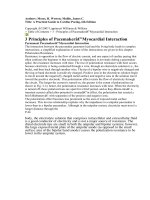
tive and positive) and in the control group at different frequencies (Table 2). Figure 1 shows
the amplication of KIR genes from one patient.
C. Lu et al.
©FUNPEC-RP www.funpecrp.com.br
Genetics and Molecular Research 11 (2) 1370-1378 (2012)
1374
Phenotype and genotype frequencies of KIRs in patients and controls
Our data showed that KIR3DL1, 2DL1, 2DL3, and 2DS4 were the most frequently
found genes in both patients and controls. The total carriage frequencies of KIR2DS1, 2DS3,
and 3DS1 in the patient groups were higher than those in the control group (P = 0.002, P <
0.001, and P = 0.011, respectively). Moreover, the frequencies of KIR2DS2 and KIR2DS5 in
KIR Control (N = 110) PTB (N = 109) OR (CI) P
n pf (%) gf (%) n pf (%) gf (%)
Inhibitory
2DL1 107 97.3 83.6 108 99.1 90.5 3.028 (0.310~29.572) 0.317
2DL2 73 66.4 42.0 83 76.1 51.1 1.618 (0.895~2.925) 0.110
2DL3 105 95.5 78.8 108 99.1 90.5 5.143 (0.591~44.763) 0.100
2DL5 77 70.0 45.2 78 71.6 46.7 1.078 (0.602~1.931) 0.800
3DL1 108 98.2 86.6 108 99.1 90.5 2.000 (0.179~22.385) 0.566
Activating
2DS1 38 34.5 19.1 60 55.0 32.9 2.320 (1.345~4.000) 0.002*
2DS2 37 33.6 18.5 40 36.7 20.4 1.144 (0.657~1.993) 0.635
2DS3 21 19.1 10.1 50 45.9 26.4 3.052 (1.958~6.588) <0.001*
2DS4 101 91.8 71.4 105 96.3 80.8 2.339 (0.698~7.837 0.158
2DS5 39 35.5 19.7 36 33.0 18.1 0.898 (0.514~1.569) 0.705
3DS1 56 50.9 30.0 74 67.9 43.3 2.039 (1.177~3.530) 0.011*
KIR = Killer cell immunoglobulin-like receptor; PTB = pulmonary tuberculosis. P value = comparison between
healthy control group and PTB group. P values were determined using chi-square test.
*P < 0.05.
Table 2. Distribution of KIR genes in healthy control group and PTB group.
Figure 1. The patient was positive for all KIR genes except for KIR2DS3 (15, 16). The corresponding digits
represent: 1,2 - KIR3DL1; 3,4 - KIR2DL5; 5,6 - KIR3DS1; 7,8 - KIR2DL1; 9,10 - KIR2DL2; 11,12 - KIR2DL3;
13,14 - KIR2DS2; 15,16 - KIR2DS3; 17,18 - KIR2DS4; 19 - KIR2DS5; 20,21 - KIR2DS1; M-DNA marker.
Association of KIR with pulmonary tuberculosis
©FUNPEC-RP www.funpecrp.com.br
Genetics and Molecular Research 11 (2) 1370-1378 (2012)
1375
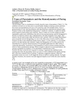
the smear-negative group were higher than those in the smear-positive group (P = 0.001 and P
< 0.001, respectively; Figure 2).
Figure 2. The frequencies of activating KIRs between sputum smear negative group and positive group.
Activating KIR genes in patients and controls
To analyze our data further, we categorized patients and controls into two groups:
Group 1 included subjects carrying less than two activating KIR genes. Group 2 included sub-
jects carrying two or more activating KIR genes (Table 3). Of 109 patients, 20.2 and 79.8%
were categorized into group 1 and group 2, respectively. On the contrary, 32.7 and 67.3% of
controls were categorized into group 1 and group 2, respectively. Moreover, we found that
genotypes containing six activating KIR genes were signicantly more frequent in the patient
group than in the control group (11.00 vs 1.80%, P = 0.005).
Activating Control (N =110) PTB (N =109) P OR (CI)
KIRs + f (%) + f (%)
Less than two 36 32.7 22 20.2 0.035
*
0.520 (0.281~0.961)
Two or more than two 74 67.3 87 79.8 0.035
*
0.520 (0.281~0.961)
+ - positive case numbers. P value was determined using chi-square test.
*P < 0.05.
Table 3. The differences between the two groups in activating gene numbers.
Increased frequency of one gene cluster in the patient group
Based on the linkage disequilibrium between certain alleles of various KIR loci, the
frequency of one gene cluster containing KIR3DS1-2DL5-2DS1-2DS5 was signicantly in-
creased in patients compared with controls (30.3 vs 15.5%, P = 0.009).
DISCUSSION
Genetic diversity is known to play an important role in PTB. Recent evidence has
suggested that T cells expressing KIR2DS2 can mediate vascular damage in patients with
C. Lu et al.
©FUNPEC-RP www.funpecrp.com.br
Genetics and Molecular Research 11 (2) 1370-1378 (2012)
1376
rheumatoid arthritis, implicating a role of activating KIR genes in rheumatoid arthritis and
other autoimmune diseases (Williams et al., 2005). Previous studies have indicated that HLA
confers susceptibility to TB (Pospelov et al., 2007). In addition, some KIR genes such as 3DS1
can bind to HLA-B27 and recruit positive signals, resulting in NK cell activation (Lanier,
1998). This recognition can affect immunomodulatory functions of NK cells (Campbell and
Purdy, 2011). Martin et al. (2002a) have reported that the activating KIR3DS1 in combination
with HLA-B alleles is associated with delayed progression to acquired immunodeciency
syndrome in individuals infected with human immunodeciency virus type 1.
In our present study, the diversity of 15 KIR genes in 109 PTB patients and 110
controls was studied to assess the effect of KIR polymorphisms on the susceptibility to PTB.
The imbalance between activating and inhibitory KIRs may affect the activation of immune
cells, contributing to the pathogenesis of diseases. KIR2DS1, 2DS3, and 3DS1 may serve as
PTB susceptibility genes. Our results were inconsistent with data described by Mendez et al.
(2006) showing that KIR2DL3 is signicant in patients compared with controls, however. The
discrepancy can be explained by the fact that they investigated a different population-each
population has a specic KIR distribution. According to data from Jiang et al. (2005), the gene
frequency of KIR2DL3 is more than 90% in the Chinese Han population; therefore, KIR2DL3
was not signicant between groups in our present study. The sample size, diversity of technol-
ogy used, linkage disequilibrium, and other limitations in our study design might also explain
the discrepancy between our data and those of Mendez et al. (2006).
Smear-positive patients are the main source of infection in a community. Only 10% of
individuals develop clinical disease. The immune response in the remaining individuals only
succeeds in containing the infection, as some bacilli escape killing by the mechanisms of im-
mune activity and remain in latent or dormant states. Clinically, the frequencies of KIR2DS2
and KIR2DS5 in the smear-positive group were lower than those in the smear-negative group.
These results suggest that activating KIRs may be involved in PTB susceptibility and the
regulation of clinical evolution during disease development. Therefore, the genetic imbalance
between activating and inhibitory KIR genes is necessary for the development of PTB. Myco-
bacterium tuberculosis is an intracellular pathogen that can persist within the host. Infection
and antibody production can lead to chronic or fatal disease. KIRs can recognize pathogen-
associated molecules and contribute to immune responses. NK cells play a crucial role in me-
diating innate immunity (Biron et al., 2002), and T cells primarily mediate the acquired immu-
nity to pathogens, such as protecting against M. tuberculosis infection (Brunstein et al., 2009).
The important point for the development of immunity against TB involves the engagement of
CD4
+
and CD8
+
lymphocyte subpopulations. CD4
+
T cells participate in the amplication and
regulation of immunity by producing cytokines. CD8
+
T cells act as cytotoxic effectors that
can break the infected target cells (Rodrigues et al., 2002). Mounting evidence suggests that
KIR gene diversity determines susceptibility to infectious diseases by sending inhibitory or
activating signals. In our study, the increased presence of activating KIRs in patients with PTB
may affect immune response. More KIR genes send activating signals to NK cells and T cells
in response to M. tuberculosis.
Taken together, our results suggest that susceptibility to PTB is mediated by complex
interactions between factors: (1) Each population has a specic KIR distribution. The poly-
morphism between ethnicity and geographical location has been described for various ethnic
diversities (Norman et al., 2001; Gutiérrez-Rodríguez et al., 2006). (2) KIR genes are orga-
Association of KIR with pulmonary tuberculosis
©FUNPEC-RP www.funpecrp.com.br
Genetics and Molecular Research 11 (2) 1370-1378 (2012)
1377
nized into a highly polymorphic, multi-gene family with considerable allelic polymorphism.
The interaction between KIR genes and other multiple genes may confer susceptibility to
PTB. (3) The frequency of one gene cluster containing KIR3DS1-2DL5-2DS1-2DS5 was sig-
nicantly increased in the patient group. Therefore, linkage disequilibrium between different
KIR loci may play an important role in disease development. (4) Differences in environmental
and cultural factors along with gene-gene interactions and even variations in M. tuberculosis
strains complicate the understanding of the observed differences. Our ndings expand the ho-
rizon of elucidating KIR gene diversity and add another level to the complex mechanisms of
NK cell/T cell-mediated response to PTB infection. Moreover, this study provides additional
understanding of KIR genes on which to base future investigations of PTB. Further functional
study of KIR genes will be helpful for clarifying the mechanism of PTB susceptibility and
providing new approaches for the rational design of vaccines and immune therapies.
ACKNOWLEDGMENTS
Research supported by the Shandong Provincial Scientic and Technological De-
velopment Projects Foundation (2009GG10002014) and Chinese National Natural Science
Foundation (grant 30371304). We thank Yu-Lian Jiao, Xue-Jin Gao, and others working in
the Department of Center Laboratory of Shandong Provincial Hospital for their skillful tech-
nical help.
REFERENCES
Berrington WR and Hawn TR (2007). Mycobacterium tuberculosis, macrophages, and the innate immune response: does
common variation matter? Immunol. Rev. 219: 167-186.
Biron CA, Nguyen KB and Pien GC (2002). Innate immune responses to LCMV infections: natural killer cells and
cytokines. Curr. Top. Microbiol. Immunol. 263: 7-27.
Brunstein CG, Wagner JE, Weisdorf DJ, Cooley S, et al. (2009). Negative effect of KIR alloreactivity in recipients of
umbilical cord blood transplant depends on transplantation conditioning intensity. Blood 113: 5628-5634.
Campbell KS and Purdy AK (2011). Structure/function of human killer cell immunoglobulin-like receptors: lessons from
polymorphisms, evolution, crystal structures and mutations. Immunology 132: 315-325.
Casanova JL and Abel L (2002). Genetic dissection of immunity to mycobacteria: the human model. Annu. Rev. Immunol.
20: 581-620.
Cooley S, Trachtenberg E, Bergemann TL, Saeteurn K, et al. (2009). Donors with group B KIR haplotypes improve
relapse-free survival after unrelated hematopoietic cell transplantation for acute myelogenous leukemia. Blood 113:
726-732.
Doherty M, Wallis RS and Zumla A (2009). Biomarkers for tuberculosis disease status and diagnosis. Curr. Opin. Pulm.
Med. 15: 181-187.
Fernando SL and Britton WJ (2006). Genetic susceptibility to mycobacterial disease in humans. Immunol. Cell Biol. 84:
125-137.
Gutiérrez-Rodríguez ME, Sandoval-Ramirez L, Diaz-Flores M, Marsh SG, et al. (2006). KIR gene in ethnic and Mestizo
populations from Mexico. Hum. Immunol. 67: 85-93.
Jiang K, Zhu FM, Lv QF and Yan LX (2005). Distribution of killer cell immunoglobulin-like receptor genes in the Chinese
Han population. Tissue Antigens 65: 556-563.
Lanier LL (1998). NK cell receptors. Annu. Rev. Immunol. 16: 359-393.
Martin MP, Gao X, Lee JH, Nelson GW, et al. (2002a). Epistatic interaction between KIR3DS1 and HLA-B delays the
progression to AIDS. Nat. Genet. 31: 429-434.
Martin MP, Nelson G, Lee JH, Pellett F, et al. (2002b). Cutting edge: susceptibility to psoriatic arthritis: inuence of
activating killer Ig-like receptor genes in the absence of specic HLA-C alleles. J. Immunol. 169: 2818-2822.
Mendez A, Granda H, Meenagh A, Contreras S, et al. (2006). Study of KIR genes in tuberculosis patients. Tissue Antigens
68: 386-389.
C. Lu et al.
©FUNPEC-RP www.funpecrp.com.br
Genetics and Molecular Research 11 (2) 1370-1378 (2012)
1378
Moretta L and Moretta A (2004). Killer immunoglobulin-like receptors. Curr. Opin. Immunol. 16: 626-633.
Norman PJ, Stephens HA, Verity DH, Chandanayingyong D, et al. (2001). Distribution of natural killer cell immunoglobulin-
like receptor sequences in three ethnic groups. Immunogenetics 52: 195-205.
Parham P (2005). MHC class I molecules and KIRs in human history, health and survival. Nat. Rev. Immunol. 5: 201-214.
Pospelov LE, Matrakshin AG, Malenko AF, Udina IG, et al. (2007). Genetic HLA markers associated with pulmonary
tuberculosis in the Barum-Khemchiksky District, Republic of Tyva. Probl. Tuberk. Bolezn. Legk. 62-64.
Rodrigues DS, Medeiros EA, Weckx LY, Bonnez W, et al. (2002). Immunophenotypic characterization of peripheral T
lymphocytes in Mycobacterium tuberculosis infection and disease. Clin. Exp. Immunol. 128: 149-154.
Seich Al Basatena NK, Macnamara A, Vine AM, Thio CL, et al. (2011). KIR2DL2 enhances protective and detrimental
HLA class I-mediated immunity in chronic viral infection. PLoS Pathog. 7: e1002270.
Sriram U, Selvaraj P, Kurian SM, Reetha AM, et al. (2001). HLA-DR2 subtypes & immune responses in pulmonary
tuberculosis. Indian J. Med. Res. 113: 117-124.
van Crevel R, Ottenhoff TH and van der Meer JW (2002). Innate immunity to Mycobacterium tuberculosis. Clin.
Microbiol. Rev. 15: 294-309.
Vidal-Castineira JR, Lopez-Vazquez A, Diaz-Pena R, Alonso-Arias R, et al. (2010). Effect of killer immunoglobulin-like
receptors in the response to combined treatment in patients with chronic hepatitis C virus infection. J. Virol. 84:
475-481.
Williams F, Meenagh A, Sleator C, Cook D, et al. (2005). Activating killer cell immunoglobulin-like receptor gene
KIR2DS1 is associated with psoriatic arthritis. Hum. Immunol. 66: 836-841.
Yen JH, Moore BE, Nakajima T, Scholl D, et al. (2001). Major histocompatibility complex class I-recognizing receptors
are disease risk genes in rheumatoid arthritis. J. Exp. Med. 193: 1159-1167.
Zhang YX, Xue Y, Liu JY and Zhao MY (2010). Association of TIRAP (MAL) gene polymorphisms with susceptibility
to tuberculosis in a Chinese population. Genet. Mol. Res. 10: 7-15.
Zhi-ming L, Yu-lian J, Zhao-lei F, Chun-xiao W, et al. (2007). Polymorphisms of killer cell immunoglobulin-like receptor
gene: possible association with susceptibility to or clearance of hepatitis B virus infection in Chinese Han population.
Croat. Med. J. 48: 800-806.