Tài liệu Tuberculosis of the chest doc
Bạn đang xem bản rút gọn của tài liệu. Xem và tải ngay bản đầy đủ của tài liệu tại đây (650.87 KB, 15 trang )
European Journal of Radiology 55 (2005) 158–172
Tuberculosis of the chest
Lu
´
ıs Curvo-Semedo
∗
,Lu
´
ısa Teixeira, Filipe Caseiro-Alves
Department of Radiology, Hospitais da Universidade de Coimbra, Praceta Mota Pinto/Avenida Bissaya Barreto,
3000-075 Coimbra, Portugal
Received 13 April 2005; received in revised form 15 April 2005; accepted 18 April 2005
Abstract
The relationship between tuberculosis and mankind has been known for many centuries, with the disease being one of the major causes of
illness anddeath. Duringthe early1980s, therewas awidespread beliefthat thedisease was beingcontrolled, but by the mid-1980s, the number
of cases increased. This change in the epidemiological picture has several causes, of which the AIDS epidemic, the progression of poverty in
developing countries, the increase in the number of elderly people with an altered immune status and the emergence of multidrug-resistant
tuberculosis are the most important.
Mainly due to this epidemiological change, the radiological patterns of the disease are also being altered, with the classical distinction
between primary and postprimary disease fading and atypical presentations in groups with an altered immune response being increasingly
reported.
Therefore, the radiologist must be able not only to recognize the classical features of primary and postprimary tuberculosis but also
to be familiar with the atypical patterns found in immuno-compromised and elderly patients, since an early diagnosis is generally asso-
ciated with a greater therapeutic efficacy. Radiologists are, in this way, presented with a new challenge at the beginning of this millen-
nium.
© 2005 Elsevier Ireland Ltd. All rights reserved.
Keywords: Tuberculosis; Pulmonary; Lung; Infection; Computed tomography (CT); Thorax; Radiography
1. Introduction
Tuberculosis (TB) is an infectious disease caused by
Mycobacterium tuberculosis, which was isolated by Robert
Koch in 1882, but has been affecting the world population
for thousands of years. In western countries, the highest
mortality and morbidity occurred in the late 1700s and early
1800s, due to the crowded environments and generalized
poverty during and after the industrial revolution [1].
Because of the improved social and economic situation
of people in the late 1800s, a spontaneous decrease of TB
was observed [2]. Improvement in diagnosing the disease
(due to discovery of X-rays), isolation of infectious cases in
sanatoria, introduction of effective antituberculous therapy
and control programs initiated after World War II, lead to an
∗
Corresponding author.
E-mail address: (L. Curvo-Semedo).
annual decrease of 5% in TB cases over the past 30 years
[3], so that, by the early 1980s, there was a strong conviction
that the disease was being controlled [2]. By the mid-1980s,
however, the number of cases was again increasing. At the
same time, in developing regions of the globe, where 90%
of TB cases of the whole world occur, the number of cases
continued to increase by more than 20% between 1984–1986
and 1989–1991 [4]. Also, the human immunodeficiency
virus (HIV) infection and the epidemics of acquired immun-
odeficiency syndrome (AIDS), together with the problem of
multidrug-resistant (MDR) TB, may have contributed to the
resurgence of the disease [5]. In 1993, the World Health As-
sociation declaredTB a“global emergency” [6], since almost
one-third ofthe world population is infected with M. tubercu-
losis. Largely because it has been neglected as a public health
issue for many years, it is estimated that between 1997 and
2020 nearly 1 billion people will become newly infected and
70 million will die from the disease at current control levels
[7].
0720-048X/$ – see front matter © 2005 Elsevier Ireland Ltd. All rights reserved.
doi:10.1016/j.ejrad.2005.04.014
L. Curvo-Semedo et al. / European Journal of Radiology 55 (2005) 158–172 159
2. Pathogenesis
2.1. Primary tuberculosis
M. tuberculosis is a strictly aerobic, acid-fast, Gram-
positive bacillus [8], transmitted via airborne droplet nuclei,
laden with afeworganisms,produced when personswith pul-
monary or laryngeal TB cough, sneeze or speak [9]. These
particles, being 1–5 m in diameter, can remain airborne for
long periods of time [7], and infection occurs when a sus-
ceptible person inhales those droplet nuclei, which in turn
deposit most commonly in the middle and lower lobes of the
lung [10]. Once in the alveoli, M. tuberculosis is ingested by
alveolar macrophages. If these cannot destroy the offending
organisms, bacilli multiply in this intracellular environment
until the macrophages burst and release them, being, in turn,
ingested by other macrophages. During this period of rapid
growth,M.tuberculosisisspreadthroughthelymphaticchan-
nels to hilar and mediastinal lymph nodes and through the
bloodstreamtoothersitesinthebody[7].Thisisarrestedwith
thedevelopmentofcell-mediatedimmunityanddelayed-type
hypersensitivity at 4–10 weeks after the initial infection. At
this time, the tuberculin reaction becomes positive [11]. The
macroscopic hallmark of hypersensitivity is the development
of caseous necrosis in the involved lymph nodes and the pul-
monary parenchymal focus, the Ghon focus [12], which, to-
gether with the enlarged draining lymph nodes, constitutes
the primary complex, also known as the Ranke or Ghon com-
plex [11]. In the immunocompetent individual, development
of specific immunity is generally adequate to limit multipli-
cation of the bacilli; the host remains asymptomatic and the
lesions heal[13], withresorption of caseous necrosis, fibrosis
and calcification. The pulmonary focus and the lymph nodes
become calcified and minimal haematogenous dissemination
may originate calcifications in lungapices (Simon’s foci) and
in extrapulmonary locations. Some bacilli in these healed
lesions remain dormant and viable, maintaining continuous
hypersensitivity to tuberculous antigen, and in situations of
immunodepression, they can reactivate. In immunocompro-
mised individuals (HIV-positives, alcoholics, diabetics, drug
addicts, elderly andpatients withchronic renal failure, malig-
nancy or undergoing immunosuppressive medication), more
widespread lymphogenic and haematogenous dissemination
occurs, resulting in lymphadenopathy and more peripheral
locations, respectively [11]. If immunity is inadequate, ac-
tive disease often develops within 5 years after initial infec-
tion, the so-called progressive primary TB, which occurs in
about 5% of infected patients [14]. In the patients with lit-
tle or no host response, disseminated (miliary) TB occurs
[15].
2.2. Postprimary tuberculosis
Postprimary disease can result from endogenous reactiva-
tion of dormant bacilli in residual foci in the lung apices [11].
Haematogenous spread andreactivationoccurs preferentially
in the upper lung zones, due to the higher oxygen tension and
impaired lymphatic drainage in those areas[16]. After reacti-
vation,the apicalfoci reachconfluence, liquefyand excavate.
Perforation of a lymph node into a bronchus may cause a tu-
berculousbronchitiswith bronchial ulceration, and aspiration
ofintraluminalbacillicancausebronchogenicdissemination;
a classic finding is an infiltrate in the subapical infraclavicu-
lar region. Postprimary disease can also occur, although less
frequently, from exogenous reinfection, particularly in coun-
tries with low infection risk [11]. Age may often determinate
the presentation of the disease: whereas neonates and chil-
dren develop primary disease, adults present with postpri-
mary TB. This picture, however, is altered by the changing
epidemiology, with atypical and “mixed” radioclinical pat-
terns occurring in adults, especially in immunocompromised
patients, with a consequent fading of the age-related distinc-
tion between primary and postprimary TB [17].
3. Clinical findings
Patients with primary TB are often asymptomatic but may
experience a symptomatic pneumonia. Young individuals
with progressive primary disease may present with cough,
haemoptysis and weight loss.
Patients with postprimary disease most commonly expe-
rience chronic productive cough and marked weight loss,
and sometimes they have hemoptysis and dyspnoea. Chest
pain can occur with extension of the inflammatory process to
the parietal pleura. Symptoms are often insidious and persist
from weeks to months [15].
Clinical features are dependent on the immune status of
the patients [18], since persons with relatively intact cel-
lular immune function have their disease localized to the
lung, whereas in those with advanced immunosupression,
pulmonary TB isfrequently accompaniedby extrapulmonary
involvement [19,20].
4. Radiological findings
In practice, it is becoming increasingly difficult to differ-
entiate between the classical primary and postprimary pat-
terns based on radiological findings, which show a consider-
able overlap in radiological manifestations [11]. Because of
the decreasing TB incidence in developed countries, many
adults have never been infected by M. tuberculosis and are at
risk for a first tuberculous infection, which may progress in
turn to active disease. One can expect a shift from the usual
pattern (endogenous reactivation) towards an unusual pattern
(progressive primary TB) similar to that observed in chil-
dren and adolescents [21]. This unusual or “atypical” pattern
includes: solitary pleural effusion, isolated mediastinal/hilar
lymphadenopathy, lower lobe TB, nodular miliary lesions,
diffuse infiltrations, atelectasis but also a normal chest plain
film [22].
160 L. Curvo-Semedo et al. / European Journal of Radiology 55 (2005) 158–172
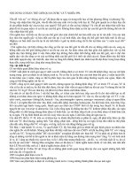
Fig. 1. Gangliopulmonary TB: on chest plain film, patchy infiltrates in the
right upper lobe and right paratracheal lymphadenopathy are detected.
4.1. Primary tuberculosis
This form of disease occurs predominantly in children,
but primary TB in the adult is increasing due to public health
measures and antituberculous therapy that lead to a decrease
in the overallincidence of disease,with aconsequentincrease
in the population of non-exposed adults [23]. Primary TB
accounts for 23–34% of all adult cases of the disease [15].
Four entities have been described: gangliopulmonary TB,
tuberculous pleuritis, miliary TB and tracheobronchial TB
[11].
4.1.1. Gangliopulmonary TB
Gangliopulmonary TB is characterized by the presence of
mediastinal and/or hilar lymphadenopathy and parenchymal
abnormalities, the Ghon focus [11].
Enlarged nodes occur in 83–96% of paediatric cases,
whereas in adult patients they arefound in 10–43% [7]. Right
paratracheal and hilar stations are the most common sites of
nodal involvement in primary TB, although other combina-
tions may also be found (bilateral hilar, isolated mediastinal)
[23–25]. Although adenopathy is usually found in associa-
tion with parenchymal consolidation or atelectasis (Fig. 1), it
can be the sole radiographic manifestation of the disease [8],
especially in early childhood (49% of cases) [24]. Computed
tomography (CT) is more sensitive than chest plain films for
detecting intrathoracic tuberculous adenopathy, and lymph
nodes greater than 2 cm in diameter may have central areas
of low attenuation associated with peripheral rim enhance-
ment and obliteration of surrounding perinodal fat (Fig. 2).
This corresponds to caseation necrosis, granulation tissue
with inflammatory hypervascularity and perinodal reaction
[25–27] and is highly suggestive of active disease [28]. Lym-
phadenopathy resolves at a slower rate than the parenchy-
mal disease, without significant radiological sequelae; nodes
Fig. 2. Tuberculouslymphadenopathy:contrast-enhanced CT showsseveral
low-density center, rim-enhancing lymph nodes in the mediastinum and left
hilum.
firstly become homogeneous and finally disappear or result
in a residual mass composed of fibrotic tissue and calcifica-
tion (Fig. 3). This develops 6 months or more after the initial
infection and is more common than parenchymal calcifica-
tion, and also more common in adultsthan children.It maybe
present in both active and inactive cases of the disease [28].
Associated pulmonary infiltrates are found on the same
side as nodal enlargement in about two-thirds of paediatric
cases of primary TB [22]. Parenchymal involvement in the
absence oflymphadenopathy occursin onlyabout 1% of pae-
diatric cases [24],whereas thispattern is muchmore common
in adults with primary disease (38–81%) [23]. Parenchymal
opacities are most often located in the periphery of the lung,
especially in the subpleural zones. These subtle infiltrates
are frequently undetected on plain chest films, so CT may
be needed to demonstrate them. Parenchymal involvement in
primary disease most commonly appears on plain films as
an area of homogeneous consolidation, with ill-defined bor-
ders and sometimes air bronchograms (Fig. 4); patchy, lin-
ear, nodular and mass-like patterns have also been reported
[23,24,29,30]. In 10% of the patients, primary disease is ap-
Fig. 3. Calcified lymphadenopathy: CT reveals conglomerates of calcified
lymph nodes in the mediastinum and both hila.
L. Curvo-Semedo et al. / European Journal of Radiology 55 (2005) 158–172 161
Fig. 4. Parenchymal disease: chest plain film shows a patchy consolidation
in the right upper lobe with ill-defined borders and air bronchograms.
parent as a single cavitary lesion [22]. Consolidation occurs
in a segmental or lobar distribution, with multifocal involve-
ment in 12–24% of the cases [24,29]. Primary TB can cause
consolidation of any lobe [8]; the most common sites are ar-
easofgreater ventilation,includingthemiddle lobe, the lower
lobes or the anterior segments of the upper lobes [31,32].
There is, however, a right-sided predominance in the distri-
bution [23,24]. On CT, a homogeneous, dense, segmental
or lobar consolidation is seen [32,33]. In two-thirds of the
cases, the parenchymal focus resolves without radiological
sequelae, although the resolution is typically slow, usually
paralleling that of lymphadenopathy [24]. A calcified scar
– the Ghon focus – is seen in 15–17% of the patients, and
together with calcified hilar or mediastinal lymph nodes con-
stitutes the Ranke complex, also known as primary or Ghon
complex [12] (Fig. 5). Calcified secondary parenchymal foci
are called Simon foci [8].
Persistent mass-like opacities predominating in the upper
lobes, corresponding to tuberculomas, are uncommon (7–9%
Fig. 6. Tuberculoma: a homogeneous, calcified nodule in the right upper
lobe is shown on the chest film.
of cases), and are thought to be a result of healed primary dis-
ease (Fig. 6). Cavitation occurs in 10–50% of these nodules,
calcification develops in up to 50% and most remain stable in
size [31]. Gangliopulmonary TB may also present with per-
foration of an adenopathy into a bronchus, retroobstructive
pneumonia and/or atelectasis (epituberculosis). Obstructive
atelectasis or overinflation due to compression by adjacent
enlarged lymph nodes occurs in 9–30% and 1–5%, respec-
tively [24], with a typical right-sided predominance.
4.1.2. Tuberculous pleuritis
Pleural TB is most frequently seen in adolescents and
adults as a complication of primary TB, being uncommon
in young children [12,24,31,34]. Pleural effusions occur in
about 10% of all primary infections and, in 5% of the cases,
effusions are the sole radiographic feature of the disease [31]
(Fig. 7). The effusion generally develops on the same side
Fig. 5. Ranke complex: CT (A) calcified hilar lymphadenopathy and (B) calcified parenchymal lesion.
162 L. Curvo-Semedo et al. / European Journal of Radiology 55 (2005) 158–172
Fig. 7. Tuberculous pleuritis: a left pleural effusion is apparent on chest
plain film.
as the initial infection and is typically unilateral, most often
in association with parenchymal and/or nodal abnormalities
[23]. It is often a late finding in primary TB and, usually,
resolves promptly with adequate therapy, but the resolution
may occur withresidual thickeningor calcification(Fig. 8). If
left untreated, it commonly leads to secondary disease [31].
Complications of pleural tuberculous involvement include
empyema formation, bronchopleural fistulae, bone erosion
and pleurocutaneous fistulae [35].
4.1.3. Miliary TB
In 2–6% of primary TB cases, the haematogenous dis-
semination of bacilli results in miliary disease [29]. The el-
derly, children younger than 2 years old and immunocom-
promised patientsare most frequently affected [12,36].Chest
plain films are usually normal at the onset of symptoms, and
the earliest finding, seen within 1–2 weeks, may be hyper-
inflation [34]. The classic finding of diffuse small (2–3 mm)
nodules, evenly distributed, witha slightlower lobe predomi-
nance,maynot appear until 6weeksor more after haematoge-
nous dissemination [12] (Fig. 9). Associated adenopathy is
Fig. 8. Tuberculous pleuritis: CT shows a right-sided encapsulated pleural
effusion with marked pleural thickening.
Fig. 9. Miliary TB: numerous well-defined, diffusely distributed, small nod-
ules (2–3 mm) are apparent on chest plain film. There is also bilateral hilar
lymphadenopathy.
presentin 95% of childrenand 12%of adults withmiliary dis-
ease, and associated parenchymal consolidation is also more
common in children (42% versus 12%) [8]. CT, particularly
high-resolution (HR) CT, can detect miliary disease before
chest plain film does, demonstrating 1–2mm nodules in a
perivascular and periseptal distribution. A nodular thicken-
ing of interlobular septa can result in a “beaded septum” ap-
pearance similar to that of carcinomatous lymphangitis [37];
rarely nodules may coalesce into parenchymal consolidation
or progress to ARDS and, occasionally, to cavitation [31,36]
(Fig. 10). With therapy, resolution is generally faster in chil-
dren than in adults.
4.1.4. Tracheobronchial TB
Tracheobronchial TBis a complicationof primary disease
that frequently originates from perforation of an adenopathy
into a bronchus; other possible ways of involvement are lym-
phogenic and haematogenic spread [11]. Chest plain films
may be normal or show parenchymal opacities in the upper
lobes andsegmental or lobar atelectasis.Airway involvement
by endobronchial TB in adults presents as areas of segmen-
tal atelectasis distal to the involved bronchi and endoluminal
or peribronchial masses, simulating a neoplasm (Fig. 11).
Endobronchically disseminated TB causes foci of ill-defined
Fig. 10. Miliary TB: CT reveals innumerable 1–3mm nodules with an even
distributionthroughoutbothlungs.Intheleft upper lobe the nodules coalesce
into parenchymal consolidation.
L. Curvo-Semedo et al. / European Journal of Radiology 55 (2005) 158–172 163
Fig. 11. Tracheobronchial TB: on CT, a nodular density is detected in the
right main bronchus (arrow).
nodular densities that may become confluent [30].OnCT,
acute tracheobronchial disease causes concentric bronchial
narrowing, wall thickening and postobstructive bronchiec-
tasis [38,39]. After healing, cicatricial bronchostenosis may
occur. Consolidation of the lower lobes is an atypical radio-
graphic pattern of endobronchial TB [40].
4.2. Postprimary tuberculosis
Also called phthisis, reactivation TB, secondary TB or
“adulthood” TB (by opposition to primary or “childhood”
TB), this form of disease develops under the influence of
acquired immunity. It is the result of reactivation of dormant
bacilliin residual foci,spread atthe time ofprimary infection;
it is, generally but not always, a disease affecting persons in
adulthood[41].When observedinthepaediatricage,itaffects
adolescents [8,12,24,42].
Postprimary TB usually manifests radiographically as
parenchymal disease andcavitation,tracheobronchial TB,tu-
berculous pleuritis and complications [8].
4.2.1. Parenchymal disease and cavitation
The earliest parenchymal finding is a heterogeneous,
poorly marginated opacity (the “exsudative” lesion) situated
in the apical and posterior segments of the upper lobes and
the superior segments of the lower lobes, radiating outwards
fromthehilumorintheperipheryofthelung[31,43].Inabout
88% of the cases more than one segment is affected, with bi-
lateral upper lobe disease seen in 32–64% of the cases [29].
Theusual progression istowardsbetter-definedreticulonodu-
lar opacities (“fibroproliferative” lesions) that may coalesce
[31,43] (Fig. 12). These lesions, when healed, may calcify
and be related to parenchymal distortion, cicatricial atelec-
tasis and traction bronchiectasis [44]. Severe fibrosis, with
upper lobe volume loss and hilar retraction is seen in up to
29% of the cases [29,31]. An apical opacity (the “apical cap”)
is seen in 41% of patients, corresponding to pleural thicken-
ing, extrapleural fat deposition and subpleural atelectatic and
fibrotic lung, as shown by CT studies [29] (Fig. 13). Whereas
active infection correlates better with “exsudative” lesions or
cavitations [31], “fibroproliferative” lesions may also indi-
Fig. 12. Parenchymal involvement: poorly-marginated nodular opacities in
the upper lobes, some of them showing confluence, are shown on chest plain
film.
cate active disease; the stability of radiographic findings for
a period longer than 6 months is the best indicator of disease
inactivity, but the radiologist should perhaps use the term
radiographically “stable” than “inactive” or “healed” [29].
Sometimes, TB may manifest as a mass-like lesion, usually
in the middle or lower lobes, which cannot be distinguished
from a neoplasm based solely on imaging studies [15].
Tuberculous cavitation usually indicates a high likelihood
of activity [42]. Cavitation is seen on chest plain films in
about 50% of the patients at some time during the course of
the disease, but chest CT is more accurate in its detection,
particularly in cases complicated by architectural distortion
[45,46]. Single or multiple cavities are more frequently seen
in MDR TB [33]. Cavities are present, in general, at mul-
tiple sites, within areas of parenchymal consolidation, and
may reach several centimetres in size [31]. Their walls are
initially thick and irregular, and progressively become thin
Fig. 13. Parenchymal disease: chest film shows evidence of significant vol-
ume loss in the right upper lobe, along with hilar retraction, cavitation and
an “apical cap”. There is also calcified mediastinal and hilar adenopathy.
164 L. Curvo-Semedo et al. / European Journal of Radiology 55 (2005) 158–172
Fig. 14. Parenchymal consolidation and cavitation: (A) CT scout film and (B) CT show multiple small nodules in both lungs, with a thin-walled cavitationin
the right upper blobe.
Fig. 15. Bronchogenic spread: HRCT shows wall thickening of the anterior
segmental bronchus of the left upper lobe (arrow) and multiple centrilobular
nodules. There is also left hilar adenopathy.
and smooth (Fig. 14); with healing, they balloon into large
emphysematous spaces [45] and resolvewith orwithout scar-
ring [8]. Air–fluid levels in cavities can be due to superim-
posed infectionby bacteriaor fungi [31,46];however,evenin
non-complicated, non-infected cavities, air–fluid levels may
be found in 9–22% of cases [47]. The differential diagno-
sis of cavities includes bullae, cysts, pneumatoceles or cystic
bronchiectasis [48].
Bronchogenic spread is the most common complication
of tuberculous cavitation, being detected radiographically in
as much as 20% of cases, and appearing as multiple ill-
defined micronodules, distributed in a segmental or lobar
fashion, usually distant from the cavity site and involving
lower lung lobes [47] (Fig. 15). HRCT is probably the most
sensitive imaging method for the detection of bronchogenic
spread of TB, which can be identified in up to 98% of cases.
Findings include centrilobular nodules 2–4 mm in size and
sharply marginated linear branching opacities (representing
caseating necrosis within and around terminal and respira-
tory bronchioles), the so-called “tree-in-bud” sign, indicat-
ing active disease and corresponding to tuberculous bron-
chitis of the small airways [45] (Fig. 16). The same lesions,
however, when surrounded by airless consolidation, may ap-
pear as fluid bronchograms [49]. Five to eight-mm poorly
marginated nodules, lobular consolidation and interlobular
septal thickening are amongthe otherHRCT featuresinbron-
chogenic spread [45].Healing with scarring,residual nodules
and parenchymal or endobronchial calcification are found in
30% [44]. Air trapping due to residual bronchiolar stenosis
leads to areas of hypoattenuation; when associated with ar-
chitectural distortion, this finding usually represents paraci-
catricial emphysema [45].
In few cases (3–6%) of postprimary TB, tuberculomas are
the predominant parenchymalfinding [43]buttheyrepresent,
most times, healed primary disease. These lesions appear
as rounded or oval sharply marginated opacities, measuring
0.5–4 cm in size (the majority remains stable in time), gener-
ally solitary and calcified (Fig. 17). Tuberculomas have ad-
Fig. 16. Bronchogenic spread: CT (A) irregular and thick-walled cavity in the anterior segment of the right upper lobe and scattered small nodules (arrowheads)
and (B) branching opacity in the peripheral lung (arrow) corresponding to dilated bronchioli filled with infected material (“tree-in-bud”).
L. Curvo-Semedo et al. / European Journal of Radiology 55 (2005) 158–172 165
Fig. 17. Tuberculoma: a well-defined, totally calcified nodule with 4 cm in
size in the right upper lobe is shown on CT.
jacent small rounded opacities (“satellite” nodules) in prox-
imity in 30% of the cases [32]. On contrast-enhanced CT,
tuberculomas may exhibit a ring-like or a central curvilinear
enhancement, with the enhancing area corresponding to a fi-
brous capsule, whereas the non-enhancing area corresponds
to caseating or liquefactive necrosis [33].
Miliary disease is seen less frequently in postprimary than
in primary TB [15]. The characteristic radiographic pattern
of multiple micronodules, scattered through both lungs, is
sometimes unseen until late in the disease, but character-
istic features of active TB (consolidation, cavitation, lym-
phadenopathy) coexist in up to 30% of the patients [50].
HRCT can detect miliary disease before it becomes appar-
ent on chest plain films [51], demonstrating both sharply and
poorly defined 1–4mm nodules, randomly distributed, often
with associated intra- and interlobular septal thickening and
areas of ground-glass opacity [51,52] (Fig. 18). Differential
diagnosis includes carcinomatous lymphangitis, bronchioli-
tis, pneumoconiosis or metastasis [37,52].
After postprimary TB, cicatricial atelectasis is relatively
common. Up to 40% of the patients have a marked fibrotic
response, with atelectasis of upper lobes, hilar retraction, hy-
perinflation of lower lobes, and mediastinal shift towards the
affected lung [11]. Extensive parenchymal destruction (the
“destroyed lung”) is sometimes the end-stage of postprimary
Fig. 18. Miliary TB: HRCT reveals multiple widespread 1–2 mm nodules,
some of them in a perivascular distribution.
Fig. 19. Tracheobronchial TB: on CT scout film, a stenosis of the right main
bronchus, due to direct extension from tuberculous lymphadenitis, is seen
(arrow).
TB, causing some difficulties in the assessment ofthe disease
activity based solely in radiographic criteria [48]. Besides,
secondary pyogenic or fungal infection may appear [11].
Mediastinal or hilarlymphadenopathy isalso rarer inpost-
primary disease (5% of patients), usually associated with
parenchymal disease and cavitation [29].
4.2.2. Tracheobronchial TB
Tracheobronchial TB is more frequently seen as a com-
plication of primary disease, but also occurs in the setting of
postprimary disease. Bronchial stenosis occurs in 10–40%
of patients and is caused by direct extension from tuber-
culous lymphadenitis, by endobronchial spread or by lym-
phatic dissemination [30] (Fig. 19). Whereas active disease
involves right and left main bronchi with equal frequency, fi-
brotic disease more commonly affects left main bronchus
[38]. On plain films, findings include segmental or lobar
atelectasis, lobar hyperinflation, mucoid impaction and ob-
structive pneumonia [30]. CT is more accurate and can show
bronchial narrowing (generally of a long segment) with ir-
regular wall thickening, luminal obstruction, and extrinsic
compression by lymphadenitis in the setting of acute dis-
ease [30,38], whereas in fibrotic disease, the wall becomes
smooth and thinner. These findings must be distinguished
from bronchogenic carcinoma involving the central airways
[38]. Bronchiectasis commonly complicates endobronchial
TB, most often occurring as a paracicatricial process (trac-
tion bronchiectasis), but also due to central bronchostenosis
and distal bronchial dilatation. Upper lobes are more fre-
quently involved [44]. Tracheal and laryngeal TB are rarer
than endobronchial disease [42].
4.2.3. Tuberculous pleuritis
Pleural disease is most often associated with primary TB,
but it may occur in postprimary disease. Small unilateral ef-
fusions, associated with parenchymal disease, are detected
in up to 18% of patients [29]. Their resolution may occur
166 L. Curvo-Semedo et al. / European Journal of Radiology 55 (2005) 158–172
Fig. 20. Tuberculous pleuritis: a right-sided, organized pleural effusion is
shown on chest plain film.
with residual thickening or calcification, as in primary dis-
ease [32].Contrast-enhanced CT scans in postprimary TB ef-
fusionsshowsmoothly thickenedvisceraland parietal pleural
leaflets, the so-called “split-pleura” sign [53]. Effusions are
typically loculated and may be stable in size for several years
(Fig. 20).
4.2.4. Complications
Bronchiectasis and residual cavities are sequelae typically
found in theupper lobes, recognizedin 71–86%and 12–22%,
respectively [54]. Fungal organisms, especially Aspergillus
species, can colonize those spaces, particularly the latter. An
earlyradiographic sign of fungalcolonization is thickening of
the cavity wall or the adjacent pleura [11]. On plain films, an
aspergilloma(a fungus ball) appears asa rounded nodule sep-
arated from the cavity wall by a crescent-shaped hyperlucent
image (“air-crescent sign”) [55]. CT features are those of a
spherical intracavitary nodule or mass, partially surrounded
by air or occupying the whole cavity [56], that may show
mobility towards the dependent position on prone and supine
scans [7] (Fig. 21). The most important consequence of as-
pergillomas, occurring in 50–70%, is haemoptysis [55].
A Rasmussen aneurysm is a pseudoaneurysm of a pul-
monary artery caused by erosion from an adjacent tubercu-
lous cavity [57], found in about 5% of patients [11] and pre-
senting with haemoptysis, sometimes massive [58]. Radio-
graphic features include an enlarging mass or a rapidly ap-
pearing parenchymal opacityrepresenting haemorrhage[57].
Broncholitiasis is an uncommon complication, resulting
from rupture of calcified lymphadenopathy into an adjacent
bronchus, with a right-sided predominance. Radiographic
manifestations include a change in the position or disappear-
ance of a calcification on serial films, development of airway
obstruction, or expiratory air trapping. CT can show, apart
from endobronchial or peribronchial calcified nodes, seg-
mental or lobar atelectasis, obstructive pneumonitis, branch-
ing linear opacities (obstructive bronchoceles), focal hyper-
inflation and bronchiectasis [59].
Hilar and mediastinal infected lymph nodes may become
fibrocaseous granulomas and coalesce, forming tuberculous
granulomas. These, in turn, may lead to reactive fibrous
changes and to acute inflammation of the mediastinum. If
the first predominate, the result is fibrosing mediastinitis and
if the latter is more relevant, tuberculous mediastinitis is the
outcome [60]. Both are, however, uncommon [39]. Radio-
graphic findings are similar to those of mediastinal tumours,
but there may also be a hilar mass or a pleural effusion.
On CT, a cluster of enlarged homo- or heterogeneously en-
hancing lymph nodes suggests the diagnosis [60] (Fig. 22);
sometimes these nodes appear as a mediastinal or hilar mass,
often with calcification [39]. Other findings include tracheo-
bronchial narrowing, pulmonary vessel encasement, superior
vena cava obstruction and pulmonary infiltrates [39], the lat-
ter due to bronchial obstruction (with resulting obstructive
pneumonia or atelectasis) or vascular obstruction (leading
to infarction) [61]. However, CT cannot always differentiate
tuberculous mediastinitis from mediastinal neoplasms [60].
Magnetic resonanceimaging (MRI)can demonstrateareas of
low signal intensity on T1-weighted images, due to the pres-
ence of fibrous and inflammatory tissue. Fibrosis may also
be hypointense on T2-weighted sequences, whereas inflam-
matory and granulomatous tissue enhances on gadolinium-
enhanced T1-weighted images [62]. Differential diagnosis
Fig. 21. Aspergilloma: (A) chest film shows two cavities, partially occupied by fungus balls, in the right upper lobe developed within an area of consolidation,
(B) HRCT demonstrates a thin-walled cavity in the right upper lobe colonized by an aspergilloma and (C) on conventional tomography (detail), intracavitary
nodular opacities are present in both upper lobes, separated from the cavity wall by a crescent of air (arrows).
L. Curvo-Semedo et al. / European Journal of Radiology 55 (2005) 158–172 167
Fig. 22. Tuberculous mediastinitis: a cluster of enlarged homogeneous
lymph nodes in the mediastinum is detected on CT.
includes sarcoidosis, lymphoma, metastatic neoplasms, thy-
moma, thymic carcinoma and malignant teratoma [60].
Tuberculous pericarditis is a complication of about 1% of
patients with TB, presenting either as a pericardial effusion,
due to exsudation of fluid with cellular proliferation, or peri-
cardial thickening, due to fibrin production and formation of
granulation tissue. CT is now the method of choice for the
evaluation of the pericardium, but in the near future may be
overtakenby MRI [63]. Pericardial thickening(>3 mm) inthe
suggestive clinical setting indicates the presence of constric-
tive pericarditis, which occurs in 10% of patients with tuber-
culous pericardialinvolvement[39]. Secondarysigns include
inferior vena cavadilatation (>3cm indiameter) secondaryto
right-sided heart failure, and angulation or tortuosity of the
interventricular septum probably due to restriction of peri-
cardial expansion. Other associated signs are the presence of
pericardial fluid in the acute form, whereas in the sub-acute
phase there is gradual absorption of fluid and caseation oc-
curs, resulting in purulent pericarditis and pericardial thick-
ening. Purulent pericarditis is probably secondary to infected
lymphnodes, and thelesions predominatealong theright bor-
der of the heart. In the chronic phase an irregularly thickened
and often calcified pericardium, without pericardial fluid, is
seen [63] (Fig. 23). Pleural effusions are secondary to the
associated haemodynamic abnormality [63] and right atrial
thrombi are due to intracardiac stasis of blood.
Pneumothorax occurs in 5% of patients with postprimary
disease, usually inthe presenceof severecavitation.It heralds
the onset of bronchopleural fistula and empyema [11]. When
tuberculous pleurisy is localized (1–4% of the cases), a tu-
berculous empyema ensues, which presents radiographically
as a loculated collection of fluid associated with parenchy-
mal disease [29,48]. On CT, a focal fluid collection with
pleural thickening and calcification, sometimes associated
with extrapleural fat proliferation, is seen [11] (Fig. 24).
Empyema may communicate with the skin – pleurocuta-
neous fistula (empyema necessitatis) – or with the bronchial
tree—bronchopleural fistula, manifested by an air–fluid level
in the pleural space; CTdemonstrates the communication be-
Fig. 23. Tuberculous pericarditis: chest film demonstrates marked pericar-
dial calcification (arrow). There is also bilateral pleural thickening with cal-
cification of the left pleura (arrowhead).
Fig. 24. Empyema:CT showsbilateral organized fluid collections with pleu-
ral calcification and extrapleural fat proliferation on the right side.
tween the pleural space and the bronchial tree [64] (Fig. 25).
Untreated empyema may also lead to bone destruction, as
well as to pleural thickening and calcification [35,48]. There
arealsoreportsabouttheassociationofchronicempyemaand
malignancy, more commonly lymphoma, squamous cell car-
cinoma and mesothelioma, presumably due to the oncogenic
action of chronic inflammation and of substances contained
Fig. 25. Bronchopleural fistula: CT demonstrates a dilated airway, which
communicates directly with an air–fluid collection in the left pleural space
(arrow). Note also thickening of both visceral and parietal pleural leaflets.
168 L. Curvo-Semedo et al. / European Journal of Radiology 55 (2005) 158–172
in the pleura. Radiographic findings include increased tho-
racic opacity, soft-tissue bulging and blurring of fat planes
in the chest wall, bone destruction and medial shift of the
calcified pleura. CT can demonstrate a soft-tissue enhancing
mass around the empyema [65].
Pulmonary TB may favour the development of bron-
chogenic carcinoma due to the oncogenic effects of chronic
inflammation and fibrosis (“scar carcinoma”)[44]. Lungcan-
cer,on theother side, maylead to ractivationof TB by eroding
quiescent focior by suppressing cellular immunity. The other
possible scenery is that TB and bronchogenic carcinoma
might be coincidentally associated [15]. Radiological fea-
tures that suggest neoplastic disease in patients with postpri-
mary TB include: progressive disease despite adequate anti-
tuberculous therapy, hilar and/or mediastinal lymphadenopa-
thy, focal mass larger than 3 cm in size and cavities with
nodular walls [66].
5. Atypical patterns
A chronic progressive parenchymal disease is observed in
5–10% of patients with primary disease. It is commonly seen
in young children, teenagers, patients with T-cellimmunode-
ficiencies and black people, in which the acquired immunity
is inadequate to contain the primary infection. The radiolog-
ical picture of progressive primary TB is similar to that of
postprimary disease [67]. Multilobar involvement with more
extensive lesions and lung necrosis is common [8], and in
some cases, destruction of a major part of a lung may re-
sult [67]. Involvement of the secondary foci within the upper
lobes is frequently observed. Endobronchial spread may re-
sult from cavitation of the tuberculous pneumonia or rupture
of diseased lymphadenopathy into bronchi, and haematoge-
nous spread may also occur [37].
In elderly individuals, in whom the cellular immune re-
sponse is altered, the presentation of TB shifts away from the
expected typical radiographic findings of postprimary dis-
ease (apical infiltrates and cavities) towards atypical presen-
tations, similar to those found in children (basal infiltrates,
mediastinal and hilar adenopathy and exsudative pleuritis),
which may be due to exogenous reinfection or to a true first
infection [68].
Impaired hostimmunity, predisposing to TB, isalso found
in diabetic patients or patients who are immunocompromised
as a result of corticosteroid therapy or malignancy. In these
patients, a higher prevalence of non-segmental distribution
and multiple smallcavities within atuberculous lesionthan in
patients without underlying disease was detected [69]. Some
authors also stress that in diabetic patients the involvement of
the lower lung zones and the anterior segments of the upper
lobes by TB is more frequent than in non-diabetic subjects
[47] (Fig. 26).
With the epidemics of AIDS, TB infection is increasing in
HIV-positiveindividuals,since thevirus-induced immunosu-
pression is apotent riskfactorfor TB [11].Followingprimary
infection, AIDS patients can have massive haematogenous
dissemination and consequently a more fulminant evolution
of disease. After infection, the risk of developing progressive
primary TB in the first year is about 30%, as compared with
3% in immunocompetent individuals [70]. HIV-infected pa-
tients are also predisposed to reactivation of the disease, due
to deficient cellular immunity. In fact, even though a frac-
tion of pulmonary TB cases in HIV-positive patients repre-
sents primary disease, it is believed that most of TB cases in
HIV patients are due to reactivation of latent infection, corre-
sponding to postprimary disease [71]. Radiographic presen-
tation in these patients, however, is more typical of primary
than of postprimary disease [20,71,72] and is dependent on
the level of immunodepression at the time of overt disease
[73,74]. A CD4 T-lymphocyte count of 200mm
−3
is consid-
ered the cut-off between those subjects who may respond in
a typical or atypical manner to M. tuberculosis infection and
indicates those at risk for atypical radiographic presentation
of TB in HIV-positive patients [75]. Patients with a relative
preservationof cell-mediatedimmunity havefindings similar
to those without HIV infection. Indeed, the typical postpri-
mary pattern of disease is seen less frequently as immunode-
pression becomes more pronounced [20]. Cavitary disease,
pulmonary infiltrates and pleural effusions are usually asso-
Fig. 26. TB in a 44-year-old diabetic man: (A) chest film and (B) CT show a huge cavity, with thick and irregular walls and an air–fluid level, in the right lower
lobe.
L. Curvo-Semedo et al. / European Journal of Radiology 55 (2005) 158–172 169
Fig. 27. TB in a 28-year-old HIV-positive man: (A) chest film reveals ground-glass opacities and areas of airspace consolidation and (B) on HRCT, the same
abnormalities are found, along with interlobular septal thickening.
ciated with higher CD4 T-lymphocyte counts [73] (Fig. 27).
At severe levels of immunosupression, 10–40% of patients
have normal chest plain films [72,75–77]; therefore, normal
radiographic findings in patients with AIDS and M. tuber-
culosis infection do not exclude active disease [77]. Other
severely immunocompromised patients present with radio-
graphic aspects found in primary disease, regardless of prior
M. tuberculosis exposition status [71]. A higher prevalence
of non-apical parenchymal infiltrates and hilar or mediasti-
nal adenopathy and a lower prevalence of cavitary disease
is found in patients with a CD4 T-lymphocyte count of less
than 200 mm
−3
[73,75]. Another feature related to severe
immunosupression is miliary disease [76]. Extrapulmonary
locations are common in HIV-infected patients. Those with a
normal chest film, positive sputum and disseminated disease
are said to have cryptogenic miliary TB [78]. CT features
of TB in HIV-positive patients with normal plain films are
usually subtle and include single or multiple nodules mea-
suring 1–10 mm in diameter and lymphadenopathy [76], re-
flecting the low sensitivity of plain films in the evaluation
of AIDS-associated TB [77]. Among the most frequent find-
ings in HIV-infected patients are nodular opacities, with an
endobronchial or miliary pattern in 57% and 17% of patients,
respectively [76]. Lymphadenopathy is found in 19–74% of
the cases [71,76,77], most often in the right paratracheal and
subcarinal regions [76] and may also exhibit the characteris-
tic peripheral rim enhancement and central hypodense area,
as in immunocompetent patients [25,76].
Myelodysplastic syndromes (MDS) are a group of blood
disorders characterized by ineffective hematopoiesis and,
consequently, pancytopenia. There are also defects in the
lymphoid system, resulting in impaired cell-mediated im-
munity, which may predispose to M. tuberculosis infection.
Patients with TB and MDS show a radiological pattern simi-
lar to that seen in patients with AIDS, commonly exhibiting
a primary pattern and frequent extrapulmonary involvement
[79].
Silicosis, asany pneumoconiosis,carries an increased risk
of TB, possiblydue tothe saturation of macrophages bysilica
particles [80]. Radiological differential diagnosis between
the two is difficult: in the former, miliary nodules predom-
inate in the upper zones, whereas in TB they may occur
everywhere. In cases where the two diseases coexist (sili-
cotuberculosis) it is sometimes impossible to recognize the
underlying pathological process. Adenopathy suggests TB,
but it is also present in silicosis (with “egg-shell” calcifica-
tion). Fibrotic conglomerate masses (massive fibrosis) in the
upper lung favours silicosis [81]. Some authors also report an
increased prevalence of TB in patients with sarcoidosis [46].
6. Follow-up
In patients undergoing adequate antituberculous
chemotherapy, a transient worsening of pre-existing lesions
or appearance of new ones may occur, what is known as
“paradoxical response”. Among the findings are the enlarge-
ment or new appearance of lymph nodes, the development of
new pulmonary infiltrates or progression of those previously
existing and the development of pleural effusions. The
transient worsening does not mean a therapeutic failure but
instead disappears with continuation of the same medication
[82]. It is now recommended that a radiographic evaluation
be made at 2–3 months after initiation of therapy [83].
Parenchymal abnormalities can be subsequently evaluated
every 2–3 months until clearance, and lymphadenopathy can
be followed every year until radiographically stable [84].
7. Specific forms of treatment
Plombage was a type of pulmonary collapse therapy used
for treatment of TB prior to the advent of antituberculous
drugs and consisted in the insertion of plastic packs (Lucite
balls) or polythene spheres in the pleural space [42](Fig. 28).
Injection of oil or paraffin (oleothorax) was also performed
[48].
Control of haemoptysis may be achieved with bronchial
embolization in cases of cavitary disease, bronchiectasis,
Rasmussen aneurysms or aspergillomas. In the latter, the in-
tracavitary injection of antifungal agents under CT guidance
is sometimes performed.
170 L. Curvo-Semedo et al. / European Journal of Radiology 55 (2005) 158–172
Fig. 28. Treatment of TB: CT shows the presence of right-sided Lucite ball
plombage.
8. Radiographic screening
The screening of TB with chest plain films aims to iden-
tify individuals with active disease [9]. Radiological screen-
ing has higher efficacy than sputum examination for detect-
ing pulmonary TB, especially when the disease is clinically
inapparent; chest plain films are recommended as effective
screening devices for pulmonary TB in populations in which
the prevalence of the disease is high [85]. Some authors ad-
vocate performing chest films in all HIV-positive contacts of
persons with positive skin tests as well as in patients selected
to undergo chemoprophylaxis to rule out active TB [72].
A normal chest plain film has a high negative predictive
value for the presence of active disease. Whereas the rate of
false positive cases approaches 1% in immunocompetent in-
dividuals [47,68], this frequency increases to 7–15% in HIV-
infected patients [76,77].
Temporal evolution allows radiographic distinction be-
tween active and inactive disease. An absence of new ra-
diographic findings over a period of 4–6 months is a reliable
indicator of inactive disease [13,43].
9. Conclusions
The chest plain film is the mainstay in the radiological
evaluationofsuspectedorprovenpulmonary TB. CT is useful
in the clarification of certain confusing findings and some
typical features should suggest the diagnosis; CT may also
be helpful in the determination of disease activity.
Primary TB is increasingly seen in the adult popula-
tion. It generally manifests as a parenchymal consolidation,
which can affect any lobe. Associated hilar and/or mediasti-
nal adenopathy is more frequent in children than in adults.
Lymphadenopathy alone is unusual.
Postprimary disease is characterized by parenchymal in-
filtrates in the upper lung, generally in association with cavi-
tation. Cavitary disease is associated with several complica-
tions (endobronchial spread, haematogenous dissemination,
pseudoaneurysm formation). Lymphadenopathy is rare.
Pleural involvement is more frequent in primary TB. Ex-
sudative pleural effusions are large and unilateral. Miliary
TB is also more often found in association with primary than
with postprimary disease.
However, the radiological presentation of TB is chang-
ing, with fading of the classical distinction between primary
andpostprimary disease. Atypicalpatterns aremore frequent,
especially in elderly and immunocompromised patients. In
these groups, there is a lower prevalence of consolidation,
cavitation and postprimary pattern and a higher prevalence
of lymphadenopathy and miliary disease in comparison with
immunocompetent subjects.
References
[1] Herzog H. History of tuberculosis. Respiration 1998;65:5–15.
[2] Van der Brande P, Vanhoenacker F, Demedts M. Tuberculosis at the
beginning of the third millennium: one disease, three epidemics. Eur
Radiol 2003;13:1767–70.
[3] Rieder HL. Epidemiology of tuberculosis in Europe. Eur Respir J
Suppl 1995;20:620–32.
[4] Lauzardo M, Ashkin D. Physiology at the dawn of the new century.
A review of tuberculosis and the prospects for its elimination. Chest
2000;117:1455–73.
[5] Espinal MA, Laszlo A, Simonsen L, et al. Global trends in resistance
to antituberculous drugs. N Engl J Med 2001;344:1294–303.
[6] Raviglione MC, Snider Jr DE, Kochi A. Global epidemiology of tu-
berculosis: morbidity and mortality of a worldwide epidemic. JAMA
1995;273:220–6.
[7] Leung AN. Pulmonary tuberculosis: the essentials. Radiology
1999;198:307–22.
[8] McAdams HP, Erasmus J, Winter JA. Radiological manifestations of
pulmonary tuberculosis. Radiol Clin North Am 1995;33:655–78.
[9] Centers for Disease Control and Prevention. Guidelines for prevent-
ing the transmission of Mycobacterium tuberculosis in health-care
facilities. MMWR Morb Mortal Wkly Rep 1994; 43(RR-13): 1–132.
[10] Haque AK. The pathology and pathophysiology of mycobacterial
infections. J Thorac Imag 1990;5:8–16.
[11] Van Dyck P, Vanhoenacker FM, Van den Brande P, De Schepper AM.
Imaging of pulmonary tuberculosis. Eur Radiol 2003;13:1771–85.
[12] Agrons GA, Markowitz RI, Kramer SS. Pulmonary tuberculosis in
children. Semin Roentgenol 1993;28:158–72.
[13] Bass Jr JR, Farer LS, Hopewell PC, Jacobs RF, Snider Jr DE. Di-
agnostic standards and classification of tuberculosis. Am Rev Respir
Dis 1990;142:725–35.
[14] Park MM, Davis AL, Schluger NW, Cohen H, Rom WN. Outcome of
MDR-TB patients, 1983–1993. Prolonged survival with appropriate
therapy. Am J Respir Crit Care Med 1996;153:317–24.
[15] Miller WT, Miller Jr WT. Tuberculosis in the normal host: radiolog-
ical findings. Semin Roentgenol 1993;28:109–18.
[16] Goodwin RA, DesPrez RM. Apical localization of pulmonary tuber-
culosis, chronic pulmonary histoplasmosis, and progressive massive
fibrosis of the lung. Chest 1983;83:801–5.
[17] Stead WW. Tuberculosis among elderly persons, as observed among
nursing home residents. Int J Tuberc Lung Dis 1998;2:S64–70.
[18] Hopewell PC. A clinical view of tuberculosis. Radiol Clin North
Am 1995;33:641–53.
[19] Telzak EE. Tuberculosis and human immunodeficiency virus infec-
tion. Med Clin N Am 1997;81:345–60.
L. Curvo-Semedo et al. / European Journal of Radiology 55 (2005) 158–172 171
[20] Barnes PF, Bloch AB, Davidson PT, Snider Jr DE. Tuberculosis in
patients with human immunodeficiency virus infection. N Engl J
Med 1991;324:1644–50.
[21] Van der Brande P, Dockx S, Valck B, Demedts M. Pulmonary tu-
berculosis in the adult in a low prevalence area: is the radiological
presentation changing? Int J Tuberc Lung Dis 1998;11:904–8.
[22] Lee KS, Song KS, Lim TH, et al. Adult-onset pulmonary tu-
berculosis: findings on chest radiographs and CT scans. AJR
1993;160:753–8.
[23] Choyke PL, Sostman HD, Curtis AM, et al. Adult-onset pulmonary
tuberculosis. Radiology 1983;148:357–62.
[24] Leung AN, Muller NL, Pineda PR, FitzGerald JM. Primary tu-
berculosis in childhood: radiographic manifestations. Radiology
1992;182:87–91.
[25] Im JG, Song KS, Kang HS, et al. Mediastinal tuberculous lym-
phadenitis: CT manifestations. Radiology 1987;164:115–9.
[26] Kim WS, Moon WK, Kim I, et al. Pulmonary tuberculosis in chil-
dren: evaluation with CT. AJR 1997;168:1005–9.
[27] Pombo F, Rodriguez E, Mato J, Perez-Fontan J, Rivera E, Valvuena
L. Patterns of contrast enhancement of tuberculous lymph nodes
demonstrated by computed tomography. Clin Radiol 1992;46:13–7.
[28] Moon WK, Im JG, Yeon KM, Han MC. Mediastinal tuberculous
lymphadenitis: CT findings of active and inactive disease. AJR
1998;170:715–8.
[29] Woodring JH, Vandiviere HM, Fried AM, Dillon ML, Williams TD,
Melvin IG. Update: the radiographic features of pulmonary tubercu-
losis. AJR 1986;146:497–506.
[30] Lee KS, Kim YH, Kim WS, Hwang SH, Kim PN, Lee BH. En-
dobronchial tuberculosis: CT features. J Comput Assist Tomogr
1991;15:424–8.
[31] Palmer PES. Pulmonary tuberculosis—usual and unusual radio-
graphic presentations. Semin Roentgenol 1979;14:204–42.
[32] Lee KS, Im JG. CT in adults with tuberculosis of the chest: char-
acteristic findings and role in management. AJR 1995;164:1361–7.
[33] Lee JY, Lee KS, Jung KJ, et al. Pulmonary tuberculosis: CT and
pathologic correlation. J Comput Assist Tomogr 2000;24:691–8.
[34] Stransberry SD. Tuberculosis in infants and children. J Thorac Imag
1990;5:17–27.
[35] Hulnick DH, Naidich DP, McCauley DI. Pleural tuberculosis evalu-
ated by computed tomography. Radiology 1983;149:759–65.
[36] Buckner CB, Walker CW. Radiological manifestations of adult tu-
berculosis. J Thorac Imag 1990;5:28–37.
[37] Webb WR, M
¨
uller NL, Naidich DP. High resolution CT of the lung.
New York: Raven Press; 2001. p. 315–25.
[38] Moon WK, Im JG, Yeon KM, Han MC. Tuberculosis of cen-
tral airways: CT findings of active and fibrotic disease. AJR
1997;169:649–53.
[39] Kim Y, Song KS, Goo JM, Lee JS, Lee KS, Lim TH. Tho-
racic sequelae and complications of tuberculosis. Radiographics
2001;21:939–58.
[40] Lee JH, Park SS, Lee DH, Shin DH, Yang SC, Yoo BM. Endo-
bronchial tuberculosis. Chest 1992;102:990–4.
[41] Lamont AC, Cremin BJ, Pelteret RM. Radiological patterns of pul-
monary tuberculosis in the paediatric age group. Paediatr Radiol
1986;16:2–7.
[42] Harisinghani MG, McLoud TC, Shepard JAO, Ko JP, Shroff
MM, Mueller PR. Tuberculosis from head to toe. Radiographics
2000;20:449–70.
[43] Miller WT, MacGregor RR. Tuberculosis: frequency of unusual ra-
diographic findings. AJR 1978;130:867–75.
[44] Fraser RS, Pare JAP, Pare PD, et al. Diagnosis of diseases of the
chest. Philadelphia: WB Saunders; 1991. p. 882–939.
[45] Im J, Itoh H, Shim Y, et al. Pulmonary tuberculosis: CT findings-
early active disease and sequential change with antituberculous ther-
apy. Radiology 1993;186:653–60.
[46] Kuhlman JE, Deutsch JH, Fishman EK, et al. CT features of thoracic
mycobacterial disease. Radiographics 1990;10:413–31.
[47] Hadlock FP, Park SK, Awe RJ, Rivera M. Unusual radiographic
findings in adult pulmonary tuberculosis. AJR 1980;134:1015–8.
[48] Winer-Muram HT, Rubin SA. Thoracic complications of tuberculo-
sis. J Thorac Imaging 1990;5:46–63.
[49] Park S, Hong YK, Joo SH, Choe KO, Cho SH. CT findings of pul-
monary tuberculosis presenting as segmental consolidation. J Comput
Assist Tomogr 1999;23:736–42.
[50] Kwong JS, Carignan S, Kang E, Muller NL, FitzGerald JM. Mil-
iary tuberculosis: diagnostic accuracy of chest radiography. Chest
1996;110:977–84.
[51] Oh Y, Kim YH, Lee NJ, et al. High-resolution CT of miliary tuber-
culosis. J Comput Assist Tomogr 1994;18:862–6.
[52] Hong SH, Im JG, Lee JS, et al. High-resolution CT findings of
miliary tuberculosis. J Comput Assist Tomogr 1998;22:220–4.
[53] Yilmaz MU, Kumcuoglu Z, Utkaner G, Yalniz O, Erkmen G. Com-
puted tomography findings of tuberculous pleurisy. Int J Tuberc Lung
Dis 1998;2:164–7.
[54] Lee KS, Hwang JW, Chung MP, Kim H, Kwon OJ. Utility of CT in
the evaluation of pulmonary tuberculosis in patients without AIDS.
Chest 1996;110:977–84.
[55] Fraser RS. Pulmonary aspegillosis: pathologic and pathogenetic fea-
tures. Pathol Annu 1993;28:231–77.
[56] Broderick LS, Conces Jr DJ, Tarver RD, Bergmann CA, Bisesi
MA. Pulmonary aspergillosis: a spectrum of disease. Crit Rev Diagn
Imaging 1996;37:491–531.
[57] Santelli ED, Katz DS, Goldschmidt AM, Thomas HA. Embolization
of multiple Rasmussen aneurysms as a treatment of hemoptysis.
Radiology 1994;193:396–8.
[58] Ramakantan R, Bandekar VG, Gandhi MS, Aulakh BG, Deshmukh
HL. Massive hemoptysis due to pulmonary tuberculosis: control with
bronchial artery embolization. Radiology 1996;200:691–4.
[59] Conces Jr DJ, Tarver RD, Vix VA. Broncholitiasis: CT features in
15 patients. AJR 1991;157:249–53.
[60] Kushihashi T, Munechika H, Motoya H, et al. CT and MRI
findings in tuberculous mediastinitis. J Comput Assist Tomogr
1995;19:379–82.
[61] Lee JY, Kim Y, Lee KS, Chung MP. Tuberculous fibrosing medias-
tinitis: radiological findings. AJR 1996;167:1598–9.
[62] Rholl KS, Levitt RG, Glazer HS. Magnetic resonance imaging of
fibrosing mediastinitis. AJR 1985;145:255–9.
[63] Suchet IB, Horwitz TA. CT in tuberculous constrictive pericarditis.
J Comput Assist Tomogr 1992;16:391–400.
[64] Westcott JL, Volpe JP. Peripheral bronchopleural fistula: CT evalu-
ation in 20 patients with pneumonia, empyema or postoperative air
leak. Radiology 1995;196:175–81.
[65] Minami M, Kawauchi N, Yoshikawa K, et al. Malignancy asso-
ciated with chronic empyema: radiological assessment. Radiology
1991;178:417–23.
[66] Ting TM, Church WR, Ravikrishnan KP. Lung carcinoma su-
perimposed on pulmonary tuberculosis. Radiology 1976;119:307–
12.
[67] Beigelman C, Sellami D, Brauner M. CT of parenchymal and
bronchial tuberculosis. Eur Radiol 2000;10:699–709.
[68] Korzeniewska-Kosela M, Krysl J, Muller N, Black W, Allen E,
Fitzgerald JM. Tuberculosis in young adults and the elderly: a
prospective comparison study. Chest 1994;106:28–32.
[69] Ikezoe J, Takeuchi N, Jonkoh T, et al. CT appearance of pul-
monary tuberculosis in diabetic and immunocompromised patients.
AJR 1992;159:1175–9.
[70] Daley CL, Small PM, Schecter GF, et al. An outbreak of tu-
berculosis with accelerated progression among persons infected
with the human immunodeficiency virus. An analysis using
restriction-fragment-length polymorphism. N Engl J Med 1992;326:
231–5.
[71] Pitchenik AE, Rubinson HA. The radiographic appearance of tu-
berculosis in patients with acquired immune deficiency syndrome
(AIDS) and pre-AIDS. Am Rev Respir Dis 1985;131:393–6.
172 L. Curvo-Semedo et al. / European Journal of Radiology 55 (2005) 158–172
[72] Fitzgerald JM, Grzybowski S, Allen EA. The impact of human im-
munodeficiency virus infection on tuberculosis and its control. Chest
1991;100:191–200.
[73] Perlman DC, El-Sadr WM, Nelson ET, et al. Variation of chest
radiographic patterns in pulmonary tuberculosis by degree of human
immunodeficiency virus-related immunosupression. Clin Infect Dis
1997;25:242–6.
[74] Goodman PC. Pulmonary tuberculosis in patients with immunodefi-
ciency syndrome. J Thorac Imaging 1990;5:38–45.
[75] Keiper MD, Beumont M, Elshami A, Langlotz CP, Miller Jr WT.
CD4 T lymphocyte count and the radiographic presentation of pul-
monary tuberculosis. Chest 1995;107:74–80.
[76] Leung AN, Brauner MW, Gamsu G, et al. Pulmonary tuberculosis:
comparison of CT findings in HIV-seropositive and HIV-seronegative
patients. Radiology 1996;198:687–91.
[77] Greenberg SD, Frager D, Suster B, Walker S, Stavropoulos C, Roth-
pearl A. Active pulmonary tuberculosis in patients with AIDS: spec-
trum of normal findings (including a normal appearance). Radiology
1994;193:115–9.
[78] Hopewell PC. Tuberculosis and HIV infection. Semin Respir Infect
1989;4:111–22.
[79] Kim HC, Goo JM, Kim HB, Lee JW, Seo JB, Im JG. Tuber-
culosis in patients with myelodysplastic syndromes. Clin Radiol
2002;57:408–14.
[80] Giron J, Couture A, Bousquet C, et al. Imagerie de la tubercu-
lose pulmonaire en 1991. Encycl M
´
ed Chir, Radiodiagnostic-Coeur-
Poumon-Larynx, 32390 A
10
, 1991. p. 1–12.
[81] Palmer PES. The imaging of tuberculosis. Berlin: Springer-Verlag;
2002. p. 5–49.
[82] Choi YW, Jeon SC, Seo HS, et al. Tuberculous pleural effusion: new
pulmonary lesions during treatment. Radiology 2002;224:493–502.
[83] Bass Jr JR, Farer LS, Hopewell PC, et al. Treatment of tuberculosis
and tuberculosis infection in adults and children. Am Rev Respir
Dis 1986;134:355–63.
[84] Abernathy RS. Tuberculosis in children and its management. Semin
Respir Infect 1989;4:232–42.
[85] Barnes PF, Verdeggem TD, Vachon LA, et al. Chest roentgenogram
in pulmonary tuberculosis. Chest 1988;94:316–20.