Tài liệu COLOR ATLAS OF DERMATOPATHOLOGY_2 pdf
Bạn đang xem bản rút gọn của tài liệu. Xem và tải ngay bản đầy đủ của tài liệu tại đây (23.49 MB, 209 trang )
17
Glandular Adnexal (Apocrine and Eccrine) Neoplasms
Timothy H. McCalmont
Department of Clinical Pathology and Dermatology and UCSF Dermatopathology Service,
University of California, San Francisco, California, U.S.A.
CONTENTS
MICROANATOMICAL AND EMBRYOLOGICAL CONSIDERATIONS
CLASSIFICATION OFGLANDULAR ADNEX AL NEOPLASMS
EXAMPLES OFADNEXAL NEOPLASMS
Apocrine Neoplasms
B Syringoma
B Poroma
B Hidradenoma
B ApocrineAdenoma
B Spiradenoma
B Cylindroma
B Adnexal Carcinoma
Eccrine Neoplasms
B Tubulopapillary (Papillary) Adenoma (and Adenocarcinoma)
The nosology of adnexal neoplasms has been confused and
confusing for decades, and much of the mystification of
the past was wrought by the lack of logical classification.
Classification proposals and inferences regarding lineage
from the authorities of the past were often contradictory,
and to a lesser extent, this problem persists at the present
time. This is in part a consequence of the fact that broad con-
clusions regarding lineage and classification were based on
enzyme histochemical attributes that lacked established
specificity and were never properly assessed in the context
of controlled trials.
Enzyme histochemistry enjoyed a brief flash of activity
in the late 1960s but has proved to be of dubious value over
time. Although the number of cases studied by enzymatic
analysis was very small and the assessment was based
mostly upon uncontrolled qualitative judgments, the
results became the basis for conclusions regarding lineage
that persisted for decades. The method has not stood the
test of time, although it lingers on in the minds of some
and in some textbook chapters. Indeed, enzyme histochem-
istry is no longer generally available as a method of analysis.
In short, enzyme histochemistry contributed to the evolution
of misguided classification schemes that have persisted in
dermatology and dermatopathology. Only recently has the
lack of credibility of enzyme analysis led to a rethinking of
this field.
Surprisingly, relatively compelling embryological and
morphological relationships among adnexal structures were
either unrecognized or overlooked by past authorities. It is
only in the last decade that some of the subtle clinical and
microscopical interrelationships displayed by these fascinat-
ing benign growths and the patients that develop them have
been more fully appreciated. In the view of this author, the
lines of evidence that best guide our thinking regarding
the classification of adnexal neoplasms include embryology,
combinations of neoplasms and associations between
neoplasms, anatomical distribution of neoplasms, and
careful microscopical observations.
MICROANATOMICAL AND EMBRYOLOGICAL CONSIDERATIONS
Although we often think of hair follicles, sebaceous glands,
and apocrine glands as distinct elements, all three com-
ponents actually stem from the same structure, which
has been termed the folliculosebaceous-apocrine unit. For
practical purposes, the terms “follicle,” “hair follicle,”
“folliculosebaceous unit,” and “folliculosebaceous-apocrine
unit” are used interchangeably. The folliculosebaceous-
apocrine unit is a structure that provides insulatory,
cosmetic, and pheromonic functions to the mammalian
organism. The eccrine unit is a completely independent
structure that serves as a thermoregulatory device via
secretion of sweat.
The follicle proper consists of an infundibulum, an
isthmus, and an underlying stem and bulb. The infundibu-
lum is the superficial most segment, in continuity with the
surface epithelium, and is composed mostly of keratinocytes
that are microscopically identical to epidermal keratinocytes.
Infundibular keratinocytes display pink cytoplasm within a
conspicuous layer analogous to the stratum spinosum and
mature via a stratum granulosum to form orthokeratin that
envelops a hair. The infundibulum forms a tunnel that
harbors and shields the projecting hair shaft. The apocrine
duct emanates from the lower infundibulum and spirals
downward through the dermis to the apocrine secretory
unit. Subjacent to the infundibulum, the follicular isthmus
is defined superiorly by the origin of the sebaceous duct
and inferiorly by the insertion of the leiomyocytes of the
arrectores pilorum musculature. The follicular isthmus is
characterized microscopically by keratinocytes with dense
pink cytoplasm that display abrupt cornification with little
intervening stratum granulosum, forming compact ortho-
keratin that is tightly arrayed around the hair shaft.
Sebaceous and apocrine glands emanate from the
primary follicle and reside within the adjacent dermis. Virtu-
ally, all follicles sport sebaceous glands, whereas apocrine
glands usually involute at most body sites, remaining detect-
able in genital and axillary sites, in periorbital and periauri-
cular skin, and sometimes in skin of the scalp. The sebaceous
duct, distinctive for the corrugated luminal border and
compact eosinophilic cuticle lining its canal, courses a
235
short distance through the adventitial dermis and links to
the adjacent sebaceous gland. The sebaceous gland proper
consists of a thin peripheral layer of seboblasts with a basa-
loid appearance, with the bulk of the gland composed of
mature sebocytes, characterized by a scalloped nuclear
border because of the presence of abundant surrounding
coarsely vacuolated cytoplasm.
In areas in which apocrine glands are preserved, the
apocrine duct juts from the lower infundibulum just
superior to the insertion of the sebaceous duct and spirals
downward to join the secretory portion of the apocrine
gland, which is situated in the deep reticular dermis and
subjacent subcutis. The secretory elements are arranged as
tubules lined by cuboidal and columnar cells with ample
eosinophilic cytoplasm that often appears finely granular
in conventional sections. At the luminal border, a papillated
or “decapitation” pattern is often present, reflecting holo-
crine secretion.
A crucial principle to keep in mind when considering
adnexal lesions is the fact that the development of the eccrine
apparatus is completely distinct from that of the folliculose-
baceous-apocrine unit. In fully developed human skin,
eccrine glands and folliculosebaceous-apocrine units are
unrelated structures, and their embryogenesis is completely
independent. Eccrine glands develop directly from the
embryonic epidermis in the early months of fetal develop-
ment, projecting as a cord of cells that subsequently entubu-
lates to form a gland. Folliculosebaceous units develop
directly from the epidermis at much the same time, but the
development of follicles differs from the development of
eccrine glands in that mesenchymal cells, precursors of the
follicular papilla, induce a follicular germ and descend
jointly into the dermis with the developing epithelial struc-
ture. Subsequently, sebaceous and apocrine glands and
their ducts elaborate as secondary structures. The folliculo-
sebaceous unit is a hamartoma-like structure from the
start, eventuating with a combination of epithelial cells of
different types and perifollicular fibrocytes, whereas the
eccrine gland is a strictly epithelial structure.
This ontogenetic principle reflects relationships that
can be observed repetitively in dermatological diseases. As
one might predict from ontogeny, follicular, sebaceous, and
apocrine differentiations are frequently observed conjointly,
and combinations of eccrine and folliculosebaceous differen-
tiations probably do not exist. Certainly, authors in the past
have described proliferations of putative mixed eccrine and
folliculosebaceous differentiation, but in the view of most
current authorities, these claims are baseless.
Combinations of adnexal neoplasms also shed light on
lineage and provide insight into the development of a logical
classification scheme. Although most adnexal neoplasms
display a relatively uniform microscopical pattern, it is not
uncommon to encounter lesions with biphasic or multipha-
sic patterns. Excepting the identification of a coincidental
“collision” between proliferations of disparate biology,
such as syringoma combined with basal cell carcinoma or
a melanocytic nevus juxtaposed on desmoplastic trichoe-
pithelioma, the elements that occur conjointly in “combined”
adnexal neoplasms can be assumed to be of related lineage.
For example, the commingling of spiradenoma and cylin-
droma is commonplace and suggests a close relationship.
Indeed, it has been suggested by some observers that these
two separately described lesions represent different patterns
of the same entity. Spiradenoma or cylindroma is occasion-
ally captured with trichoepithelioma. Adnexal neoplasms
also develop, singly or in combination, in association with
nevus sebaceus, which is not an adnexal neoplasm itself
but rather a folliculosebaceous-apocrine hamartoma.
It is clear from such combinations that cylindroma and
spiradenoma are of the same lineage. The nonsensical his-
torical notion (present in most textbooks of dermatology
and dermatopathology) that spiradenoma is “eccrine”
and cylindroma is “apocrine” is pure poppycock. This
conclusion can be based not only on the fact that the two
neoplasms occur intertwined, but also on their relationship
in common with both trichoepithelioma and nevus sebaceus.
Although not in direct combination, adnexal neo-
plasms can also occur jointly (in multiplicity) in the same
patient in the context of a genetic disorder, typically a dom-
inantly inherited syndrome. Multiple trichoepitheliomas
occurring in concert with multiple cylindromas and/or spir-
adenomas, the so-called Brooke–Spiegler syndrome, is a
common connection. Spiradenoma and cylindroma have
also been observed jointly in multiplicity, as has the triad
of spiradenoma, cylindroma, and trichoepithelioma. These
observations assert that the lineage of cylindroma, spirade-
noma, and trichoepithelioma is linked and that cylindroma,
spiradenoma, and trichoepithelioma are best classified as
folliculosebaceous-apocrine neoplasms.
The topographic distribution of adnexal structures
also offers insight into logical classification. There is striking
variation in anatomic distribution among adnexal neo-
plasms, and some of these differences hold implications
with respect to logical assignment of lineage. Historically,
poroma has been considered so thoroughly eccrine that
many dermatologists do not even refer to it as poroma.
Rather, the designation “eccrine poroma” is used as its
formal name. Poromas are routinely observed on the
palms and soles, sites rife with eccrine structures, as one
would expect of a neoplasm of eccrine lineage. However,
the clinical presentation of poroma is broad and is not
limited to glabrous lesions. Poromas present not uncom-
monly on the scalp and in axillary and inguinal skin, sites
where apocrine elements are prominent. Poromas also
develop as secondary neoplasms within nevus sebaceus, a
folliculosebaceous-apocrine hamartoma. The most parsimo-
nious explanation for the distribution of poroma is not that
poroma is eccrine, but rather that poroma may be of either
eccrine or apocrine lineage. Similarly, the distribution of
syringoma is at odds with historical classification schemes.
Purportedly an eccrine neoplasm, syringomata virtually
never develop at sites replete with eccrine elements, such
as the palm or sole. Acral syringomata are a rarity. Instead,
syringomata are found almost exclusively on the periorbital
face and genitalia, sites at which apocrine elements are
identifiable. This topographic evidence suggests that syrin-
gomas are probably apocrine in nature, most of the time.
Microscopy and other morphological tools, including
the wide array of available special stains, also play a role
in the assessment of lineage. However, if microscopists are
to use their observations as the foundation for a system of
classification, they must be certain that the microscopical
features chosen for tabulation are determinate of a specific
line of differentiation. For some lines of differentiation, the
meanings attributed to specific microscopical findings are
indisputable. The presence of cells with coarsely vacuolated
cytoplasm and scalloped nuclei clearly indicates sebaceous
differentiation. There is consensus that follicular (germina-
tive) differentiation is established if a proliferation contains
basaloid cells resembling the follicular bulb and adjacent
mesenchymal cells resembling the papilla. Other unequivo-
cal marks of follicular differentiation include anucleate
236
McCalmont
matrical cells (“shadow” cells), a palisade of pallid cells with
an adjacent thickened basement membrane, an attribute of
the follicular outer sheath (trichilemma), and bright pink
intracytoplasmic trichohyalin granules, typical of matrical
corneocytes of the inner sheath. In contrast to these univer-
sally accepted attributes, the features that indicate glandular
lineage lack specificity. Decapitation secretion is rightly held
as the pathognomonic marker of apocrine differentiation, yet
an essentially indistinguishable microscopical pattern can be
encountered at times in occluded eccrine glands or in neo-
plasms of postulated eccrine lineage. Ducts with a compact
eosinophilic cuticle have been wrongly interpreted as a
specific indicator of eccrine differentiation, as identical struc-
tures can reflect apocrine or even sebaceous lineage in the
ducts of the folliculosebaceous-apocrine unit.
What then are the specific microscopical features of
eccrine glands that, when observed within a neoplasm,
confirm eccrine lineage? There are none. Apocrine lineage
can be suspected on the basis of recognition of decapitation
secretion, but a judgment as to whether a process exhibits
eccrine or apocrine differentiation cannot be based on the
presence of ducts, as eccrine and apocrine ducts are indistin-
guishable. In short, microscopical assessment is invaluable
in the specific recognition of follicular and sebaceous differ-
entiation and is sometimes sufficient to suspect apocrine
differentiation. Microscopy alone is insufficient to establish
eccrine lineage, save for the exclusion of other modes of
differentiation.
Other morphological tools for assessing lineage, such
as electron microscopy and enzyme histochemistry, have
been suggested but have been proven to be of little value
and will not be addressed further. Immunoperoxidase
staining has clarified the classification and lineage of many
neoplasms, especially lymphomas, and still holds hope as
an arbiter of adnexal lineage. To date, however, immuno-
peroxidase stains have resolved few, if any, of the conun-
drums of adnexal classification owing to lack of specificity.
Carcinoembryonic antigen (CEA) was among the earliest
reagents assessed. Although CEA nimbly labels areas of
luminal differentiation, the pattern observed in both eccrine
and apocrine ducts (and in eccrine and apocrine lesions) is
identical. The situation is much the same for other reagents,
including gross cystic disease fluid protein (GCDFP-15), epi-
thelial membrane antigen, and various anti-keratins, all of
which have been found at times to stain both eccrine and
apocrine elements, whether normal or neoplastic.
CLASSIFICATION OF GLANDULAR ADNEXAL NEOPLASMS
Historically, adnexal neoplasms have been classified into
four broad categories, namely follicular, sebaceous, apocrine,
and eccrine. In light of the embryological considerations
discussed previously, a logical ontogenetic classification
yields but two (folliculosebaceous-apocrine and eccrine).
This condensation is of no consequence for an established
entity with a singular line of differentiation, such as sebaceous
adenoma. The classification schemes of the past placed sebac-
eous adenoma as a tumor of sebaceous lineage, and sebaceous
adenoma fits neatly under the rubric of folliculosebaceous-
apocrine tumors in a modern classification scheme.
The advantage of ontogenetic classification relates to
neoplasms with mixed or allegedly “divergent” differen-
tiation, such as microcystic adnexal carcinoma (MAC).
There is no need to debate whether MAC should be
classified as a follicular neoplasm or a glandular neoplasm,
and there is no need to force it, arbitrarily and uncomforta-
bly, into one category or another. If a classification scheme
places MAC as a glandular neoplasm, then what is to be
done to acknowledge its follicular attributes? With a
broadly conceived classification scheme, MAC can be desig-
nated in good conscience as a low-grade form carcinoma of
folliculosebaceous-apocrine lineage, a categorization that
reflects its heterogeneous differentiation.
In the discussion that follows, proliferations of follicu-
losebaceous-apocrine lineage will be limited to lesions with
apocrine differentiation, as follicular and sebaceous lesions
are discussed in other sections. The discussion of prolifer-
ations of eccrine lineage will be relatively brief; the fact
that there are fewer eccrine proliferations is unsurprising,
as eccrine glands lack anatomical complexity and have low
proliferative potential.
In most textbooks, adnexal neoplasms with glandular
and ductular differentiation have been rigorously separated
into eccrine and apocrine neoplasms. Commonly, the dis-
tinction was based upon enzyme histochemical data from
an imprecise technique that is no longer available. There
has also been an illogical desire to lump all adnexal neo-
plasms of a certain type together and presume that all
were of the same lineage. For example, syringoma, which
shows distal ductular differentiation, was historically inter-
preted as exclusively eccrine, although common sense
suggests that both apocrine and eccrine syringomata
would likely occur. A few authorities have responded to
this shortsightedness of the past by grouping apocrine and
eccrine neoplasms together in recognition of the fact that it
is impossible to determine whether a given lesion, such as
a given syringoma, is of apocrine or eccrine lineage. In the
presentation that follows, the traditional categorization as
“apocrine” or “eccrine” will be maintained, but areas of
overlap will be expressly noted. For entities such as syrin-
goma and poroma, which can be of either apocrine or
eccrine lineage, the bulk of the presentation will be included
in the discussion of apocrine lesions, which is presented first.
EXAMPLES OFADNEXAL NEOPLASMS
APOCRINE NEOPLASMS
SYRINGOMA
Clinical Presentation:
B
Small firm papule with similar coloration to surrounding
normal skin (Fig. 1).
B
May occur at any site, but prone to occur in the peri-
orbital area.
B
Commonly multiple (Fig. 1).
B
May occur in “eruptive” fashion, involving the trunk or
extremities, including the palms and soles, extensively.
Histopathology:
B
Small, symmetrical, and well circumscribed.
B
Usually confined to the upper reticular dermis.
B
Composed of uniform nests of epithelial cells with pale
or pinkish cytoplasm, many with central cuticulated
ducts or tubules (Fig. 2).
Chapter 17: Glandular Adnexal Neoplasms 237
B
Nests may resemble a comma or a tadpole, sometimes
(Fig. 2).
B
Associated sclerosis (commonly) (Fig. 2).
B
Pronounced clear cell change (sometimes) (Fig. 2).
B
Squamous metaplasia or cornification, rarely.
Clinicopathologic Correlation:
The firm papular nature of syringoma stems from associated
sclerosis.
Pathophysiology:
Syringoma is a benign adnexal neoplasm with negligible
proliferative capacity. Clinically, syringomata are small
stable papules. Although historically interpreted as a
lesion of eccrine lineage, at present, it seems clear that syrin-
gomata may be of apocrine or eccrine lineage. Most are prob-
ably apocrine, as they occur at “apocrine” sites such as the
periorbital area. Acral syringomata also occur and can
involve the palm or sole, either singly or in multiplicity.
Differential Diagnosis:
The superficial aspects of a syringoma may be difficult to dis-
tinguish from the superficial aspects of an MAC, especially
in a shave biopsy. Sometimes, a dermatopathologist will
find it necessary to defer to a deeper biopsy for a definitive
diagnosis to be rendered. When the interpreter of a super-
ficial biopsy is strongly considering the diagnosis of syrin-
goma in a lesion that shows cornification and involvement
of the deep biopsy margin, the clue of the “sesame seed
bun” should be considered. To understand this clue, one
must consider an analogy between the Big Mac
w
, a hambur-
ger produced by one of the world’s powerhouse fast food
chains, and MAC. Just as one cannot deduce that a Big
Mac
w
includes two all-beef patties, special sauce, lettuce,
cheese, pickles, and onions merely by gazing at the sesame
seed bun on the surface, a dermatopathologist often may
also find it difficult to recognize MAC in a superficial biopsy.
Desmoplastic trichoepithelioma may also resemble
syringoma. Although both share in common a background
of sclerosis, syringoma differs from desmoplastic tricho-
epithelioma in that it is not composed of basaloid (follicular
germinative) cells. Furthermore, the small cystic spaces in
a syringoma represent areas of ductular differentiation,
whereas the cystic spaces in a trichoepithelioma represent
superficial follicular cornification.
Referenc e:
1. McCalmont TH. A call for logic in the classification of adnexal
neoplasms. Am J Dermatopathol 1996; 18:103–109.
POROMA
Synonyms for poroma include hidroacanthoma simplex and
dermal duct tumor. The late Elson Helwig of the Armed
Forces Institute of Pathology utilized the designation acros-
piroma to refer to a broad spectrum encompassing both
poroma and hidradenoma.
ClinicalPresentation:
B
Solitary (almost always); multiple (rarely), known as
poromatosis.
B
Pigmented (sometimes).
B
Highly vascularized (Fig. 3).
B
Presents in hyperkeratotic or crusted fashion (some-
times) (Fig. 3).
B
Favored sites include palm, sole, and genital or axillary
skin.
B
Occasionally found as a secondary neoplasm within
nevus sebaceous.
Histopathology:
B
Circumscribed proliferation of compact cuboidal
(“poroid”) keratinocytes with monomorphous nuclei
and scant eosinophilic cytoplasm (Fig. 4).
B
Highly vascularized and inflamed stroma (almost
always) (Fig. 4).
B
Stromal sclerosis (sometimes).
B
Confined to the epidermis (hidroacanthoma simplex)
(sometimes).
B
Present in broad continuity with the epidermis, with
extension into the papillary dermis (juxtaepidermal
poroma) (sometimes).
B
Present wholly (or nearly so) within the dermis (dermal
duct tumor) (sometimes).
B
Conspicuous ductal differentiation (almost always) (Fig. 5).
B
Intracytoplasmic lumen formation (sometimes).
B
Clear cell change (commonly). Necrosis en masse (focal)
(commonly).
B
Overt apocrine differentiation (sometimes). Focal sebocy-
tic differentiation (sometimes).
Clinicopathologic Correlation:
Clinical Feature Correlating Microscopic Feature
Vascular (pyogenic
granuloma-like) appearance
Highly vasculized stroma and
superjacent crust
Firm nodule Stromal sclerosis
Overlying scale Intra-epidermal involvement
(hidroacanthoma simplex)
Pathophysiology:
Poroma is a benign adnexal neoplasm with low proliferative
capacity, and lesions tend to be clinically stable. Rarely, a
poroma will undergo malignant transformation with resul-
tant porocarcinoma. Although historically interpreted as a
lesion of eccrine lineage, current information clearly indi-
cates that poromata may be of either apocrine or eccrine
lineage. Most are probably eccrine, involving glabrous
sites. Apocrine poromata may occur at virtually any site,
but are prone to occur in axillary, genital, or scalp skin,
where apocrine elements can be found. Poromata
with sebaceous differentiation are probably best thought
of as being lesions of folliculosebaceous-apocrine lineage.
In support of this conclusion, this author’s experience
indicates that sebaceous poromata commonly show apocrine
differentiation as well.
Differential Diagnosis:
When intra-epidermal or juxtaepidermal, poroma may
closely simulate the configuration of a seborrheic keratosis.
Recognizing areas of ductular differentiation and associated
highly vascularized stroma are helpful in making the distinc-
tion. Hidradenoma is always in the differential diagnosis of
poroma and differs in that the epithelial cells that comprise
238
McCalmont
it tend to be larger and often have ample pale or clear cyto-
plasm, in contrast to the compact cuboidal cells of poroma.
In addition, ductular differentiation tends to be prominent
in poroma, yet may be inconspicuous in hidradenoma.
Referenc es:
1. Kamiya H, Oyama Z, Kitajima Y. “Apocrine” poroma: review of
the literature and case report. J Cutan Pathol 2001; 28:101–104.
2. Harvell JD, Kerschmann RL, LeBoit PE. Eccrine or apocrine
poroma? Six poromas with divergent adnexal differentiation.
Am J Dermatopathol 1996; 18:1–9.
HIDRADENOMA
Hidradenoma is a close relative of poroma. Some authorities
use the broad designation acrospiroma to refer to hidrade-
noma and poroma jointly.
ClinicalPresentation:
B
Solitary (almost always).
B
Cystic (sometimes) [“solid-cystic” hidradenoma (Fig. 6)].
B
Pigmented (rarely) (Fig. 6).
B
Highly vascularized (sometimes).
B
Favored sites include genital, axillary, or inguinal skin.
B
Occasionally found as a secondary neoplasm within
nevus sebaceous.
Histopathology:
B
Nodular and sharply circumscribed in pattern.
B
Solid or cystic or, commonly, a combination of the two
(Fig. 7).
B
Composed of cells with ample pale or pink cytoplasm
(Figs. 7 and 8).
B
Overt clear cell change (commonly) (“clear cell” hidrade-
noma).
B
Juxtaepidermal configuration with multifocal attach-
ment to the epidermis (sometimes).
B
Ductular/tubular differentiation (often), with varying
prominence (Fig. 7).
B
Stromal sclerosis (commonly) (Fig. 8).
B
Highly vascularized stroma (sometimes).
B
Overt apocrine differentiation (“decapitation secretion”)
(sometimes) (Fig. 8).
B
Focal sebocytic differentiation (sometimes).
Clinicopathologic Correlation:
Clinical Feature Correlating Microscopic Feature
Vascular appearance Highly vasculized stroma
Firmness Stromal sclerosis
Cystic appearance (“solid-
cystic” hidradenoma)
Cystic dilatation of neoplastic
epithelium
Pigmentation Either lesional hemorrhage or interca-
lated pigmented dendritic melanocytes
Pathophysiology:
Hidradenoma is a benign adnexal neoplasm with very low
proliferative capacity. As a result, lesions tend to be clinically
stable. Rarely, a hidradenoma will undergo malignant
transformation with resultant hidradenocarcinoma.
Although historically interpreted as a lesion of eccrine
lineage, current information clearly indicates that hidrade-
nomata may be of either apocrine or eccrine lineage. Most
are probably of apocrine lineage. Apocrine hidradenomata
may occur at virtually any site, but lesions are prone to
occur in axillary, genital, or scalp skin, where apocrine
elements can be found. Hidradenomata with sebaceous
differentiation are probably best thought of as reflecting fol-
liculosebaceous-apocrine lineage.
Differential Diagnosis:
Hidradenoma and trichilemmoma show overlapping find-
ings in that both commonly display a lobular or nodular
profile and are composed of pale or clear cells. Trichilem-
moma tends to show verrucous surface changes and a
surrounding-thickened basal lamina, as is typical of the
follicular outer sheath, whereas hidradenoma lacks those
attributes. In contrast, hidradenoma may show focal ductu-
lar differentiation or cystic alteration, neither of which is
commonly found in trichilemmoma. The cells of poroma
tend to be compact with scant cytoplasm, in contrast to the
larger pale cells of a hidradenoma.
References:
1. Liu HN, Chang YT, Chen CC, Huang CH. Histopathological and
immunohistochemical studies of poroid hidradenoma. Arch
Dermatol Res 2006; 297:319–323.
2. Gianotti R, Alessi E. Clear cell hidradenoma associated with the
folliculosebaceous-apocrine unit: histologic study of five cases.
Am J Dermatopathol 1997; 19:351–357.
APOCRINE ADENOMA
Any benign neoplasm with apocrine differentiation, includ-
ing poroma and hidradenoma and even spiradenoma
and cylindroma, could legitimately be termed an apocrine
adenoma in a generic sense. However, on a practical basis,
only adenomas with conspicuous apocrine glandular
differentiation are included in this category. Entities
within this spectrum include tubular adenoma, papillary
adenoma, syringocystadenoma papilliferum, and hidrade-
noma papilliferum.
Clinical Presentation:
B
Solitary (virtually always).
B
Favored sites include axillary, inguinal, genital, and peri-
auricular skin, as well as the scalp; syringocystadenoma,
in particular, favors the head and neck area.
B
Surface crust and exudate (sometimes) (especially in
association with syringocystadenoma) (Fig. 9).
B
Commonly found as a secondary neoplasm within nevus
sebaceous.
B
Linear configuration, especially when in concert with
nevus sebaceous.
Histopathology:
B
Nodular and circumscribed from scanning magnification.
B
Verrucous surface changes, especially with syringocysta-
denoma (Fig. 10).
B
Tubular, papillary, or tubulopapillary internal structure
(Figs. 10 and 11).
Chapter 17: Glandular Adnexal Neoplasms 239
B
Frond-like internal structure, especially with hidrade-
noma papilliferum (Figs. 10 and 11).
B
Decapitation secretion along lumina border.
B
Glands lined by a bilayer of cells with a well-formed
myoepithelial layer.
B
Mucin-producing cells (sometimes).
B
Stromal plasma cells, especially with syringocystade-
noma (Fig. 11).
Clinicopathologic Correlation:
Clinical Feature Correlating Microscopic Feature
Verrucous surface Glandular crypts in continuity with surface
squamous epithelium
Surface crust and
exudate
Glandular secretions
Pathophysiology:
Apocrine adenomas represent a group of benign adnexal neo-
plasms with low proliferative potential. As a result, lesions
tend to be clinically stable and do not pose a threat for malig-
nant transformation. Apocrine adenomata may occur at vir-
tually any site, but lesions are prone to occur in the so-called
“apocrine” sites such as axillary or genital skin, where apoc-
rine elements are commonly found. Apocrine adenomas are
also a common secondary occurrence within nevus sebaceus.
Differential Diagnosis:
Syringocystadenoma differs from hidradenoma papilliferum
and tubulopapillary adenoma in that it presents in
verrucous or plaque-like rather than nodular fashion. Hidra-
denoma papilliferum requires distinction from conventional
hidradenoma. Although both present in nodular fashion
clinically, hidradenoma papilliferum is distinctive for its
strikingly papillary internal structure with obvious apocrine
glandular differentiation. In contrast, conventional hidrade-
noma usually has a “solid” microscopical appearance from
low magnification and typically demonstrates only focal or
inconspicuous glandular or ductular differentiation.
Referenc es:
1. Hsu PJ, Liu CH, Huang CJ. Mixed tubulopapillary hidradenoma
and syringocystadenoma papilliferum occurring as a verrucous
tumor. J Cutan Pathol 2003; 30:206–210.
2. Ishiko A, Shimizu H, Inamoto N, Nakmura K. Is tubular apoc-
rine adenoma a distinct clinical entity? Am J Dermatopathol
1993; 15:482–487.
SPIRADENOMA
Spiradenoma connotes a type of undifferentiated benign
adnexal neoplasm that has been historically interpreted as
an eccrine lesion, although modern reassessment clearly
indicates apocrine lineage.
ClinicalPresentation:
B
Papular or nodular.
B
Rare “giant” lesions may achieve a diameter of several
centimeters.
B
Bluish coloration (sometimes).
B
Painful sometimes.
B
Multiple sometimes, especially in the context of Brooke–
Spiegler syndrome.
B
Present in concert with cylindroma or trichoblastoma,
(sometimes).
Histopathology:
B
Nodular or multinodular pattern from scanning magnifi-
cation (Fig. 12).
B
Large individual nodules, often positioned within both
dermis and subcutis.
B
Sharply circumscribed individual nodules (Fig. 12).
B
Trabecular internal structure, with compact (dark) cells
bordering trabecula and cells with ample pale cytoplasm
(pale cells) within trabecular centers (Fig. 13).
B
Obvious ductal or apocrine glandular (decapitation)
differentiation (sometimes).
B
Small lymphocytes scattered throughout trabecular areas
(commonly).
B
Central cystic degeneration (sometimes).
B
Striking-associated vascular ectasia (sometimes).
B
Compact eosinophilic periodic acid-Schiff (PAS)-D-
positive basement membrane material within or bordering
trabecula (sometimes).
Clinicopathologic Correlation:
Clinical Feature Correlating Microscopic Feature
Blue coloration Deep location, ectatic vascular spaces in
stroma, or intralesional hemorrhage
Nodular morphology Large collections of undifferentiated adnexal
glandular cells
Cystic morphology Degeneration of adnexal epithelium or stroma or
profound vascular ectasia
Pathophysiology:
Spiradenoma is a benign adnexal neoplasm with low prolif-
erative capacity, and recurrence is uncommon after simple
enucleation. Rarely, accelerated proliferation with secondary
transformation into spiradenocarcinoma may be observed.
Although historically interpreted as an “eccrine” lesion
based mostly upon long since defunct enzyme histochemical
analysis, spiradenoma is now accepted as an apocrine neo-
plasm that is closely related to cylindroma. Sadly, the desig-
nation “eccrine spiradenoma” is entrenched in the language of
dermatology and dermatopathology, so much so that spirade-
noma is not even indexed under the letter “S” in virtually any
textbook of dermatology. Rather, spiradenoma is commonly
and wrongly indexed under the letter “E.” Stunningly, articles
including the term “eccrine” spiradenoma continue to wriggle
into the medical literature.
Differential Diagnosis:
Spiradenoma can sometimes be misinterpreted as basal cell
carcinoma, although the distinction is typically easily made
by an experienced observer. Basal cell carcinoma is envel-
oped by fibromyxoid stroma and commonly shows necrosis
of single cells or areas of necrosis en masse, all of which are
lacking in spiradenomata. Cylindroma and spiradenoma
are closely related and sometimes occur in synchrony, yet
the two lesions remain distinguishable. Spiradenoma is
240
McCalmont
typically composed of large nodules of undifferentiated
“basaloid” cells, while small nests of cylindroma cells are
juxtaposed in puzzle-like fashion to form larger nodular
collections. In addition, the small nests of cylindroma are
often enveloped by a band of periodic acid Schiff after dia-
stase (PAS-D) positive basement membrane material, an
attribute that is often absent in spiradenomata.
Referenc es:
1. Michal M, Lamovec J, Mukensnabl P, Pizinger K.
Spiradenocylindromas of the skin: tumors with morphological
features of spiradenoma and cylindroma in the same lesion:
report of 12 cases. Pathol Int 1999; 49:419–425.
2. Michal M. Spiradenoma associated with apocrine adenoma
component. Pathol Res Pract 1996; 192:1135–1139.
CYLINDROMA
ClinicalPresentation:
B
Single (sporadic) (sometimes).
B
Multiple and confluent (often); multiple lesion may
occur in mosaic fashion, a clinical pattern that has been
dubbed “turban tumor.”
B
Favored sites include the head and neck area, especially
the scalp and periauricular area, as well as the trunk or
genitalia.
Histopathology:
B
Nodular or papular at scanning magnification, some-
times with dumbbell-shaped nodules in the dermis
and/or subcutis.
B
Larger nodules are composed of nests of undifferentiated
basaloid cells in close apposition, arrayed in puzzle-like
fashion (Fig. 14).
B
A rim of densely eosinophilic, PAS-D-positive basement
membrane material commonly envelops individual nests
(Fig. 14).
B
Small dot-like “droplets” of basement membrane material
often punctuate the centers of small basaloid (Fig. 14).
B
Foci of ductular or apocrine glandular differentiation
(sometimes).
Clinicopathologic Correlation:
Clinical Feature Correlating Microscopic Feature
Nodular
morphology
Large collections of undifferentiated adnexal
glandular cells
Turban tumor Confluent array of dermal and subcutaneous
nodules
Pathophysiology:
Cylindroma is a benign adnexal neoplasm with low prolif-
erative capacity. Because the nodules of cylindroma are com-
posed of many individual nests, simple enucleation can be
difficult and local persistence after biopsy is not
uncommon. As a distinct rarity, secondary transformation
into cylindrocarcinoma can be observed. With respect to
lineage, cylindroma has been generally accepted as an
apocrine neoplasm and is viewed as being closely related
to cylindroma. Brooke–Spiegler syndrome is an autosomal
dominant disease in which multiple cylindromas can occur,
as well as multiple spiradenomas and trichoblastomas. The
gene has been mapped to 16q, and mutations in the
CYLD gene have been identified in Brooke–Spiegler families.
The CYLD gene is believed to be causative of multiple cylin-
dromas, spiradenomas, and trichoblastomas and may rep-
resent the underlying cause of solitary sporadic cylindroma
as well.
Differential Diagnosis:
Cylindroma and spiradenoma are closely related and some-
times occur conjointly, especially in the context of Brooke–
Spiegler syndrome. Despite overlapping features and joint
occurrence, the two lesions remain distinguishable. Spirade-
noma typically presents as large uniform nodules, whereas
cylindroma manifests as a puzzle-like array of small nests
that coalesce to form larger nodules. In addition, the small
nests of cylindroma are often typically enveloped by a
band of PAS-D-positive basement membrane material, an
attribute that is only occasionally present in spiradenomata.
References:
1. Lian F, Cockerell CJ. Cutaneous appendage tumors: familial
cylindromatosis and associated tumors update. Adv Dermatol
2005; 21:217–234.
2. Oiso N, Mizuno N, Fukai K, Nakagawa K, Ishii M. Mild pheno-
type of familial cylindromatosis associated with an R758X non-
sense mutation in the CYLD tumour suppressor gene. Br J
Dermatol 2004; 151:1084–1086.
ADNEXAL CARCINOMA
Adnexal carcinomas (adenocarcinomas) are relatively
uncommon, and their rarity has contributed to confusion
with respect to diagnosis, classification, and therapy.
Because of their infrequency, description of adnexal carci-
nomas has often come in the form of case reports, and
large series that could serve as the foundation for lucid
conclusions regarding behavior and therapy has been
difficult for investigators to assemble. The literature is
probably also skewed by inclusion of extraordinary lesions
diagnosed late in their development, which has contributed
to a general sense by dermatologists, perhaps unwarranted,
that adnexal carcinomas are clinically aggressive. Clearly,
additional study, preferably of thoughtfully stratified
clinicopathologic entities, is warranted to determine the
biological behavior and malignant potential of this group
of skin cancers, especially in the early stages of development.
Adnexal carcinomas can develop de novo or can arise
in association with an existent benign adnexal neoplasm. For
some types of adnexal carcinoma, such as spiradenocarci-
noma or cylindrocarcinoma, the adenocarcinomas that
develop from the benign neoplasm typically lack a decisive
pattern of differentiation and are only specifically diagnosa-
ble through recognition of the residual benign lesion. Other
forms of adnexal adenocarcinoma, such as porocarcinoma
and MAC, display distinctive differentiation that is recogniz-
able whether occurring de novo or developing within a
pre-existent benign lesion.
In general, the presentation of adnexal adenocarcino-
mas is not distinctive. For simplicity, this brief section
includes information referencing porocarcinoma and MAC.
Chapter 17: Glandular Adnexal Neoplasms 241
ClinicalPresentation:
B
Plaque-like or nodular, sometimes with ulceration
(Fig. 15).
B
Often first noted in young or middle-aged adults,
commonly women.
B
Slow enlargement over years (often).
B
Limited mobility (fixed to contiguous structures), (some-
times).
B
Often misdiagnosed prior to definitive recognition.
B
Left-sided predominance noted in the largest US series
(of MAC).
Histopathology:
B
Architectural attributes of malignancy include asymme-
try, lack of circumscription, and an infiltrative pattern
(Fig. 16).
B
Associated stromal sclerosis (often) (Fig. 16). Varied cyto-
logical atypicality, sometimes pronounced (in porocarci-
noma) but often only subtle (in both porocarcinoma and
MAC) (Fig. 17).
B
Neurotropism (sometimes).
B
Muscular invasion (sometimes).
B
Superficial follicular differentiation, often with pale
collections of outer sheath (trichilemmal) cells or small
cornifying cysts (only in MAC).
B
Superficial and deep foci of ductular (syringoma-like and
poroma-like) differentiation (Fig. 17).
Clinicopathologic Correlation:
Clinical Feature Correlating Microscopic Feature
Nodular or plaque-like
morphology
Collections of neoplastic cells arrayed broadly
and deeply in the dermis and/or subcutis
Limited mobility Deep infiltration (sometimes of fat or muscle or
nerve) and associated stromal sclerosis
Pathophysiology:
Adnexal carcinomas are rare lesions that can develop
de novo (such as MAC) or in association with a pre-existent
benign adnexal neoplasm (such as spiradenocarcinoma or
hidradenocarcinoma). The precise genetic or molecular
mechanisms that underlie the evolution of various forms
of adnexal carcinoma are not yet understood. One large
series illustrated that microcystic adnexal shows a left-
sided predilection, suggesting that ultraviolet irradiation
could play a role in the evolution of some cancers.
Differential Diagnosis:
Desmoplastic trichoepithelioma and MAC show extensive
overlap in microscopical findings, as both show a back-
ground of reticular dermal sclerosis and both are punctuated
by many small superficial cornifying (infundibular) micro-
cysts. Desmoplastic trichoepithelioma differs from MAC in
that it is composed mostly of basaloid (follicular germina-
tive) cells, whereas MAC is composed of nests of pale cells
with ductal, superficial follicular, or follicular outer sheath
differentiation. Porocarcinoma may be misconstrued as
poroma, its benign analogue, but is usually differentiable
on the basis of parameters such as larger size, infiltrative
pattern, and greater nuclear atypicality.
References:
1. Robson A, Greene J, Ansari N, et al. Eccrine porocarcinoma
(malignant eccrine poroma): a clinicopathologic study of 69
cases. Am J Surg Pathol 2001; 25:710–720.
2. Chiller K, Passaro D, Scheuller M, Singer M, McCalmont T,
Grekin RC. Microcystic adnexal carcinoma: forty-eight cases,
their treatment, and their outcome. Arch Dermatol 2000;
136:1355–1359.
3. LeBoit PE, Sexton M. Microcystic adnexal carcinoma of the skin.
A reappraisal of the differentiation and differential diagnosis of
an underrecognized neoplasm. J Am Acad Dermatol 1993;
29:609–618.
ECCRINE NEOPLASMS
As noted previously, there is extensive overlap between
eccrine and apocrine neoplasms. For discussion of common
eccrine neoplasms such as syringoma and poroma, please
refer to the appropriate segment in the preceding discussion
of apocrine neoplasms.
TUBULOPAPILLARY (PAPILLARY) ADENOMA
(AND ADENOCARCINOMA)
Clinical Presentation:
B
Solitary (almost always).
B
Papular or nodular morphology.
B
Striking acral predilection; favored sites include fingers,
toes, palms, and soles.
B
Recent rapid enlargement (not uncommonly).
B
Recurrence/persistence after incomplete removal
(commonly).
Histopathology:
B
Nodular and often asymmetrical from scanning magnifi-
cation.
B
Solid with papillary areas (commonly).
B
Cystic, tubular, or cribriform foci (sometimes).
B
High cellularity (commonly).
B
Nuclear hyperchromatism (often).
B
Numerous mitotic figures (commonly).
B
Necrosis of single cells or necrosis en masse (sometimes).
Clinical Feature Correlating Microscopic Feature
Persistence/
recurrence
High cellularity with many mitotic figures
Cystic appearance Cystic dilatation of neoplastic epithelium
Pathophysiology:
The distinction between papillary adenoma and papillary
adenocarcinoma can be challenging. Papillary adenoma of
the digit was initially reported as “aggressive digital papil-
lary adenoma” and was deemed “aggressive” because the
neoplasms were prone to local recurrence if not completely
excised, and some lesions were found to erode bone or infil-
trate adjacent soft tissue. Some lesions originally classified as
“aggressive papillary adenoma” eventuated with metastasis.
Subsequently, such lesions have generally been interpreted
242
McCalmont
as papillary adenocarcinomas. At present, it is unclear
whether there is a spectrum that includes both digital adeno-
mas and adenocarcinomas, or whether virtually all acral
papillary lesions represent carcinomas. It seems likely to
this author that this spectrum includes both adenomas and
adenocarcinomas, but that adenocarcinomas are more
common. At best, it is difficult to predict the clinical
course of lesions within this spectrum on the basis of the
microscopical pattern, and thus a conservative approach to
management is warranted.
Differential Diagnosis:
Because of their papillary morphology, papillary adenomas
and adenocarcinomas can simulate apocrine adenomas.
Apocrine adenomas are typically sharply circumscribed,
include a well-formed myoepithelial layer around glands,
and show low proliferation, with infrequent mitoses. In con-
trast, papillary adenomas and adenocarcinomas are cellular
lesions in which mitotic figures are commonly found and in
which a myoepithelial layer is often lacking. The distinction
of digital papillary adenoma/adenocarcinoma from apoc-
rine adenoma is also typically easily made because of differ-
ing topography. Because of high cellularity and frequent
mitoses, the differential diagnosis of papillary adenoma/
adenocarcinoma also includes metastatic adenocarcinoma.
In this setting, the exclusion of carcinoma requires careful
clinicopathologic correlation.
References:
1. Gorva AD. Digital papillary adenoma and aggressive digital
papillary Adenocarcinoma. Am J Dermatopathol 2005; 27:
546–547.
2. Duke WH, Sherrod TT, Lupton GP. Aggressive digital papillary
adenocarcinoma (aggressive digital papillary adenoma and ade-
nocarcinoma revisited). Am J Surg Pathol 2000; 24:775–784.
3. Kao GF, Helwig EB, Graham JH. Aggressive digital papillary
adenoma and adenocarcinoma. A clinicopathological study
of 57 patients, with histochemical, immunopathological, and
ultrastructural observations. J Cutan Pathol 1987; 14:129–146.
Chapter 17: Glandular Adnexal Neoplasms
243
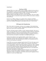
Figure 1 Syringoma. There are multiple,
small, firm, skin-colored papules in the
axillary vault.
Figure 2 Syringoma. This magnified view
shows small nests of cells with clear
cytoplasm and small nuclei. Some of the
nests are shaped like commas or tadpoles,
and some display central ducts lined by a
compact eosinophilic cuticle, as is stereo-
typical of syringoma. There is also associated
dermal sclerosis, accounting for the firm
clinical quality of a syringoma.
Figure 3 Poroma. This exophytic, scaly
papule of the toe assumes a highly vascular
and wart-like appearance.
Figure 4 Poroma. At low magnification, this
poroma displays an exophytic profile and
shows superjacent parakeratosis and crust.
The associated stroma is highly vascularized
and inflamed.
Figure 5 Poroma. This high magnification
view demonstrates a nest of poroid cells with
monomophous, small, round or ovoid nuclei,
and scant eosinophilic cytoplasm. Conspi-
cuous central ductal differentiation is evident.
Figure 6 Hidradenoma. This multinodular
lesion is partially pigmented and partially
cystic and compressible.
Figure 7 Hidradenoma. At scanning magnifi-
cation, both solid and cystic areas are clearly
evident within a larger circumscribed nodule.
Even at low magnification, glandular areas
and foci of clear cell change can be seen.
Figure 8 Hidradenoma. This high magnifi-
cation view highlights keratinocytes with pale
or eosinophilic cytoplasm flanking an area of
glandular differentiation, with hints of decapi-
tation secretion at the luminal border. The
contiguous stroma is sclerotic.
Figure 9 Syringocystadenoma papilliferum.
There is a linear array of crusted, slightly
verrucous papules on the upper thigh. This
linear syringocystadenoma did not occur in
concert with nevus sebaceus, but the combi-
nation of the two is commonplace.
244 McCalmont
Figure 11 Syringocystadenoma papilliferum.
A high magnification view demonstrates
obvious apocrine epithelium at the luminal
border of a papilla and also highlights many
plasma cells within its inflamed “core.”
Figure 12 Spiradenoma. Within the subcutis,
there is a circumscribed multinodular array of
sizable collections of undifferentiated benign
(basaloid) glandular cells. Spiradenoma
is typified by an oligonodular array of sizable
collections of basaloid cells, whereas
cylindroma is characterized by numerous
small nests of similar cells.
Figure 10 Syringocystadenoma papilliferum.
The surface of the biopsy is eroded, with a
subjacent papillary array of broad fronds lined
by the combination of columnar apocrine epi-
thelium and attenuated squamous epithelium.
Figure 13 Spiradenoma. At higher magnifi-
cation, nodules of spiradenoma demonstrate a
trabecular internal configuration, with two
types of cells present. There are small cells
with scant cytoplasm (comprising the so-
called “dark” cells) at the borders of trabe-
cula and cells with pale cytoplasm (so-called
“light” cells) centrally within trabecula. In
actual fact, there are three cell types present,
as a sprinkling of superimposed lymphocytes
is also a stereotypical finding. Although most
spiradenomata show no clear differentiation,
foci of apocrine glandular or ductal differen-
tiation can be found at times (a duct is
evident in this image).
Figure 14 Cylindroma. This magnified view
demonstrates many small, closely juxtaposed
nests of cylindroma. Most of the nests are
encircled by a thickened and [periodic acid
Schiff (PAS)-D-positive] basement membrane.
A few scattered small dots of PAS-positive
material can also be found within the compact
nests.
Figure 15 Porocarcinoma. This large asym-
metrical ulcerated malignancy was clinically
firm and showed limited mobility, reflecting
its infiltrative nature.
Chapter 17: Glandular Adnexal Neoplasms
245
Figure 16 Porocarcinoma. This scanning
magnification view demonstrates the deeply
infiltrative pattern of this carcinoma and also
highlights associated dermal sclerosis. There
is overlying crust as a consequence of
erosion/ulceration.
Figure 17 Porocarcinoma. At high magnifi-
cation, the nests of carcinoma cells vary in
size and shape, and areas of distal ductal
differentiation (with a cuticulated luminal
border) are easily found. Much like microcys-
tic adnexal carcinoma, many examples of
porocarcinoma show only modest or slight
nuclear atypicality, and thus the distinction
from benign lesions must be based upon
careful assessment of architectural par-
ameters, including lesional circumscription.
246 McCalmont
18
Benign Melanocytic Neoplasms
Raymond L. Barnhill, Stephen Vernon, and Harold S. Rabinovitz
Departments of Dermatology and Pathology, University of Miami Miller School of Medicine, Miami, Florida, U.S.A.
CONTENTS
B Lent igo Simplex
B Common Acquired Melanocytic Nevi
B Halo Melanocytic N evus
B Melanocytic Nevus of Acral Skin
B Recurrent/Persisten t Melanocytic Nevus
B Genital/Flexural Nevi
B Small and Inte rmediate-Sized Congenital Nevi
B Large or Giant Congenital Nevi
B SpitzTumor
B SpitzTumor with Atypical Features
B Desmoplastic SpitzTumor
B Pigmented Spindle Cell MelanocyticTumor
B Dermal Melanocytoses
B Common Blue Nevus
B Cellular Blue Nevus
B Combined Nevus
B Plexiform Pigmented Spindle Cell Nevus/Tumor(Deep-Penet rating
Nevus)
B The Clinically and HistologicallyAtypical Melanocytic Nevi
(Th e So-Called ‘‘Dysplastic’’ Nevus)
Benign melanocytic neoplasms constitute an increasingly
important and diverse group of cutaneous lesions. Their
importance is derived from their relationship to malignant
melanoma as simulants, risk markers and precursors to
melanoma. As such they pose a significant diagnostic chal-
lenge to both clinicians and pathologists because of their
profound heterogeneity and capacity to mimic melanoma.
There is also evidence that along with cutaneous melanoma
melanocytic nevi are increasing in frequency worldwide.
Melanocytic neoplasms originate from melano-
cytes: neural crest-derived cells defined by their unique
property of synthesis of melanin pigments. The melanins
are synthesized in unique organelles: the melanosomes,
which are also transferred to keratinocytes. Melanocytes
seem to originate from pluripotential cells that migrate
from the neural crest to the skin via the paraspinal ganglia
and their peripheral nerves and become terminally differen-
tiated after migration to the local microenvironment of the
dermis and basal layer of the epidermis.
Beyond establishing the embryonic origin of melano-
cytic nevi from neural crest-derived cells, the histogenesis
of these melanocytic proliferations has not been adequately
elucidated. The conventional viewpoint is that nevi arise
from proliferation of intraepidermal melanocytes within
junctional nests or theques. According to this model, nevus
cells are considered a morphological variant of melanocytes
that have assumed a morphology that is more epithelioid,
and less dendritic. With evolution of the lesions, it is held
that cells “drop off” (Abtropfung of Unna) into the dermis.
The Abtropfung hypothesis derives from cross-sectional
observations correlating histological findings in nevi with
chronological aging.
Alternative hypotheses regarding the genesis of nevi
include the proposal that nevus cells arise from cutaneous
nerves, from a pluripotential cell of nerve sheath origin, or
by contributions from both neural and non-neural dermal
sources. However neural crest cells may phenotypically
display both melanocytic and neural differentiation.
Whether melanocytic nevi are hamartomas or neoplasms
has been subject to long-standing debate. The common
finding of other tissue elements in excess within nevi, such
as epidermal hyperplasia, hypertrophy of adnexal struc-
tures, and connective tissue alterations, indeed suggest
that nevi are developmental malformations; thus, the term
nevus is often used synonymously with malformation or
hamartoma.
On the other hand accumulating data suggest that the
melanocytic nevus is clonal, hence a neoplasm and, puta-
tively, the first stage in tumor progression of the melanocy-
tic system. Stages in the putative progression model may
not be obligate precursors to the subsequent stages,
but rather could represent end stages at any point in
the process. In support of this model are the gross morpho-
logical and cytological differences between melanocytes
and nevus cells; the expression on nevus cells of markers
of tumor progression that are not present on intraepider-
mal basilar melanocytes by immunophenotyping; and
the growth advantages of nevus cells over epidermal
melanocytes in cell culture.
Both genetic and environmental factors clearly influ-
ence the development of melanocytic nevi. Increased
numbers of nevi aggregate in some families, and the pheno-
type of multiple nevi and/or enlarged nevi is linked
to melanoma-prone kindreds and, in fact, is the strongest
epidemiological risk factor for melanoma. These familial
associations indicate a genetic basis for the growth and
development of nevi. Quantification of total nevus number
and total nevus density in melanoma kindreds has also
shown familial (hereditary) correlations, but the nevus
phenotype does not readily model genetically as a simple
mendelian trait resulting from the transmission of a domi-
nant locus. With respect to environmental factors, sun
exposure, especially during early childhood, promotes the
initiation and development of nevi in susceptible individ-
uals. This effect is reflected in the observation that nevi
have a predilection for sun-exposed sites, especially those
sites receiving intermittent, but occasionally intense, ultra-
violet exposure. Moreover, nevus counts are higher in tropi-
cal than at temperate latitudes. From these empirical
247
observations, nevi may be viewed as clonal proliferations
of initiated cells with a growth advantage over their
progenitors, the intraepidermal melanocytes.
A reasonable hypothesis regarding the natural history
of melanocytic nevi is that they arise as a lentiginous (i.e.,
lentigo-like) proliferation of single cell units along the
basal zone of elongated and hyperpigmented rete ridges.
At some point thereafter, the melanocytes undergo a mor-
phological transition into the epithelioid nevus cells with
their propensity to aggregate as junctional nests (junctional
nevus). Following this stage of development as a junctional
nevus, further cellular development and proliferation
results in the migration or “dropping off” of nevus cells
and their organization into nests within the papillary
dermis (compound nevus). According to this generally
accepted model, eventually all intraepidermal proliferation
of melanocytes ceases, and the nevus becomes entirely intra-
dermal (dermal nevus). Nevus cells residing within the
dermis have reduced proliferative and metabolic activity,
except for the formation of melanosomes. With the decline
of replication, the nevus cell population is gradually
replaced by mesenchymal elements, including fibrous
matrix, glycosaminoglycans, and adipose tissue. Most
dermal nevi are believed to undergo progressive involution,
some eventuating as acrochordons and others shedding.
This developmental (or maturational or differentiation)
sequence may, presumably, be arrested at any stage,
such that a lentigo, junctional nevus, or compound nevus
may persist indefinitely. Because the model has been
developed from largely cross sectional data, alternative
theories of development have been proposed, including a
model invoking a reverse order of development.
Classification and Criteria for Benign
Melanocytic Neoplasms:
Benign melanocytic neoplasms constitute a heterogenous
spectrum of lesions that are classified according to a number
of clinical, histological, and other attributes (Tables 1 and 2).
As with any classification there is controversy as to the
basis for defining and including the various entities in
such a classification. The scheme outlined in Table 3
will be utilized in this chapter. Major considerations for
classification include age of onset of the lesion, size, ana-
tomic site, other gross morphologic features, location of
melanocytes in the skin, the spatial relationships of melano-
cytes, cytological features of melanocytes, stromal attributes,
and finally abnormal features such as atypical architecture,
cytological atypia, and proliferation rate.
In the routine evaluation of melanocytic lesions one is
continually faced with the decision as to whether a lesion is
benign or malignant. In approaching this problem one has
to apply a number of criteria for this interpretation (Table 4)
since no single criterion is sufficient. At present there is no uni-
versal consensus as to which criteria should be included in
this exercise, or the relative importance or relative weight of
each criterion. It is certain that this latter exercise should
take into consideration clinical information, organizational,
cytological, and cell proliferation-related properties of the
individual lesion. It must be emphasized that there are excep-
tions to each criterion, and the failure to consider this may
result in both over- and underdiagnosis of melanoma.
An important aspect of the interpretation of melano-
cytic lesions is recognizing the subjectivity of such evalu-
ation and the imperfect state of knowledge at present. It
cannot be overemphasized that despite having criteria for
diagnosis a certain percentage of melanocytic lesions
cannot be easily interpreted as benign or malignant. Conse-
quently the author utilizes a third or intermediate category
reserved for melanocytic lesions occupying the continuum
between benign and malignant. An intermediate category
avoids overdiagnosis of melanoma and also the under-recog-
nition of abnormal or indeterminate lesions that require
additional therapy and close monitoring, rather than being
pronounced “benign” without further qualification. The cri-
teria and nomenclature for (and some would argue even the
very legitimacy of) such intermediate lesions are presently a
source of considerable controversy and debate. The various
terms suggested for such intermediate lesions have not as
yet been standardized, as evidenced by nomenclature such
as “atypical” nevi, “dysplastic” nevi, nevi with architectural
disorder and cytological atypia, Spitz tumors with atypical
features (atypical Spitz tumors), atypical cellular blue nevi,
and so on. The authors believe that additional research and
substantial effort are needed to standardize the terminology
of benign (and malignant) melanocytic neoplasms. For the
time being the authors suggest a provisional terminology
(Table 3).
Definition ofTerms:
Melanocyte: Melanocytes are the “clear cells” in the basal
layer of the epidermis owing to retraction of their cytoplasms.
They have dendritic cellular processes and uniform intensely
basophilic nuclei slightly smaller than those of nearby
keratinocytes. The melanocyte has the unique property of
synthesizing the complex molecules, the melanins, in specific
organelles, the melanosomes, and transferring them to kera-
tinocytes. Melanocytes seem to originate from pluripotential
cells that travel from the neural crest to the skin via the
paraspinal ganglia and their peripheral nerves and become
terminally differentiated after migration to the local micro-
environment of the dermis and basal layer of the epidermis.
Table 1 Clinical Criteria Used for the Classification of Benign
Melanocytic Neoplasms
Age of onset: congenital or acquired
Size
Small congenital nevus: ,1.5 cm
Medium sized congenital nevus: .1.5–20 cm
Large congenital nevus: .20 cm
Garment or bathing trunk nevus
Segmental nevus
Anatomic location
Nonglabrous skin
Glabrous/acral
Mucosal
Genital/flexural
Other sites such as breast, scalp, ear, etc.
Appearance
Border characteristics (symmetry, circumscription)
Surface topography (macular, papular, papillomatous, verrucoid)
Pattern of coloration: variegated or homogeneous
Colors present: flesh, tan, brown, black, blue, gray, white, pink, red
Speckled, targetoid, agminated, zosteriform
248 Barnhill et al.
Nevus Cell: This somewhat confusing and archaic term refers
to the melanocytes present in melanocytic nevi. Although
“nevus cells” share properties with melanocytes, they are
currently thought to be melanocytes in the initial stage of
tumor progression to melanoma. These modified melano-
cytes are characterized by syncytial aggregation in nests
within the epidermis and/or dermis, loss of dendritic pro-
cesses, and a progressive sequence of differentiation with
descent into the dermis termed “maturation.”
Type A or epithelioid nevus cells are melanocytes resid-
ing in junctional (intraepidermal) or superficial dermal nests.
These polygonal cells have the appearance of epithelial cells
because of relatively abundant eosinophilic cytoplasms and
often the syncytial appearance in aggregate referred to earlier.
The nuclei are slight larger than those of basilar melanocytes.
Type B or lymphocytoid nevus cells constitute the next
slightly deeper population of dermal melanocytes in this
maturational sequence. Thus these cells are commonly small
Table 2 Histological Criteria for the Classification of Benign
Melanocytic Neoplasms
Location of melanocytes in the skin (depth)
Superficial
Intraepidermal
Papillary dermis
Upper half of reticular dermis
Deep
Lower half of reticular dermis
Subcutaneous
Fascial
Disposition of melanocytes
Intraepidermal
Basilar melanocytes (single cell pattern)
Normal numbers
Increased frequency
– With elongated rete (lentiginous)
– Without elongated rete
Pagetoid pattern
Nested pattern
– With lentiginous pattern
– Without lentiginous pattern
Dermal
Diffuse, interstitial
Patchy perivascular, periadnexal, perineurial
Wedge pattern (deep apex of nests, fascicles of melanocytes extend into
reticular dermis or subcutaneous fat)
Plexiform pattern (discreet nests, fascicles associated with
neurovascular or adnexal structures of reticular dermis with
intervening normal dermis)
Bulbous aggregates, nodules (cellular nests or fascicles with rounded
contours, usually extending into reticular dermis, subcutis)
Alveolar pattern
Maturation/differentiation
Stroma
Desmoplasia (sclerosis)
Cell type
Small round or oval cell
Spindle cell
Epithelioid cell (abundant cytoplasm, overall enlarged)
Dendritic cell (lengthy, delicate cellular processes)
Large spindle cell
Large epithelioid cell
All with varying degrees of melanization
Table 3 Practical Classification of Benign Melanocytic
Neoplasms
Benign melanocytic lesions without clinical or histological atypia
:
Circumscribed lentiginous melanocytic proliferations
Lentigo simplex
Common acquired melanocytic nevi and variants
Balloon cell nevus
Halo nevus
Lentiginous junctional and compound nevi
Neural nevus (neurotized nevus)
Nevus spilus
Recurrent/persistent melanocytic nevus
Particular anatomic sites:
Acral
Genital/flexural
Breast
Scalp
Congenital melanocytic nevi
Small congenital nevus
Intermediate congenital nevus
Large or giant congenital nevus
Spitz tumors and variants
Desmoplastic Spitz tumor
Pigmented spindle cell tumor
Dermal melanocytoses, blue nevi, and variants
Mongolian spot
Nevus of Ito and Ota
Common blue nevus
Epithelioid blue nevus
Cellular blue nevus
Plexiform pigmented spindle cell nevus/tumor (deep-penetrating nevus)
Melanocytic nevi with phenotypic heterogeneity (combined nevi)
The clinically and histologically atypical melanocytic nevi/tumors/
neoplasms including those with indeterminate biological potential
:
Clinically atypical nevi/neoplasms
Histologically atypical nevi/neoplasms (melanocytic nevus with architectural
disorder and cytological atypia; nevus with atypical features) including other
melanocytic nevi with atypical features (acral, genital, etc.)
Congenital nevi with atypical features
Spitz tumor with atypical features
Cellular blue nevus with atypical features
Plexiform pigmented spindle cell nevus/tumor (deep-penetrating nevus) with
atypical features
Melanocytic nevus with phenotypic heterogeneity (combined nevus) with
atypical features
Chapter 18: Benign Melanocytic Neoplasms 249
and round, have lesser amounts of cytoplasm, and slightly
smaller nuclei hence their resemblance to lymphocytes.
Type C or spindled nevus cells are the final or terminal
stage of differentiation or maturation. This population of mel-
anocytes is usually the most deeply situated ones in a nevus
and is characterized by schwannian (or neural) differentiation.
These are often tapered spindle cells with striking resemblance
to Schwann cells. They commonly form nerve-like structures
that have been termed neural tubules.
Melanophage: Macrophages, containing coarse melanin
pigment, that are present in pigmented melanocytic neo-
plasms. These macrophages have polygonal morphologies
and eccentrically-placed nuclei within the cell.
Lentigin ous Melanocytic Proliferation: The proliferative pattern
of melanocytes arrayed as single cells in the basal layer of
squamous epithelium. The melanocytes are usually most
concentrated at the tips of the epidermal rete and least fre-
quent above the dermal papillae. This pattern is observed
in lentigines, lentiginous junctional and compound nevi, aty-
pical lentiginous melanocytic proliferations, and lentiginous
melanomas.
Pagetoid Melanocytosis (Pagetoid Infiltration; Pagetoi d
Spread):
Single melanocytes and nests of melanocytes scat-
tered throughout the spinous and granular layer of the
squamous epithelium in a pattern mimicking Paget’s
disease of the breast. Pagetoid melanocytosis strictly
defined involves only the most superficial spinous layer,
that is, the squamous epitheilium above the plane of the
most superficial extensions of the dermal papillae.
Although a major criterion for melanoma, pagetoid melano-
cytosis also may occur in the following benign melanocytic
Table 4 Histopathologic and Clinical Criteria for Melanoma Vs. Benign Melanocytic Lesion
Melanoma Benign Lesion
Size !6 mm, often !10 mm ,6 mm small-diameter
melanoma, metastatic melanoma
,5 or 6 mm often
Symmetry Usually asymmetrical with respect to epidermal
thickness, melanocytic elements, melanin distri-
bution, host response
Usually symmetrical
Circumscription Often poorly-circumscribed at peripheries with
single-cell intraepithelial patterns
Often well circumscribed with well-defined
nests at periphery
Heterogeneity Often heterogenous with two or more cellular
phenotypes, or variable cellular populations
Often homogeneous cellular populations
Intraepidermal patterns Loss of rete-oriented pattern Single cells on elongated rete ridges
Cells scattered in pagetoid patterns above the level
of dermal papillae
Little or no pagetoid spread
Single cells reaching confluence along dermal
epidermal junction
Regular, uniform nesting, cohesive, relatively
small nests
Irregular and haphazard nesting
Discohesive and large nests
Dermal patterns Confluence of cells with little or no maturation
(sheet-like patterns of cells)
Regular spacing and maturation with depth
Cellularity, cellular density High cellular density, crowding of cells Lower cellular density
Melanin synthesis Variable or no loss of synthesis with depth Loss of synthesis with depth
Epidermal reaction Hyperplasia, thinning ulceration; variable epidermal
thickness
Often uniform thickness of epidermis
Stratum corneum shows alteration: hyperkeratosis,
parakeratosis, scale-crust
Stratum corneum basket weave or unaffected
Maturation (differentiation) Melanoma cells fail to exhibit diminished cellular
and nuclear sizes and overall cellular density with
depth in dermis
Melanocytes exhibit diminished cellular and
nuclear sizes and overall cellular density with
depth in dermis;
Progression from polygonal cells to lymphocyte-like
cells to schwannian cells (neurotization) with depth
Mitosis Likelihood of melanoma increases with absolute
number in dermis and depth of mitoses
Usually not present in dermal component
Atypical mitoses
Necrosis Single cells or confluent necrosis Usually not present
Host response Often band-like and asymmetrical infiltrates with
superficially invasive melanoma
Little or no inflammation, perivascular infiltrates,
band-like infiltrates in halo nevi
Diminished inflammation with increasing tumor
thickness
Regression Often all stages: early, intermediate, late; multifocal
common in thin melanoma; often asymmetrical
Uncommon, often symmetrical
250 Barnhill et al.
neoplasms: traumatized, solar-irradiated and persistent/
recurrent melanocytic nevi; congenital nevi; nevi from chil-
dren; acral nevi; pigmented spindle cell melanocytic
tumors; and Spitz tumors.
Maturation: The sequential cytological alterations (terminal
differentiation, or perhaps even senescence) of “nevus
cells” or melanocytes (in a melanocytic nevus or Spitz
tumor), as outlined earlier under “nevus cells,” with pro-
gressive descent into the dermis. The assessment of the
dermal component of a melanocytic lesion for maturation
is often an important exercise in deciding whether the
lesion is likely to be benign or malignant. It is important to
keep in mind that a given melanocytic nevus may be arrested
in only one or two stages of maturation, rather than display-
ing all three stages. To complicate matters, some proportion
of melanomas may show “pseudomaturation.”
Melanocytic Nev us: The term nevus historically is a cognate
for hamartoma or developmental anomaly, hence melanocy-
tic hamartoma. In common usage the term has come to
signify virtually all benign melanocytic lesions, irrespective
of whether they are hamartomas (developmental anomalies)
or neoplasms. In fact, this distinction has been obscured by
increasing the evidence that melanocytic nevi, in general,
are clonal and consequently neoplastic.
Referenc es:
1. Masson P. My conception of cellular nevi. Cancer 1951; 4:9–38.
2. Stegmaier OC. Natural regression of the melanocytic nevus.
J Invest Dermatol 1959; 32:413–419.
3. LeDouarin N. Migration and differentiation of neural crest cells.
Curr Top Devel Biol 1980; 16:31–85.
4. Quevedo WC, Fleischman RD. Developmental biology of mam-
malian melanocytes. J Invest Dermatol 1980; 75:116 –120.
5. Hu F. Melanocyte cytology in normal skin, melanocytic nevi, and
malignant melanomas. In: Ackerman AB, ed. Pathology of malig-
nant melanoma. New York: Masson Publishing USA, 1981:1–21.
6. BarnhillRL,FitzpatrickTB,FandreyK,KenetRO,MihmMCJr,Sober
AJ. The pigmented lesion clinic: a color atlas and synopsis of benign
and pigmented lesions. New York: McGraw-Hill, Inc., 1995.
7. Barnhill RL. The pathology of melanocytic nevi and malignant
melanoma. Boston: Butterworth-Heineman, 1995.
8. Barnhill RL, Piepkorn M, Busam KJ. The pathology of melanocytic
nevi and malignant melanoma. 2nd ed. New York: Springer, 2004.
LENTIGO SIMPLEX
Lentigo simplex is a circumscribed, uniformly pigmented tan,
brown, or dark brown macule usually 1 to 5 mm in diameter
occurring either as a solitary lesion or as multiple lesions,
possibly in the context of an inherited syndrome, anywhere
on the cutaneous or mucocutaneous surface (Fig. 1). These
lesions demonstrate regular and well-defined borders. Histo-
logically, they show increased numbers of solitary basilar
melanocytes and increased epidermal basal layer and poss-
ibly suprabasal layer melanin (Table 5) (Fig. 1B).
Synonyms: Simple lentigo; genital lentiginosis.
Clinical Features:
B
Lentigo simplex onsets childhood, adolescence, and in
some cases later
B
All skin phototypes affected
B
Some probably related to sun exposure; persistant
B
Usually 1 to 5 mm macules, which are light-, medium-,
dark-brown, or black in color
B
Homogenous pigment pattern
B
Round and oval
B
Symmetric
B
Regular borders
B
Well circumscribed
B
Located on any cutaneous surface including sun-
protected skin and mucocutaneous sites
Histopathology:
B
Elongated, club-shaped epidermal rete ridges
B
Basal layer hyperpigmentation, accentuated lower poles
of rete
B
Increased numbers of basilar melanocytes concentrated
on tips of rete ridges
B
Melanophages in papillary dermis
Table 5 Differential Diagnosis: Lentigo Simplex
Lentigo
Simplex Freckle Cafe
´
-au-lait Macule Becker’s Nevus Solar Lentigo
Lentiginous
Junctional Nevus
Size 1–5 mm 1–3 mm 2–5 cm 3–12 cm or more 5–15 mm ,5,6 mm
Elongated, club-shaped
epidermal rete ridges
Present Normal configur-
ation usually
Normal configur-
ation usually
Present or absent,
sometimes papillomatous
epidermal hyperplasia
Often present but
may be absent
Present
Basal layer hyperpig-
mentation, accentuated
lower poles of rete
Present Present Present Present Present Present
Increased numbers of
basilar melanocytes
concentrated on tips of
rete ridges
Present Usually absent or
slightly increased
Usually absent or
slightly increased
Usually absent or slightly
increased
Present or absent Present with
junctional nests
of melanocytes
Smooth muscle
hamartoma
Absent Absent Absent May be present Absent Absent
Solar elastosis Usually
absent or
slightly
increased
Often present Usually absent or
slightly increased
Usually absent or slightly
increased
Present May be present
Chapter 18: Benign Melanocytic Neoplasms 251
Clinicopathologic Correlation:
Differential Diagnosis:
B
Freckle
B
Cafe
´
-au-lait macule
B
Becker’s nevus (melanosis)
B
Solar lentigo
B
Lentiginous junctional nevus with or without atypia
(Table 5)
Referenc es:
1. Barnhill RL, Fitzpatrick TB, Fandrey K, Kenet RO, Mihm MC Jr,
Sober AJ. The pigmented lesion clinic: a color atlas and synopsis
of benign and pigmented lesions. New York: McGraw-Hill, Inc.,
1995.
2. Barnhill RL, Piepkorn M, Busam KJ. The pathology of melano-
cytic nevi and malignant melanoma. 2nd Ed. Springer,
New York, 2004.
3. Barnhill RL, Albert LS, Shama SK, Goldenhersh MA, Rhodes AR,
Sober AJ. Genital lentiginosis: a clinical and histopathologic
study. J Am Acad Dermatol 1990; 22:453–460.
COMMON ACQUIRED MELANOCYTICNEVI
Common acquired melanocytic nevi are the most prevalent
benign melanocytic lesions. They are clonal proliferations
of melanocytes usually developing in childhood and adoles-
cence but also at any age in adults and defined by their local-
ization to the epidermis (junctional), both epidermis and
dermis (compound), or dermis only (dermal or intradermal).
In general, common acquired nevi measure about 3 to 6 mm
in diameter, are symmetrical, have regular and well-defined
borders, and have uniform pink, tan, brown, or dark-brown
color (Table 6; Figs. 2 and 3).
A number of subtypes of common acquired nevi have
been described and are generally related to a number of
factors that may in some way influence or alter the basic
morphology of acquired nevi. Some of these determinates
include anatomic site, host response, external trauma, and
other poorly understood developmental and genetic factors.
Synonym: Common mole.
ClinicalFeatures:
Histopathology:
Clinicopathologic Correlation:
Differential Diagnosis:
See Table 6.
B
Clinically atypical nevi
B
Histologically atypical melanocytic nevi
B
Malignant melanoma
Pathophysiology:
Melanocytic nevi are derived from pluripotential cells that
migrate from the neural crest and take residence in the
basal layer of the epidermis, the dermis, and possibly of
sites such as regional lymph nodes. There has been a long-
standing debate as to the developmental nature of melanocy-
tic nevi, that is, are they hamartomas or neoplasms, and so
on? As already mentioned there is increasing evidence that
melanocytic nevi in general are clonal lesions thus support-
ing a neoplastic basis. However this may not be applicable to
all melanocytic nevi.
References:
1. Lund HZ, Stobbe GD. The natural history of the pigmented
nevus; factors of age and anatomic location. Am J Pathol 1949;
25:1117–1155.
2. Stegmaier OC, Montgomery H. Histopathologic studies of
pigmented nevi in children. J Invest Dermatol 1953;
20:51–62.
Junctional Nevus Compound Nevus Dermal Nevus
Onset childhood,
adolescence
Onset childhood, ado-
lescence, third decade
Onset first, second, third
decades or later
2 to 5 mm macule 3 to 6 mm papule 3 to 6 mm papule, dome-
shaped or papillomatous
Round, oval Round, oval Round, oval
Symmetrical Symmetrical Symmetrical
Well defined,
regular borders
Well defined, regular
borders
Well defined, regular
borders
Homogeneous
brown, dark brown
Homogeneous tan to
dark brown
Light brown to flesh tones
Junctional Nevus Compound Nevus Dermal Nevus
Symmetry Symmetry Symmetry
Well circumscribed Well circumscribed Well circumscribed
Regular junctional
nesting
Regular junctional
nesting
Dome shaped or
papillomatous
Uniform size, shape,
and placement of nests
Uniform size, shape,
and placement of
nests
Orderly arrange-
ment of nevus cells
in dermis
Nests often at tips of
retia
Nests often at tips of
retia
Transition from
epithelioid to
lymphocytoid to
spindled cells with
dermal descent
Cohesive nests usually Cohesive nests usually
No or little pagetoid
scatter
No or little pagetoid
scatter
Lentiginous prolifer-
ation common
Nevus cells often con-
fined to papillary or
superficial reticular
dermis
Nevus cells often
confined to papil-
lary or superficial
reticular dermis
Transition from
epithelioid to lympho-
cytoid
to spindled cells with
dermal descent
Transition from
epithelioid to
lymphocytoid to
spindled cells with
dermal descent
Mitotic figures rare in
dermis
Mitotic figures rare
in dermis
Minimal nuclear
pleomorphism
Minimal
pleomorphism
Clinical Feature Pathologic Feature
Tan to dark brown color Melanin present in the basal and
possibly the suprabasal squamous
epithelium, and possibly in dermal
melanophages
Clinical Feature Pathologic Feature
Tan or brown color Junctional nests of melanocytes
Papular appearance Melanocytes in dermis
252 Barnhill et al.
3. Stegmaier OC, Becker SW Jr. Incidence of melanocytic nevi in
young adults. J Invest Dermatol 1960; 34:125– 129.
4. Maize JC, Foster G. Age-related changes in melanocytic naevi.
Clin Exp Dermatol 1979; 4:49–58.
HALO MELANOCYTIC NEVUS
Halo nevi arecommon nevi exhibitinga peripheral vitiligo-like
annulus or “halo” of hypopigmentation or depigmentation
surrounding a central nevus (Fig. 4A). Histologically halo
nevi demonstrate a dense lymphocytic infiltrate associated
with the central nevus and a peripheral zone of hypo- to depig-
mentation of the epidermis, corresponding to the clinical
halo (Fig. 4B). Halo melanocytic nevi may exhibit both clini-
cally and histologically atypical features and thus raise
concern for melanoma (Tables 4 and 7).
Synonyms: Suttton’s nevus; leukoderma acquisitum centri-
fugum.
ClinicalFeatures:
B
Children, adolescents, and young adults most often
affected.
B
Multiple or solitary.
B
Trunk especially upper back.
B
Central nevus 3 to 6 mm in size.
B
Symmetry of central nevus and surrounding halo of
depigmentation.
B
Regular borders.
B
Uniform color of central nevus—tan, brown, or pink, and
halo—hypo- or depigmentation.
Histopathology:
B
More than 5 to 6 mm.
B
Symmetry.
B
Usually well circumscribed.
B
Mononuclear cell infiltrate orderly with well-defined
inferior margin.
B
Maturation/differentiation of nevus elements.
B
Apoptosis of nevus cells common.
B
Rete ridges maintained. Low-grade or no cytologic
atypia commonly.
B
Few mitoses in dermal component (usually ,2to3/mm
2
).
B
Atypical mitoses usually absent.
Clinicopathologic Correlation:
Differential Diagnosis:
See Table 4, Table 7, and Histopathology under the section
“Common Acquired Melanocytic Nevi.”
Pathophysiology:
Although poorly understood, the halo phenomenon is
thought to represent progressive destruction of nevi by the
host immune response, possibly both cell- and antibody-
mediated. The infiltrating lymphocytes in halo nevi are
predominately T lymphocytes. The presence of both CD8-
positive and antigen-presenting cells suggest a cytotoxic
destruction of nevus cells. In addition, individuals with
halo nevi harbor both activated lymphocytes and antibodies
against neoplastic melanocytes in their peripheral blood.
References:
1. Kopf A, Morrill S. I. S. Broad spectrum of leukoderma acquisi-
tum centrifugum. Arch Dermatol 1965; 92:14–35.
2. Wayte DM, Helwig EB. Halo nevi. Cancer 1968; 22:69–90.
3. Mitchell MS, Nordlund JJ, Lerner AB. Comparison of cell-
mediated immunity to melanoma cells in patients with vitiligo,
halo nevi or melanoma. J Invest Dermatol 1980; 75:144–147.
4. Zeff RA, Freitag A, Grin CM, Grant-Kels JM. The immune
response in halo nevi. J Am Acad Dermatol 1997; 37:620– 624.
MELANOCYTICNEVUS OFACRAL SKIN
Melanocytic nevi involving nonhair-bearing skin of the
palms, soles, and digits share characteristics of other acquired
nevi but nonetheless manifest properties unique to this
portion of the skin (Fig. 4C). Historically, such nevi have
often raised concern for melanoma. The increased frequency
Table 6 Differential Diagnosis: Common Acquired Melanocytic Nevi
Common Acquired Melanocytic Nevi Histologically Atypical Melanocytic Nevi Malignant Melanoma
Size Usually ,5, 6 mm 4 to 12 mm or more (any size) Usually .5, 6 mm, often .10 mm (any size)
Symmetry Usually present Often present, may be absent Often absent
Circumscription Well circumscribed Usually poorly circumscribed Usually poorly circumscribed
Junctional nesting Regular Disordered: variation in size, shape,
placement of nests on epidermal rete
More disordered often with pagetoid
melanocytosis
Lentiginous melanocytic
proliferation
May be present May be present with greater frequency
of melanocytes
May be present with contiguous proliferation
of melanocytes
Cytological atypia of intra-
epidermal melanocytes
Usually absent Present and variable, ranges from slight
to severe
Present, uniform pattern of atypia at least
moderate to severe
Maturation Usually present Often present, may be diminished Often absent but “pseudomaturation” may
be present
Dermal mitoses Usually absent, rarely present in
small numbers, e.g., 1 to 3/mm
2
Usually absent, rarely present in small
numbers, e.g., 1 to 3/mm
2
Commonly present
Usually superficial and not deep Usually superficial and not deep Deeply located mitoses often present
Clinical Feature Pathologic Feature
Halo of hypo- or
depigmentation
Diminished or absent epidermal
basilar melanocytes and
melanin
Chapter 18: Benign Melanocytic Neoplasms 253
of particular histological characteristics, lentiginous melano-
cytic proliferation, and pagetoid melanocytosis in acral nevi
often suggest melanoma (Fig. 4D). However, other histologi-
cal features of benign nevi, especially the lack of significant
cytological atypia of melanocytes, usually allow distinction
from melanoma (Table 8).
ClinicalFeatures:
B
All ages
B
Size usually 3 to 6 mm
B
Macular or only slightly raised
B
Uniform brown or dark brown pigmentation
B
Symmetry
B
Regular and well-defined borders
Histopathology:
B
Symmetrical, well demarcated silhouette.
B
Regular, evenly spaced nesting at the junctional zone.
B
Lentiginous melanocytic proliferation along the basal
layer of elongated rete.
B
Pagetoid scatter of cells common but orderly.
B
Little or no inflammatory reaction within the stroma.
B
When of compound type, the dermal nests are well
formed and cells mature with dermal descent.
Clinicopathologic Correlation:
Table 7 Differential Diagnosis: Halo Melanocytic Nevus
Halo Nevi Histologically Atypical Nevus Melanoma
Size Usually ,5, 6 mm 4 to 12 mm or more (any size) Usually .5, 6 mm, often
.10 mm (any size)
Symmetry Usually present Often present, may be absent Often absent
Circumscription Well circumscribed Usually poorly circumscribed Usually poorly circumscribed
Junctional nesting Regular Disordered: variation in size,
shape, placement of nests on
epidermal rete
More disordered often with
pagetoid melanocytosis
Mononuclear cell infiltrate
orderly with well-defined inferior
margin
Present Often present Greater asymmetry
Cytological atypia of intraepi-
dermal melanocytes
Usually absent, reactive hypertro-
phy of melanocytes and nuclear
variation may be present
Present and variable, ranges from
slight to severe
Present, uniform pattern of aty-
pia at least moderate to severe
Maturation Usually present Often present, may be diminished Often absent but “pseudoma-
turation” may be present
Dermal mitoses Usually absent, rarely present in
small numbers, e.g., 1 to 3/mm
2
Usually superficial and not deep
Usually absent, rarely present in
small numbers, e.g., 1 to 3/mm
2
Usually superficial and not deep
Commonly present
Deeply located mitoses often
present
Clinical Feature Pathologic Feature
Parallel rows of pigmentation
within nevus
Discrete vertical columns of melanin
pigment in stratum corneum
Table 8 Differential Diagnosis: Melanocytic Nevus of Acral Skin
Acral Melanocytic Nevi Histologically Atypical Acral Nevi Acral Melanoma
Size Usually ,7 mm 4 to 12 mm or more (any size) Usually .6, 7 mm, often .10 mm
(any size)
Symmetry Usually present Often present, may be absent Often absent
Circumscription Well circumscribed Usually poorly circumscribed Usually poorly circumscribed
Junctional nesting Regular Disordered: variation in size, shape,
placement of nests on epidermal
rete
More disordered often with pagetoid
melanocytosis
Lentiginous melanocytic
proliferation
Often present, localized to
epidermal rete
May be present with greater
frequency of melanocytes
Usually present with contiguous
proliferation of melanocytes
Pagetoid melanocytosis May be present, sparsely cellular
density often
May be present, sparsely cellular
density often
Often present, greater cellular
density compared to nevi and with
significant cytological atypia
Cytological atypia of
intraepidermal
melanocytes
Usually absent Present and variable, ranges from
slight to severe
Present, uniform pattern of atypia at
least moderate to severe
Maturation Usually present Often present, may be diminished Often absent but “pseudomatura-
tion” may be present
Dermal mitoses Usually absent, rarely present in
small numbers, e.g., 1 to 3/mm
2
Usually absent, rarely present in
small numbers, e.g., 1 to 3/mm
2
Commonly present
Usually superficial and not deep Usually superficial and not deep Deeply located mitoses often
present
254 Barnhill et al.
Differential Diagnosis:
See Table 8.
B
Atypical nevi
B
Acral lentiginous melanoma
Referenc es:
1. McCalmont TH, Brinsko R, LeBoit PE. Melanocytic acral nevi
with intraepidermal ascent of cells (MANIACs): a reappraisal of
melanocytic lesions from acral sites [abstr]. J Cutan Pathol 1991;
18:378.
2. Boyd AS, Rapini RP. Acral melanocytic neoplasms: a histologic
analysis of 158 lesions. J Am Acad Dermatol 1994; 31:740–745.
3. Fallowfield ME, Collina G, Cook MG. Melanocytic lesions of the
palm and sole. Histopathol 1994; 24:463–467.
4. Clemente C, Zurrida S, Bartoli C, Bono A, Collini P, Rilke F.
Acral-lentiginous naevus of plantar skin. Histopathology 1995;
27:549–555.
RECURRENT/PERSISTENT MEL ANOCYTIC NEVUS
The recurrent (persistent) nevus is defined as the appearance
of macular pigmentation within the confines of the clinical
scar of a previously biopsied (usually by shave technique)
melanocytic nevus, usually after the passage of about six
weeks to six months (Fig. 5A). Histologically regenerative
and often irregular single cell and nested intraepidermal
melanocytic proliferation, sometimes mimicking melanoma,
overlies the dermal scar (Table 9) (Fig. 5B). On occasion, the
latter intraepidermal component may show cytological
atypia of melanocytes (Fig. 5C) and may extend into the
dermal cicatrix. Commonly, a residual dermal nevus (corre-
sponding to the original nevus) involves or resides at the
base of the scar. It is important to verify the nature of the
lesion originally biopsied and exclude malignant melanoma.
Synonyms: Pseudomelanoma; nevus recurrens.
ClinicalFeatures:
B
Onsets approximately six weeks to six months after
previous surgery
B
Macular pigmentation
B
Limited to scar
B
4 to 6 mm (usually ,1.5 cm)
B
Some irregularity of borders often
Histopathology:
B
Effacement of epidermis.
B
Dermal scar.
B
Intraepidermal melanocytic proliferation limited to area
above scar.
B
Lentiginous or nested pattern of intraepidermal melano-
cytes.
B
Variable cytologic atypia, usually low-grade.
B
Dermal nevus remnant often beneath dermal scar.
Clinicopathologic Correlation:
Differential Diagnosis:
See Table 9.
B
Lentigo-like regenerative melanosis overlying dermal
scars from biopsy of either melanocytic or nonmelano-
cytic lesions
B
Atypical (dysplastic) nevus
B
Melanoma
Pathophysiology:
The regenerative proliferation observed in recurrent melano-
cytic nevi is poorly understood but has parallels with other
traumas incurred by nevi such as ultraviolet irradiation
and physical injury not otherwise specified. There is specu-
lation that the intraepidermal melanocytic proliferation orig-
inates from the melanocytes in skin appendages.
References:
1. Kornberg R, Ackerman AB. Pseudomelanoma. Arch Dermatol
1975; 111:1588–1590.
2. Park HK, Leonard DD, Arrington JH, Lund HZ. Recurrent mel-
anocytic nevi: Clinical and histologic review of 175 cases. J Am
Acad Dermatol 1987; 17:285–292.
Clinical Feature Pathologic Feature
Macular pigmentation within the
perimeter of a dermal scar
Intraepidermal (and sometimes
dermal) proliferation of often
pigmented melanocytes above
dermal cicatrix
Table 9 Differential Diagnosis: Recurrent/Persistent Melanocytic Nevus
The Recurrent
(Persistent) Nevus
Lentigo-Like Regenerative Melano-
sis Overlying Dermal Scars from
Biopsy of Either Melanocytic or
Nonmelanocytic Lesions Histologically Atypical Nevus Melanoma
Effacement of epidermis Present Present Usually absent Often present
Dermal scar Present Present Usually absent Usually absent
Intraepidermal melanocy-
tic proliferation limited to
area above scar
Present Present May extend beyond scar
if present
Usually extends beyond
scar if present
Pagetoid melanocytosis Sometimes present
but with little if any
cytological atypia
Usually absent Usually absent Often present, greater cellular
density compared to nevi and
with significant cytological atypia
Cytological atypia of
intraepidermal
melanocytes
Usually absent or slight
to moderate (rarely
severe)
Usually absent Present and variable, ranges
from slight to severe
Present, uniform pattern of
atypia at least moderate to
severe
Dermal nevus remnant
often beneath dermal scar
Usually present Absent Usually absent Usually absent
Chapter 18: Benign Melanocytic Neoplasms 255
GENITAL/FLEXURAL NEVI
These variants of common acquired nevi are largely defined
by particular clinical and histological features related to
anatomic site, whether this is genitalia (vulva, perineum,
and male genitalia), or umbilicus, and other flexural sites
such as the axillae. Such nevi may be slightly larger in size
and may share some clinical and histological characteristics
with other atypical (“dysplastic”) nevi such as architectural
disorder and cytological atypia (Table 10) (Fig. 6). The
relationship of this group of nevi to other “atypical” (so
called dysplastic) nevi with respect to nomenclature, mela-
noma risk, and so on, has not been definitively elucidated.
Synonyms: Atypical melancytic nevi of the genital type;
melanocytic nevi of “special” sites.
ClinicalFeatures:
B
Occurrence in premenopausal women (range 14–40
years of age)
B
Often enlarged, up to 10 mm in diameter
B
Other features not well studied
Histopathology:
B
Symmetric, occasionally asymmetric
B
Well circumscribed usually
B
Enlarged junctional nests with diminished cohesion
B
Lentiginous melanocytic proliferation
B
Confluence of cells, nests along dermal epidermal junction
B
Variation in size, shape, and position of junctional nests
B
Extension of intraepidermal component along adnexal
epithelium
B
Generally no pagetoid spread
B
Generally no lateral extension
B
Fibroplasia common, often lamellar
B
Lymphocytic infiltrates
B
Cytologic atypia, often slight to moderate
B
Somewhat enlarged intraepidermal melanocytes, often
abundant cytoplasm
B
Multinucleate nevus giant cells
B
Maturation of nevus cells common
Clinicopathologic Correlation:
See Clinicopathologic Correlation under the section
“Common Acquired Melanocytic Nevi.”
Differential Diagnosis:
B
Vulvar and other melanomas
B
Spitz tumor
B
Clinically and histologically atypical nevus
See Table 10.
Pathophysiology:
How the regional cutaneous anatomy influences the his-
tology of melanocytic nevi is poorly understood. Factors
such as sun exposure, physical trauma, hormonal influences,
type and frequency of skin appendages, and blood and
vascular supply would seem operative.
References:
1. Christensen WN, Friedman KJ, Woodruff JD, Hood AF. Histo-
logic characteristics of vulvar nevocellular nevi. J Cutan Pathol
1986; 14:87–91.
2. Clark WH Jr, Hood AJ, Tucker MA, Jampel RM. Atypical mela-
nocytic nevi of the genital type with a discussion of reciprocal
parenchymal-stromal interactions in the biology of neoplasia.
Hum Pathol 1998; 29(Suppl 1):S1–S24.
3. Rongioletti F, Ball RA, Marcus R, Barnhill RL. Histopathological
features of flexural melanocytic nevi: a study of 40 cases. J Cutan
Pathol 2000; 27:215– 217.
SMALL AND INTERMEDIATE-SIZED CONGENITAL NEVI
In general congenital melanocytic nevi (CMN) are defined
by their presence at birth or appearance up to about three
Table 10 Differential Diagnosis: Genital/Flexural Nevi
Genital/Flexural Nevi Histologically Atypical Nevus Vulvar and Other Melanomas
Size Usually ,10 mm, often .5, 6 mm 4 to 12 mm or more (any size) Usually .5,6 mm, often .10 mm (any
size)
Symmetry Usually present Often present, may be absent Often absent
Circumscription Often well circumscribed Usually poorly circumscribed Usually poorly circumscribed
Junctional nesting Regular but often large hypercellular
junctional nests with diminished cohe-
sion; striking horizontal confluence of
nests along dermal-epidermal junction
Disordered: variation in size, shape,
placement of nests on epidermal rete
More disordered often with pagetoid
melanocytosis
Lentiginous melanocytic
proliferation
May be present May be present with greater frequency
of melanocytes
May be present with contiguous
proliferation of melanocytes
Large “bulky” dermal
component
Present commonly in vulvar nevi Usually absent Invasive melanoma may be present
Cytological atypia of
intraepidermal
melanocytes
Often present with enlarged epithelioid
melanocytes, atypia often slight to
moderate; multinucleate giant cells
common
Present and variable, ranges from
slight to severe
Present, uniform pattern of atypia at
least moderate to severe
Maturation Usually present Often present, may be diminished Often absent but “pseudomaturation”
may be present
Dermal mitoses Usually absent, rarely present in small
numbers, e.g., 1 to 3/mm
2
Usually absent, rarely present in small
numbers, e.g., 1 to 3/mm
2
Commonly present
Usually superficial and not deep Usually superficial and not deep Deeply located mitoses often present
256 Barnhill et al.
months of age postpartum (congenital nevus tardive).
Clinically and histologically they constitute a continuum;
however, they have arbitrarily been classified according to
size as small: up to 1.5 cm in diameter and medium: 1.5 to
19.9 cm (Fig. 7A). A proportion of small congenital nevi
may show overlap with acquired nevi (some are termed
“congenital-pattern” nevi based their involvement of the
upper reticular dermis). With increasing size congenital
nevi exhibit progressively more distinctive features allowing
their definitive recognition (Table 11) (Figs. 7B–D). Increas-
ing evidence suggests that melanoma risk associated with
small and medium-sized CMN is not significant, as was
previously thought.
ClinicalFeatures:
B
May occur anywhere but head and neck is common.
B
Symmetry.
B
Small congenital nevus: ,1.5 cm; medium-sized: .1.5 to
20 cm.
B
Round, oval, and elongate in shape.
B
Tan, brown, dark brown, often mahogany; or black in
color.
B
Slightly raised plaque.
B
Pebbled or rugose surface.
B
Coarse hairs often.
Histopathology:
B
Lentiginous melanocytic hyperplasia often
B
Junctional, compound or dermal
B
Small congenital nevi (,1.5 cm)
B
Involvement of upper half of reticular dermis common
B
Interstitial pattern
B
Perivascular, periadnexal pattern
B
Medium-sized congenital nevi (1.5–20 cm)
B
Involvement of reticular dermis, particularly lower
half
– Diffuse dermal involvement by nevus cells
– Interstitial pattern
– “Inflammatory” pattern
B
Nevus cells within appendages, blood vessels, and
nerves
Clinicopathologic Correlation:
Differential Diagnosis:
See Table 11.
B
Becker’s nevus
B
Atypical (dysplastic) nevus
B
Epidermal nevus
B
Congenital lentigo
B
Melanoma
LARGE ORGIANTCONGENITAL NEVI
Large or giant congenital nevi have been defined as nevi
with diameters of at least 20 cm to those occupying a
major portion of the cutaneous surface of an individual.
These lesions are clearly distinctive because of their grossly
Clinical Feature Pathologic Feature
Tan, brown, or dark brown color Melanin in epidermis, superficial
dermis
Rugose or mamillated topography Melanocytes in dermis, subcutis or
deeper
Table 11 Differential Diagnosis: Small and Intermediate-Sized Congenital Nevi
Small and
Intermediate-Sized
Congenital Nevi Becker’s Nevus Epidermal Nevus Congenital Lentigo
Histologically
Atypical Nevus Melanoma
Size Small: ,1.5 cm
Medium-sized:
1.5–19.9 cm
3–12 cm or more 1–2 cm or more Any size 3–12 mm or more Usually .5, 6 mm,
often .10 mm
Rugose or mamil-
lated tan to dark
brown slightly
raised lesion with
well-defined and
regular borders
Often unilateral
solitary tan to
brown patch with
serrated borders
and prominent
coarse hair
Unilateral some-
times linear or sys-
tematized verrucoid
lesions
Tan to brown
macular lesion with
well-defined
borders
Often irregular and/
or ill-defined
borders, irregular
coloration
Often ABCDEs
Elongated, club-
shaped epidermal
rete ridges
Often present Present or absent,
sometimes papillo-
matous epidermal
hyperplasia
Papillomatous epi-
dermal hyperplasia
Usually present Often present Often absent
Basal layer hyper-
pigmentation,
accentuated lower
poles of rete
Often present May be present Usually absent Often present Often present Present or absent
Increased numbers
of basilar melano-
cytes concentrated
on tips of rete
ridges
Often present Usually absent or
slightly increased
Absent Present Usually present Usually contiguous
proliferation
of atypical
melanocytes
Junctional and/or
dermal nesting of
melanocytes
(nevus cells)
Present Absent Absent Absent Present Usually nesting of
atypical
melanocytes
Chapter 18: Benign Melanocytic Neoplasms 257
disfiguring appearances and sometimes pose immediate
threat to the patient. These lesions also show unique clinical
and histological attributes often not shared with smaller con-
genital nevi (Table 12). Some of these unique properties
include: deep tissue involvement (Figs. 7C and 7D), neuro-
cutaneous melanosis (central nervous system involvement),
clearly increased melanoma risk, and greater range of other
deformities and neoplasia.
Dermal and subcutaneous melanocytic nodules and
atypical nodular melanocytic proliferations occurring in con-
genital nevi may suggest melanoma (Table 13B) (Fig. 8).
However, the vast majority of such nodular proliferations
are immature or atypical growths and not true biological mel-
anoma, particularly in infants below the age of two. Such pro-
liferative nodules often show transition to the surrounding
congenital nevus versus an abrupt interface with the
surrounding nevus in melanoma, lack the degree of cytological
atypia observed in melanoma, and generally have low mitotic
rates (,1to3mitoses/mm
2
). However, both biologically inde-
terminate nodular proliferations and melanoma may be
observed. Strikingly, confluent nodules with high-grade cyto-
logical atypia and more significant mitotic rates, for example,
more than 6 mitoses/mm
2
, raise concern for melanoma.
Synonyms: Bathing trunk nevi; garment-type nevi.
ClinicalFeatures:
B
Symmetry
B
Greater than 20 cm.
B
Involvement of major anatomic area, segmental
B
Often dorsal involvement
B
Occasional congenital deformities
B
Well-defined borders
B
Brown, dark brown, and black
B
“Animal pelt” feature, rugose, doughy
B
Soft tissue hypertrophy
B
Hypertrichosis
B
Scattered satellite nevi distant from giant nevus
B
Involvement of meninges common for head and neck nevi
Histopathology:
B
Lentiginous melanocytic hyperplasia common
B
Usually compound or dermal
B
Reticular dermal involvement, superficial and deep,
usually
B
Diffuse
B
Interstitial
B
Perivascular, periadnexal
B
Subcutaneous involvement, septal)lobular
B
Maturation
B
Neural differentiation, neuroid or neurofibroma-like
pattern, on occasion
B
Wagner-Meissner-like corpuscles
B
Fascial or muscle involvement
B
Cellular nodules in reticular dermis, on occasion
B
Blue nevus component, on occasion
B
Spindle and epithelioid cell nevus component, on occasion
B
Hamartomatous elements
B
Cartilaginous differentiation
B
Adipose differentiation
Clinicopathologic Correlation:
Differential Diagnosis:
See Table 12.
B
Becker’s nevus
B
Neurofibromatosis
B
Melanoma
B
Peripheral nerve sheath tumors
Intraepidermaland Dermal Proliferations Developing
in Large/Giant Congenital Nevi:
Clinical Feature Pathologic Feature
Tan, brown, or dark brown color Melanin in epidermis, superficial dermis
Rugose or “doughy” texture Melanocytes in dermis, subcutis or
deeper
Palpable nodules Proliferative nodules of melanocytes in
dermis and/or subcutis
Benign Indeterminate/Malignant
Epithelioid cell
Intraepidermal
Pagetoid spread in nevi Pagetoid melanoma
Dermal
Expansile nodule of epithelioid
cells
Melanoma, epithelioid cell type
Epithelioid schwannoma Malignant epithelioid
schwannoma
Pigmented spindle cell
Dermal
Expansile nodule of spindle cells Melanoma, pigmented spindle
cell type
Spindle cell with schwannian/
perineurial differentiation
Dermal
Neurofibroma-like tumor
Schwannoma Malignant peripheral nerve
sheath tumor
Small round cell
Dermal
Expansile nodule of small cells Small cell melanoma
(Continued)
Table 12 Differential Diagnosis: Large or Giant Congenital Nevi
Large or Giant
Congenital Nevi Becker’s Nevus Neurofibromatosis Melanoma
Peripheral Nerve
Sheath Tumors
Size .20 cm or major
portion of skin
surface
3–12 cm or more Any size Usually ,20 cm 1–20 cm or more
Junctional and/or
dermal or deeper
nesting of melano-
cytes (nevus cells)
Present Absent Absent (presence of
Schwann cells,
perineurial fibro-
blasts, etc.)
Atypical
melanocytes
Absent (presence of
Schwann cells,
perineurial
fibroblasts, etc.)
258 Barnhill et al.
Pathophysiology:
The biological basis for the development of congenital nevi has
not been established. As compared to acquired nevi, the
migration of neural crest-derived (pluripotential) cells to their
destinations would seem to begin at a much earlier stage in
utero. This earlier migration of neurocristic cells may be one
reason that may account for the larger sizes of congenital nevi
later in life. If the initial end points of migration are comparable
in terms of surface area for both congenital and acquired
nevi, the enormous expansion in skin surface area from the
embryonic stage to that of ex utero could potentially explain
the differences in size between congenital and acquired nevi.
Referenc es:
1. Mark GJ, Mihm MC, Liteplo MG, Reed RJ, Clark WH. Congenital
melanocytic nevi of the small and garment type. Hum Pathol 1973;
4:395–418.
2. Hendrickson MR, Ross JC. Neoplasms arising in congenital
giant nevi: morphologic study of seven cases and a review of
the literature. Am J Surg Pathol 1981; 5:109–135.
3. Stenn KS, Arons M, Hurwitz S. Patterns of congenital nevocellu-
lar nevi. J Am Acad Dermatol 1983; 9:388–393.
4. Rhodes AR, Silberman RA, Harrist TJ, Melski JW. A histologic
comparison of congenital and acquired nevomelanocytic nevi.
Arch Dermatol 1985; 121:1266–1273.
5. Barnhill RL, Fleischli M. Histologic features of congenital mela-
nocytic nevi in infants less than a year of age. J Am Acad Derma-
tol 1995; 33:780– 785.
SPITZ TUMOR
This unique melanocytic neoplasm developing most com-
monly in young individuals (but also at birth and in older
persons) is defined by a cytologically distinctive large
epithelioid and/or spindle-shaped melanocyte in the appro-
priate clinical and organizational context. Spitz tumors may
represent a form of melanocytic neoplasia quite apart from
other conventional nevi and melanomas. A number of clini-
cal and histological variants have been described including
polypoid, plaque-type, desmoplastic, halo, pagetoid, pig-
mented, plexiform, combined, and finally atypical variants
(Figs. 9 and 10). Of particular note is the frequent difficulty
of distinguishing (or not being able to distinguish) atypical
variants of Spitz tumor from melanoma (see subsequently).
Synonyms: Spitz nevus; spindle and epithelioid cell nevus;
benign juvenile melanoma.
Clinical Features:
B
Majority in children, adolescents, young adults but any age
B
Location on face and extremities (especially thigh) most
common
B
Usually solitary; rare multiple or agminated forms occur
B
Commonly asymptomatic; rarely pruritic
B
History of growth in months; usually less than a year
B
Often less than 5 mm, usually less than 10 mm
B
Plaque, papule or nodule, often dome-shaped
B
Smooth surface topography often
B
Pink/red; pigmented forms occur.
Histopathology:
Cytologic features
Spindle and/or epithelioid cell type
a
Overall monomorphous population of cells
a
Occasional striking pleomorphism in a minority of cells
Architectural features
Symmetry
a
Sharp lateral demarcation
a
Zonation in depth (e.g., “maturation”)
a
Orderly nondisruptive infiltration of collagen by Spitz nevus
a
cells
Other helpful diagnostic features
Absent or rare, but not atypical, mitoses in deep parts
a
Giant nevus cells
Irregular contours of growth at deep margin
a
Kamino bodies
Paucity or absence of single-cell upward spread
Junctional clefts
Loss of cohesion between cells (retraction spaces)
Perivascular or diffuse inflammatory infiltrate
Superficial distribution of pigmentation
Telangiectasia and edema
Epidermal hyperplasia
a
Most helpful features.
Clinicopathologic Correlation:
Differential Diagnosis:
B
Melanocytic lesions
B
Malignant melanoma
B
Atypical nevi with features of Spitz nevus
B
Variants of nevi with spindle and/or epithelioid cells
B
Pigmented spindle cell nevus
B
Desmoplastic Spitz tumor
B
Plexiform spindle cell nevus/deep penetrating
nevus
B
Cellular blue nevus
B
Various “combined” nevi
B
Nonmelanocytic lesions
B
Epithelioid cell histiocytoma
B
Reticulohistiocytoma
B
Cellular neurothekeoma
Clinical Feature Pathologic Feature
Raised pink nodule Epidermal hyperplasia, proliferation of
epithelioid and or spindle cells in epidermis
and/dermis, vascular ectasia, edema
Intraepidermaland Dermal Proliferations Developing
in Large/Giant Congenital Nevi: Continued
Benign Indeterminate/Malignant
Specific mesenchymal
(“ectomesenchymal”)
differentiation
Cartilage
Lipoma Rhabdomyosarcoma
Hemangioma
Neuronal elements
Ganglioneuroma Ganglioneuroblastoma
Unclassified or undifferentiated
neoplasms
Undifferentiated sarcoma
Chapter 18: Benign Melanocytic Neoplasms 259