Tài liệu Báo cáo khoa học: Animal models of amyloid-b-related pathologies in Alzheimer’s disease docx
Bạn đang xem bản rút gọn của tài liệu. Xem và tải ngay bản đầy đủ của tài liệu tại đây (324.45 KB, 21 trang )
REVIEW ARTICLE
Animal models of amyloid-b-related pathologies in
Alzheimer’s disease
Ola Philipson
1
, Anna Lord
2
, Astrid Gumucio
1
, Paul O’Callaghan
1
, Lars Lannfelt
1
and Lars
N.G. Nilsson
1
1 Department of Public Health and Caring Sciences ⁄ Molecular Geriatrics, Uppsala University, Sweden
2 BioArctic Neuroscience AB, Stockholm, Sweden
Introduction
Alzheimer’s disease (AD) accounts for 60–70% of
all dementia cases. Prevalence increases with age from
1% in the 60 to 64-year age group, to 24–33% in
those aged > 85 years. There is an insidious onset
with an initial loss of short-term memory, followed by
progressive impairment of multiple cognitive functions
that affect the activities of daily living. The AD diag-
nosis is based on a patient’s medical history, neurolog-
ical assessment and neuropsychiatric testing of
cognitive functions. Neuroimaging techniques and
biomarkers in cerebrospinal fluid (CSF) are invaluable
in differential diagnosis.
The neuropathological diagnosis takes into account
the regional distribution and frequency of histopatho-
logical hallmarks; specifically, extracellular neuritic pla-
ques and intracellular neurofibrillary tangles (NFTs) in
postmortem brain. Neuritic plaques mainly consist of
b-sheet-containing fibrils of amyloid-b (Ab) that are
surrounded by dystrophic neurites and reactive glial
cells. Diffuse Ab deposits are also present, but these
Keywords
Alzheimer’s disease; amyloid beta-protein;
amyloid beta-precursor protein; animal
model; apolipoprotein E; neuropathology;
presenilin-1; presenilin-2; tau proteins;
transgenic mice
Correspondence
L. Nilsson, Department of Public Health and
Caring Sciences, Molecular Geriatrics,
Uppsala University, Rudbeck Laboratory,
Dag Hammarskjo
¨
lds va
¨
g 20, SE-751 85
Uppsala, Sweden
Fax: +46 18 471 4808
Tel: +46 18 471 5039
E-mail:
(Received 5 October 2009, revised 29
November 2009, accepted 30 December
2009)
doi:10.1111/j.1742-4658.2010.07564.x
In the early 1990s, breakthrough discoveries on the genetics of Alzheimer’s
disease led to the identification of missense mutations in the amyloid-b
precursor protein gene. Research findings quickly followed, giving insights
into molecular pathogenesis and possibilities for the development of new
types of animal models. The complete toolbox of transgenic techniques,
including pronuclear oocyte injection and homologous recombination, has
been applied in the Alzheimer’s disease field, to produce overexpressors,
knockouts, knockins and regulatable transgenics. Transgenic models have
dramatically advanced our understanding of pathogenic mechanisms and
allowed therapeutic approaches to be tested. Following a brief introduction
to Alzheimer’s disease, various nontransgenic and transgenic animal models
are described in terms of their values and limitations with respect to patho-
genic, therapeutic and functional understandings of the human disease.
Abbreviations
AD, Alzheimer’s disease; ApoE, apolipoprotein E; APP, amyloid-b precursor protein; Ab, Amyloid-b; BACE-1, b-site APP cleaving enzyme-1;
CAA, cerebral amyloid angiopathy; CCR2, chemokine (C-C motif) receptor 2; CSF, cerebrospinal fluid; MWM, Morris water maze; NFTs,
neurofibrillary tangles; PDGF, platelet-derived growth factor; PS, presenilin; SMC, smooth muscle cells; wt, wild-type.
FEBS Journal 277 (2010) 1389–1409 ª 2010 The Authors Journal compilation ª 2010 FEBS 1389
lack b-sheet structure and are therefore by definition
not amyloid. Cerebral amyloid angiopathy (CAA)
results in the degeneration of vessel walls and hemor-
rhages. CAA is found in 80% of AD brains, but is
not a diagnostic criterion. NFTs are intracellular fila-
mentous lesions with amyloid properties. They contain
hyperphosphorylated and aggregated forms of tau, a
microtubule-associated protein that normally serves to
assemble and stabilize microtubules.
Genetics and risk factors implicated in
Alzheimer’s disease pathogenesis
Familial forms of AD, with an autosomal dominant
mode of inheritance, account for < 2% of all AD
cases. Onset is most often before 65 years of age, and
the penetrance is nearly always complete. The purifica-
tion and partial sequencing of Ab from amyloid depos-
its of AD brain in the 1980s [1], led to the cloning and
localization of the amyloid-b precursor protein (APP)
gene on chromosome 21 [2]. The first identified AD
mutation was located in the APP gene [3], although
the majority of mutations were caused by genetic
lesions in the presenilin (PS) genes, PS1 and PS2. The
mutations either enhance the steady-state level of Ab,
like the Swedish APP mutation (K670N ⁄ M671L) [4],
or selectively increase the level of Ab42 and⁄ or alter
the Ab42 ⁄ Ab40-ratio, like the PS and London-type
APP mutations do [5]. Ab is liberated following
cleavage of APP by b-site APP-cleaving enzyme-1
(BACE-1) and the c-secretase complex, in which
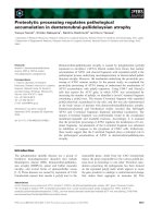
presenilin contributes to the catalytic activity (Fig. 1).
However, only a fraction of Ab in postmortem
AD brain is full-length Ab1-42 or Ab1-40. N- and
C-terminally truncated variants are prevalent, and Ab
can undergo racemization, isomerization [6] and pyro-
glutamyl modification [7]. The biochemical processes
generating all these Ab species and their significance
to AD pathogenesis are only partially understood.
Early-onset AD can also arise as a result of increased
APP gene dosage caused by APP gene duplication [8]
and Down’s syndrome with trisomy 21. Virtually all
Down’s syndrome patients aged 35–40 years develop
AD neuropathology, and most experience dementia by
60–70 years of age [9].
The major genetic risk factor for developing late-
onset AD is the apolipoprotein E (ApoE) e4 allele
[10,11]. One ApoE e4 allele increases the risk of AD by
two- to threefold, and two e4 alleles confer a 12-fold
increase in risk. In the brain, ApoE is primarily synthe-
sized by astrocytes and serves to regulate the transport
of cholesterol-containing lipoprotein particles. ApoE
binds to Ab and becomes a component of amyloid in
AD senile plaques. The pathogenic mechanism of
ApoE likely relates to altered deposition and ⁄ or clear-
ance of Ab in the brain, although the details are still
not fully understood [12]. A large number of other dis-
ease-related loci and candidate genes have been pro-
posed, but not generally verified, indicating that these
genes have a modest impact on the pathogenesis. The
major risk factors for AD are age and a family history
of the disease. Low education or cognitive reserve
capacity, female gender, head trauma, hypertension,
cardiovascular disease and a high-cholesterol diet are
proposed risk factors for AD [13] (Fig. 2).
Nontransgenic animal models
Based on the cholinergic hypothesis, scopolamine-
induced amnesia, excitotoxic lesions of the basal
forebrain and aged primates have been used to assess
cognitive deficits. Current symptomatic drugs for AD
were successfully evaluated in these models, but their
etiological relevance is low [14]. Nontransgenic rodents
Fig. 1. Disease-causing APP mutations used in transgenic models. The Swedish mutation (1) favors b-secretase (b) cleavage, while the
Flemish mutation (2) partly disfavors cleavage of APP at the a-secretase (a) site. The Arctic, Dutch and Iowa mutations (3), which are located
in the Ab-domain, mainly increase aggregation. The London-type APP mutations (4) alter c-secretase (c) cleavage to increase Ab42 or the
Ab42 ⁄ Ab40 ratio.
Animal models of Alzheimer’s disease O. Philipson et al.
1390 FEBS Journal 277 (2010) 1389–1409 ª 2010 The Authors Journal compilation ª 2010 FEBS
are poor natural animal models of AD, but intracere-
broventricular infusion of Ab [15] or lipopolysaccharide
in such animals has been used. The latter leads to
neuroinflammation with hippocampal neurodegenera-
tion and spatial memory deficits [16]. The models
require attention to methodological detail and are
difficult to standardize. Senescence-accelerated mice
were selectively bred from AKR ⁄ J mice. In short-lived
SAM-P8, there is an age-related increase in diffuse Ab
deposits and cyclin-dependent kinase 5, cholinergic
deficits and increased blood–brain barrier permeability.
The phenotypes likely relate to oxidative stress and
mitochondrial dysfunctions [17]. Mice with segmental
trisomy of chromosome 16 have primarily been used to
dissect the genetic mechanisms of Down’s syndrome
phenotypes. Ts65Dn mice [18], the most frequently
used model, have interesting synaptic and cognitive
phenotypes with degeneration of cholinergic neurons
that depend on APP gene dosage.
Following observations in postmortem brain from
patients with coronary artery disease, rabbits fed a
cholesterol-enriched diet were used as an animal model
[19]. They are impared in classical eyelid conditioning
and show diffuse Ab deposits and vascular inflamma-
tion. Aged dogs (> 10 years) can show impaired atten-
tion, spatial disorientation and disturbed diurnal
rhythm. Cognitive dysfunction in old dogs is associated
with diffuse Ab deposits [20], neuritic dystrophy and gli-
osis, but few amyloid plaques and no NFTs. Ventricular
dilation, cortical and hippocampal atrophy, CAA with
degeneration of smooth muscle cells (SMC) and hemor-
rhages can all be found in aged canine brain. Interest in
nonhuman primate models has grown following the fail-
ure to predict meningoencephalitis as a side-effect of the
AN1792 vaccination trial from transgenic studies. The
efficacy and safety of an Ab vaccine has been tested in
the Carribean vervet monkey [21]. Alternatives are aged
lemurs [22], cotton-top tamarins [23], rhesus monkeys
[24] or squirrel monkeys [25]. An aged chimpanzee with
complete AD neuropathology, including neuritic
plaques and paired helical filament-containing NFTs,
was recently reported [26].
Fig. 2. AD pathogenesis according to the
amyloid cascade hypothesis. This theory
suggests that altered metabolism of Ab,in
particular aggregation-prone Ab species like
Ab42, initiates AD pathogenesis. Oligomeric
assemblies of Ab trigger aggregation of tau
and the formation of NFTs, but also inflam-
mation and oxidative stress, by rather
unclear mechanisms. These downstream
processes give rise to progressive neurode-
generation, which ultimately results in
dementia. The main pathogenic pathway of
AD is illustrated with red arrows, whereas
minor contributory pathways are shown
with thinner brown arrows. The experimen-
tal support for the hypothesis comes mainly
from studies of families in which AD is
inherited as a dominant trait due to muta-
tions in APP, PS1 or PS2. The evidence that
the theory applies to sporadic AD is less
solid, although risk factors such as age and
ApoE genotype both strongly impact on Ab
aggregation in transgenic models and post
mortem AD brain.
O. Philipson et al. Animal models of Alzheimer’s disease
FEBS Journal 277 (2010) 1389–1409 ª 2010 The Authors Journal compilation ª 2010 FEBS 1391
Transgenic animal models
Models devoid of any disease-causing APP
mutations
Animal models expressing wild-type (wt) human APP
are of interest because the great majority of sporadic
AD patients do not carry any disease causing APP
mutation. In early transgenic attempts, APP processing
was bypassed altogether and human Ab was directly
expressed under a promoter. Natural inclusions in the
brain were mistakenly identified as amyloid-like fibrils
in these mice [27]. Fusion proteins, in which the signal
peptide and C-terminal fragment (C99) of wild-type
APP were joined and expressed under the control of
the cytomegalus enhancer ⁄ chick b-actin promoter,
were also generated. Ab levels in plasma from these
transgenic mice were in the nm range, but Ab deposits
did not form in the brain. Instead, intracellular Ab
aggregates or amyloid deposists were found in the pan-
creas [28], intestine [29] or skeletal muscles [30]. The
level of plasma Ab in C99-based models was similar to
Tg2576, an APP transgenic model with high peripheral
promoter activity.
In an alternative strategy, a yeast artificial chromo-
some, harboring the whole APPwt gene, was used to
maintain transcriptional regulation, alternative splicing
and normal APP processing. In these Py8.9 mice,
proper APP protein synthesis and alternative splicing
was demonstrated, but the brain was devoid of neuro-
pathology and the levels of Ab were low [31]. How-
ever, when wild-type human APP was expressed at
very high levels, under the Thy1 promoter, sparse
parenchymal and vascular amyloid deposits were
found in aged mice [32]. Thus a pathogenic APP muta-
tion is not a prerequisite for amyloid deposition.
Instead it seems to depend upon producing sufficient
Ab levels in the brain to ensure fibrillization. To
explore the pathogenic impact of individual Ab spe-
cies, a fusion protein, BRI–wt-Ab42, was designed
from which Ab was released by furin-like enzymes on
the cell surface. BRI is a transmembrane protein that
is involved in amyloid deposition in British familial
dementia. The fusion design permitted the synthesis of
high Ab levels in the brain in a manner similar to APP
transgenic mice, but in the absence of APP overexpres-
sion. Transgenic mice expressing BRI–wt-Ab42 devel-
oped extensive vascular and parenchymal amyloid
pathology, accompanied by dystrophic neurites and
astrogliosis. In contrast to many APP transgenic mod-
els, amyloid deposition in BRI–wt-Ab42 mice began in
the cerebellum, where both furin and the transgene
were highly expressed. This illustrates how the anatom-
ical location of AD neuropathology can be manipu-
lated simply by enhancing the regional dosage of
amyloidogenic proteins and enzymes regulating their
metabolism. In contrast to BRI–wt-Ab42, no neuropa-
thology was found in aged BRI–wt-Ab40 transgenic
mice although they had higher Ab levels when they
were young. Thus the identity of Ab determines if
neuropathology will develop [33].
Models with the London-type APP mutation
The London mutation (V717I) was the first genetic
lesion to be discovered in a family with AD [3], and
shortly thereafter the Indiana mutation (V717F) was
found in an American pedigree [34]. Patients with the
Indiana mutation develop short-term learning impair-
ments in the fifth decade, followed by progressive
cognitive impairment and dementia with typical AD
neuropathology. An abundance of NFTs and senile
plaques was observed at autopsy, as well as mild CAA
[35]. Games et al. described the neuropathology of
PDAPP mice, the first transgenic AD model [36]. A
mini-gene encompassing a human APP cDNA with the
Indiana mutation interposed with introns had been
designed. Alternative splicing and synthesis of all three
isoforms APP
695
, APP
751
and APP
770
, with strong and
selective neuronal expression was enabled by the plate-
let derived growth factor (PDGF)b promoter. Impor-
tantly, young PDAPP mice produced high Ab42 levels
in the brain, particularly in the hippocampus. The ani-
mals preferentially accumulated Ab42 peptides and
developed senile plaques, but also a substantial num-
ber of diffuse Ab deposits at 9–10 months of age [37].
Plaque formation began in the cingulate cortex and
was accompanied by phospho-tau immunoreactive
dystrophic neurites, synaptic loss and gliosis in the
adjacent tissue, but not by overt neuronal loss [38,39].
Ultrastructural analyses revealed neurons in close
proximity to senile plaques and amyloid fibrils. The
latter had a diameter of 9–11 nm and were surrounded
by neuronal membranes and vesicles [40]. Young
PDAPP mice showed deficits in spatial learning and
memory, which worsened with increasing age and Ab
burden, although their performance in a novel
object-recognition task was unimpaired [41]. By
contrast, others found age-dependent deficits in object
recognition and place learning impairments that were
independent of age [42]. These discrepancies could be
because of differences in experimental procedures or
unintentional genetic drift of mouse colonies. PDAPP
mice are typically bred on a mixed genetic background
(Swiss Webster, DBA ⁄ 2 and C57Bl ⁄ 6). Hippocampal
volume and corpus callosum length is reduced in
Animal models of Alzheimer’s disease O. Philipson et al.
1392 FEBS Journal 277 (2010) 1389–1409 ª 2010 The Authors Journal compilation ª 2010 FEBS
PDAPP mice and this depends on APP gene dosage,
but it is unrelated to age-dependent Ab accumulation
[43]. Certainly this abnormality could impact on the
behavior of PDAPP mice, but the molecular mecha-
nism and its relevance to macroscopic atrophy in AD
(if any) is still unclear.
Van Leuven et al. generated several transgenic mod-
els, including mice with only the London mutation,
APP-London. As expected, a markedly increased level
of Ab42 was found in young mice and predominantly
Ab42-immunoreactive diffuse and neuritic plaques in
aged animals, compared with models harboring the
Swedish mutation. Impaired long-term potentiation in
hippocampal slices and deficits in spatial learning and
memory in the Morris water maze (MWM) were
reported. The mice were on a FVB ⁄ N background and
displayed neophobic behavior. They were sensitive to
glutamate antagonists and died prematurely. These
phenotypes were noted in young mice prior to the
onset of plaque formation, and could possibly be
caused by the combination of APP overexpression and
FVB ⁄ N genetic background [44]. In older mice
(> 15 months), CAA was found in the arteries and
pial arterioles in association with disruption of external
elastic lamina and the formation of aneurysm. Further-
more, the ratio of Ab42 ⁄ Ab40 levels in leptomeninges
was eight times lower than in neocortical tissue
extracts. Ab42 could still have initiated deposition of
Ab in vessels, because some focal lesions were only
Ab42-immunoreactive [45]. By contrast, brains from
patients with the London mutation contained mainly
Ab-immunoreactive plaques and cytoskeletal pathol-
ogy, but only modest or little CAA [46]. Thus, the
CAA phenotype in the transgenic mice might not have
been caused by the London mutation. Instead it may
be the result of strong APP expression, advanced age,
strain background (FVB ⁄ N) and ⁄ or differences in APP
processing between species.
Models with the Swedish APP mutation
The Swedish mutation (KM670 ⁄ 671NL) is located just
outside the N-terminus of the Ab domain in APP. It
was identified in 1992 [47] and shown to increase Ab
levels by six- to eightfold [4]. These discoveries created
intense interest in APP processing and paved the way
for the development of more sophisticated ELISAs to
selectively measure Ab40 and Ab42 [48]. Later, the
Swedish mutation became essential in the identification
and characterization of BACE-1 [49]. The clinical and
neuropathological features associated with the Swedish
mutation are those of typical AD [47,50]. Tg2576
mice, the most frequently used APP transgenic model,
harbor the Swedish mutation and display both AD-like
Ab neuropathology and cognitive deficits [51]. The
Swedish mutation redirects APP processing to secre-
tory vesicles en route to the cell surface in cell culture
[52], whereas APPwt is largely processed in recycling
endosomes [53]. This difference may be largely irrele-
vant in the brain, because Ab synthesis along the
endolysosomal pathway is clearly important in Tg2576
[54]. A substantial amount of CAA is often found in
transgenic mice with the Swedish mutation, which is
likely to be because of the high rate of synthesis and
accumulation of Ab1-40.
In Tg2576, more than fivefold overexpression of the
human APP
695
isoform with the Swedish mutation is
generated by the prion promoter. APP cDNA was
cloned into a 40 kb genomic fragment (cosSHaPrP)
[55] from the hamster prion protein gene. A significant
proportion of Tg2576 mice die at a young age, and the
severity of this phenotype depends upon the genetic
background. It has been found that colonies are best
maintained by mating heterozygous C57BL ⁄ 6 males
with B6SJLF1 females. At around 11 months of
age, Tg2576 mice show extracellular Ab deposits which
are largely soluble in SDS and mainly contain Ab40
( 75%). CSF levels of Ab42, but not Ab40, decrease
with age and amyloid deposition [56], and pathogenesis
is accelerated in female Tg2576 mice [57]. Both these
observations fit well with biochemical and epidemio-
logical findings in AD [13].
Borchelt et al. used the prion promoter to generate
line C3-3 [58]. A chimeric cDNA clone encoding
murine APP
695
was used, in which the region in and
around the murine Ab domain was replaced with the
human Ab sequence and the Swedish mutation. The
MoPrP.Xho vector was much smaller than the cos-
SHaPrP, and selectively directed twofold overexpres-
sion of APP to the brain [58,59]. In an even more
refined strategy, the murine Ab sequence was human-
ized and the Swedish mutation introduced with gene
targeting. In this knockin model, APP
NLh ⁄ NLh
, only
five single amino acids were altered in the entire mur-
ine genome. Consequently, APP protein synthesis
remained unchanged in terms of its spatial and tempo-
ral expression pattern and mRNA localization. The
Swedish mutation led to markedly enhanced b-secre-
tase activity and a ninefold increase in Ab level,
compared with normal aged human brain [60]. By
22 months of age, APP
NLh ⁄ NLh
mice had not devel-
oped Ab neuropathology [61], but young mice were
elegantly used to estimate the turnover of Ab, APP
and APP fragments in vivo [62]. In another genomic
approach, a 650 kb yeast artificial chromosome vector
harboring the whole human APP gene locus with the
O. Philipson et al. Animal models of Alzheimer’s disease
FEBS Journal 277 (2010) 1389–1409 ª 2010 The Authors Journal compilation ª 2010 FEBS 1393
Swedish mutation was used. In the homozygous mice
(R.1.40), in which Ab42 levels were 15 to 20-fold
higher than in mice expressing wild-type human APP,
fibrillar A b deposition began at 14–15 months of age.
There were region-dependent differences with more
cortical Ab deposits in old R.1.40 mice than cDNA-
based Tg2576 mice, despite lower Ab40 levels in young
mice [63]. Heterozygous aged R.1.40 mice had only
diffuse Ab deposits, illustrating how a small change in
Ab levels in adolescence influences the speed and type
of AD-like pathology [64].
The APP23 model was generated by inserting human
APP
751
with the Swedish mutation into the murine
Thy1 cassette. It led to strong and highly specific
expression in postmitotic neurons and at 7 months
of age the animals developed a progressively increasing
burden of Congo Red-positive plaques. The plaques
were surrounded by gliosis and distorted neurites that
were immunopositive for hyperphosphorylated tau
[65]. APP23 mice have often been used to study CAA
pathogenesis. It is most frequent at the adventitial sur-
face of SMC in arteries ⁄ arterioles and is accompanied
by degeneration of vascular SMCs, disruption of the
blood–brain barrier and microhemorrhages in severely
amyloid-laden vessels [66,67]. Behavioral and cognitive
effects with changes in activity levels that depended on
circadian rhythm were apparent from 3 months of age
in APP23 mice. By 25 months, the mice underper-
formed in passive avoidance and small MWM tasks
[68].
Models with both Swedish and London-type APP
mutations
Patients never inherit multiple pathogenic mutations in
APP, presenilin, tau or a-synuclein genes, nor do they
overexpress chimeric APP mRNA under a heterolo-
gous promoter. Thus none of the transgenic models
fully mimic the genetics of familial AD. By combining
genetic lesions one can accelerate Ab aggregation and
lower the cost of research. One can also confer certain
characteristics to Ab and dissect molecular interac-
tions. The Swedish mutation has often been used
together with other mutations in transgenic models
because it is located outside the Ab domain and serves
to enhance Ab levels.
No Ab pathology was evident at 24 months of age
in J.1.96 homozygous transgenic mice when a genomic
vector with both the Swedish and London mutations
was introduced, despite life-long exposure to a four- to
sixfold increased levels of Ab42, compared to human
APPwt [64]. In contrast, APP22 mice presented with
diffuse Ab deposits and few amyloid plaques when the
mutations were combined in a cDNA-strategy to
create twofold overexpression under the human Thy1
promoter [65].
The Tg-CRND8 model was designed by inserting
human APP
695
with Swedish and Indiana mutations in
the cosSHaPrP vector [55]. It resulted in an aggressive
neuropathology with onset of amyloid deposition and
place learning impairment as early as 3 months of age.
There was postnatal lethality, like many other APP
transgenic models, which could be mitigated by main-
taining colonies on a favorable genetic background
[69]. Lines J9 and J20, developed by Mucke et al., also
combined these two mutations, but the expression was
regulated by the PDGFb promoter. There was a loss
of presynaptic synaptophysin immunoreactivity which
was unrelated to plaques, and it could be shown that
not only the level of Ab 42, but also the ratio of
Ab42 ⁄ Ab40, determined the onset of plaque formation
[70]. Consistent with this idea, Ab40 inhibited amyloid
formation when uncoupled from APP processing in
transgenic mice expressing BRI–Ab fusion proteins
[71].
Tetracycline-regulated systems have not often been
used in the Ab-based transgenic models, perhaps
because of their complicated nature with technical
caveats regarding ‘leakage’. They offer a way to tightly
control the induction or repression of a transgene. In
TetO-APP-Swe ⁄ Ind (line 107), a tetracycline-responsive
promoter was linked to a chimeric mouse ⁄ human
APP
695
isoform, designed like the C3-3 line, but with
both the Swedish and Indiana mutations. The mice
were crossed with animals expressing the tetracycline
transactivator under the control of the calcium-
calmodulin kinase II a promoter. Repression of the
transgene was thereby restricted to the forebrain. Amy-
loid deposition was found in crossed rTA ⁄ APP mice
from 8 weeks of age, a consequence of 10 to 30-fold
increased APP expression and two pathogenic APP
mutations. APP transgene expression was suppressed
> 95% when 4-week-old mice were given doxycycline
for 2 weeks. Relative to doxycycline-free mice, Ab in
PBS- and SDS-soluble pools were efficiently cleared,
although Ab42 partially remained in the formic acid
soluble pool. By contrast, brains of animals reared on
doxycycline from birth to 6 weeks of age contained
essentially no human Ab, suggesting a very early onset
of Ab aggregation and a tightly controlled transgene
expression with little leakage. Interestingly, suppression
of the transgene in 6-month-old mice arrested amyloid
deposition, but did not promote clearance. Moreover,
astrogliosis and ubiquitin-positive dystrophic neurites
in the vicinity of senile plaques were unchanged. Thus,
the endogenous clearance systems were unable to
Animal models of Alzheimer’s disease O. Philipson et al.
1394 FEBS Journal 277 (2010) 1389–1409 ª 2010 The Authors Journal compilation ª 2010 FEBS
eliminate existing Ab aggregates and secondary pathol-
ogy, at least in this aggressive transgenic model [72].
Models with the Flemish, Arctic, Dutch or Iowa
APP mutation inside the Ab domain
Mutations at positions 21–23 in the Ab domain of
APP, near the hydrophobic cluster, are a heterogeneous
group of genetic lesions. They affect Ab aggregation
and degradation, but also APP processing. The Dutch
(E693Q) [73] and Iowa (D694N) [74] mutations are
associated with CAA and diffuse Ab deposits, resulting
in hemorrhagic strokes and ⁄ or infarcts and dementia.
The neuropathology also consists of leukoencephalopa-
thy, degenerating neurites and NFTs [73,74]. Transgene
expression in APPDutch mice, with only the Dutch
mutation, is regulated by the neuron-specific Thy1
promoter [32]. The ratio of Ab40 ⁄ Ab42 was increased
in APPDutch mice, compared with APPwt, with the
Dutch mutation favoring the production and ⁄ or
increased resistance of Ab40 to proteolysis. In aged
animals there was an extensive accumulation of CAA
in leptomeningeal and cortical vessels with few diffuse
plaques, severe loss of SMCs, weakening of vessel walls
leading to hemorrhage and perivascular microgliosis
and astrocytosis. The Dutch and Iowa mutations both
reduce the negative charge of Ab and stimulate the
formation of amyloid fibrils and attachment to the cell
surface of human cerebrovascular SMCs. The two
mutations were combined with the Swedish mutation
in the SweDI transgenic model [75], because in vitro
studies had shown that Ab peptides with both muta-
tions induced greater vascular SMC degeneration [76].
The SweDI mice, with a low transgene expression
regulated by the Thy1.2 promoter, accumulated large
amounts of Ab in microvessels. Severe CAA was
observed at 3 months of age with predominantly
diffuse parenchymal Ab deposits [75], and the mice
were cognitively impaired from a young age [77]. The
vessel density in the hippocampus and thalamus was
reduced in parallel with increasing CAA, but was
not reduced in the frontotemporal cortex where
mainly diffuse parenchymal Ab deposits accumulated.
The microvascular pathology was accompanied by
astro- and microgliosis as well as increased levels of
proinflammatory cytokines.
The Flemish (A692G) [78] mutation can start either
with presenile dementia or CAA. In contrast to the
other intra-Ab mutations, it makes APP an inferior
substrate for a-secretase and increases Ab levels
[79,80]. APP cleavage by b-secretase was favored in
transgenic mice with the Flemish mutation (APP ⁄ Fl),
with modestly increased Ab1-40 levels. There was
spongiosis and gliosis in APP ⁄ Fl mice, but no Ab or
tau pathology. Male APP ⁄ Fl mice, which were bred on
the FVB background, were aggressive and suffered
premature death and seizures. APP expression was
likely insufficient to generate neuropathology and it is
unclear if the findings in APP ⁄ Fl were specifically
caused by the Flemish mutation. An APP ⁄ Du model,
developed in parallel, displayed similar phenotypes
[81].
The Arctic APP mutation (E693G) [79] is associated
with clinical features of early-onset AD commencing at
52–62 years. There are NFTs, severe CAA in the
absence of hemorrhage and an abundance of paren-
chymal Ab deposits lacking amyloid cores in postmor-
tem brain [82]. The Arctic mutation promotes Ab
protofibril and fibril formation, but also favors intra-
cellular b-secretase processing of APP [79,83,84].
Tg-ArcSwe models with both the Swedish and Arctic
mutation were developed by two independent groups.
In young mice, the Arctic mutation increased intraneu-
ronal A b accumulation in an age-dependent manner
[85,86]. Tg-ArcSwe mice without Ab deposition
showed cognitive deficits in MWM and two-way active
avoidance [85,87], and their performance correlated
inversely with soluble Ab protofibril levels [87]. Build-
ing on the combinatorial principle, lines Arc6 and
Arc48 with Swedish, Arctic and Indiana mutations
were generated. Again, the Arctic mutation accelerated
amyloid formation despite a reduced proportion of
Ab42 in young mice [88]. Recently, a mouse model
expressing human APP with only the Arctic mutation
(APParc) under the control of the neuron-specific
Thy1 promoter was reported [89]. In old APParc mice,
both parenchymal and vascular congophilic Ab depos-
its were found in the subiculum and thalamus. In
contrast to transgenic models with both the Arctic and
Swedish mutation [85,86], APParc mice did not show
any punctate intraneuronal immunoreactivity. Loco-
motor activity and exploratory behavior of APParc
mice was normal, although aged female mice displayed
spatial learning and memory deficits. Accelerated
amyloid pathology in female APParc mice is consistent
with findings in Tg2576 mice [57].
APP transgenic models harboring a presenilin,
tau or a-synuclein transgene
Presenilin transgenic mice were generated in response
to the identification of the presenilin-1 (PS-1) and
presenilin-2 (PS-2) AD mutations. Metabolic Ab42
levels were selectively increased in PS-1 transgenic
mice [58,90] and, in comparison with APP transgenic
mice, amyloid deposition was markedly accelerated in
O. Philipson et al. Animal models of Alzheimer’s disease
FEBS Journal 277 (2010) 1389–1409 ª 2010 The Authors Journal compilation ª 2010 FEBS 1395
bigenic PS-1 · APP transgenic mice [91,92]. Borchelt
et al. generated PS-1 transgenic models expressing
mutant protein (M146L, A246E or PS1DE9), which
were cross-bred with APP transgenic mice with the
Swedish mutation, line C3-3 [58,91]. Other researchers
cross-bred Tg2576 with PS-1 transgenic mice, in which
PS-1 cDNA (M146L or M146V) had been linked to
the PDGFb2 promoter [90], resulting in the PSAPP
model [92]. PS2APP mice, generated by crossing PS2
(N141I) and APP-Swe, also displayed an aggressive
Ab pathology with age-dependent spatial learning and
memory deficits [93].
Autosomal dominant mutations in tau causing
frontotemporal lobe dementia were quickly utilized for
transgenic experiments. The JNPL3 model, expressing
4R0N tau isoform with the P301L mutation, was the
first model with Gallyas-positive NFTs. When double-
transgenic mice were created by cross-breeding with
Tg2576 a more complete AD neuropathology was
generated. Moreover tau pathology, but not Ab patho-
logy, was enhanced in these mice, suggesting that effects
on tau are downstream of Ab in AD pathogenesis [94]
(Fig. 2). However triple-transgenic mice, which were
generated by crossing mice producing the wild-type tau
isoform (3R0N) with mice carrying the Swedish and
London APP mutations and a PS-1 mutation (M146L),
only resulted in somadendritic accumulation of tau and
cytoskeletal changes. Thus Ab-driven transgene expres-
sion failed to facilitate NFT formation in the presence
of human wild-type tau [95]. In a similar strategy, the
pathogenic interaction between Ab and a-synuclein
was investigated by crossing PDGF–a-synuclein
and APP-SweInd, which led to a 1.6-fold increase in
a-synuclein inclusions in comparison with transgenic
mice that expressed only wild-type human a-synuclein.
Similar to crossed APP · Tau mice, the level of accumu-
lated Ab in brain was similar in single- and double-
transgenic mice [96]. The findings are relevant to the
Lewy body variant of AD with a-synuclein inclusions.
Instead of cross-breeding two single-transgenic mod-
els, several vector constructs can be coinjected into
fertilized oocytes. The transgenes will typically cointe-
grate at one location in the genome, and thus be inher-
ited as a single transgene ([97] and references therein).
The approach saves time and generates transgenic mice
on a homogenous genetic background, which reduces
variability and the number of animals needed for
experiments. LaFerla et al. developed 3xTg-AD by
coinjecting transgenes encoding both APP
695
-Swedish
and Tau isoform 4R0N with the P301L mutation into
pronuclei of PS-1 (M146V) knockin mice. Both transg-
enes were subcloned into the Thy1.2 expression
cassette. Intraneuronal Ab was visible in 3-month-old
mice and extracellular plaques in 6 to 12-month-old
animals. Phospho-tau immunoreactivity was detected
in 12 to 15-month-old mice, whereas paired helical
filament-1 immunoreactivity and Gallyas staining,
which indicate NFT formation, were not seen until
18 months of age [98]. In more recent studies of this
model, amyloid deposition commenced at 15 months
in the hippocampus and was widespread > 18 months
[99]. Triple-transgenic mice have been elegantly used to
study interactions between Ab and tau pathologies and
their impact on phenotypes of synaptic and cognitive
dysfunction [100,101].
Advanced animal models have recently been gener-
ated in which neuronal degeneration is clearly evident.
In the 5xFAD transgenic model, Thy1 promoter-driven
transgenes of APP (with the Swedish, Florida and
London AD mutations) and PS-1 (with the AD muta-
tions M146L and L286V) were coinjected into pronu-
clei of C57BL ⁄ 6xSJL mice. The model was made in an
effort to alter the Ab42 ⁄ Ab40 ratio in favor of Ab42
synthesis ([102] and references therein). Indeed the
strategy resulted in a high level of Ab42 and an
Ab42 ⁄ Ab40 synthesis ratio of 25 : 1 in young mice, in
comparison with 0.1–0.2 : 1 in Tg2576 mice with only
the Swedish APP mutation. Amyloid deposits formed
within 2 months, and the mice also developed intran-
euronal Ab aggregates. The intraneuronal deposits
were in a b-pleated sheet conformation, and located to
large pyramidal neurons of cerebral cortex layer V.
Interestingly, in 9-month-old 5xFAD-mice there was a
selective loss of these neurons and a decrease of several
synaptic markers. Importantly, these phenotypes and
age-dependent cognitive deficits seemed to depend
upon Ab because they did not occur in aged 5xFAD ⁄
BACE-KO mice [103]. Neuronal loss was also found
in APP ⁄ PS1 KI, homozygous PS-1 knockin mice
(M233T ⁄ L235P) with a Thy1 promoter-driven APP
transgene harboring the Swedish and London
mutation. In young mice intraneuronal Ab aggregates,
positive for thioflavine S, were found in neurons that
degenerated with aging, as in the 5xFAD model. Their
brains contained a substantial amount of N-truncated
and modified Ab peptides [104].
Insight into AD pathogenesis from
experiments with transgenic models
Although far from perfect animal models of AD,
transgenic mice have contributed significantly to the
understanding of molecular pathogenesis. Steady-state
levels of Ab in brain and CSF, and the ratio between
them in young transgenic mice are quite similar
between the different models and conditions in healthy
Animal models of Alzheimer’s disease O. Philipson et al.
1396 FEBS Journal 277 (2010) 1389–1409 ª 2010 The Authors Journal compilation ª 2010 FEBS
humans (Table 1). It has been proposed that Ab
exists in a state of dynamic equilibrium between the
plasma and central nervous system (brain and CSF)
[105]. If so, the ratio of [Ab]
brain
⁄ [Ab]
plasma
or
[Ab]
CSF
⁄ [Ab]
plasma
should be the same in all trans-
genic models. A much higher plasma Ab level in
Tg2576 mice than in APP23, but a comparable
central nervous system Ab level, is inconsistent with
the idea of a dynamic equilibrium. The higher plasma
Ab levels in Tg2576 is most likely explained by the
stronger peripheral activity of the hamster PrP
promoter, compared with the neuron-specific Thy1
promoter in APP23. This emphasises the influence of
promoter selection on differential expression patterns
of APP and steady-state Ab levels in the central
nervous system and in peripheral tissue; consequently,
interpretations from transgenic models regarding Ab
dynamics should be made with caution.
In the 1980s it was debated whether Ab amyloid
deposits, and in particular CAA at the cerebral vessel
wall, had a central nervous system or a peripheral
source. Models driven by the Thy1 promoter, like
APPDutch transgenic mice, with almost exclusive
neuronal central nervous system expression of APP
develop almost only CAA, but by introducing a
presenilin transgene and raising the ratio of
Ab42 ⁄ Ab40 synthesis instead mainly parenchymal
senile plaques develop [32]. By contrast, models with
peripheral APP synthesis and high plasma Ab levels
present with amyloid deposits in peripheral organs
and neither CAA nor Ab plaques are found in the
brain of such mice [28–30]. Thus, studies in transgenic
mice strongly suggest that neuronal Ab produced in
the brain generates cerebrovascular Ab neuropathol-
ogy. With live imaging, the arrangement of vascular
SMC is found to be disrupted by CAA in transgenic
mice. It leads to impaired vasodilator reactivity, dis-
tinct loss of SMC and hemorrhages [106], which is
similar to the pathogenesis in human brain [107].
Enthorhinal cortex lesion or transection of the per-
forant pathway have been used to demonstrate that
senile plaque formation depends on the synaptic
release of Ab and anterograde axonal transport of
APP [108,109]. Senile plaque formation can also be
induced in APP transgenic mice if Ab-containing
brain extracts from plaque-laden mice or AD brain
are brought in direct contact with the central nervous
system. The Ab phenotype then depends on both the
seeding agent and the host environment, similar to
prion disorders [110,111]. The growth and stability of
dense-cored plaques have been investigated using
open skull window surgery and multiphoton micros-
copy [112]. The great majority of amyloid plaques
Table 1. Metabolic levels of amyloid-b (Ab) in brain, cerebrospinal fluid (CSF) and plasma of young transgenic mice and healthy human. Measures in PDAPP refer to Ab
1–x
, in Tg2576,
APP23 and humans to the sum of Ab
1–40
and Ab
1–42
, and in tgArcSwe they refer to Ab
1–40
. A density 1 gÆL
)1
for brain tissue has been assumed when the ratio of brain Ab ⁄ plasma Ab
has been calculated. Thus, for example, 20 pmolÆg
)1
20 nM. Studies on the initial description and on metabolic levels of brain, CSF and plasma Ab that were used as sources of informa-
tion are cited. APP, amyloid-b precursor protein; CAA, cerebral amyloid angiopathy; NFT, neurofibrillary tangles; PDGF, platelet-derived growth factor.
Model Transgene Promoter Neuropathology
Age-of
-onset
(months)
Brain
Ab
(pmolÆg
)1
)
Plasma
Ab
Ratio
brain Ab ⁄
plasma Ab
CSF
Ab
Ratio
brain Ab ⁄
CSF Ab Contact
PDAPP [36,37,105] APP minigene V717F
(Indiana)
PDGF Diffuse and neuritic
plaques, little CAA
6–9 20 40 p
M 500 4 nM 5 Elan ⁄ Eli Lilly
Tg2576 [51,56] APP
695
cDNA
KM670 ⁄ 671NL (Swedish)
Hamster PrP Neuritic plaques
substantial CAA
9–10 40 4.5 nM 913nM 3 Taconic
APP23 [65] APP
751
cDNA
KM670 ⁄ 671NL (Swedish)
Murine Thy1 Neuritic plaques
profound CAA
some neuron loss
64080pM 500 11 nM 4 Novartis
TgArcSwe [86] APP
695
cDNA
KM670 ⁄ 671NL (Swedish)
+ E693G (Arctic)
Murine Thy1 Neuritic plaques
and CAA
5–6 3 10 pM 300 1.5 nM 2 BioArctic
Healthy [164,165]
human
Diffuse and neuritic
plaques and
CAA NFTs, atrophy
20 90 p
M 220 1.6 nM 12
O. Philipson et al. Animal models of Alzheimer’s disease
FEBS Journal 277 (2010) 1389–1409 ª 2010 The Authors Journal compilation ª 2010 FEBS 1397
formed very rapidly, within 1–2 days, reaching a size
that was surprisingly stable. Within 1 week, early
changes were accompanied by the recruitment of
reactive microglia, and shortly thereafter by neuritic
dystrophy [113]. However, in a subsequent study,
plaque growth occurred over a period of weeks when a
thinned-skull cranial window was used instead [114].
There is also a local neurotoxic effect on nerve endings
near amyloid plaques in APP transgenic models and in
AD postmortem brain [115], whereby dendritic spines
decrease in density but do not change in structure
[116]. Loss of CA1 pyramidal neurons in the hippo-
campus was reported in aged APP23 mice with a high
plaque load [117], although subtle cell loss is difficult
to distinguish from physical displacement. Today,
almost every research article in the AD field contains
an introductory statement in which the neurotoxicity
of Ab is described as a well-established fact, yet analy-
ses of mouse brain in which large amounts of Ab have
accumulated provide no or very sparse support for this
hypothesis. Neurodegenerative mechanisms of proteop-
athies are still largely unknown. Perhaps the neurotox-
icity is sparse because APP trafficking and subcellular
Ab accumulation in AD brain is poorly mimicked in
most models, as chimeric APP mRNAs are overexpres-
sed under heterologous promoters. This hypothesis is,
however, inconsistent with knockin mice, APP
NLh ⁄ NLh
,
showing no neurodegeneration. Murine neurons could
be devoid of the downstream pathways necessary for
Ab to induce toxicity, and a prime suspect is of course
the processes leading to tau aggregation and NFTs in
AD brain. More than 10 years ago modified and trun-
cated Ab peptides were demonstrated in AD brain
[6,7], but the observations were partially ignored. It
could be that only certain species of Ab are neurotoxic
and that by using mutations linked to familial AD we
poorly replicate the processes of Ab production and
aggregation in sporadic AD brain. There is now a
renewed interest in studying APP processing in
sporadic AD brain, and in understanding the mecha-
nisms of Ab truncation and modification. Most trans-
genic models produce mainly full-length Ab, but by
manipulating the regulatory mechanisms or by uncou-
pling Ab synthesis from APP processing one can
generate transgenic models producing certain Ab
species. These can indeed be neurotoxic, for example,
Ab3(pE)-42 in TBA2 mice [118], and thus not only in
a cell culture.
The effect of ApoE on Ab neuropathology was first
examined in APP transgenic mice lacking murine
ApoE.Ab burden, and more markedly amyloid
burden, was reduced in a gene-dose-dependent manner
[119]. The human ApoE isoforms (e2, e3 and e4) were
expressed under the glial fibrillary acidic protein
promoter in PDAPP mice lacking murine ApoE.Ab
burden was then accelerated by the risk allele ApoE e4
and decelerated by the protective allele ApoE e2, rela-
tive to the ApoE e3 allele [120]. These findings fit well
with observations in postmortem AD brain [121].
However, although murine ApoE facilitates Ab deposi-
tion in a gene-dose-dependent manner, human ApoE
decelerates Ab deposition compared with murine
ApoE [122]. This may be because a human transgene
was introduced into a complex feedback network
involving murine lipoprotein receptors. Alternatively,
ApoE may affect both Ab clearance and deposition. It
illustrates the complexity of detailed mechanistic stud-
ies. Deletion of apolipoprotein J, which also binds to
Ab, decelerated amyloid formation [123], whereas abla-
tion of both ApoE and apolipoprotein J strongly
increased Ab deposition [124]. Possibly the lipoprotein
metabolism in the brain is altered when two abun-
dantly expressed apolipoproteins, E and J, are both
absent. Lipidation of ApoE-containing lipoparticles via
the ATP-binding cassette family of active transporters
regulates Ab deposition. PDAPP mice overexpressing
murine ATP-binding cassette family of active transport-
ers 1 are phenotypically similar to those devoid of
murine ApoE. In contrast, Ab and amyloid deposition
is accelerated in APP transgenic mice devoid of ATP-
binding cassette family of active transporters 1 ([12] and
references therein).
Neuroinflammation has been suggested to influence
AD pathogenesis. Astroglial expression of a1-anti-
chymotrypsin, a constituent of senile plaques in AD
brain [125], accelerated both diffuse and senile plaque
formation [126–128]. Transforming growth factor b1, a
multifunctional cytokine, increased the level of extra-
cellular matrix proteins and induced CAA in aged
mice. When glial fibrillary acidic protein ⁄ transforming
growth factor b1 mice were crossed with PDGF–APP
transgenic mice (lines H6 or J9) [70] CAA was
increased and parenchymal amyloid deposition
reduced. In postmortem AD brain, the extent of CAA
correlated with expression of transforming growth
factor b1 [129,130]. Bone marrow-derived microglia
can reduce both the size and number of senile plaques
in transgenic mice [131,132]. The chemokine (C-C
motif) ligand 2, and its receptor CCR2, is a key system
in the recruitment of mononuclear phagocytes into the
CNS. Astroglial overexpression of chemokine (C-C
motif) ligand 2 led to microgliosis and more diffuse Ab
deposits [133]. When CCR2 was deleted, microglial
activation was mitigated and perivascular Ab deposi-
tion accelerated in crossed Tg2576 ⁄ CCR2-knockout
mice [134].
Animal models of Alzheimer’s disease O. Philipson et al.
1398 FEBS Journal 277 (2010) 1389–1409 ª 2010 The Authors Journal compilation ª 2010 FEBS
There is considerable in vitro evidence that metal
ions like Zn
2+
and Cu
2+
play a role in Ab biology,
and possibly in AD pathogenesis. Both ions bind APP
and Ab with high affinity, and stimulate Ab aggrega-
tion and oxidizing effects of Ab in vitro. By deleting
the gene encoding a zinc transporter, ZnT3, the endog-
enous pool of synaptic Zn
2+
was depleted. Senile
plaque deposition was then markedly decelerated in
Tg2576 mice in a dose-dependent manner, whereas
soluble Ab was modestly increased. Synaptic zinc and
ZnT3 changed in response to ovariectomy and estro-
gen replacement possibly explaining why increased Ab
burden is often observed in female APP transgenic
mice [57,135,136].
As described above, knowledge on pathogenic
mechanisms has often been gained by removing or
expressing normal or mutant genes and examining
APP ⁄ Ab-related phenotypes. A major caveat when
interpreting the results from a cross-breeding experi-
ment is the influence of the genetic background. This
problem can be circumvented by expressing a trans-
gene at a specific location in the brain with a lentiviral
vector [137]. The relevance of cross-breeding experi-
ments is strengthened if expression and deletion of the
gene(s) results in the opposite outcome, or if the results
are substantiated by in vitro studies or analyses of
clinical samples. The ability to track pathogenic events
in living animals with intravital multiphoton micros-
copy, small animal PET cameras and microdialysis
has considerably enhanced our understanding of AD
pathogenesis.
Therapeutic studies in AD models
Access to good animal models is crucial to success in
developing disease-modifying therapeutics. However,
AD neuropathology is incomplete in the Ab-expressing
animal models discussed, and their ability to predict
the outcome of clinical studies is limited. There are
publications on a great variety of therapeutic strategies
in APP transgenics and most often they have had a
positive outcome [13]. Does this mean that virtually
anything can clear Ab deposits in transgenic mice and
that the models lack predictive validity altogether? We
would argue that it does not, but that many therapeu-
tic studies have, in fact, been poorly designed and have
had insufficient power and that their appearance in the
literature is caused by publication bias. If experimental
groups are not randomized it can result in the drug-
treated group of mice having by chance lower Ab
burden prior to the therapeutic intervention. Well-
known to many researchers, but seldom discussed in
the literature, are both gender- [57] and litter-depen-
dent differences in the speed of Ab accumulation.
Moreover, transgene expression can differ across gen-
erations and give rise to unstable Ab phenotypes
because of loss of transgene copies or more complex
genetic mechanisms. Transgenic mice are often bred on
a mixed background, and complex genetics affects the
steady-state level of Ab and Ab accumulation [138]. A
difference in genetic background between drug- and
placebo-treated groups can then lead to a systematic
error that is mistakenly interpreted as evidence of ther-
apeutic efficacy. Many APP transgenic models also
suffer spontaneous death, which drives selection in a
cohort of animals by unknown mechanisms. Conse-
quently, a drug under investigation might modulate
spontaneous death and not AD-pathogenesis. Ran-
domizing experimental groups, matching them for gen-
der, and having sufficient power are therefore
extremely important in minimizing the influence of
confounding factors arising from inherent problems
associated with breeding APP transgenic mice and
maintaining stable phenotypes. We would argue that a
single report that has not been replicated, preferably
by other researchers, provides weak evidence of thera-
peutic efficacy.
Knowledge of the pathway one intends to target
and the pharmacological mechanism of the drug are
equally important. In vitro and in vivo pharmacology
of the drug should preferably be carried out before
efficacy is tested in APP transgenic mice. Unfortu-
nately, many drug candidates or dietary supplements
are simply directly tested for in vivo efficacy in the
absence of prior pharmacological and pharmacoki-
netic experiments, and without mechanistic knowl-
edge. One also needs to carefully consider which
transgenes to express ⁄ suppress and what pathogenic
AD mutation to include in the animal model. For
example, a putative BACE-1 inhibitor can be evalu-
ated in a transgenic model with the Swedish mutation
because Ab levels would be high and even a modest
reduction by a drug candidate would be easy to
detect. The conditions would not, however, reflect
those of sporadic AD in terms of APP trafficking and
enzyme substrate, since BACE-1 cleaves APP with the
Swedish mutation much more efficiently than wild-
type APP. Another example are c-secretase modula-
tors whose efficacy differs between wild-type and
mutant PS, and also among different PS mutations
[139]. The choice of transgene and mutation can also
markedly affect the solubility of Ab deposits [140].
Thus, testing a drug that stimulates clearance in an
animal model carrying the Swedish mutation may
generate a positive outcome, but then fail in
patients, because AD deposits are far more resilient.
O. Philipson et al. Animal models of Alzheimer’s disease
FEBS Journal 277 (2010) 1389–1409 ª 2010 The Authors Journal compilation ª 2010 FEBS 1399
Complementary studies in AD models where plaques
are more resistant to degradation, for example,
Tg-ArcSwe [140] or PSAPP transgenic mice [72] may
then be worthwhile. A substance that has been proven
to target Ab aggregation ⁄ clearance or APP processing
in vitro in convincing dose–response experiments is a
good candidate for an efficacy study in APP trans-
genic mice. One can then investigate drug metabolism,
determine if relevant concentrations reach its target in
the brain and compare dose-response findings in vivo
and in vitro.
It is also important to consider how the pathological
Ab-lesions relate to dementia in AD patients. Unfortu-
nately, in a clinical trial AD patients (and likely some
healthy subjects) will have substantial amounts of Ab
and tau pathology at the commencement of treatment.
Protein aggregation has often only been prevented in
APP transgenic mice, but it is far more difficult to
clear existing Ab deposits. This has also been seen in,
for example, superoxide dismutase 1 transgenic mice
models of amyotrophic lateral sclerosis [141]. There-
fore, to avoid overinterpreting therapeutic studies in
transgenic mice, it is important to record the age and
evaluate the stage of neuropathology when the animals
were first given the drug. Here we present a few exam-
ples of Ab-based drug candidates that are in preclinical
or clinical development.
Ab immunotherapy is perhaps the most promising
disease-modifying treatment strategy for AD, and it
also illustrates the potential clinical value of transgenic
mice. Immunization would probably never have been
pursued if APP transgenic mice had not been avail-
able. Human clinical trials of active immunization with
fibrillar Ab
1–42
(AN1792) were rapidly initiated when
biochemical and functional efficacy had been proven in
APP transgenic mice [142–144]. Studies were halted in
phase II, because meningoencephalitis developed in a
subgroup of patients [145]. Encouragingly, there was
evidence of Ab plaque clearance in postmortem brain
of vaccinated patients, resembling that of transgenic
mice [146]. Passive immunization, i.e. direct adminis-
tration of N-terminal Ab antibodies has proven effica-
cious and possibly safer [147]. It permits dosage
control and circumvents problems of insufficient
humoral immune response among the elderly [148].
Reduced Ab pathology has been shown with several
types of Ab antibodies in APP transgenic mice
[147,149,150]. The outcome of ongoing phase III trials
will likely decisively influence the future of immuno-
therapy, and also the procedures whereby immunother-
apeutic strategies are evaluated at the preclinical stage.
Reduction of Ab synthesis with inhibitors of b- and
c-secretase was pursued long before the molecular
identities of the drug targets were known or APP
transgenic mice were available. With robust and sensi-
tive Ab ELISAs, the efficacy of b- and c-secretase can
be tested in nontransgenic animals. Pharmacological
studies with the c-secretase inhibitor semagacestat
(LY450139) showed that temporal changes in plasma
and CSF Ab in patients, were better reflected by obser-
vations in beagle dogs than in PDAPP mice [151–
153]. This illustrates that nontransgenic animal mod-
els are needed, at least as a complement in the drug-
development process. Nonetheless, pharmacological
evaluation of b- and c-secretase inhibitors in trans-
genic mice permits investigators to determine if a
treatment is tolerable at a dosage which impacts on
neuropathology and cognitive dysfunction. It should
also be remembered that transgenic models have
contributed significantly to research on b- and c-sec-
retase inhibitors by identifying potential side effects
and providing suggestive biomarkers to be used in
clinical trials. Examples of pioneering studies are the
demonstration of impaired remyelination of sciatic
nerves and reduced cleavage of the neuregulin-1 pre-
cursor in BACE-1 knockout mice [154], and lethal phe-
notypes observed in PS-1 knockout mice. The latter
experiments led to the realization that the c-secretase
complex also regulates the Notch cell signaling
pathway [155].
Functional studies with AD models
We regard behavioral studies with APP transgenic
mice as being relevant to prove that a certain Ab
species or a pathological lesion has a functional effect
on neurotransmission. Because our knowledge of the
mechanisms and neuropathology of AD are both
incomplete, it is not possible to predict if a new
drug will impact on AD symptomatology based on
functional studies with APP transgenic mice.
Effects of the promoter, transgene overexpression
and strain background all need to be considered when
interpreting functional studies. These parameters can
have a major impact on behavior and also generate
variability. It is also difficult to distinguish between
deficits caused by Ab or APP overexpression. A trans-
genic model should ideally first be investigated in a
comprehensive battery of cognitive and sensorimotor
tests [156], which is labor intensive but can be reward-
ing if combined with statistical analyses [157]. The
MWM task is one of the most widely used cognitive
tests. It depends on hippocampal formation, which is
damaged by pathological lesions early in AD. The ani-
mal swims in a pool and is required to find and
remember where a submerged platform is hidden, by
Animal models of Alzheimer’s disease O. Philipson et al.
1400 FEBS Journal 277 (2010) 1389–1409 ª 2010 The Authors Journal compilation ª 2010 FEBS
using distal visual cues [158]. This is believed to trigger
increased activity of place cells in the hippocampus,
whereby a spatial map is engrained [159]. By analyzing
the swim speed and pattern or by elevating the
platform above the surface (visual learning), one can
exclude sensorimotor disturbances or motivational
shortage. More challenging protocols of MWM, in
which the platform location alternates [41], or the
radial arm water maze [160] will increase the demand
on working memory. Spatial alternation tasks (e.g.
Y- and T-maze) are other hippocampal-dependent
behavioral tests in which spontaneous or rewarded
exploration behaviour and working memory can be
assessed.
Functional study results are often difficult to repro-
duce between laboratories, and even between cohorts
of an animal model in the same laboratory. Conven-
tional tests are also time-consuming and greatly
influenced by individual handlers. IntelliCages are
automated learning cages where animals carrying
transponders are housed in groups and trained in
learning corners. Each individual is recognized by a
distinct antenna signal [161]. Standardized and
validated protocols with the Intellicage system, or a
similar system [162], could circumvent practical
drawbacks and limit variability associated with
conventional behavioral tasks. These systems should
facilitate reproducible functional studies with animal
models of AD.
Concluding remarks and future
perspectives
Transgenic techniques have revolutionized our ability
to develop animal models of AD, and also contributed
significantly to the understanding of molecular patho-
genesis. Today a wide range of animal models are
available for mechanistic, therapeutic and functional
studies. They offer an appealing means to rapidly
move from simplistic in vitro experiments to clinical
trials. It is important to understand the strengths and
limitations of the models (Table 2). We foresee that
technical advances in RNA interference and gene tar-
geting with, for example, zinc-finger nucleases will be
increasingly utilized in the future. This could lead to
new animal models of AD where proteins are not
overexpressed and also to more sophisticated studies
of pathogenic mechanisms in APP transgenic mice.
Moreover, chimeric proteins will frequently be
designed to target transgene expression to certain sub-
cellular locations in postmitotic neurons and to then
express only a defined Ab peptide. The first transgenic
nonhuman primate model of AD will be developed, as
a result of recent successes in modeling Huntington’s
disease [163]. This will enable limited therapeutic stud-
ies where effects of a new drug on higher cognitive
functions can be better evaluated with high-resolution
imaging and neuropsychology. Animal models will
continue to be crucial in translational research, but
Table 2. Neuropathological characteristics of some common transgenic mouse models. +++, extensive phenotype; +, detectable; ), not
detected; nr, not reported.
Model [Ref]
Age of
plaque
onset (mo)
Neuritic
plaques
Diffuse
plaques CAA
Intraneuronal
Ab
accumulation
CNS
specific
expression
Neuro-
degeneration
N-terminal
truncated
Ab
Bri-wt-Ab42A [33] 3 +++ +++ + nr + ) nr
PDAPP [36] 6–8 +++ +++ + nr +++ ) nr
APP-London [44] > 12 +++ +++ + nr +++ ) nr
Tg2576 [51] 9–11 +++ + + + + ) +
APP
NLh ⁄ NLh
[60,61] > 22 nr nr nr nr )) nr
C3-3 [58] 18 + + nr nr + ) nr
R.1.40 [64] 14 +++ +++ + nr )) nr
APP23 [65] 6 +++ + + nr +++ ) nr
Tg-CRND8 [69] 3 +++ + + nr + ) nr
APPDutch [32] 22–25 ) + +++ + +++ ) nr
SweDI [75] 3 ) +++ +++ nr +++ ) nr
Tg-ArcSwe [86] 6 +++ + + +++ +++ ) nr
APParc [89] > 12 + + + ) +++ ) nr
PSAPP [92] 6 +++ + + nr + ) nr
3xTg-AD [98] 6 +++ +++ + +++ +++ ) nr
5xFAD [102] 2 +++ + nr +++ +++ + nr
APP ⁄ PS1 KI [104] 2–3 +++ + nr +++ +++ + +++
TBA2 [118] 2 nr +++ ) +++ +++ +++ +++
O. Philipson et al. Animal models of Alzheimer’s disease
FEBS Journal 277 (2010) 1389–1409 ª 2010 The Authors Journal compilation ª 2010 FEBS 1401
careful in vitro experiments and advanced early clinical
studies will provide important contributions to the
development of the first approved disease-modifying
drug for AD.
Acknowledgements
Mattias Staufenbiel and Thomas Bayer are greatly
acknowledged for providing information on the
APP23 and TBA ⁄ 2 models, and the Swedish Research
Council (2009-4567, LL; 2009-4389, LN) provided
financial support.
References
1 Glenner GG & Wong CW (1984) Alzheimer’s disease:
initial report of the purification and characterization
of a novel cerebrovascular amyloid protein. Biochem
Biophys Res Commun 120, 885–890.
2 Kang J, Lemaire HG, Unterbeck A, Salbaum JM,
Masters CL, Grzeschik KH, Multhaup G, Beyreuther
K & Muller-Hill B (1987) The precursor of
Alzheimer’s disease amyloid A4 protein resembles a
cell-surface receptor. Nature 325, 733–736.
3 Goate A, Chartier-Harlin MC, Mullan M, Brown J,
Crawford F, Fidani L, Giuffra L, Haynes A, Irving N,
James L et al. (1991) Segregation of a missense muta-
tion in the amyloid precursor protein gene with famil-
ial Alzheimer’s disease. Nature 349, 704–706.
4 Citron M, Oltersdorf T, Haass C, McConlogue L,
Hung AY, Seubert P, Vigo-Pelfrey C, Lieberburg I &
Selkoe DJ (1992) Mutation of the beta-amyloid
precursor protein in familial Alzheimer’s disease
increases beta-protein production. Nature 360, 672–674.
5 Price DL & Sisodia SS (1998) Mutant genes in familial
Alzheimer’s disease and transgenic models. Annu Rev
Neurosci 21, 479–505.
6 Roher AE, Lowenson JD, Clarke S, Wolkow C,
Wang R, Cotter RJ, Reardon IM, Zurcher-Neely HA,
Heinrikson RL et al. (1993) Structural alterations in
the peptide backbone of beta-amyloid core protein
may account for its deposition and stability in
Alzheimer’s disease. J Biol Chem 268, 3072–3083.
7 Saido TC, Iwatsubo T, Mann DM, Shimada H, Ihara
Y & Kawashima S (1995) Dominant and differential
deposition of distinct beta-amyloid peptide species,
A beta N3(pE), in senile plaques. Neuron 14, 457–466.
8 Rovelet-Lecrux A, Hannequin D, Raux G, Le Meur N,
Laquerriere A, Vital A, Dumanchin C, Feuillette S,
Brice A et al. (2006) APP locus duplication causes
autosomal dominant early-onset Alzheimer disease with
cerebral amyloid angiopathy. Nat Genet 38, 24–26.
9 Olson MI & Shaw CM (1969) Presenile dementia and
Alzheimer’s disease in mongolism. Brain 92 , 147–156.
10 Corder EH, Saunders AM, Strittmatter WJ, Schmechel
DE, Gaskell PC, Small GW, Roses AD, Haines JL &
Pericak-Vance MA (1993) Gene dose of apolipoprotein
E type 4 allele and the risk of Alzheimer’s disease in
late onset families. Science 261, 921–923.
11 Strittmatter WJ, Saunders AM, Schmechel D, Pericak-
Vance M, Enghild J, Salvesen GS & Roses AD (1993)
Apolipoprotein E: high-avidity binding to beta-amy-
loid and increased frequency of type 4 allele in
late-onset familial Alzheimer disease. Proc Natl Acad
Sci USA 90, 1977–1981.
12 Kim J, Basak JM & Holtzman DM (2009) The role of
apolipoprotein E in Alzheimer’s disease. Neuron 63,
287–303.
13 Blennow K, de Leon MJ & Zetterberg H (2006)
Alzheimer’s disease. Lancet 368, 387–403.
14 Bartus RT (2000) On neurodegenerative diseases,
models, and treatment strategies: lessons learned and
lessons forgotten a generation following the cholinergic
hypothesis. Exp Neurol 163, 495–529.
15 Geula C, Wu CK, Saroff D, Lorenzo A, Yuan M &
Yankner BA (1998) Aging renders the brain vulnerable
to amyloid beta-protein neurotoxicity. Nat Med 4,
827–831.
16 Hauss-Wegrzyniak B, Dobrzanski P, Stoehr JD &
Wenk GL (1998) Chronic neuroinflammation in rats
reproduces components of the neurobiology of
Alzheimer’s disease. Brain Res 780, 294–303.
17 Takeda T (2009) Senescence-accelerated mouse (SAM)
with special references to neurodegeneration models,
SAMP8 and SAMP10 mice. Neurochem Res 34, 639–
659.
18 Reeves RH, Irving NG, Moran TH, Wohn A, Kitt C,
Sisodia SS, Schmidt C, Bronson RT & Davisson MT
(1995) A mouse model for Down syndrome exhibits
learning and behaviour deficits. Nat Genet 11, 177–184.
19 Sparks DL (2008) The early and ongoing experience
with the cholesterol-fed rabbit as a model of
Alzheimer’s disease: the old, the new and the pilot.
J Alzheimers Dis 15, 641–656.
20 Cotman CW & Head E (2008) The canine (dog) model
of human aging and disease: dietary, environmental
and immunotherapy approaches. J Alzheimers Dis 15,
685–707.
21 Lemere CA, Beierschmitt A, Iglesias M, Spooner ET,
Bloom JK, Leverone JF, Zheng JB, Seabrook TJ,
Louard D et al. (2004) Alzheimer’s disease abeta
vaccine reduces central nervous system abeta levels in
a non-human primate, the Caribbean vervet. Am J
Pathol 165, 283–297.
22 Bons N, Rieger F, Prudhomme D, Fisher A & Krause
KH (2006) Microcebus murinus: a useful primate model
for human cerebral aging and Alzheimer’s disease?
Genes Brain Behav 5, 120–130.
Animal models of Alzheimer’s disease O. Philipson et al.
1402 FEBS Journal 277 (2010) 1389–1409 ª 2010 The Authors Journal compilation ª 2010 FEBS
23 Lemere CA, Oh J, Stanish HA, Peng Y, Pepivani I,
Fagan AM, Yamaguchi H, Westmoreland SV &
Mansfield KG (2008) Cerebral amyloid-beta protein
accumulation with aging in cotton-top tamarins:
a model of early Alzheimer’s disease? Rejuvenation Res
11, 321–332.
24 Gandy S, DeMattos RB, Lemere CA, Heppner FL,
Leverone J, Aguzzi A, Ershler WB, Dai J, Fraser P
et al. (2004) Alzheimer’s Abeta vaccination of rhesus
monkeys (Macaca mulatta). Mech Ageing Dev 125,
149–151.
25 Elfenbein HA, Rosen RF, Stephens SL, Switzer RC,
Smith Y, Pare J, Mehta PD, Warzok R & Walker LC
(2007) Cerebral beta-amyloid angiopathy in aged
squirrel monkeys. Histol Histopathol 22, 155–167.
26 Rosen RF, Farberg AS, Gearing M, Dooyema J, Long
PM, Anderson DC, Davis-Turak J, Coppola G,
Geschwind DH et al. (2008) Tauopathy with paired
helical filaments in an aged chimpanzee. J Comp
Neurol 509, 259–270.
27 Wirak D, Bayney R, Ramabhadran TV, Fracasso RP,
Hart JT, Hauer PE, Hsiau P, Pekar SK, Scangos GA
et al. (1992) Age-associated inclusions in normal and
transgenic mouse brain. Science 255, 1445.
28 Kawarabayashi T, Shoji M, Sato M, Sasaki A, Ho L,
Eckman CB, Prada CM, Younkin SG, Kobayashi T
et al. (1996) Accumulation of beta-amyloid fibrils in
pancreas of transgenic mice. Neurobiol Aging 17,
215–222.
29 Fukuchi K, Ho L, Younkin SG, Kunkel DD, Ogburn
CE, LeBoeuf RC, Furlong CE, Deeb SS, Nochlin D
et al. (1996) High levels of circulating beta-amyloid
peptide do not cause cerebral beta-amyloidosis in
transgenic mice. Am J Pathol 149, 219–227.
30 Fukuchi K, Pham D, Hart M, Li L & Lindsey JR
(1998) Amyloid-beta deposition in skeletal muscle of
transgenic mice: possible model of inclusion body
myopathy. Am J Pathol 153, 1687–1693.
31 Lamb BT, Sisodia SS, Lawler AM, Slunt HH, Kitt
CA, Kearns WG, Pearson PL, Price DL & Gearhart
JD (1993) Introduction and expression of the 400 kilo-
base amyloid precursor protein gene in transgenic mice
[corrected]. Nat Genet 5, 22–30.
32 Herzig MC, Winkler DT, Burgermeister P, Pfeifer M,
Kohler E, Schmidt SD, Danner S, Abramowski D,
Sturchler-Pierrat C et al. (2004) Abeta is targeted to
the vasculature in a mouse model of hereditary
cerebral hemorrhage with amyloidosis. Nat Neurosci 7,
954–960.
33 McGowan E, Pickford F, Kim J, Onstead L, Eriksen
J, Yu C, Skipper L, Murphy MP, Beard J et al. (2005)
Abeta42 is essential for parenchymal and vascular
amyloid deposition in mice. Neuron 47, 191–199.
34 Murrell J, Farlow M, Ghetti B & Benson MD (1991)
A mutation in the amyloid precursor protein
associated with hereditary Alzheimer’s disease. Science
254, 97–99.
35 Ghetti B, Murrell J, Benson MD & Farlow MR (1992)
Spectrum of amyloid beta-protein immunoreactivity in
hereditary Alzheimer disease with a guanine to
thymine missense change at position 1924 of the APP
gene. Brain Res 571, 133–139.
36 Games D, Adams D, Alessandrini R, Barbour R,
Berthelette P, Blackwell C, Carr T, Clemens J,
Donaldson T et al. (1995) Alzheimer-type neuropatho-
logy in transgenic mice overexpressing V717F
beta-amyloid precursor protein. Nature 373, 523–527.
37 Johnson-Wood K, Lee M, Motter R, Hu K, Gordon
G, Barbour R, Khan K, Gordon M, Tan H et al.
(1997) Amyloid precursor protein processing and A
beta42 deposition in a transgenic mouse model of
Alzheimer disease. Proc Natl Acad Sci USA 94, 1550–
1555.
38 Irizarry MC, Soriano F, McNamara M, Page KJ,
Schenk D, Games D & Hyman BT (1997) Abeta depo-
sition is associated with neuropil changes, but not with
overt neuronal loss in the human amyloid precursor
protein V717F (PDAPP) transgenic mouse. J Neurosci
17, 7053–7059.
39 Masliah E, Sisk A, Mallory M & Games D (2001)
Neurofibrillary pathology in transgenic mice
overexpressing V717F beta-amyloid precursor protein.
J Neuropathol Exp Neurol 60, 357–368.
40 Masliah E, Sisk A, Mallory M, Mucke L, Schenk D &
Games D (1996) Comparison of neurodegenerative
pathology in transgenic mice overexpressing V717F
beta-amyloid precursor protein and Alzheimer’s
disease. J Neurosci 16, 5795–5811.
41 Chen G, Chen KS, Knox J, Inglis J, Bernard A,
Martin SJ, Justice A, McConlogue L, Games D et al.
(2000) A learning deficit related to age and beta-
amyloid plaques in a mouse model of Alzheimer’s
disease. Nature 408, 975–979.
42 Dodart JC, Meziane H, Mathis C, Bales KR, Paul
SM & Ungerer A (1999) Behavioral disturbances
in transgenic mice overexpressing the V717F
beta-amyloid precursor protein. Behav Neurosci 113,
982–990.
43 Redwine JM, Kosofsky B, Jacobs RE, Games D,
Reilly JF, Morrison JH, Young WG & Bloom FE
(2003) Dentate gyrus volume is reduced before onset
of plaque formation in PDAPP mice: a magnetic
resonance microscopy and stereologic analysis.
Proc Natl Acad Sci USA 100, 1381–1386.
44 Moechars D, Dewachter I, Lorent K, Reverse D,
Baekelandt V, Naidu A, Tesseur I, Spittaels K,
Haute CV et al. (1999) Early phenotypic changes in
transgenic mice that overexpress different mutants of
amyloid precursor protein in brain. J Biol Chem 274,
6483–6492.
O. Philipson et al. Animal models of Alzheimer’s disease
FEBS Journal 277 (2010) 1389–1409 ª 2010 The Authors Journal compilation ª 2010 FEBS 1403
45 Van Dorpe J, Smeijers L, Dewachter I, Nuyens D,
Spittaels K, Van Den Haute C, Mercken M, Moechars
D, Laenen I et al. (2000) Prominent cerebral amyloid
angiopathy in transgenic mice overexpressing the
London mutant of human APP in neurons. Am J
Pathol 157, 1283–1298.
46 Mullan M, Tsuji S, Miki T, Katsuya T, Naruse S,
Kaneko K, Shimizu T, Kojima T, Nakano I et al.
(1993) Clinical comparison of Alzheimer’s disease in
pedigrees with the codon 717 Val fi Ile mutation in
the amyloid precursor protein gene. Neurobiol Aging
14, 407–419.
47 Mullan M, Crawford F, Axelman K, Houlden H,
Lilius L, Winblad B & Lannfelt L (1992) A pathogenic
mutation for probable Alzheimer’s disease in the APP
gene at the N-terminus of beta-amyloid. Nat Genet 1,
345–347.
48 Cai XD, Golde TE & Younkin SG (1993) Release of
excess amyloid beta protein from a mutant amyloid
beta protein precursor. Science 259, 514–516.
49 Vassar R, Bennett BD, Babu-Khan S, Kahn S,
Mendiaz EA, Denis P, Teplow DB, Ross S, Amarante P
et al. (1999) Beta-secretase cleavage of Alzheimer’s
amyloid precursor protein by the transmembrane
aspartic protease BACE. Science 286, 735–741.
50 Mann DM, Iwatsubo T, Ihara Y, Cairns NJ, Lantos
PL, Bogdanovic N, Lannfelt L, Winblad B, Maat-
Schieman ML et al. (1996) Predominant deposition of
amyloid-beta 42(43) in plaques in cases of Alzheimer’s
disease and hereditary cerebral hemorrhage associated
with mutations in the amyloid precursor protein gene.
Am J Pathol 148, 1257–1266.
51 Hsiao K, Chapman P, Nilsen S, Eckman C, Harigaya
Y, Younkin S, Yang F & Cole G (1996) Correlative
memory deficits, Abeta elevation, and amyloid plaques
in transgenic mice. Science 274, 99–102.
52 Haass C, Lemere CA, Capell A, Citron M, Seubert P,
Schenk D, Lannfelt L & Selkoe DJ (1995) The Swed-
ish mutation causes early-onset Alzheimer’s disease by
beta-secretase cleavage within the secretory pathway.
Nat Med 1, 1291–1296.
53 Perez RG, Soriano S, Hayes JD, Ostaszewski B, Xia
W, Selkoe DJ, Chen X, Stokin GB & Koo EH (1999)
Mutagenesis identifies new signals for beta-amyloid
precursor protein endocytosis, turnover, and the gener-
ation of secreted fragments, including Abeta42. J Biol
Chem 274, 18851–18856.
54 Cirrito JR, Kang JE, Lee J, Stewart FR, Verges DK,
Silverio LM, Bu G, Mennerick S & Holtzman DM
(2008) Endocytosis is required for synaptic activity-
dependent release of amyloid-beta in vivo. Neuron 58,
42–51.
55 Scott MR, Kohler R, Foster D & Prusiner SB (1992)
Chimeric prion protein expression in cultured cells and
transgenic mice. Protein Sci 1, 986–997.
56 Kawarabayashi T, Younkin LH, Saido TC, Shoji M,
Ashe KH & Younkin SG (2001) Age-dependent
changes in brain, CSF, and plasma amyloid (beta)
protein in the Tg2576 transgenic mouse model of
Alzheimer’s disease. J Neurosci 21, 372–381.
57 Callahan MJ, Lipinski WJ, Bian F, Durham RA,
Pack A & Walker LC (2001) Augmented senile plaque
load in aged female beta-amyloid precursor protein-
transgenic mice. Am J Pathol 158, 1173–1177.
58 Borchelt DR, Thinakaran G, Eckman CB, Lee MK,
Davenport F, Ratovitsky T, Prada CM, Kim G,
Seekins S et al. (1996) Familial Alzheimer’s disease-
linked presenilin 1 variants elevate Abeta1-42 ⁄ 1-40
ratio in vitro and in vivo. Neuron 17, 1005–1013.
59 Borchelt DR, Davis J, Fischer M, Lee MK, Slunt HH,
Ratovitsky T, Regard J, Copeland NG, Jenkins NA
et al. (1996) A vector for expressing foreign genes in
the brains and hearts of transgenic mice. Genet Anal
13, 159–163.
60 Reaume AG, Howland DS, Trusko SP, Savage MJ,
Lang DM, Greenberg BD, Siman R & Scott RW
(1996) Enhanced amyloidogenic processing of the
beta-amyloid precursor protein in gene-targeted mice
bearing the Swedish familial Alzheimer’s disease
mutations and a ‘humanized’ Abeta sequence. J Biol
Chem 271, 23380–23388.
61 Flood DG, Reaume AG, Dorfman KS, Lin YG, Lang
DM, Trusko SP, Savage MJ, Annaert WG, De
Strooper B et al. (2002) FAD mutant PS-1 gene-
targeted mice: increased A beta 42 and A beta
deposition without APP overproduction. Neurobiol
Aging 23, 335–348.
62 Savage MJ, Trusko SP, Howland DS, Pinsker LR,
Mistretta S, Reaume AG, Greenberg BD, Siman R &
Scott RW (1998) Turnover of amyloid beta-protein in
mouse brain and acute reduction of its level by
phorbol ester. J Neurosci 18, 1743–1752.
63 Lehman EJ, Kulnane LS & Lamb BT (2003)
Alterations in beta-amyloid production and deposition
in brain regions of two transgenic models. Neurobiol
Aging 24, 645–653.
64 Lamb BT, Bardel KA, Kulnane LS, Anderson JJ,
Holtz G, Wagner SL, Sisodia SS & Hoeger EJ (1999)
Amyloid production and deposition in mutant amyloid
precursor protein and presenilin-1 yeast artificial
chromosome transgenic mice. Nat Neurosci 2, 695–697.
65 Sturchler-Pierrat C, Abramowski D, Duke M,
Wiederhold KH, Mistl C, Rothacher S, Ledermann
B, Burki K, Frey P et al. (1997) Two amyloid
precursor protein transgenic mouse models with
Alzheimer disease-like pathology. Proc Natl Acad Sci
USA 94, 13287–13292.
66 Calhoun ME, Burgermeister P, Phinney AL, Stalder
M, Tolnay M, Wiederhold KH, Abramowski D,
Sturchler-Pierrat C, Sommer B et al. (1999) Neuronal
Animal models of Alzheimer’s disease O. Philipson et al.
1404 FEBS Journal 277 (2010) 1389–1409 ª 2010 The Authors Journal compilation ª 2010 FEBS
overexpression of mutant amyloid precursor protein
results in prominent deposition of cerebrovascular
amyloid. Proc Natl Acad Sci USA 96, 14088–14093.
67 Winkler DT, Bondolfi L, Herzig MC, Jann L, Calhoun
ME, Wiederhold KH, Tolnay M, Staufenbiel M &
Jucker M (2001) Spontaneous hemorrhagic stroke in a
mouse model of cerebral amyloid angiopathy. J Neuro-
sci 21, 1619–1627.
68 Van Dam D, D’Hooge R, Staufenbiel M, Van Ginne-
ken C, Van Meir F & De Deyn PP (2003) Age-depen-
dent cognitive decline in the APP23 model precedes
amyloid deposition. Eur J Neurosci 17, 388–396.
69 Chishti MA, Yang DS, Janus C, Phinney AL, Horne
P, Pearson J, Strome R, Zuker N, Loukides J et al.
(2001) Early-onset amyloid deposition and cognitive
deficits in transgenic mice expressing a double mutant
form of amyloid precursor protein 695. J Biol Chem
276, 21562–21570.
70 Mucke L, Masliah E, Yu GQ, Mallory M,
Rockenstein EM, Tatsuno G, Hu K, Kholodenko D,
Johnson-Wood K et al. (2000) High-level neuronal
expression of abeta 1-42 in wild-type human amyloid
protein precursor transgenic mice: synaptotoxicity
without plaque formation. J Neurosci 20, 4050–4058.
71 Kim J, Onstead L, Randle S, Price R, Smithson L,
Zwizinski C, Dickson DW, Golde T & McGowan E
(2007) Abeta40 inhibits amyloid deposition in vivo.
J Neurosci 27, 627–633.
72 Jankowsky JL, Slunt HH, Gonzales V, Savonenko
AV, Wen JC, Jenkins NA, Copeland NG, Younkin
LH, Lester HA et al. (2005) Persistent amyloidosis
following suppression of Abeta production in a
transgenic model of Alzheimer disease. PLoS Med 2,
e355.
73 Levy E, Carman MD, Fernandez-Madrid IJ, Power
MD, Lieberburg I, van Duinen SG, Bots GT,
Luyendijk W & Frangione B (1990) Mutation of the
Alzheimer’s disease amyloid gene in hereditary cerebral
hemorrhage, Dutch type. Science 248, 1124–1126.
74 Grabowski TJ, Cho HS, Vonsattel JP, Rebeck GW
& Greenberg SM (2001) Novel amyloid precursor
protein mutation in an Iowa family with dementia
and severe cerebral amyloid angiopathy. Ann Neurol
49, 697–705.
75 Davis J, Xu F, Deane R, Romanov G, Previti ML,
Zeigler K, Zlokovic BV & Van Nostrand WE (2004)
Early-onset and robust cerebral microvascular accumu-
lation of amyloid beta-protein in transgenic mice
expressing low levels of a vasculotropic Dutch ⁄ Iowa
mutant form of amyloid beta-protein precursor. J Biol
Chem 279, 20296–20306.
76 Van Nostrand WE, Melchor JP, Cho HS, Greenberg
SM & Rebeck GW (2001) Pathogenic effects of D23N
Iowa mutant amyloid beta-protein. J Biol Chem 276,
32860–32866.
77 Xu F, Grande AM, Robinson JK, Previti ML, Vasek
M, Davis J & Van Nostrand WE (2007) Early-onset
subicular microvascular amyloid and neuroinflamma-
tion correlate with behavioral deficits in vasculotropic
mutant amyloid beta-protein precursor transgenic
mice. Neuroscience 146, 98–107.
78 Hendriks L, van Duijn CM, Cras P, Cruts M, Van
Hul W, van Harskamp F, Warren A, McInnis MG,
Antonarakis SE et al. (1992) Presenile dementia and
cerebral haemorrhage linked to a mutation at codon
692 of the beta-amyloid precursor protein gene. Nat
Genet 1, 218–221.
79 Nilsberth C, Westlind-Danielsson A, Eckman CB,
Condron MM, Axelman K, Forsell C, Stenh C, Luth-
man J, Teplow DB et al. (2001) The ‘Arctic’ APP
mutation (E693G) causes Alzheimer’s disease by
enhanced Abeta protofibril formation. Nat Neurosci 4,
887–893.
80 Lammich S, Kojro E, Postina R, Gilbert S, Pfeiffer R,
Jasionowski M, Haass C & Fahrenholz F (1999)
Constitutive and regulated alpha-secretase cleavage of
Alzheimer’s amyloid precursor protein by a disintegrin
metalloprotease. Proc Natl Acad Sci USA 96, 3922–
3927.
81 Kumar-Singh S, Dewachter I, Moechars D, Lubke U,
De Jonghe C, Ceuterick C, Checler F, Naidu A,
Cordell B et al. (2000) Behavioral disturbances without
amyloid deposits in mice overexpressing human
amyloid precursor protein with Flemish (A692G) or
Dutch (E693Q) mutation. Neurobiol Dis 7, 9–22.
82 Basun H, Bogdanovic N, Ingelsson M, Almkvist O,
Naslund J, Axelman K, Bird TD, Nochlin D,
Schellenberg GD et al. (2008) Clinical and
neuropathological features of the Arctic APP gene
mutation causing early-onset Alzheimer disease.
Arch Neurol 65, 499–505.
83 Stenh C, Nilsberth C, Hammarback J, Engvall B,
Naslund J & Lannfelt L (2002) The Arctic mutation
interferes with processing of the amyloid precursor
protein. Neuroreport 13, 1857–1860.
84 Sahlin C, Lord A, Magnusson K, Englund H, Almeida
CG, Greengard P, Nyberg F, Gouras GK, Lannfelt L
et al. (2007) The Arctic Alzheimer mutation favors
intracellular amyloid-beta production by making amy-
loid precursor protein less available to alpha-secretase.
J Neurochem 101, 854–862.
85 Knobloch M, Konietzko U, Krebs DC & Nitsch RM
(2007) Intracellular Abeta and cognitive deficits
precede beta-amyloid deposition in transgenic
arcAbeta mice. Neurobiol Aging 28, 1297–1306.
86 Lord A, Kalimo H, Eckman C, Zhang XQ, Lannfelt L
& Nilsson LN (2006) The Arctic Alzheimer mutation
facilitates early intraneuronal Abeta aggregation and
senile plaque formation in transgenic mice. Neurobiol
Aging 27, 67–77.
O. Philipson et al. Animal models of Alzheimer’s disease
FEBS Journal 277 (2010) 1389–1409 ª 2010 The Authors Journal compilation ª 2010 FEBS 1405
87 Lord A, Englund H, Soderberg L, Tucker S, Clausen
F, Hillered L, Gordon M, Morgan D, Lannfelt L et al.
(2009) Amyloid-beta protofibril levels correlate with
spatial learning in Arctic Alzheimer’s disease trans-
genic mice. FEBS J 276, 995–1006.
88 Cheng IH, Palop JJ, Esposito LA, Bien-Ly N, Yan F
& Mucke L (2004) Aggressive amyloidosis in mice
expressing human amyloid peptides with the Arctic
mutation. Nat Med 10, 1190–1192.
89 Ronnback A, Zhu S, Dillner K, Aoki M, Lilius L,
Naslund J, Winblad B & Graff C (2009) Progressive
neuropathology and cognitive decline in a single Arctic
APP transgenic mouse model. Neurobiol Aging,
doi:10.1016/j.neurobiolaging.2009.02.021.
90 Duff K, Eckman C, Zehr C, Yu X, Prada CM, Perez-
tur J, Hutton M, Buee L, Harigaya Y et al. (1996)
Increased amyloid-beta42(43) in brains of mice
expressing mutant presenilin 1. Nature 383, 710–713.
91 Borchelt DR, Ratovitski T, van Lare J, Lee MK,
Gonzales V, Jenkins NA, Copeland NG, Price DL &
Sisodia SS (1997) Accelerated amyloid deposition in
the brains of transgenic mice coexpressing mutant
presenilin 1 and amyloid precursor proteins. Neuron
19, 939–945.
92 Holcomb L, Gordon MN, McGowan E, Yu X,
Benkovic S, Jantzen P, Wright K, Saad I, Mueller R
et al. (1998) Accelerated Alzheimer-type phenotype in
transgenic mice carrying both mutant amyloid
precursor protein and presenilin 1 transgenes.
Nat Med 4, 97–100.
93 Richards JG, Higgins GA, Ouagazzal AM, Ozmen L,
Kew JN, Bohrmann B, Malherbe P, Brockhaus M,
Loetscher H et al. (2003) PS2APP transgenic mice,
coexpressing hPS2mut and hAPPswe, show age-related
cognitive deficits associated with discrete brain amyloid
deposition and inflammation. J Neurosci 23, 8989–9003.
94 Lewis J, Dickson DW, Lin WL, Chisholm L, Corral
A, Jones G, Yen SH, Sahara N, Skipper L et al.
(2001) Enhanced neurofibrillary degeneration in trans-
genic mice expressing mutant tau and APP. Science
293, 1487–1491.
95 Boutajangout A, Authelet M, Blanchard V, Touchet
N, Tremp G, Pradier L & Brion JP (2004) Characteri-
sation of cytoskeletal abnormalities in mice transgenic
for wild-type human tau and familial Alzheimer’s
disease mutants of APP and presenilin-1. Neurobiol
Dis 15, 47–60.
96 Masliah E, Rockenstein E, Veinbergs I, Sagara Y,
Mallory M, Hashimoto M & Mucke L (2001) beta-
Amyloid peptides enhance alpha-synuclein accumula-
tion and neuronal deficits in a transgenic mouse model
linking Alzheimer’s disease and Parkinson’s disease.
Proc Natl Acad Sci USA 98, 12245–12250.
97 Jankowsky JL, Slunt HH, Ratovitski T, Jenkins NA,
Copeland NG & Borchelt DR (2001) Co-expression of
multiple transgenes in mouse CNS: a comparison of
strategies. Biomol Eng 17, 157–165.
98 Oddo S, Caccamo A, Shepherd JD, Murphy MP,
Golde TE, Kayed R, Metherate R, Mattson MP,
Akbari Y et al. (2003) Triple-transgenic model of
Alzheimer’s disease with plaques and tangles: intra-
cellular Abeta and synaptic dysfunction. Neuron 39,
409–421.
99 Mastrangelo MA & Bowers WJ (2008) Detailed
immunohistochemical characterization of temporal and
spatial progression of Alzheimer’s disease-related
pathologies in male triple-transgenic mice. BMC
Neurosci 9, 81.
100 Oddo S, Vasilevko V, Caccamo A, Kitazawa M,
Cribbs DH & LaFerla FM (2006) Reduction of
soluble Abeta and tau, but not soluble Abeta alone,
ameliorates cognitive decline in transgenic mice with
plaques and tangles. J Biol Chem 281, 39413–39423.
101 Oddo S, Billings L, Kesslak JP, Cribbs DH & LaFerla
FM (2004) Abeta immunotherapy leads to clearance of
early, but not late, hyperphosphorylated tau aggregates
via the proteasome. Neuron 43
, 321–332.
102 Oakley H, Cole SL, Logan S, Maus E, Shao P, Craft
J, Guillozet-Bongaarts A, Ohno M, Disterhoft J et al.
(2006) Intraneuronal beta-amyloid aggregates,
neurodegeneration, and neuron loss in transgenic mice
with five familial Alzheimer’s disease mutations: poten-
tial factors in amyloid plaque formation. J Neurosci
26, 10129–10140.
103 Ohno M, Cole SL, Yasvoina M, Zhao J, Citron M,
Berry R, Disterhoft JF & Vassar R (2007) BACE1
gene deletion prevents neuron loss and memory deficits
in 5XFAD APP ⁄ PS1 transgenic mice. Neurobiol Dis
26, 134–145.
104 Casas C, Sergeant N, Itier JM, Blanchard V, Wirths
O, van der Kolk N, Vingtdeux V, van de Steeg E, Ret
G et al. (2004) Massive CA1 ⁄ 2 neuronal loss with
intraneuronal and N-terminal truncated Abeta42 accu-
mulation in a novel Alzheimer transgenic model. Am J
Pathol 165, 1289–1300.
105 DeMattos RB, Bales KR, Parsadanian M, O’Dell MA,
Foss EM, Paul SM & Holtzman DM (2002) Plaque-
associated disruption of CSF and plasma amyloid-beta
(Abeta) equilibrium in a mouse model of Alzheimer’s
disease. J Neurochem 81, 229–236.
106 Christie R, Yamada M, Moskowitz M & Hyman B
(2001) Structural and functional disruption of vascular
smooth muscle cells in a transgenic mouse model of
amyloid angiopathy. Am J Pathol 158, 1065–1071.
107 Maeda A, Yamada M, Itoh Y, Otomo E, Hayakawa
M & Miyatake T (1993) Computer-assisted three-
dimensional image analysis of cerebral amyloid
angiopathy. Stroke 24, 1857–1864.
108 Lazarov O, Lee M, Peterson DA & Sisodia SS
(2002) Evidence that synaptically released
Animal models of Alzheimer’s disease O. Philipson et al.
1406 FEBS Journal 277 (2010) 1389–1409 ª 2010 The Authors Journal compilation ª 2010 FEBS
beta-amyloid accumulates as extracellular deposits in
the hippocampus of transgenic mice. J Neurosci 22,
9785–9793.
109 Sheng JG, Price DL & Koliatsos VE (2002) Disruption
of corticocortical connections ameliorates amyloid
burden in terminal fields in a transgenic model of
Abeta amyloidosis. J Neurosci 22, 9794–9799.
110 Meyer-Luehmann M, Coomaraswamy J, Bolmont T,
Kaeser S, Schaefer C, Kilger E, Neuenschwander A,
Abramowski D, Frey P et al. (2006) Exogenous
induction of cerebral beta-amyloidogenesis is governed
by agent and host. Science 313, 1781–1784.
111 Kane MD, Lipinski WJ, Callahan MJ, Bian F,
Durham RA, Schwarz RD, Roher AE & Walker LC
(2000) Evidence for seeding of beta-amyloid by
intracerebral infusion of Alzheimer brain extracts in
beta-amyloid precursor protein-transgenic mice.
J Neurosci 20, 3606–3611.
112 Christie RH, Bacskai BJ, Zipfel WR, Williams RM,
Kajdasz ST, Webb WW & Hyman BT (2001) Growth
arrest of individual senile plaques in a model of
Alzheimer’s disease observed by in vivo multiphoton
microscopy. J Neurosci 21, 858–864.
113 Meyer-Luehmann M, Spires-Jones TL, Prada C,
Garcia-Alloza M, de Calignon A, Rozkalne A,
Koenigsknecht-Talboo J, Holtzman DM, Bacskai BJ
et al. (2008) Rapid appearance and local toxicity of
amyloid-beta plaques in a mouse model of Alzheimer’s
disease. Nature 451, 720–724.
114 Yan P, Bero AW, Cirrito JR, Xiao Q, Hu X, Wang Y,
Gonzales E, Holtzman DM & Lee JM (2009) Charac-
terizing the appearance and growth of amyloid plaques
in APP ⁄ PS1 mice. J Neurosci 29, 10706–10714.
115 Knowles RB, Wyart C, Buldyrev SV, Cruz L, Urbanc
B, Hasselmo ME, Stanley HE & Hyman BT (1999)
Plaque-induced neurite abnormalities: implications for
disruption of neural networks in Alzheimer’s disease.
Proc Natl Acad Sci USA 96, 5274–5279.
116 Spires TL, Meyer-Luehmann M, Stern EA, McLean
PJ, Skoch J, Nguyen PT, Bacskai BJ & Hyman BT
(2005) Dendritic spine abnormalities in amyloid
precursor protein transgenic mice demonstrated by
gene transfer and intravital multiphoton microscopy.
J Neurosci 25, 7278–7287.
117 Calhoun ME, Wiederhold KH, Abramowski D,
Phinney AL, Probst A, Sturchler-Pierrat C,
Staufenbiel M, Sommer B & Jucker M (1998) Neuron
loss in APP transgenic mice. Nature 395, 755–756.
118 Wirths O, Breyhan H, Cynis H, Schilling S, Demuth
HU & Bayer TA (2009) Intraneuronal pyroglutamate-
Abeta 3-42 triggers neurodegeneration and lethal
neurological deficits in a transgenic mouse model.
Acta Neuropathol 118, 487–496.
119 Bales KR, Verina T, Dodel RC, Du Y, Altstiel L,
Bender M, Hyslop P, Johnstone EM, Little SP et al.
(1997) Lack of apolipoprotein E dramatically reduces
amyloid beta-peptide deposition. Nat Genet 17, 263–
264.
120 Fagan AM, Watson M, Parsadanian M, Bales KR,
Paul SM & Holtzman DM (2002) Human and murine
ApoE markedly alters A beta metabolism before and
after plaque formation in a mouse model of Alzhei-
mer’s disease. Neurobiol Dis 9, 305–318.
121 Schmechel DE, Saunders AM, Strittmatter WJ, Crain
BJ, Hulette CM, Joo SH, Pericak-Vance MA, Gold-
gaber D & Roses AD (1993) Increased amyloid beta-
peptide deposition in cerebral cortex as a consequence
of apolipoprotein E genotype in late-onset Alzheimer
disease. Proc Natl Acad Sci USA 90, 9649–9653.
122 Holtzman DM, Bales KR, Wu S, Bhat P, Parsadanian
M, Fagan AM, Chang LK, Sun Y & Paul SM (1999)
Expression of human apolipoprotein E reduces amy-
loid-beta deposition in a mouse model of Alzheimer’s
disease. J Clin Invest 103, R15–R21.
123 DeMattos RB, O’Dell MA, Parsadanian M, Taylor
JW, Harmony JA, Bales KR, Paul SM, Aronow BJ &
Holtzman DM (2002) Clusterin promotes amyloid pla-
que formation and is critical for neuritic toxicity in a
mouse model of Alzheimer’s disease. Proc Natl Acad
Sci USA 99, 10843–10848.
124 DeMattos RB, Cirrito JR, Parsadanian M, May PC,
O’Dell MA, Taylor JW, Harmony JA, Aronow BJ,
Bales KR et al. (2004) ApoE and clusterin coopera-
tively suppress Abeta levels and deposition: evidence
that ApoE regulates extracellular Abeta metabolism in
vivo. Neuron 41, 193–202.
125 Abraham CR, Selkoe DJ & Potter H (1988) Immuno-
chemical identification of the serine protease inhibitor
alpha 1-antichymotrypsin in the brain amyloid depos-
its of Alzheimer’s disease. Cell 52, 487–501.
126 Nilsson LN, Arendash GW, Leighty RE, Costa DA,
Low MA, Garcia MF, Cracciolo JR, Rojiani A, Wu X
et al. (2004) Cognitive impairment in PDAPP mice
depends on ApoE and ACT-catalyzed amyloid
formation. Neurobiol Aging 25, 1153–1167.
127 Nilsson LN, Bales KR, DiCarlo G, Gordon MN,
Morgan D, Paul SM & Potter H (2001) Alpha-1-antic-
hymotrypsin promotes beta-sheet amyloid plaque
deposition in a transgenic mouse model of Alzheimer’s
disease. J Neurosci 21, 1444–1451.
128 Mucke L, Yu GQ, McConlogue L, Rockenstein EM,
Abraham CR & Masliah E (2000) Astroglial expres-
sion of human alpha(1)-antichymotrypsin enhances
alzheimer-like pathology in amyloid protein precursor
transgenic mice. Am J Pathol 157, 2003–2010.
129 Wyss-Coray T, Lin C, Yan F, Yu GQ, Rohde M,
McConlogue L, Masliah E & Mucke L (2001) TGF-
beta1 promotes microglial amyloid-beta clearance and
reduces plaque burden in transgenic mice. Nat Med 7,
612–618.
O. Philipson et al. Animal models of Alzheimer’s disease
FEBS Journal 277 (2010) 1389–1409 ª 2010 The Authors Journal compilation ª 2010 FEBS 1407
130 Wyss-Coray T, Masliah E, Mallory M, McConlogue
L, Johnson-Wood K, Lin C & Mucke L (1997)
Amyloidogenic role of cytokine TGF-beta1 in
transgenic mice and in Alzheimer’s disease. Nature
389, 603–606.
131 Simard AR, Soulet D, Gowing G, Julien JP & Rivest
S (2006) Bone marrow-derived microglia play a critical
role in restricting senile plaque formation in Alzhei-
mer’s disease. Neuron 49, 489–502.
132 Stalder AK, Ermini F, Bondolfi L, Krenger W,
Burbach GJ, Deller T, Coomaraswamy J,
Staufenbiel M, Landmann R et al. (2005) Invasion of
hematopoietic cells into the brain of amyloid precursor
protein transgenic mice. J Neurosci 25, 11125–11132.
133 Yamamoto M, Horiba M, Buescher JL, Huang D,
Gendelman HE, Ransohoff RM & Ikezu T (2005)
Overexpression of monocyte chemotactic protein-
1 ⁄ CCL2 in beta-amyloid precursor protein transgenic
mice show accelerated diffuse beta-amyloid deposition.
Am J Pathol 166, 1475–1485.
134 El Khoury J, Toft M, Hickman SE, Means TK,
Terada K, Geula C & Luster AD (2007) Ccr2
deficiency impairs microglial accumulation and
accelerates progression of Alzheimer-like disease.
Nat Med 13, 432–438.
135 Lee JY, Cole TB, Palmiter RD, Suh SW & Koh JY
(2002) Contribution by synaptic zinc to the gender-
disparate plaque formation in human Swedish mutant
APP transgenic mice. Proc Natl Acad Sci USA 99,
7705–7710.
136 Lee JY, Kim JH, Hong SH, Lee JY, Cherny RA, Bush
AI, Palmiter RD & Koh JY (2004) Estrogen decreases
zinc transporter 3 expression and synaptic vesicle zinc
levels in mouse brain. J Biol Chem 279, 8602–8607.
137 Marr RA, Rockenstein E, Mukherjee A, Kindy MS,
Hersh LB, Gage FH, Verma IM & Masliah E (2003)
Neprilysin gene transfer reduces human amyloid
pathology in transgenic mice. J Neurosci 23, 1992–
1996.
138 Sebastiani G, Krzywkowski P, Dudal S, Yu M,
Paquette J, Malo D, Gervais F & Tremblay P (2006)
Mapping genetic modulators of amyloid plaque
deposition in Tg-CRND8 transgenic mice. Hum Mol
Genet 15, 2313–2323.
139 Page RM, Baumann K, Tomioka M, Perez-Revuelta
BI, Fukumori A, Jacobsen H, Flohr A, Luebbers T,
Ozmen L et al. (2008) Generation of Abeta38 and
Abeta42 is independently and differentially affected by
familial Alzheimer disease-associated presenilin
mutations and gamma-secretase modulation. J Biol
Chem 283, 677–683.
140 Philipson O, Hammarstrom P, Nilsson KP, Portelius
E, Olofsson T, Ingelsson M, Hyman BT, Blennow K,
Lannfelt L et al. (2009) A highly insoluble state of
Abeta similar to that of Alzheimer’s disease brain is
found in Arctic APP transgenic mice. Neurobiol Aging
30, 1393–1405.
141 Schnabel J (2008) Neuroscience: standard model.
Nature 454, 682–685.
142 Janus C, Pearson J, McLaurin J, Mathews PM,
Jiang Y, Schmidt SD, Chishti MA, Horne P,
Heslin D et al. (2000) A beta peptide immunization
reduces behavioural impairment and plaques in
a model of Alzheimer’s disease. Nature 408, 979–
982.
143 Morgan D, Diamond DM, Gottschall PE, Ugen KE,
Dickey C, Hardy J, Duff K, Jantzen P, DiCarlo G
et al. (2000) A beta peptide vaccination prevents
memory loss in an animal model of Alzheimer’s
disease. Nature 408, 982–985.
144 Schenk D, Barbour R, Dunn W, Gordon G, Grajeda
H, Guido T, Hu K, Huang J, Johnson-Wood K et al.
(1999) Immunization with amyloid-beta attenuates
Alzheimer-disease-like pathology in the PDAPP
mouse. Nature 400, 173–177.
145 Orgogozo JM, Gilman S, Dartigues JF, Laurent B,
Puel M, Kirby LC, Jouanny P, Dubois B, Eisner L
et al. (2003) Subacute meningoencephalitis in a subset
of patients with AD after Abeta42 immunization.
Neurology 61, 46–54.
146 Nicoll JA, Barton E, Boche D, Neal JW, Ferrer I,
Thompson P, Vlachouli C, Wilkinson D, Bayer A
et al. (2006) Abeta species removal after abeta42
immunization. J Neuropathol Exp Neurol 65 ,
1040–1048.
147 Bard F, Cannon C, Barbour R, Burke RL, Games D,
Grajeda H, Guido T, Hu K, Huang J et al. (2000)
Peripherally administered antibodies against amyloid
beta-peptide enter the central nervous system and
reduce pathology in a mouse model of Alzheimer
disease. Nat Med 6, 916–919.
148 Haynes L & Eaton SM (2005) The effect of age on the
cognate function of CD4+ T cells. Immunol Rev 205,
220–228.
149 DeMattos RB, Bales KR, Cummins DJ, Dodart JC,
Paul SM & Holtzman DM (2001) Peripheral anti-A
beta antibody alters CNS and plasma A beta clearance
and decreases brain A beta burden in a mouse model
of Alzheimer’s disease. Proc Natl Acad Sci USA 98,
8850–8855.
150 Lord A, Gumucio A, Englund H, Sehlin D, Sundquist
VS, Soderberg L, Moller C, Gellerfors P, Lannfelt L
et al. (2009) An amyloid-beta protofibril-selective anti-
body prevents amyloid formation in a mouse model of
Alzheimer’s disease. Neurobiol Dis 36, 425–434.
151 Siemers ER, Dean RA, Friedrich S, Ferguson-Sells L,
Gonzales C, Farlow MR & May PC (2007) Safety,
tolerability, and effects on plasma and cerebrospinal
fluid amyloid-beta after inhibition of gamma-secretase.
Clin Neuropharmacol 30, 317–325.
Animal models of Alzheimer’s disease O. Philipson et al.
1408 FEBS Journal 277 (2010) 1389–1409 ª 2010 The Authors Journal compilation ª 2010 FEBS
152 Henley DB, May PC, Dean RA & Siemers ER (2009)
Development of semagacestat (LY450139), a func-
tional gamma-secretase inhibitor, for the treatment of
Alzheimer’s disease. Expert Opin Pharmacother 10,
1657–1664.
153 May PC, Yang ZX, Li WY, Hyslop PA, Siemers E &
Boggs LN (2004) Multi-compartmental pharmacody-
namic assessment of the functional gamma-secretase
inhibitor LY450139 in PDAPP transgenic mice and
non-transgenic mice. Neurobiol Aging 25, S65–S65.
154 Willem M, Garratt AN, Novak B, Citron M,
Kaufmann S, Rittger A, DeStrooper B, Saftig P,
Birchmeier C et al. (2006) Control of peripheral nerve
myelination by the beta-secretase BACE1. Science 314,
664–666.
155 Shen J, Bronson RT, Chen DF, Xia W, Selkoe DJ &
Tonegawa S (1997) Skeletal and CNS defects in prese-
nilin-1-deficient mice. Cell 89, 629–639.
156 Crawley JN (2008) Behavioral phenotyping strategies
for mutant mice. Neuron 57, 809–818.
157 Leighty RE, Nilsson LN, Potter H, Costa DA, Low
MA, Bales KR, Paul SM & Arendash GW (2004) Use
of multimetric statistical analysis to characterize and
discriminate between the performance of four
Alzheimer’s transgenic mouse lines differing in Abeta
deposition. Behav Brain Res 153, 107–121.
158 Morris RG, Garrud P, Rawlins JN & O’Keefe J
(1982) Place navigation impaired in rats with
hippocampal lesions. Nature 297, 681–683.
159 O’Keefe J & Dostrovsky J (1971) The hippocampus as
a spatial map. Preliminary evidence from unit activity
in the freely-moving rat. Brain Res 34, 171–175.
160 Arendash GW, King DL, Gordon MN, Morgan D,
Hatcher JM, Hope CE & Diamond DM (2001)
Progressive, age-related behavioral impairments in
transgenic mice carrying both mutant amyloid
precursor protein and presenilin-1 transgenes. Brain
Res 891, 42–53.
161 Galsworthy MJ, Amrein I, Kuptsov PA, Poletaeva II,
Zinn P, Rau A, Vyssotski A & Lipp HP (2005) A
comparison of wild-caught wood mice and bank voles
in the Intellicage: assessing exploration, daily activity
patterns and place learning paradigms. Behav Brain
Res 157, 211–217.
162 Lewejohann L, Hoppmann AM, Kegel P, Kritzler M,
Kruger A & Sachser N (2009) Behavioral phenotyping
of a murine model of Alzheimer’s disease in a seminat-
uralistic environment using RFID tracking. Behav Res
Methods 41, 850–856.
163 Yang SH, Cheng PH, Banta H, Piotrowska-Nitsche K,
Yang JJ, Cheng EC, Snyder B, Larkin K, Liu J
et al. (2008) Towards a transgenic model of
Huntington’s disease in a non-human primate. Nature
453, 921–924.
164 Tremblay C, Pilote M, Phivilay A, Emond V, Bennett
DA & Calon F (2007) Biochemical characterization of
Abeta and tau pathologies in mild cognitive impair-
ment and Alzheimer’s disease. J Alzheimers Dis 12,
377–390.
165 Giedraitis V, Sundelof J, Irizarry MC, Garevik N,
Hyman BT, Wahlund LO, Ingelsson M & Lannfelt L
(2007) The normal equilibrium between CSF and
plasma amyloid beta levels is disrupted in Alzheimer’s
disease. Neurosci Lett 427, 127–131.
O. Philipson et al. Animal models of Alzheimer’s disease
FEBS Journal 277 (2010) 1389–1409 ª 2010 The Authors Journal compilation ª 2010 FEBS 1409