Tài liệu The Protein Data Bank: a historical perspective ppt
Bạn đang xem bản rút gọn của tài liệu. Xem và tải ngay bản đầy đủ của tài liệu tại đây (1.23 MB, 8 trang )
feature articles
88 doi:10.1107/S0108767307035623 Acta Cryst. (2008). A64, 88–95
Acta Crystallographica Section A
Foundations of
Crystallography
ISSN 0108-7673
Received 1 June 2007
Accepted 20 July 2007
# 2008 International Union of Crystallography
Printed in Singapore – all rights reserved
The Protein Data Bank: a historical perspective
Helen M. Berman
Rutgers, The State University of New Jersey, USA. Correspondence e-mail:
The Protein Data Bank began as a grassroots effort in 1971. It has grown from a
small archive containing a dozen structures to a major international resource for
structural biology containing more than 40 000 entries. The interplay of science,
technology and attitudes about data sharing have all played a role in the growth
of this resource.
1. The history of the Protein Data Bank
The establishment of the Protein Data Bank (PDB) began in
the 1970’s as a grassroots effort. A group of (then) young
crystallographers, including Edgar Meyer, Gerson Cohen and
myself, began discussing the idea of establishing a central
repository for coordinate data at an American Crystal-
lographic Association (ACA) meeting in Ottawa, Canada, in
1970. Those conversations were continued with a larger group
at the ACA meeting in Columbia, South Carolina, USA, in
1971. At that meeting, a petition was written and a proposal
was submitted to the United States National Committee for
Crystallography (USNCCr). Later that year, the Cold Spring
Harbor (CSH) Symposium was held on ‘Structure and Func-
tion of Proteins at the Three Dimensional Level’ (Cold Spring
Laboratory Press, 1972). This meeting, characterized by David
Phillips as a ‘coming of age’, heralded a new era in biology.
The discussions within the meeting room, on the lawn, and on
the beach were exciting and intense. In an informal meeting
convened by Max Perutz, protein crystallographers discussed
how best to collect and distribute data. Until that point,
coordinates for individual entries had only been exchanged
among a few research laboratories using punched cards. Since
each atom was represented by a single card, an exchange of a
structure the size of myoglobin required more than 1000 cards.
By providing a central repository for these data, the PDB
would make such an exchange possible for anyone.
Walter Hamilton was also in attendance. A chemist at
Brookhaven National Laboratory (BNL) and a leader in the
crystallographic community, Hamilton had begun to focus on
two new science and technology projects. In collaboration with
postdoctoral fellow Tom Koetzle and others, he was working
on the determination of the structures of all the amino acids
using neutron diffraction (Lehmann et al., 1972). In another
collaboration, he was developing new computer technologies
for graphics and for remote computing with Edgar Meyer
(Meyer, 1997). During the CSH meeting, Hamilton was
approached with the idea that had been discussed within the
ACA community – a public data bank of protein structures. At
an ad hoc meeting of protein crystallographers attending the
Symposium, it was proposed that there should be a repository
with identical files in the United Kingdom and in the USA.
Hamilton volunteered to set up the American data bank at
Brookhaven.
When Max Perutz returned to England, he discussed this
proposal with Olga Kennard, who was the founder of the
Cambridge Crystallographic Data Centre (CCDC) (Kennard
et al., 1972; Allen et al., 1973), and had wide experience in
assembling and archiving crystallographic data. Walter
Hamilton wrote to her with an offer of collaboration and
proposed to meet and discuss some of the details of coordi-
nating the activities. He visited England that summer and, by
October 1971, the establishment of the Protein Data Bank
archive, jointly operated by the CCDC and BNL, was
announced in Nature New Biology (Protein Data Bank, 1971).
After Hamilton’s untimely death in 1973, Koetzle took over
the direction of the PDB, and with the support of key
members of the community – most especially Michael Ross-
mann and Fred Richards – the PDB was able to survive. In
1974, the first PDB Newsletter was distributed to describe the
details of data deposition and remote access. At this point,
thirteen structures were ready for distribution and four were
pending.
According to the January 1976 report to the ACA Council,
the PDB archive contained 23 structures and 375 data sets had
been distributed to 31 laboratories in that year. A grant for
USD 33000 from the National Science Foundation was
awarded, and an Advisory Board consisting of David Davies,
Fred Richards and Ken Neet had been established. The
project, which began as a dream of a community, finally had all
the components of a fully fledged international resource
(Bernstein et al., 1977).
The PDB remained in Brookhaven until 1998. In 1999, the
management changed to a consortium called the Research
Collaboratory of Structural Bioinformatics (RCSB PDB)
consisting of Rutgers, The State University of New Jersey, the
San Diego Supercomputer Center at the University of Cali-
fornia San Diego (UCSD) and the National Institute of
Standards and Technology (Berman et al., 2000). In 2003, the
Worldwide PDB (wwPDB) formalized the existing interna-
tional collaborations and an agreement was made among the
RCSB PDB, the Macromolecular Structure Database at the
European Bioinformatics Institute (MSD-EBI) and PDB
Japan (PDBj) at the Institute for Protein Research at Osaka
University, Japan (Berman et al., 2003). This set the stage for
the PDB to remain a single uniform global resource of
structural biology data.
2. Impact of technology and community action
The first decade of the PDB was characterized by attempts to
capture the interest of the community in depositing their
structures. Because there were relatively few structural
biology groups at that time, it was possible to know virtually
every member of the structural biology community. Letters
were written and phone calls made to authors of articles
reporting structures requesting that they deposit their coor-
dinates in the PDB archive.
The rate of structure determinations began to change as
technology became more advanced. The 1980’s saw rapid
development in all aspects of the structure determination
pipeline. Molecular biology made it possible to clone genes
and express proteins. Crystallization methods began to evolve
and sparse-matrix methods were introduced (Jancarik & Kim,
1991). Synchrotron beamlines made it possible to obtain
extremely intense X-rays (Helliwell, 1983). Detection methods
evolved from collecting one reflection at a time to multiwire
detectors to CCDs. Computer speeds and storage capacities
increased dramatically. Graphics computers made it possible
to visualize molecules and electron density. New computa-
tional methods for all stages of structure determination were
developed. Multiple anomalous dispersion (MAD) using
synchrotron radiation enabled the direct determination of
structures (Hendrickson, 1991). Programs such as FRODO
(Jones, 1978) and O (Jones et al., 1991) allowed computerized
electron-density fitting. Methods to refine structures evolved
with the use of geometrical and energy restraints
(Hendrickson & Konnert, 1981; Bru
¨
nger, 1990).
As it became easier to determine structures, more and more
structures were published. Soon it became commonplace
to see pictures of structures on the covers of journals such
as Nature and Science. New journals, including Acta
Acta Cryst. (2008). A64, 88–95 Helen M. Berman
The Protein Data Bank 89
feature articles
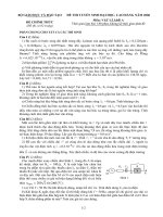
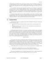
Figure 1
The growth of the number of structures in the PDB archive 1972–2006.
Crystallographica Section D and Nature Stru ctural Biology
(now Nature Structural & Molecular Biology), were estab-
lished to report the results of the analyses of biological
macromolecules. Many now believed that structural biology
could give definitive information that would be key to
understanding the molecular basis of biology and medicine.
As the value of structural biology became more obvious to
other biologists, several committees were formed to look at
the new demands from the community for required data
deposition. The IUCr Commission on Biological Macro-
molecules set up a committee to determine exact policies. The
ACA and the USNCCr also set up a committee. Fred Richards
created an ad hoc group of crystallographers who felt strongly
about creating a policy that would require data deposition.
These groups worked for a few years to hammer out the exact
guidelines. The timing of deposition of coordinates and of
structure factors was discussed at length. During this period,
commentaries were published in journals, letters to the editor
were written and there was intense debate in the community.
By the end of the decade, a petition resulting from the action
of the Richards committee was produced and, in 1989, a
formal guideline for data deposition was published (Interna-
tional Union of Crystallography, 1989). The guidelines stated
that coordinates should be deposited at the time of publication
and released within one year; structure factors should be
deposited and released within four years. Most major journals
adopted these guidelines and the National Institute for
General Medical Sciences made the bold step of saying that
funding would be contingent on the open sharing of structural
data.
3. The content of the PDB
By the end of the 1980’s, the number of structures in the PDB
began to increase dramatically (Fig. 1) and that growth
continues to date. Nuclear magnetic resonance (NMR)
methods began to be used to determine structures, thus
providing additional types of information to be archived in the
PDB. These structures now make up about 15% of the
structures in the archive.
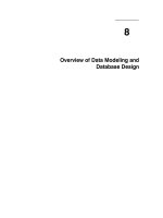
In addition, the complexity of the structures that could be
determined grew. The use of flash freezing and highly intense
synchrotron radiation during data collection combined with
the sophisticated use of non-crystallographic symmetry for
structure determination made it possible to solve virus
structures (Arnold & Rossmann, 1988) (Fig. 2). Even larger
structures, including molecular machines such as the ribosome
particles, became amenable to the methods of X-ray crystal-
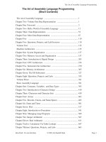
lography (Moore, 2001). The ability to freeze single particles
also allowed structures to be determined using cryoelectron
microscopy (cryoEM) and models for these structures began
to appear in the PDB archive (Fig. 3).
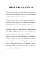
By the year 2000, the diversity of molecules and complexes
in the PDB archive (Fig. 4) was so great that the possibility of
actually understanding biology and medicine at a molecular
level was not just a far off dream but something that might
actually be realized.
Could this be achieved even more rapidly? Enter structural
genomics. Analysis of the PDB archive showed that, from the
point of view of amino acid sequences, there was about a
sevenfold sequence redundancy (Hobohm et al., 1992). The
number of new protein folds deposited in the PDB archive was
relatively low (10% since 2000). At the same time, bioinfor-
matics analysis of the newly determined gene sequences
showed that the coverage of all protein families was uneven
and incomplete. Would it be possible to very carefully select
target sequences that would be novel (<30% sequence simi-
larity), determine their structures and, through homology
modeling, obtain structures for the rest of the protein
families? Several projects ensued which focused on high-
throughput methods for structure determination of these
unique targets (Nature Structure Biology, 2000; Levitt, 2007).
To date, structural genomics centres worldwide have been
responsible for adding more than 5500 new structures of which
50.5% are novel.
4. Challenges to the PDB
When the PDB began there were relatively few structures,
the structures were small, and only X-ray crystallographic
methods were used to determine the structures. Today, the
PDB has structures with molecular weights of more than two
million, and new structure-determination methods are waiting
in the wings to join NMR and cryoEM in the PDB Exchange
Dictionary. To meet the challenges of handling these data and
making them accessible to a broad community of scientists,
methods had to be developed for data representation, acqui-
sition and processing, management and distribution. The
information in the PDB must be tied to other data resources,
such as GenBank (Benson et al., 2000), UniProt (Apweiler et
al., 2004), model organism databases and many, many others.
In addition, the global nature of science makes it essential to
coordinate these data efforts at an international level.
4.1. Data representation
In addition to the xyz coordinates, an entry in the PDB
contains information about the chemistry of the macro-
molecule, the small-molecule ligands, some details of the data
collection and structure refinement, and some structural
descriptors. In all, a typical PDB entry has about 400 unique
items of data. The PDB file format that was devised in 1976 is
simple, easy to read by humans and used by many computer
applications. However, as the PDB format is based on the
original 80 column punched-card format, the number of atoms
and number of residues that can be represented is limited. This
means that large macromolecular complexes must be repre-
sented in more than one data file in the PDB file format.
Additionally, there are implicit assumptions made in this file
format that limit its use by modern computer applications. In
an effort to remedy these shortcomings, the macromolecular
crystallographic information file (mmCIF) was created. The
mmCIF dictionary contains well over 3000 definitions of
every aspect of the crystallographic experiment and results
feature articles
90 Helen M. Berman
The Protein Data Bank Acta Cryst. (2008). A64, 88–95
Acta Cryst. (2008). A64, 88–95 Helen M. Berman
The Protein Data Bank 91
feature articles
Figure 2
A selection of the more than 250 icosahedral virus structures currently available in the PDB archive. The representation of these entries has recently
undergone a major facelift.
(Fitzgerald et al., 2005). In this format, each data item is
completely defined, along with the relationships among the
data items. mmCIF is completely computer readable and can
be used to create a relational database. To accommodate data
from NMR and cryoEM experiments, a PDB Exchange
Dictionary (PDBx) was created that has the same syntax as
mmCIF but contains all the definitions needed to handle the
data that are now part of the PDB (Westbrook, Henrick et al.,
2005). In addition, the mmCIF data files have been translated
into PDBML-XML files that can be managed by off-the-shelf
tools (Westbrook, Ito et al., 2005).
4.2. Data acquisition and processing
In the earliest days of the PDB at BNL, data were sent by
mail on either punched cards or magnetic tape. A form was
completed that contained information about the structure.
Annotators examined the data and obvious errors were
corrected. Checking the geometry was done with a set of
special-purpose computer programs. Letters were sent
through the post office to authors with reports of the checks
and, after author agreement, the data were entered in the
computer archive. It was not until the 1990’s that fully elec-
tronic submission was possible. AutoDep allowed the author
to submit files online (Lin et al., 2000). Some checks were done
automatically and others were done manually.
When the PDB changed management in 1998, data acqui-
sition and processing began to evolve. EBI reengineered
AutoDep and made it more automatic (Keller et al., 1998). The
RCSB PDB created the AutoDep Input Tool (ADIT)
(Berman et al., 2000) which was based on mmCIF. PDBj also
adopted this tool for data collection. The newest wwPDB
member, the BMRB (), uses ADIT-
NMR to collect experimental and coordinate data at the same
time. Data processing, although done with different tools
around the world, uses the same principles and algorithms. All
feature articles
92 Helen M. Berman
The Protein Data Bank Acta Cryst. (2008). A64, 88–95
Figure 3
Growth of the number of cryoEM structures in the PDB and the number of related maps in the Electron Microscopy Database (EMDB) (Henrick et al.,
2003).
Acta Cryst. (2008). A64, 88–95 Helen M. Berman
The Protein Data Bank 93
feature articles
Figure 4
A look at the diversity of structures in the PDB archive. These images, shown to scale, were created by David S. Goodsell (The Scripps Research
Institute), who also writes and illustrates the RCSB PDB’s Molecule of the Month feature. An expanded version of this figure is available for download
from .
PDB files are checked for accuracy of the geometry, chemistry
of the polymer and ligands, nomenclature, and the likely
biological assembly. In recent years, structure factors and
NMR constraint files are deposited with the majority of data
files so that now it is possible to calculate the agreement with
experimental data using SFCheck (Vaguine et al., 1999). To
ensure complete uniformity, the wwPDB has reviewed and
documented all data processing practices among the member
sites. The wwPDB has also taken on the task of reviewing all
the files in the archive and has very recently created a new,
more uniform, archive at (Henrick et al.,
2008)
4.3. Data distribution and query
In the early days of the PDB, data were distributed via
magnetic tape and later by CD-ROM. Now there is an ftp site
that contains the data in three formats: PDB, mmCIF-PDBx
and PDBML-XML. Distributing these data on media would
require more than 15 DVD disks. The ftp site is updated
weekly and each wwPDB center maintains a mirror of the site.
The RCSB PDB website alone is accessed by about 100 000
unique visitors per month from nearly 140 different countries.
More than 500 GB of data are transferred each month. On a
typical weekday, two pages from the site are viewed every
second. Data are accessed via the website, ftp servers
(supporting ftp and rsync access), web services and RSS feeds.
Until the 1990’s, interactive query was not possible from a
central site. The World Wide Web changed all that. The first
web-accessible interface was made available at BNL and
allowed many useful queries (Prilusky, 1996). Now the
wwPDB sites offer numerous services including simple and
complex searches as well as a variety of visualization methods.
In addition, the RCSB PDB () provides
browsing capabilities across external resources, a Structure
Summary page for each entry and a Molecule of the Month
feature highlighting a particular structure (Berman et al.,
2000). PDBj () provides several services,
including Alignment of Structural Homologs (Standley et al.,
2007). The services of the MSD-EBI ( />msd) include analyses of macromolecule ligand interactions,
statistical analyses and residue-based analyses (Golovin et al.,
2004).
The accessibility of the data and the growing importance of
understanding the data has meant that the PDB’s user
community has grown from the community of crystal-
lographers that banded together to form the archive. The PDB
archive is a critical resource for researchers in academia and
industry, working in subjects such as structural biology,
computational biology, biophysics, biochemistry, genetics, cell
biology and molecular biology. The PDB is also a tool used by
educators and students from middle school to graduate school.
5. Applications of PDB data in academia and industry
The PDB is very widely used. For example, an average of
211515 files are downloaded from the ftp site each day. To
date, there are more than 5000 references to the first RCSB
PDB publication (Berman et al., 2000), making it one of the
most-cited papers in all of biology. While the early users of the
PDB were mostly crystallographers who used the resource to
store their data and to review other structures for comparison
with their own, now more than half of the papers that cite this
one publication alone describe bioinformatic and computa-
tional analyses of structural data. Enormous efforts are under
way to be able to understand protein folding so that perhaps
someday it will be possible to predict structure from sequence
(Moult, 2005). While the PDB is considered an archival data
resource that stores and distributes primary data, there are
hundreds of derivative databases that catalog the data in
different ways. For example, CATH (Orengo et al., 1997) and
SCOP (Conte et al., 2000) provide classifications of the folds
found in proteins. More recently, there have also been efforts
to understand protein–protein interactions (Janin et al., 2003)
and again to try to predict these. There are specialty databases
such as the Nucleic Acid Database (Berman et al., 1992) and
the HIV Protease Structural Database (Ravichandran et al.,
2002) that create in-depth resources for researchers in nucleic
acids and HIV, respectively.
When the PDB updates the ftp site each week, most phar-
maceutical companies download the new data for inclusion in
their own in-house databases. These structural data are used
to aid the discovery of new pharmaceuticals. Indeed, the ready
availability of the structure of HIV protease (Navia et al.,
1989) enabled many companies to concentrate their efforts on
the development of effective protease inhibitors that are now
the basis of AIDS treatment.
6. Future of the PDB
The PDB archive is a key example of a community resource
that has evolved over its 36 year history. Its evolution has been
driven by changes in science and technology used to deter-
mine structures, the nature of the structures that are deter-
mined, community attitudes about data sharing, and the
nature of the communities that are interested in structural
data.
In the short term, there will be several new challenges.
There needs to be better representation for disordered
structures, for X-ray structures refined with multiple models
(Furnham et al., 2006) and for very large macromolecular
complexes. New annotations will be required to describe
function. All of these changes must be done in consultation
with depositors, software developers and users of the data.
Annotation practices used by the different data centers will
continue to be examined and standardized so as to keep the
archive uniform. It is likely that the wwPDB sites will develop
a single deposition system. In the long term, the wwPDB will
be able to develop new joint services for analysis and browsing
of the rich data contained within the archive.
As the PDB continues to evolve, in addition to being able to
use these data to perhaps predict structure, an even greater
challenge will be to determine function by knowing the
structure. Once accomplished, the long-term vision of
feature articles
94 Helen M. Berman
The Protein Data Bank Acta Cryst. (2008). A64, 88–95
enabling a molecular view of biology and medicine will
become a reality.
Many people have been key in the development and
maintenance of the PDB in its 36 year history. These include
previous directors Tom Koetzle and Joel L. Sussman, and the
BNL staff including Enrique Abola and Frances Bernstein.
John Westbrook and Phil Bourne have been instrumental to
the development of the RCSB PDB resources. MSD-EBI
leader Kim Henrick and PDBj leader Haruki Nakamura have
worked hard to ensure that the PDB remains a global
resource. The many staff members of the wwPDB data centers
continue to contribute to the continued viability and quality of
the PDB. The RCSB PDB is supported by DBI 0312718.
References
Allen, F. H., Kennard, O., Motherwell, W. D. S., Town, W. G. &
Watson, D. G. (1973). J. Chem. Doc. 13, 119–123.
Apweiler, R., Bairoch, A., Wu, C. H., Barker, W. C., Boeckmann, B.,
Ferro, S., Gasteiger, E., Huang, H., Lopez, R., Magrane, M., Martin,
M. J., Natale, D. A., O’Donovan, C., Redaschi, N. & Yeh, L. S.
(2004). Nucleic Acids Res. 32, Database issue, D115–D119.
Arnold, E. & Rossmann, M. G. (1988). Acta Cryst. A44, 270–283.
Benson, D. A., Karsch-Mizrachi, I., Lipman, D. J., Ostell, J., Rapp,
B. A. & Wheeler, D. L. (2000). Nucleic Acids Res. 28, 15–18.
Berman, H. M., Henrick, K. & Nakamura, H. (2003). Nature Struct.
Biol. 10, 980.
Berman, H. M., Olson, W. K., Beveridge, D. L., Westbrook, J., Gelbin,
A., Demeny, T., Hsieh, S. H., Srinivasan, A. R. & Schneider, B.
(1992). Biophys. J. 63, 751–759.
Berman, H. M., Westbrook, J., Feng, Z., Gilliland, G., Bhat, T. N.,
Weissig, H., Shindyalov, I. N. & Bourne, P. E. (2000). Nucleic Acids
Res. 28, 235–242.
Bernstein, F. C., Koetzle, T. F., Williams, G. J. B., Meyer, E. F. Jr, Brice,
M. D., Rodgers, J. R., Kennard, O., Shimanouchi, T. & Tasumi, M.
(1977). J. Mol. Biol. 112, 535–542.
Bru
¨
nger, A. T. (1990). X-PLOR. A System for Crystallography and
NMR, Version 2.1. New Haven, CT, USA: Yale University.
Cold Spring Laboratory Press (1972). Cold Spring Harbor Symposia
on Quantitative Biology, Vol. 36.
Conte, L., Bart, A., Hubbard, T., Brenner, S., Murzin, A. & Chothia,
C. (2000). Nucleic Acids Res. 28, 257–259.
Fitzgerald, P. M. D., Westbrook, J. D., Bourne, P. E., McMahon, B.,
Watenpaugh, K. D. & Berman, H. M. (2005). International Tables
for Crystallography, Vol. G. Definition and Exchange of Crystal-
lographic Data, edited by S. R. Hall & B. McMahon, ch. 4.5,
Macromolecular Dictionary (mmCIF), pp. 295–443. Dordrecht:
Springer.
Furnham, N., Blundell, T. L., DePristo, M. A. & Terwilliger, T. C.
(2006). Nature Struct. Mol. Biol. 13, 184–185; discussion p. 185.
Golovin, A., Oldfield, T. J., Tate, J. G., Velankar, S., Barton, G. J.,
Boutselakis, H., Dimitropoulos, D., Fillon, J., Hussain, A., Ionides,
J. M., John, M., Keller, P. A., Krissinel, E., McNeil, P., Naim, A. et al.
(2004). Nucleic Acids Res. 32, Database issue, D211–D216.
Helliwell, J. (1983). Acta Radiol. Suppl. 365, 35–37.
Hendrickson, W. A. (1991). Science, 254, 51–58.
Hendrickson, W. A. & Konnert, J. H. (1981). PROLSQ,Vol.1,
Biomolecular Structure, Conformation, Function and Evolution,
edited by R. Srinivasan, E. Subramanian & N. Yathindra, pp. 43–57.
Oxford: Pergamon Press.
Henrick, K., Feng, Z., Bluhm, W., Dimitropoulos, D., Doreleijers, J. F.,
Dutta, S., Flippen-Anderson, J. L., Ionides, J., Kamada, C.,
Krissinel, E., Lawson, C. L., Markley, J. L., Nakamura, H.,
Newman, R., Shimizu, Y. et al. (2008).
Nucleic Acids Res. Database
Issue. In the press.
Henrick, K., Newman, R., Tagari, M. & Chagoyen, M. (2003). J.
Struct. Biol. 144, 228–237.
Hobohm, U., Scharf, M., Schneider, R. & Sander, C. (1992). Protein
Sci. 1, 409–417.
International Union of Crystallography (1989). Acta Cryst. A45, 658.
Jancarik, J. & Kim, S H. (1991). J. Appl. Cryst. 24, 409–411.
Janin, J., Henrick, K., Moult, J., Eyck, L. T., Sternberg, M. J., Vajda, S.,
Vakser, I. & Wodak, S. J. (2003). Proteins, 52, 2–9.
Jones, T. A. (1978). J. Appl. Cryst. 11, 268–272.
Jones, T. A., Zou, J Y., Cowan, S. W. & Kjeldgaard, M. (1991). Acta
Cryst.A47, 110–119.
Keller, P. A., Henrick, K., McNeil, P., Moodie, S. & Barton, G. J.
(1998). Acta Cryst. D54, 1105–1108.
Kennard, O., Watson, D. G. & Town, W. G. (1972). J. Chem. Doc. 12,
14–19.
Lehmann, M. S., Koetzle, T. F. & Hamilton, W. C. (1972). J. Am.
Chem. Soc. 94, 2657–2660.
Levitt, M. (2007). Proc. Natl Acad. Sci. USA, 104, 3183–3188.
Lin, D., Manning, N. O., Jiang, J., Abola, E. E., Stampf, D., Prilusky, J.
& Sussman, J. L. (2000). Acta Cryst. D56, 828–841.
Meyer, E. F. (1997). Protein Sci. 6, 1591–1597.
Moore, P. (2001). Biochemistry, 40, 3243–3250.
Moult, J. (2005). Curr. Opin. Struct. Biol. 15, 285–289.
Nature Structural Biology (2000). Archive – Nature Structural
Biology, 7:11s, />index.html.
Navia, M. A., Fitzgerald, P. M., McKeever, B. M., Leu, C. T.,
Heimbach, J. C., Herber, W. K., Sigal, I. S., Darke, P. L. & Springer,
J. P. (1989). Nature (London), 337, 615–620.
Orengo, C. A., Michie, A. D., Jones, S., Jones, D. T., Swindells, M. B. &
Thornton, J. M. (1997). Structure, 5, 1093–1108.
Prilusky, J. (1996). OCA, a Browser-Database for Protein Structure/
Function, and mirrors worldwide.
Protein Data Bank (1971). Nature New Biol. 233, 223.
Ravichandran, V., Vondrasek, J., Gilliland, G., Bhat, T. N. &
Wlodawer, A. (2002). CSB Proceedings of the IEEE Computer
Society Conference on Bioinformatics 340. Washington: IEEE
Computer Society.
Standley, D. M., Toh, H. & Nakamura, H. (2007). BMC Bioinfor-
matics, 8, 116.
Vaguine, A. A., Richelle, J. & Wodak, S. J. (1999). Acta Cryst. D55,
191–205.
Westbrook, J., Henrick, K., Ulrich, E. L. & Berman, H. M. (2005).
International Tables for Crystallography,Vol.G.Definition and
Exchange of Crystallographic Data , edited by S. R. Hall & B.
McMahon, ch. 3.6.2, The Protein Data Bank Exchange Data
Dictionary, pp. 195–198. Dordrecht: Springer.
Westbrook, J., Ito, N., Nakamura, H., Henrick, K. & Berman, H. M.
(2005). Bioinformatics, 21, 988–992.
Acta Cryst. (2008). A64, 88–95 Helen M. Berman
The Protein Data Bank 95
feature articles