Tài liệu Báo cáo khoa học: Mixed lineage leukemia: a structure–function perspective of the MLL1 protein ppt
Bạn đang xem bản rút gọn của tài liệu. Xem và tải ngay bản đầy đủ của tài liệu tại đây (521.2 KB, 11 trang )
MINIREVIEW
Mixed lineage leukemia: a structure–function perspective
of the MLL1 protein
Michael S. Cosgrove and Anamika Patel
Department of Biology, Syracuse University, NY, USA
Introduction
Chromosomal translocations that disrupt the mixed
lineage leukemia protein-1 gene (MLL1, ALL1, HRX,
Htrx) are associated with a unique subset of acute
lymphoblastic or myelogenous leukemias [1–4]. The
product of the MLL1 gene is a large protein that func-
tions as a transcriptional co-activator required for the
maintenance of Hox gene expression patterns during
hematopoiesis and development [5–8]. The transcrip-
tional co-activator activity of MLL1 is mediated in
part by its histone H3 lysine 4 (H3K4) methyltransfer-
ase activity [6], an epigenetic mark correlated with
transcriptionally active forms of chromatin [9,10].
MLL1 complexes catalyze mono-, di- and trimethyla-
tion of H3K4, regulation of which can have distinct
functional consequences. MLL1 contains a number of
conserved functional domains that work together for
the assembly of multiprotein complexes that influence
the appropriate targeting and regulation of the H3K4
methylation activity of MLL1. In this minireview, we
summarize recent structural and functional studies that
are beginning to provide a picture of how these
domains are used to regulate the targeting, assembly
and enzymatic activity of MLL1 complexes.
The MLL protein
The MLL1 gene encodes a large protein of 3969 amino
acid residues that contains several conserved domains
with functions implicated in chromatin-mediated tran-
scriptional regulation [11] (Fig. 1). Domains include
DNA-binding AT hooks, a cysteine-rich CXXC
domain with homology to DNA methyltransferases,
plant homeodomain (PHD) finger motifs, a bromo
domain (BD), a transactivation domain (TAD), a
Keywords
Ash2L; CXXC; H3K4; methylation; MLL;
RbBP5; SET; TAD; WDR5; Win motif
Correspondence
M. S. Cosgrove, Syracuse University, 340
Life Sciences Complex, 107 College Place,
Syracuse, NY 13244, USA
Fax: +1 315 443 2012
Tel: +1 315 443 2964
E-mail:
(Received 16 November 2009,
accepted 3 February 2010)
doi:10.1111/j.1742-4658.2010.07609.x
Several acute lymphoblastic and myelogenous leukemias are correlated with
alterations in the human mixed lineage leukemia protein-1 (MLL1) gene.
MLL1 is a member of the evolutionarily conserved SET1 family of histone
H3 lysine 4 (H3K4) methyltransferases, which are required for the regula-
tion of distinct groups of developmentally regulated genes in metazoans.
Despite the important biological role of SET1 family enzymes and their
involvement in human leukemias, relatively little is understood about how
these enzymes work. Here we review several recent structural and biochem-
ical studies that are beginning to shed light on the molecular mechanisms
for the regulation of H3K4 methylation by the human MLL1 enzyme.
Abbreviations
AdoHyc, S-adenosyl-homocysteine; BD, bromo domain; CBP, CREB-binding protein; CREB, cAMP response element-binding;
H3K4, histone H3 lysine 4; HMT, histone methyltransferase; MLL1, mixed lineage leukemia protein-1; PHD, plant homeodomain;
TAD, transactivation domain.
1832 FEBS Journal 277 (2010) 1832–1842 ª 2010 The Authors Journal compilation ª 2010 FEBS
nuclear receptor interaction motif (NR box), a WDR5
interaction or Win motif and a C-terminal SET
domain, which is responsible for MLL1’s histone
methyltransferase (HMT) activity [6,12,13]. Upon
normal expression of the MLL1 gene, the full-length
protein is proteolytically processed into two fragments,
MLL-N and MLL-C, which associate to form a
complex in vivo (Fig. 1A) [14,15]. The mature protein
assembles with numerous regulatory proteins into
multimolecular complexes important for MLL1’s
transcriptional co-activator activity [12,16–21].
Because of its large size, full-length MLL1 protein
has thus far proven refractory to structural analysis.
However, the modular nature of MLL1 has allowed
structural analysis of some individual domains alone
or in complex with functionally relevant ligands
(Fig. 1B). Structures that have been determined include
the MLL1 CXXC domain [22], a portion of the MLL1
TAD bound to the KIX domain of the cAMP
response element-binding (CREB) binding protein
(CBP) [23], a peptide from the Win motif of MLL1
bound to the WD40 repeat protein, WDR5 [24,25] and
the C-terminal SET domain in the presence and
absence of histone peptides and the cofactor product,
S-adenosyl-homocysteine (AdoHcy) [26] (Fig. 1B).
These structures provide clues as to how MLL1 is tar-
geted to MLL1-dependent genes and how MLL1’s
enzymatic activity is regulated.
CXXC domain
The molecular mechanisms by which the MLL1 protein
is recruited to specific target genes are poorly under-
stood. The CXXC domain of MLL1 binds selectively to
nonmethyl CpG DNA [27], and is essential for target
gene recognition, transactivation and myeloid transfor-
mation in MLL1 fusion proteins [28]. Because the pro-
moters of active genes in vertebrates are generally
hypomethylated [29], the CXXC domain of MLL1 may
play a role in targeting MLL1 to active genes. To
1143
1215
3790
3762
3770
MLL1 CXXC
1LLMniamod
SET
domain
WDR5
MLL1
Win
motif
2838
2879
CBP-KIX
domain
SET
TAD
CXXC
1-
-3969
3969
Win
MLL1 TAD domain
c-Myb
SETFYRCTADFYRNBDPHD
CXXC
AT-Hooks
1-
-3969
Win
1-
SET
-3969
Menin
HCF
CBP MOF
WDR5
RbBP5
Ash2L
DPY30
Cleavage by Taspase 1
Cleavage site
Break point
SET
B
A
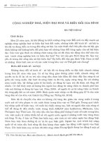
Fig. 1. Schematic representation showing
the domain architecture of the MLL1
protein. (A) The full-length MLL1 protein is
rapidly processed by the Taspase 1 enzyme
into MLL-N and MLL-C fragments, which
reassociate through FYRN and FYRC motifs
to form a stable complex. This mature
protein then assembles with a number of
proteins to form MLL1 complexes in the
cell. (B) Known 3D structures of conserved
MLL1 domains (colored green in each
image). On the top, from left to right is the
CXXC domain (PDB code: 2j2s) and the
C-terminal SET domain (PDB code: 2w5z).
On the bottom, from left to right is the
MLL1 TAD domain (green) bound to
the CBP : c-Myb complex (orange and blue,
respectively; PDB code: 2agh); and the
MLL1 Win motif (green) bound to the
WDR5 protein (purple; PDB code: 3eg6).
M. S. Cosgrove and A. Patel MLL1: a structure–function perspective
FEBS Journal 277 (2010) 1832–1842 ª 2010 The Authors Journal compilation ª 2010 FEBS 1833
identify the molecular basis of DNA recognition by the
MLL1 CXXC domain, Allen et al. [22] determined the
solution structure of the MLL1 CXXC domain consist-
ing of amino acid residues 1146–1214, and used chemi-
cal-shift mapping and site-directed mutagenesis to
identify residues involved in DNA recognition. The
overall structure adopts an extended crescent-like shape
that coordinates two zinc ions using the two conserved
CGXCXXC motifs (Fig. 2A). The zinc ions are required
for the structural integrity of the protein, as mutation of
any of the cysteine residues involved in zinc coordina-
tion results in protein unfolding [22]. The structure con-
tains a positively charged surface groove containing a
number of residues that were shown using chemical-
shift mapping and site-directed mutagenesis to be
important for DNA binding (Fig. 2A). The MLL1
CXXC domain binds to unmethylated CpG DNA with
a dissociation constant of 4 lm, as measured by iso-
thermal titration calorimetry [22], but does not bind to
similar DNA-containing methyl-CpG dinucleotides –
consistent with previous observations [27,28]. These
studies suggest a model in which the phospho-backbone
of DNA binds to the positively charged groove on the
CXXC domain, whereas residues from the extended
loop insert into the major groove to interact with the
CpG dinucleotide [22]. It is hypothesized that methyla-
tion of the CpG prevents the extended loop from inter-
acting with the CpG dinucleotide, resulting in reduced
affinity for DNA.
Although recognition of unmethylated CpG
dinucleotides by the CXXC domain of MLL1 likely
contributes to MLL1 targeting, as previously noted [7],
several genes that are not regulated by MLL1 also
contain unmethylated CpG dinucleotides in their pro-
moters, indicating that other mechanisms contribute to
target gene recognition by MLL1. A more recent struc-
ture of the TAD of MLL1 bound to the CBP protein
describes one such additional mechanism that could
also be involved in targeting MLL1 to specific loci.
TAD
The CBP protein and its homolog p300 are general
transcriptional co-activators that contain histone and
transcription factor acetylation activities [30]. In addi-
tion, CBP contains a number of protein-binding
domains that mediate transcription factor recruitment.
The MLL1 TAD interaction with CBP was originally
identified in a yeast three-hybrid screen using the
CREB–CBP complex as bait [31], and was shown to
be important for MLL1-mediated transcriptional
activation [31]. Domain mapping experiments localize
Extended loop
DNA
Binding
Surface
MLL1 CXXC domain
E666
E665
K291
R294
C-Myb
CBP-KIX domain
MLL1-TAD
domain
Ternary complex
α3
α1
α2
α1
α3
α2
E666
E665
K291
R294
C-Myb
CBP-KIX domain
Binary complex
CBA
Fig. 2. The CXXC and TAD domains of MLL1 help recruit MLL1 to target loci. (A) Transparent surface representation of the MLL1 CXXC
domain (purple) determined by heteronuclear NMR spectroscopy (PDB code: 2j2s). A cartoon of the protein backbone is shown with zinc
ions represented as spheres. The surfaces of amino acid residues perturbed by DNA binding in chemical shift and mutagenesis experiments
are indicated in blue. The location of the extended loop is indicated with an arrow. (B) The CBP–KIX domain : cMyb binary complex. The
CBP–KIX domain is shown in orange and the c-Myb transactivation domain is shown in blue (drawn from PDB code: 1sb0). Positions
of E665 and E555 of the CBP–KIX domain, and residues K291 and R294 of the c-Myb transactivation domain are indicated. (C) The
CBP–KIX:cMyb:MLL1 TAD ternary complex (drawn form PDB code: 2AGH). The MLL1 TAD is shown in green and the colors for
the CBP–KIX:cMyb are as in (B). Upon formation of the ternary complex, residues E665 and E666 of the CBP–KIX domain become ordered
and interact with the c-Myb transactivation domain (indicated with the arrow).
MLL1: a structure–function perspective M. S. Cosgrove and A. Patel
1834 FEBS Journal 277 (2010) 1832–1842 ª 2010 The Authors Journal compilation ª 2010 FEBS
MLL1’s interaction to the KIX or CREB-binding
domain of CBP [31]. The KIX domain of CBP is a
structural platform that is capable of binding several
different families of transcriptional activators [30], and
evidence indicates that the KIX domain has the ability
to simultaneously interact with at least two different
polypeptides in a cooperative manner [31,32]. To iden-
tify the molecular basis of cooperative transcription
factor binding by CBP, De Guzman et al. [23] deter-
mined the solution structure of a peptide derived from
the MLL1 TAD bound to the KIX domain:c-Myb
binary complex.
The overall structure of the c-Myb:KIX binary com-
plex resembles a four-helix bundle in which the c-Myb
peptide adopts a helical conformation that binds to
helices a1 and a3 of KIX (Fig. 2B) [33]. When the
MLL1 TAD peptide is added to the binary complex,
the TAD peptide adopts a helical conformation in
which the conserved residues of MLL1 TAD (residues
2845–2853) bind in a hydrophobic groove on the
opposite side of the KIX domain between helices a2
and a3 (Fig. 2C) [23]. No direct interaction between
the c-Myb and MLL1 peptides are observed when
bound to the KIX domain, suggesting that the mecha-
nism of cooperative transcription factor binding is
transmitted through subtle conformational changes in
the KIX domain [23]. Consistent with allosteric
binding, residues of the a3 helix of KIX that are disor-
dered in the binary complex become ordered when
MLL1 binds (see arrow in Fig. 2C). This conforma-
tional change results in the placement of conserved
KIX domain amino acids E665 and E666 into
positions for optimal electrostatic interactions with
conserved residues R294 and K291 of the c-Myb
transactivation domain, respectively. Thermodynamic
binding experiments show that interaction of MLL1
with the KIX domain increases CBP’s affinity for
the c-Myb transactivation peptide by approximately
two-fold [32].
These experiments begin to provide a picture of how
the recruitment of MLL1 can increase the binding of
other important transcriptional activators that ulti-
mately could result in the synergistic activation of gene
transcription. In addition, cooperative transcription
factor binding through CBP could provide a mecha-
nism to help MLL1 recognize its target genes. MLL1
recruitment to chromatin results in the methylation of
H3K4 by the SET domain of MLL1, an activity that
is regulated in part by a core complex of proteins that
includes WDR5, RbBP5, Ash2L and DPY-30 [26,34–
37]. H3K4 methylation is an epigenetic mark corre-
lated with transcriptionally active forms of chromatin
[10]. Several recent investigations have provided struc-
tural and functional information that describe how the
HMT activity of MLL1 is regulated.
SET domain
MLL1 contains an evolutionarily conserved SET
domain which is found in a number of chromatin-
associated proteins with diverse transcriptional activi-
ties [38]. The SET domain is a HMT motif named
for its presence in Drosophila chromatin regulators
SuVar3-9, E(z), and Trx [39]. SET domain proteins
can be classified into several families that differ with
respect to substrate specificity, processivity and the
presence of associated domains, and include the
SUV39, SET1, SET2, E(z), Riz, SMYD and SUV2-20
families [40]. MLL1 belongs to the SET1 family of
SET domain proteins, which are found in conserved
multisubunit complexes that regulate cellular H3K4
methylation levels [9,41]. Because of the role of
H3K4 methylation in diverse cellular processes rang-
ing from stem cell differentiation to metazoan devel-
opment and cancer, there has been an intense interest
in understanding how SET1 family enzymes regulate
H3K4 methylation.
To understand the structural basis of H3K4 methyl-
ation by the MLL1 SET domain, Southhall et al. [26]
determined the X-ray crystal structures of a minimal
MLL1 SET domain fragment in complex with its
cofactor product AdoHyc in the presence and absence
of a peptide mimicking the methylated histone H3
N-terminal tail (Fig. 3). Much like other SET domains
where the structures have been determined [42], the
overall structure of the MLL1 SET domain contains
two canonical conserved regions, SET-N and SET-C,
that are separated by a less conserved insert region
(SET-I) (expanded region in Fig. 3A). The MLL1 SET
domain is flanked on the C-terminus by a 22-amino
acid post-SET domain, which provides several con-
served residues that coordinate a zinc atom that is
required for enzymatic activity (A Patel & MS
Cosgrove, unpublished observation). In the ternary
complex, the histone H3 peptide binds in a deep chan-
nel that divides a pair of acidic lobes, one of which is
composed of residues from the SET-I region and the
other of residues from the SET-C and post-SET
regions. Lysine 4 of histone H3 is inserted into a chan-
nel, at the end of which is the AdoHcy binding site,
which is composed of residues from SET-N, SET-C
and the post-SET domain (Fig. 3A).
In published 3D structures of other SET domain
proteins that also contain the canonical post-SET
domain [43–46], formation of the ternary complex
results in ordering of the post-SET domain, so that the
M. S. Cosgrove and A. Patel MLL1: a structure–function perspective
FEBS Journal 277 (2010) 1832–1842 ª 2010 The Authors Journal compilation ª 2010 FEBS 1835
two lobes that flank the peptide-binding site close
around the peptide, presumably to exclude solvent
from the active site. However, comparison of the
binary and ternary complexes of the MLL1 SET
domain crystal structures reveals that the two lobes
remain in a relatively open conformation, which is not
optimal for catalysis [26]. It has been suggested on the
basis of this observation that proteins that interact
with the SET domain are required to induce the cor-
rect conformation of the active site [26], which is con-
sistent with the poor catalytic activity of the isolated
MLL1 SET domain. However, an analysis of crystal
packing forces suggests that the SET-I lobe may be
constrained in an unnatural conformation in the crys-
talline state by residues from the N-terminus of a sym-
metry related molecule (Fig. 3B). It therefore remains
to be determined to what extent the observed confor-
mation of the isolated MLL1 SET domain in the crys-
tal structure represents the range of possible
conformations that may exist in solution.
Consistent with the conformational change hypothe-
sis, Southhall et al. [26] observed that addition of other
components of the MLL1 core complex, namely
WDR5, RbBP5, Ash2L and DPY-30, increases H3K4
methylation by 20-fold compared with that of the
isolated MLL1 SET domain. However, the extent to
which this 20-fold increase in enzymatic activity is
because of a conformational change in the MLL1 SET
domain is unclear at present. This is because the con-
struct used to determine the structure of the MLL1
SET domain lacks the evolutionarily conserved Win
motif in the region flanking the N-terminus of the SET
domain [26], which has been shown to be essential for
the assembly and dimethyltransferase activity of the
MLL1 core complex [24,25,36]. In addition, recent
work from our laboratory indicates that the non-SET
domain components of the MLL1 core complex pos-
sess a previously unrecognized H3K4 methyltransferase
activity that is independent of the MLL1 SET domain
[35] (see below). It is therefore possible that the
SET-N
SET-I SET-C
Post-SET
3969
3785
SETFYRCTADFYRNBDPHD
CXXC
AT-Hooks
1-
-3969
Win
etis
e
gavaelCtniop kaerB
AdoHcy
H3 peptide
A
B
Fig. 3. X-Ray crystal structure of the C-terminal MLL1 SET domain bound to AdoHcy (yellow) and histone H3 peptide (purple) (PDB code:
2W5Z). (A) At the top is a schematic representation of the full-length MLL1 protein and blown up is the construct used for crystallization of
the MLL1 SET domain (residues 3785–3969). The SET-N, SET-I and SET-C sub-domains are colored in blue, yellow and green, respectively.
The post-SET domain is colored in grey, and the N-flanking region is colored white. The positions of histone H3 and AdoHcy are indicated.
(B) Crystal packing constrains the MLL1 SET domain into an open conformation. Surface representation of the MLL1 SET domain (grey)
shown with a symmetry related molecule in red. The N-terminus of the symmetry related molecule interacts extensively with the SET-I
region – constraining the MLL1 SET domain in an open conformation.
MLL1: a structure–function perspective M. S. Cosgrove and A. Patel
1836 FEBS Journal 277 (2010) 1832–1842 ª 2010 The Authors Journal compilation ª 2010 FEBS
increase in H3K4 methylation activity observed by
Southhall et al. [26] is due, at least in part, to the inde-
pendent activities of the MLL1 SET domain and the
sub-complex containing WDR5, RbBP5, ASH2L and
DPY-30, which do not significantly interact in the
absence of the MLL1 Win motif [25,36].
Win motif
The WD40 repeat protein WDR5 is a conserved com-
ponent of SET1 family complexes ranging from yeast
to humans and has been shown to be important for
H3K4 methylation and HOX gene expression in hema-
topoiesis and development [47]. Recent studies have
shown that WDR5 interacts directly with MLL1 or
other SET1 family members and functions to bridge
interactions between MLL1 and other components of
the MLL1 core complex [20,48]. It has also been sug-
gested that WDR5 functions within the MLL1 core
complex as a histone-binding module that presents his-
tone H3 for further methylation by MLL1 [47,49]. In
an effort to identify the WDR5-binding surface in
MLL1, two independent groups mapped the WDR5-
binding site in MLL1 to a short six-residue conserved
sequence in the N-flanking region of the MLL1 SET
domain [25,36]. This sequence, called the Win or
WDR5 interaction motif, is highly conserved among
metazoan MLL1 orthologs and other SET1 family
members [36]. To determine the structural basis for the
interaction between MLL1 and WDR5, two groups
independently determined high-resolution crystal struc-
tures of WDR5 bound to peptides derived from the
MLL1 Win motif [24,25]. Surprisingly, the structures
reveal that the MLL1 Win motif forms a 3
10
-helix that
binds to the central opening of WDR5, the same site
that was previously suggested to bind histone H3
(Fig. 4). Conserved arginine 3765 of the MLL1 Win
motif inserts into the central opening and is stabilized
by a number of hydrogen bond, cation–Pi and Pi–Pi
interactions with conserved residues from WDR5.
Consistent with a central role for the MLL1 Win motif
in the interaction by WDR5, substitution of arginine
3765 with alanine in MLL1 abolishes the interaction
between MLL1 and WDR5 [25,36]. Furthermore, the
same amino acid substitution, or a synthetic peptide
derived from the MLL1 Win motif abolishes the inter-
action between MLL1 and the WDR5–RbBP5–Ash2L
sub-complex, which also results in loss of the H3K4
dimethylation activity of the MLL1 core complex [36].
These results have led to a model in which the con-
served Win motif of MLL1 and other metazoan SET1
family members functions to bind the WDR5 compo-
nent of the WDR5–RbBP5–Ash2L sub-complex, which
is required for the assembly and H3K4 dimethylation
activity of the MLL1 core complex [36]. These results
also suggest that Win motif peptides or related com-
pounds could have therapeutic value as inhibitors of
SET1 family complexes.
Binding of the MLL1 Win motif to the central argi-
nine-binding pocket of WDR5 raises questions about
the proposed role of WDR5 in binding histone H3, at
least while WDR5 is incorporated into the MLL1 core
SET
FYRCTAD
FYRNBDPHD
CXXC
AT-Hooks
1-
-3969
Win
e
t
i
s
egav
ae
lC
tniop k
a
e
r
B
R3765
Win
motif
A3764
S3763G3762
R3765
A3766
E3767
V3768
H3769
L3770
90°
WDR5
Fig. 4. X-Ray crystal structure of the MLL1 Win motif peptide in complex with WDR5. At top the domain architecture of full-length MLL1 is
shown. The blown up portion shows a cut-away view of the MLL1 Win motif (green) bound to the central opening of WDR5 (PDB code
3EG6). The position of the conserved Arg 3765 is indicated. On the left, a stick representation is used to show the position of the MLL1
Win motif residues 3762–3770 (green) bound to the central opening of WDR5. MLL Win motif residue numbers are indicated.
M. S. Cosgrove and A. Patel MLL1: a structure–function perspective
FEBS Journal 277 (2010) 1832–1842 ª 2010 The Authors Journal compilation ª 2010 FEBS 1837
complex. This is because structure–function studies
show that histone H3 and MLL1 compete for the same
binding site on WDR5 (for a review see [48]). To rec-
oncile these models, it has been suggested that the
WDR5–MLL1 interaction in the MLL1 core complex
may be displaced by the mono- or dimethylated H3K4
product of the MLL1 core complex in a potential feed-
back mechanism [25]. Indeed, it has been demonstrated
that H3 peptides that are mono- or dimethylated at
H3K4 more efficiently disrupt the interaction between
MLL1 and WDR5 than similar peptides that are
unmodified or trimethylated at H3K4 [25]. Because
WDR5 is required for assembly of the MLL1 core
complex [34,36], this model predicts that the mono-
and dimethylated forms of H3K4 could potentially
regulate assembly of the MLL1 core complex at spe-
cific loci [48]. However, this hypothesis is difficult to
reconcile with the high-affinity interaction between
WDR5–MLL1 (estimated at 120 nm measured by ana-
lytical ultracentrifugation) [36], with the relatively
weaker binding of the mono- and dimethyl H3K4 pep-
tides to WDR5, for which a broad range of estimated
dissociation constants have been reported in solution
( 7-115 lm for H3K4me1 and 5-77 lm for
H3K4me2, as measured by isothermal titration calo-
rimetry [50,51]). It remains to be determined if the
H3K4me1 and H3K4me2 peptides can displace
the WDR5–MLL1 interaction within the context of
the holo-MLL1 core complex.
Mechanism of multiple lysine methylation by
MLL1
SET domain enzymes differ in their ability to add one,
two or three methyl groups to the epsilon amino group
of a lysine side chain, a phenomenon that has been
termed ‘product specificity’ [44]. Structure–function
studies have demonstrated that product specificity of
SET domain enzymes is determined by the presence of
a phenylalanine or tyrosine at a key position in the
SET domain active site, called the Phe ⁄ Tyr switch
position [44,52–55]. Enzymes with a Phe at the switch
position have a relatively larger active site volume that
can accommodate the addition of more than one
methyl group to the lysine side chain. By contrast,
SET domain enzymes with a tyrosine at the switch
position have a relatively smaller active site volume
and are predominantly monomethyltransferases.
Although site-directed mutagenesis studies have vali-
dated the Phe ⁄ Tyr switch hypothesis for a number of
SET domain enzymes [44,52], SET1 family enzymes
appear to contradict this rule [52]. This is because
SET1 family enzymes are predicted to be mono-
methyltransferases based on the presence of a con-
served tyrosine at the Phe ⁄ Tyr switch position.
However, mono-, di- and trimethylation activities have
been attributed to SET1 family complexes in vivo and
in vitro [52]. To resolve this paradox, it has been pro-
posed that the product specificity of SET1 family
enzymes is regulated by proteins that bind to and alter
the conformation of the SET domain active site
[26,56].
To test the conformational change hypothesis, we
have developed an in vitro system to examine the
enzymatic activity and product specificity of the
MLL1 SET domain in the presence and absence of
MLL1-interacting proteins WDR5, RbBP5, Ash2L
and DPY-30 [35]. This analysis reveals that the iso-
lated MLL1 SET domain is a relatively slow H3K4
monomethyltransferase, which is consistent with the
predictions of the Phe ⁄ Tyr switch hypothesis [35].
Substitution of Tyr 3942 with phenylalanine in MLL1
converts MLL1 into a mono-, di- and trimethyltrans-
ferase [35], suggesting that Tyr 3942 largely limits the
product specificity of wild-type MLL1 to that of a
monomethyltransferase. By contrast, when WDR5,
RbBP5, Ash2L and DPY-30 are added to the MLL1
SET domain, enzymatic activity increases 600-fold,
but only to the dimethyl form of histone H3 [35],
suggesting that the product specificity of the MLL1
core complex is that of a dimethyltransferase. Con-
trary to expectations, kinetic experiments suggest that
the mechanism of multiple lysine methylation is dis-
tinct from that expected from a conformational
change in the SET domain active site [35]. To test
the alternative hypothesis that one of the other
components of the MLL1 core complex catalyzes
dimethylation of H3K4, we assembled the MLL1 core
complex with a catalytically inactive MLL1 SET
domain variant, and discovered that the non-SET
domain components of the MLL1 core complex
possess a previously unrecognized HMT activity that
catalyzes H3K4 dimethylation within the MLL1 core
complex [35]. In addition, it was shown that the non-
SET domain components of the MLL1 core complex
[WDR5, RbBP5, Ash2L and DPY-30 (WRAD)] pos-
sesses an H3K4 monomethyltransferase activity in the
absence of the MLL1 SET domain [35]. Because the
WRAD components lack homology to a conserved
SET or DOT1-like methyltransferase fold (Fig. 5A),
WRAD represents a new class of non-SET domain
HMTs. These results suggest that the mechanism of
multiple lysine methylation by the MLL1 core com-
plex involves the sequential addition of two methyl
groups at two distinct active sites within the same
complex (Fig. 5B).
MLL1: a structure–function perspective M. S. Cosgrove and A. Patel
1838 FEBS Journal 277 (2010) 1832–1842 ª 2010 The Authors Journal compilation ª 2010 FEBS
The lack of H3K4 trimethylation by the in vitro
assembled MLL1 core complex is surprising [35]. This
observation is in contrast to previous results suggesting
that an insect cell immunoprecipitated complex con-
taining MLL1, WDR5, RbBP5 and Ash2L represents
the minimal complex required for H3K4 trimethylation
activity [34,37]. A possible reason for this discrepancy
could be the different assays used to quantitate the
degree of H3K4 methylation in enzymatic reactions
[35]. In previous investigations [34,37], the degree of
H3K4 methylation was monitored with methylation-
state-specific antibodies, which can sometimes provide
misleading results because of antibody cross-reactivity
[57]. Indeed, we and others [58] have observed signifi-
cant cross-reactivity of a-H3K4me3 antibodies with
H3K4me2 epitopes in enzymatic assays. By contrast,
in the investigation by Patel et al. [35], MALDI-TOF
MS was used to quantitate the degree of H3K4 meth-
ylation, which shows an accumulation of the dimethyl
from of H3K4 with little evidence for H3K4 trimethy-
lation under the assayed conditions. These results sug-
gest that an additional unidentified protein or post-
translational modification may be required for H3K4
trimethylation by the MLL1 core complex [35]. The
possibility that an additional enzyme is required for
H3K4 trimethylation is strengthened by the existence
of a SET domain enzyme [PRDM9 (Meisetz)] that can
trimethylate H3K4, but not mono- or dimethylate
H3K4 [59]. Further experimentation with more quanti-
tative techniques to assess the degree of H3K4 methyl-
ation will be required to understand how H3K4
trimethylation is regulated by the MLL1 core complex.
Future prospects
The regulatory mechanisms in the pathways that con-
trol eukaryotic transcription remain poorly under-
stood. Analysis of the molecular mechanisms
regulating the enzymes that introduce covalent modifi-
cations into histones is expected lead to a deeper
understanding of how transcription initiation, elonga-
tion and termination are controlled in the context of
chromatin. It is likely that the key enzymes in these
pathways have evolved to integrate cellular, extracellu-
lar and feedback signals in mechanisms that result in
exquisite control over enzymatic activity. Defects in
this process are expected to be highly detrimental for
the development of an organism or in the specification
WD-40
WD-40
PHD SPRY
DPY-30 motif
A
B
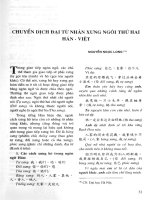
Fig. 5. New model for the mechanism of multiple lysine methylation by the MLL1 core complex (adapted from Patel [36]). (A) The MLL1
core complex is composed of two distinct H3K4 methyltransferases each possessing H3K4 monomethylation activity on their own. The
dashed oval on the WDR5–RbBP5–Ash2L–DPY-30 sub-complex indicates that the catalytic motif is currently unknown, and may be shared
between subunits. (B). WDR5’s recognition of the MLL1 Win motif results in the assembly of the MLL1 core complex, which possesses
H3K4 dimethyltransferase activity. We suggest that the MLL1 SET domain catalyzes monomethylation of histone H3 at lysine 4, which is fol-
lowed by transfer of the monomethylated histone H3 to a second active site on the WRAD sub-complex, where H3K4 dimethylation occurs.
We propose that mechanisms that control the assembly of the MLL1 core complex will be important for the regulation of H3K4 methylation
states in the cell.
M. S. Cosgrove and A. Patel MLL1: a structure–function perspective
FEBS Journal 277 (2010) 1832–1842 ª 2010 The Authors Journal compilation ª 2010 FEBS 1839
of cell fate. Leukemias associated with loss-of-function
or gain-of-function variants of MLL1 are prime exam-
ples of the importance of maintaining the enzymatic
activity of MLL1 under tight control. Identifying the
protein structural features that account for the enzy-
matic activity of MLL1 and other SET1 family mem-
bers will be essential for understanding how the
regulation of H3K4 methylation is integrated into
eukaryotic transcriptional circuits. A future challenge
is defining, from a structure–function perspective, how
MLL1 interacts with and is regulated by other proto-
oncoproteins including the multiple endocrine neopla-
sia type 1 (Men1) [60] or the integrase interactor 1
(Ini ⁄ hSNF5) [61] tumor suppressor proteins. An addi-
tional challenge is in understanding the molecular basis
for how all the different MLL fusion proteins disrupt
normal MLL functioning and contribute to cellular
transformation. In addition, important questions that
remain unanswered include: How does MLL1 regulate
the trimethyl form of histone H3? Does the regulation
of H3K4 methylation involve posttranslational modifi-
cations in MLL1 or other proteins that regulate the
assembly of the MLL1 core complex? How does
MLL1 discriminate among potential target genes? It is
expected that such knowledge will be valuable for the
development of new therapeutic strategies for the treat-
ment of some forms leukemia and other aggressive
cancers.
Acknowledgements
This work is supported in part by a Research Scholar
Grant (RSC-09-245-01-DMC) from the American Can-
cer Society and by NIH grant number R01CA140522
from the National Cancer Institute (to MSC). We
thank Venkat Dharmarajan for a critical reading of
this manuscript. We would also like to dedicate this
manuscript to the memory of Warren DeLano, the cre-
ator of the molecular graphics program pymol, which
was used to create the figures in this manuscript.
References
1 Ziemin-van der Poel S, McCabe NR, Gill HJ, Espinosa
R III, Patel Y, Harden A, Rubinelli P, Smith SD,
LeBeau MM, Rowley JD et al. (1991) Identification of
a gene, MLL, that spans the breakpoint in 11q23 trans-
locations associated with human leukemias. Proc Natl
Acad Sci USA 88, 10735–10739.
2 Leegte B, Kerstjens-Frederikse WS, Deelstra K, Begeer
JH & van Essen AJ (1999) 11q-syndrome: three
cases and a review of the literature. Genet Couns 10,
305–313.
3 Canaani E, Nakamura T, Rozovskaia T, Smith ST,
Mori T, Croce CM & Mazo A (2004) ALL-1 ⁄ MLL1,
a homologue of Drosophila TRITHORAX, modifies
chromatin and is directly involved in infant acute
leukaemia. Br J Cancer 90, 756–760.
4 Marschalek R (2010) Mixed lineage leukemia: roles in
human malignancies and potential therapy. FEBS J
doi:10.1111/j.1742-4658.2010.07608.x.
5 Yu BD, Hess JL, Horning SE, Brown GA &
Korsmeyer SJ (1995) Altered Hox expression and
segmental identity in Mll-mutant mice. Nature 378,
505–508.
6 Milne TA, Briggs SD, Brock HW, Martin ME,
Gibbs D, Allis CD & Hess JL (2002) MLL targets SET
domain methyltransferase activity to Hox gene promot-
ers. Mol Cell 10, 1107–1117.
7 Milne TA, Dou Y, Martin ME, Brock HW, Roeder
RG & Hess JL (2005) MLL associates specifically with
a subset of transcriptionally active target genes. Proc
Natl Acad Sci USA 102, 14765–14770.
8 Terranova R, Agherbi H, Boned A, Meresse S &
Djabali M (2006) Histone and DNA methylation
defects at Hox genes in mice expressing a SET domain-
truncated form of Mll. Proc Natl Acad Sci USA 103,
6629–6634.
9 Malik S & Bhaumik SR (2010) Mixed lineage leukemia:
histone H3 lysine 4 methyltransferases from yeast to
human. FEBS J doi:10.1111/j.1742-4658.2010.07607.x.
10 Strahl BD, Ohba R, Cook RG & Allis CD (1999)
Methylation of histone H3 at lysine 4 is highly
conserved and correlates with transcriptionally active
nuclei in Tetrahymena. Proc Natl Acad Sci USA 96,
14967–14972.
11 Rasio D, Schichman SA, Negrini M, Canaani E &
Croce CM (1996) Complete exon structure of the ALL1
gene. Cancer Res 56, 1766–1769.
12 Nakamura T, Mori T, Tada S, Krajewski W,
Rozovskaia T, Wassell R, Dubois G, Mazo A, Croce
CM & Canaani E (2002) ALL-1 is a histone methyl-
transferase that assembles a supercomplex of proteins
involved in transcriptional regulation. Mol Cell 10,
1119–1128.
13 Ansari KI & Mandal SS (2010) Mixed lineage leukemia:
role in gene expression, hormone signaling and mRNA
processing. FEBS J doi:10.1111/j.1742-4658.2010.
07606.x.
14 Yokoyama A, Kitabayashi I, Ayton PM, Cleary ML &
Ohki M (2002) Leukemia proto-oncoprotein MLL is
proteolytically processed into 2 fragments with opposite
transcriptional properties. Blood 100, 3710–3718.
15 Hsieh JJ, Ernst P, Erdjument-Bromage H, Tempst P &
Korsmeyer SJ (2003) Proteolytic cleavage of MLL
generates a complex of N- and C-terminal fragments
that confers protein stability and subnuclear
localization. Mol Cell Biol 23, 186–194.
MLL1: a structure–function perspective M. S. Cosgrove and A. Patel
1840 FEBS Journal 277 (2010) 1832–1842 ª 2010 The Authors Journal compilation ª 2010 FEBS
16 Dou Y, Milne TA, Tackett AJ, Smith ER, Fukuda A,
Wysocka J, Allis CD, Chait BT, Hess JL & Roeder RG
(2005) Physical association and coordinate function of
the H3 K4 methyltransferase MLL1 and the H4 K16
acetyltransferase MOF. Cell 121, 873–885.
17 Yokoyama A, Wang Z, Wysocka J, Sanyal M, Aufiero
DJ, Kitabayashi I, Herr W & Cleary ML (2004)
Leukemia proto-oncoprotein MLL forms a SET1-like
histone methyltransferase complex with menin to regu-
late Hox gene expression. Mol Cell Biol 24, 5639–5649.
18 Wysocka J, Myers MP, Laherty CD, Eisenman RN &
Herr W (2003) Human Sin3 deacetylase and trithorax-
related Set1 ⁄ Ash2 histone H3-K4 methyltransferase are
tethered together selectively by the cell-proliferation fac-
tor HCF-1. Genes Dev 17, 896–911.
19 Lee JH, Tate CM, You JS & Skalnik DG (2007) Identi-
fication and characterization of the human Set1B his-
tone H3-Lys4 methyltransferase complex. J Biol Chem
282, 13419–13428.
20 Cho YW, Hong T, Hong S, Guo H, Yu H, Kim D,
Guszczynski T, Dressler GR, Copeland TD, Kalkum M
et al. (2007) PTIP associates with MLL3- and
MLL4-containing histone H3 lysine 4 methyltransferase
complex. J Biol Chem 282, 20395–20406.
21 Hughes CM, Rozenblatt-Rosen O, Milne TA, Copeland
TD, Levine SS, Lee JC, Hayes DN, Shanmugam KS,
Bhattacharjee A, Biondi CA et al. (2004) Menin
associates with a trithorax family histone methyltrans-
ferase complex and with the hoxc8 locus. Mol Cell 13,
587–597.
22 Allen MD, Grummitt CG, Hilcenko C, Min SY,
Tonkin LM, Johnson CM, Freund SM, Bycroft M &
Warren AJ (2006) Solution structure of the nonmethyl-
CpG-binding CXXC domain of the leukaemia-associ-
ated MLL histone methyltransferase. EMBO J 25,
4503–4512.
23 De Guzman RN, Goto NK, Dyson HJ & Wright PE
(2006) Structural basis for cooperative transcription
factor binding to the CBP coactivator. J Mol Biol 355,
1005–1013.
24 Patel A, Dharmarajan V & Cosgrove MS (2008)
Structure of WDR5 bound to Mixed Lineage Leukemia
Protein-1 peptide. J Biol Chem 283, 32158–32161.
25 Song JJ & Kingston RE (2008) WDR5 interacts with
mixed lineage leukemia (MLL) protein via the histone
H3-binding pocket. J Biol Chem 283, 35258–35264.
26 Southhall SM, Wong P, Odho Z, Roe SM & Wilson JR
(2009) Structural basis for the recruitment of additional
factors for MLL1 SET domain activity and recognition
of epigenetic marks. Mol Cell 33, 181–191.
27 Birke M, Schreiner S, Garcia-Cuellar MP, Mahr K,
Titgemeyer F & Slany RK (2002) The MT domain of
the proto-oncoprotein MLL binds to CpG-containing
DNA and discriminates against methylation. Nucleic
Acids Res 30, 958–965.
28 Ayton PM, Chen EH & Cleary ML (2004) Binding to
nonmethylated CpG DNA is essential for target
recognition, transactivation, and myeloid transforma-
tion by an MLL oncoprotein. Mol Cell Biol 24, 10470–
10478.
29 Cross SH & Bird AP (1995) CpG islands and genes.
Curr Opin Genet Dev 5, 309–314.
30 Goodman RH & Smolik S (2000) CBP ⁄ p300 in cell
growth, transformation, and development. Genes Dev
14, 1553–1577.
31 Ernst P, Wang J, Huang M, Goodman RH &
Korsmeyer SJ (2001) MLL and CREB bind coopera-
tively to the nuclear coactivator CREB-binding protein.
Mol Cell Biol 21, 2249–2258.
32 Goto NK, Zor T, Martinez-Yamout M, Dyson HJ &
Wright PE (2002) Cooperativity in transcription factor
binding to the coactivator CREB-binding protein
(CBP). The mixed lineage leukemia protein (MLL)
activation domain binds to an allosteric site on the KIX
domain. J Biol Chem 277, 43168–43174.
33 Zor T, De Guzman RN, Dyson HJ & Wright PE
(2004) Solution structure of the KIX domain of CBP
bound to the transactivation domain of c-Myb. J Mol
Biol 337, 521–534.
34 Dou Y, Milne TA, Ruthenburg AJ, Lee S, Lee JW,
Verdine GL, Allis CD & Roeder RG (2006) Regulation
of MLL1 H3K4 methyltransferase activity by its core
components. Nat Struct Mol Biol 13, 713–719.
35 Patel A, Dharmarajan V, Vought VE & Cosgrove MS,
(2009) On the mechanism of multiple lysine methyla-
tion by the human mixed lineage leukemia protein-1
(MLL1) core complex. J Biol Chem 284, 24242–
24256.
36 Patel A, Vought V, Dharmarajan V & Cosgrove MS
(2008) A conserved arginine containing motif crucial for
the assembly and enzymatic activity of the Mixed Line-
age Leukemia protein-1 core complex. J Biol Chem 283,
32162–32175.
37 Steward MM, Lee JS, O’Donovan A, Wyatt M,
Bernstein BE & Shilatifard A (2006) Molecular
regulation of H3K4 trimethylation by ASH2L, a shared
subunit of MLL complexes. Nat Struct Mol Biol 13,
852–854.
38 Jenuwein T, Laible G, Dorn R & Reuter G (1998)
SET domain proteins modulate chromatin domains
in eu- and heterochromatin. Cell Mol Life Sci 54,
80–93.
39 Rea S, Eisenhaber F, O’Carroll D, Strahl BD, Sun ZW,
Schmid M, Opravil S, Mechtler K, Ponting CP & Allis
CD (2000) Regulation of chromatin structure by
site-specific histone H3 methyltransferases. Nature 406,
593–599.
40 Dillon SC, Zhang X, Trievel RC & Cheng X (2005)
The SET-domain protein superfamily: protein lysine
methyltransferases. Genome Biol 6, 227.
M. S. Cosgrove and A. Patel MLL1: a structure–function perspective
FEBS Journal 277 (2010) 1832–1842 ª 2010 The Authors Journal compilation ª 2010 FEBS 1841
41 Shilatifard A (2008) Molecular implementation and
physiological roles for histone H3 lysine 4 (H3K4)
methylation. Curr Opin Cell Biol 20, 341–348.
42 Xiao B, Wilson JR & Gamblin SJ (2003) SET
domains and histone methylation. Curr Opin Struct
Biol 13, 699–705.
43 Zhang X, Tamaru H, Khan SI, Horton JR, Keefe LJ,
Selker EU & Cheng X (2002) Structure of the Neuros-
pora SET domain protein DIM-5, a histone H3 lysine
methyltransferase. Cell 111, 117–127.
44 Zhang X, Yang Z, Khan SI, Horton JR, Tamaru H,
Selker EU & Cheng X (2003) Structural basis for the
product specificity of histone lysine methyltransferases.
Mol Cell 12, 177–185.
45 Chang Y, Zhang X, Horton JR, Upadhyay AK,
Spannhoff A, Liu J, Snyder JP, Bedford MT &
Cheng X (2009) Structural basis for G9a-like protein
lysine methyltransferase inhibition by BIX-01294.
Nat Struct Mol Biol 16, 312–317.
46 Min J, Zhang X, Cheng X, Grewal SI & Xu RM
(2002) Structure of the SET domain histone lysine
methyltransferase Clr4. Nat Struct Biol 9 , 828–832.
47 Wysocka J, Swigut T, Milne TA, Dou Y, Zhang X,
Burlingame AL, Roeder RG, Brivanlou AH & Allis CD
(2005) WDR5 associates with histone H3 methylated at
K4 and is essential for H3 K4 methylation and verte-
brate development. Cell 121, 859–872.
48 Trievel RC & Shilatifard A (2009) WDR5, a complexed
protein. Nat Struct Mol Biol 16, 678–680.
49 Ruthenburg AJ, Wang W, Graybosch DM, Li H,
Allis CD, Patel DJ & Verdine GL (2006) Histone H3
recognition and presentation by the WDR5 module of
the MLL1 complex. Nat Struct Mol Biol 13, 704–712.
50 Couture JF, Collazo E & Trievel RC (2006) Molecular
recognition of histone H3 by the WD40 protein WDR5.
Nat Struct Mol Biol 13, 698–703.
51 Schuetz A, Allali-Hassani A, Martin F, Loppnau P,
Vedadi M, Bochkarev A, Plotnikov AN, Arrowsmith
CH & Min J (2006) Structural basis for molecular rec-
ognition and presentation of histone H3 by WDR5.
EMBO J 25, 4245–4252.
52 Collins RE, Tachibana M, Tamaru H, Smith KM,
Jia D, Zhang X, Selker EU, Shinkai Y & Cheng X
(2005) In vitro and in vivo analyses of a Phe ⁄ Tyr switch
controlling product specificity of histone lysine
methyltransferases. J Biol Chem 280, 5563–5570.
53 Qian C, Wang X, Manzur K, Sachchidanand, Farooq
A, Zeng L, Wang R & Zhou MM (2006) Structural
insights of the specificity and catalysis of a viral
histone H3 lysine 27 methyltransferase. J Mol Biol 359,
86–96.
54 Trievel RC, Flynn EM, Houtz RL & Hurley JH (2003)
Mechanism of multiple lysine methylation by the SET
domain enzyme Rubisco LSMT. Nat Struct Biol 10,
545–552.
55 Xiao B, Jing C, Kelly G, Walker PA, Muskett FW,
Frenkiel TA, Martin SR, Sarma K, Reinberg D,
Gamblin SJ et al. (2005) Specificity and mechanism of
the histone methyltransferase Pr-Set7. Genes Dev 19,
1444–1454.
56 Takahashi YH, Lee JS, Swanson SK, Saraf A,
Florens L, Washburn MP, Trievel RC & Shilatifard A
(2009) Regulation of H3K4 trimethylation via Cps40
(Spp1) of COMPASS is monoubiquitination indepen-
dent: implication for a Phe ⁄ Tyr switch by the catalytic
domain of Set1. Mol Cell Biol
29, 3478–3486.
57 Cheung P (2004) Generation and characterization of
antibodies directed against di-modified histones, and
comments on antibody and epitope recognition. Meth-
ods Enzymol 376, 221–234.
58 Kohlmaier A, Savarese F, Lachner M, Martens J,
Jenuwein T & Wutz A (2004) A chromosomal memory
triggered by Xist regulates histone methylation in X
inactivation. PLoS Biol 2, E171.
59 Hayashi K, Yoshida K & Matsui Y (2005) A histone
H3 methyltransferase controls epigenetic events required
for meiotic prophase. Nature 438, 374–378.
60 Yokoyama A, Somervaille TC, Smith KS, Rozenblatt-
Rosen O, Meyerson M & Cleary ML (2005) The menin
tumor suppressor protein is an essential oncogenic co-
factor for MLL-associated leukemogenesis. Cell 123,
207–218.
61 Rozenblatt-Rosen O, Rozovskaia T, Burakov D,
Sedkov Y, Tillib S, Blechman J, Nakamura T, Croce
CM, Mazo A & Canaani E (1998) The C-terminal SET
domains of ALL-1 and TRITHORAX interact with the
INI1 and SNR1 proteins, components of the SWI ⁄ SNF
complex. Proc Natl Acad Sci USA 95, 4152–4157.
MLL1: a structure–function perspective M. S. Cosgrove and A. Patel
1842 FEBS Journal 277 (2010) 1832–1842 ª 2010 The Authors Journal compilation ª 2010 FEBS