Tài liệu Báo cáo khoa học: Mitochondrial chaperone tumour necrosis factor receptor-associated protein 1 protects cardiomyocytes from hypoxic injury by regulating mitochondrial permeability transition pore opening docx
Bạn đang xem bản rút gọn của tài liệu. Xem và tải ngay bản đầy đủ của tài liệu tại đây (621.78 KB, 10 trang )
Mitochondrial chaperone tumour necrosis factor
receptor-associated protein 1 protects cardiomyocytes
from hypoxic injury by regulating mitochondrial
permeability transition pore opening
Fei Xiang, Yue-Sheng Huang, Xiao-Hua Shi and Qiong Zhang
Institute of Burn Research, State Key Laboratory of Trauma, Burns and Combined Injury, Southwest Hospital, Third Military Medical
University, Chongqing, China
Introduction
Hypoxia is one of the main causes of myocardial
damage after the receipt of a burn. In the early stages
after a severe burn, myocardial damage not only
causes cardiac insufficiency, but also induces or
aggravates burn shock, which can cause or aggravate
ischaemic ⁄ hypoxic injury to other organs [1,2]. Hence,
it is important to protect cardiomyocytes from hypoxic
damage. Mitochondria are the primary target of
hypoxic damage in cardiomyocytes. Several inter-
related factors, including calcium overload, an increase
in reactive oxygen species (ROS) and a decrease in
adenine nucleotides, contribute to mitochondrial
impairment during hypoxia and ischaemia [3]. Mito-
chondrial dysfunction in cardiomyocytes can also
Keywords
cardiomyocytes; cell damage; hypoxia;
mitochondrial permeability transition pore;
tumour necrosis factor receptor-associated
protein 1
Correspondence
Y S. Huang, Institute of Burn Research,
State Key Laboratory of Trauma, Burns and
Combined Injury, Southwest Hospital, Third
Military Medical University, Chongqing
400038, China
Fax: +86 23 65461696
Tel: +86 23 65461696
E-mail:
(Received 3 December 2009, revised 3
February 2010, accepted 11 February
2010)
doi:10.1111/j.1742-4658.2010.07615.x
Tumour necrosis factor receptor-associated protein 1 (TRAP1) is a mito-
chondrial chaperone that plays a role in maintaining mitochondrial func-
tion and regulating cell apoptosis. The opening of the mitochondrial
permeability transition pore (MPTP) is a key step in cell death after
hypoxia. However, it is still unclear whether TRAP1 protects cardiomyo-
cytes from hypoxic damage by regulating the opening of the pore. In the
present study, primary cultured cardiomyocytes from neonatal rats were
used to investigate changes in TRAP1 expression after hypoxia treatment
as well as the mechanism and effect of TRAP1 on hypoxic damage. The
results obtained showed that TRAP1 expression increased after 1 h of
hypoxia and continued to increase for up to 12 h of treatment. Hypoxia
caused an increase in cell death and decreased cell viability and mitochon-
drial membrane potential; overexpressing TRAP1 prevented hypoxia-
induced damage to cardiomyocytes. The silencing of TRAP1 induced an
increase in cell death and decreased both cell viability and mitochondrial
membrane potential in cardiomyocytes under normoxic and hypoxic condi-
tions. Furthermore, cell damage induced by the silencing of TRAP1
was prevented by the mitochondrial permeability transition pore inhibitor,
cyclosporin A. These data demonstrate that hypoxia induces an increase in
TRAP1 expression in cardiomyocytes, and that TRAP1 plays a protective
role by regulating the opening of the mitochondrial permeability transition
pore.
Abbreviations
Ad-TRAP1, recombinant adenovirus vector for TRAP1 overexpression; CsA, cyclosporin A; CypD, cyclophilin D; GFP, green fluorescent
protein; HSP, heat shock protein; MPTP, mitochondrial permeability transition pore; ROS, reactive oxygen species; siRNA, small interfering
RNA; TRAP1, tumour necrosis factor receptor-associated protein 1.
FEBS Journal 277 (2010) 1929–1938 ª 2010 The Authors Journal compilation ª 2010 FEBS 1929
directly lead to cell death after hypoxia. The mitochon-
drial permeability transition pore (MPTP) is a nonspe-
cific pore that opens during the time of calcium
overload, oxidative stress, adenine nucleotide depletion
and elevated phosphate levels. Many studies have dem-
onstrated the role of MPTP opening during an ischae-
mia ⁄ reperfusion injury to the heart and other organs
[4–6]. We have also demonstrated that more MPTPs
open in cardiomyocytes after hypoxia compared to
normoxic conditions [7]. Once the pore opens, the
membrane potential and pH gradient dissipate, pre-
venting ATP generation by oxidative phosphorylation.
Ultimately, these changes lead to cell death through
the activation of phospholipases, nucleases and prote-
ases [8]. Indeed, the irreversible mitochondrial injury
caused by MPTP opening is the key step in cell death
that occurs during hypoxia and other conditions [9].
Tumour necrosis factor receptor-associated protein 1
(TRAP1) localizes to the mitochondria and its targeting
sequence, which is found in the N-terminus of the pro-
tein, is for mitochondria matrix. An analysis of the
cDNA sequences reveals that TRAP1 is identical to
heart shock protein (HSP) 75, which is a member of the
HSP90 family [10]. HSP90 comprises an important
molecular chaperone that is involved in many cellular
processes. After hypoxia treatment, HSP90 expression
increases, and this plays a protective role against dam-
age [11]. However, the changes in TRAP1 in cardiomyo-
cytes under hypoxic conditions remain unclear. TRAP1
comprises a mitochondrial chaperone that is critical for
importing proteins into the mitochondrial matrix [12]. A
previous study showed that up-regulation of TRAP1
expression suppressed arsenite-induced apoptosis in
lung epithelium cells [13]. Apoptogenic inducers, such as
the protein-tyrosine kinase inhibitor b-hydroxyisovaler-
ylshikonin or the topoisomerase II inhibitor VP16, can
decrease TRAP1 expression [14]. At the same time,
TRAP1 antagonizes ROS production and protects
tumour cells from granzyme M-mediated apoptosis [15].
A recent study also demonstrated that TRAP1 over-
expression preserves the mitochondrial membrane
potential (Dw) and maintains ATP levels and cell viabil-
ity during ischaemic-like injury in vivo [16]. These data
suggest that TRAP1 may play an important role in
maintaining mitochondrial function. As noted above,
MPTP is recognized as a key player in cell death. How-
ever, whether TRAP1 can protect cells from hypoxic
damage by regulating MPTP opening in cardiomyocytes
has remained unclear until now.
The present study aimed to observe changes in
TRAP1 expression after hypoxia treatment and to
investigate the effect of TRAP1 on cell death and
MPTP opening in primary cardiomyocytes.
Results
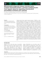
Hypoxia increases TRAP1 expression in
cardiomyocytes
Western blot analysis was used to investigate TRAP1
expression after hypoxia treatment in cardiomyocytes.
TRAP1 content increased after 1 h of hypoxia and
continued to increase until for up to 12 h compared to
the normoxic group. At the same time, longer hypoxic
treatments yielded higher TRPA1 expression
(Fig. 1A,B). We then examined TRAP1 immunoreac-
tivity with an immunofluorescence assay. After 1 h of
hypoxia, TRAP1 fluorescence intensity was brighter in
hypoxic cells than in normoxic cells, which meant that
TRAP1 expression increased after 1 h of hypoxia
(Fig. 1C,D). Furthermore, increases in TRAP1 fluores-
cence intensity became greater with an extension of
hypoxic treatment time (Figs 1E–G and 2I). The
results obtained were similar to those observed with
the western blot.
TRAP1 overexpression decreases hypoxic
damage to cardiomyocytes
Because TRAP1 expression of cardiomyocytes was
increased after hypoxia treatment, we performed exper-
iments to determine whether the increase in TRAP1
expression plays a protective role in hypoxic cardio-
myocytes. We constructed a recombinant adenovirus
vector for TRAP1 overexpression (Ad-TRAP1) and
transfected the cardiomyocytes. After 48 h of infection,
infection efficiency was visualized by the expression of
green fluorescent protein (GFP), and more than 90%
of the cardiomyocytes were infected (Fig. 2A). Protein
was then harvested and the results obtained by western
blotting revealed that TRAP1 expression increased sig-
nificantly in cardiomyocytes infected with Ad-TRAP1
compared to the expression in negative vector-trans-
duced cardiomyocytes and to endogenous TRAP1
levels in normoxic cells (Fig. 2B).
To evaluate the role of TRAP1 overexpression in
cardiomyocytes under hypoxic conditions, we investi-
gated cell viability, Dw and cell death. After 6 h of
hypoxia, cell viability and Dw were significantly lower
in the uninfected and vector-infected cardiomyocytes
compared to normoxic cells. By contrast, TRAP1
overexpression increased hypoxic cell viability
(Fig. 2C) and preserved Dw (Fig. 2D). Additionally,
propidium iodide staining was used to investigate the
effect of TRAP1 overexpression on cell death. As
shown in Fig. 3, hypoxia treatment resulted in
increased cell death, which was reduced by TRAP1
TRAP1 protects cells from hypoxic injury by MPTP F. Xiang et al.
1930 FEBS Journal 277 (2010) 1929–1938 ª 2010 The Authors Journal compilation ª 2010 FEBS
overexpression. At the same time, infection with
the negative vector had no effect on hypoxia-induced
cell death.
Silencing of TRAP1 expression induces
cardiomyocyte damage
After demonstrating that TRAP1 overexpression can
prevent hypoxic damage in cardiomyocytes, we next
examined whether silencing TRAP1 expression induced
damage in cardiomyocytes. After infection with
TRAP1-small interfering RNA (siRNA) or control
vector adenovirus for 4 days, more than 90% of the
cardiomyocytes were determined to be infected by
observing GFP expression using a fluorescent micro-
scope (Fig. 4A). The effective silencing of endogenous
TRAP1 by TRAP1-siRNA adenovirus infection was
also confirmed by western blotting (Fig. 4B).
After TRAP1-siRNA infection, the viability of the
cardiomyocytes was significantly decreased compared
to that of normoxic cells and vector-infected cells
(Fig. 4C). Furthermore, silencing TRAP1 expression
induced a decrease in Dw of cardiomyocytes under
normoxic conditions and aggravated Dw loss induced
by hypoxia (Fig. 4D). As shown in Fig. 5, TRAP1
depletion also induced a significant increase in cardio-
myocytes death, whereas there was very little cell death
in the normoxic cardiomyocytes and vector-infected
cardiomyocytes.
In addition, we also observed the effect of silencing
TRAP1 expression on cardiomyocyte damage under
hypoxic conditions. It was found that hypoxia induced
more injuries in cardiomyocytes in terms of both
viability and cell death after TRAP1-siRNA infection
(Fig. 6A,B).
MPTP mediates the TRAP1 effect
TRAP1 is a mitochondria chaperon and plays a role
in maintaining mitochondrial homeostasis, whereas
MPTP opening is a key step in the process of cell
death. Therefore, we aimed to determine whether
MPTP opening mediates TRAP1 behaviour. After
cardiomyocytes were infected with TRAP1-siRNA or
negative vector for 2 days, cyclosporin A (CsA;
2 lm), a selective inhibitor of MPTP opening, was
added to the cardiomyocytes. Cells were then infected
for an additional 2 days (4 days in total). Treatment
with CsA prevented the decrease in cardiomyocyte
viability and the increase in cell death induced by
TRAP1-siRNA infection under normoxic conditions
(Fig. 7). However, there were no differences between
vector-infected cells and vector-infected cells after
CsA treatment (Fig. 7).
Because silencing TRAP1 expression aggravated
hypoxic damage of cardiomyocytes, we next investi-
gated the effect of CsA on cell viability and cell death
A
B
I
CDE
FGH
40
30
20
10
fluorescence intensity
(arbitrarty units)
0
Normoxia
*
*
*
*
13 6
H
yp
oxia treatment (h)
12
0.4
TRAP1
β-actin
0.3
0.2
0.1
TRAP1/β-actin
0
Normoxia
*
*
*
*
136
Hypoxia treatment (h)
Hypoxia treatment (h)
12
Normoxia
13 612
75 kD
a
43 kD
a
Fig. 1. Effects of hypoxia on the TRAP1 levels in primary cultured
cardiomyocytes. (A) Western blots show TRAP1 immunoreactivity
in normoxic or hypoxic cells at the indicated times. b-actin was
used as an internal control. (B) TRAP1 levels were normalized with
b-actin under normoxic or hypoxic conditions. (C–G) TRAP1 expres-
sion detected by immunofluorescence under normoxic conditions
(C) and hypoxic conditions for 1 h (D), 3 h (E), 6 h (F) and 12 h (G).
TRAP1 primary antibody was omitted as a negative control (H).
(I) Differences in fluorescence intensity of TRAP1 in normoxic or
hypoxic cells. Data are the mean
± SEM. Scale bar = 25 lm.
*P < 0.05 compared to the normoxic group. The experiment was
repeated three times.
F. Xiang et al. TRAP1 protects cells from hypoxic injury by MPTP
FEBS Journal 277 (2010) 1929–1938 ª 2010 The Authors Journal compilation ª 2010 FEBS 1931
after TRAP1-siRNA infection under hypoxic condi-
tions. After 6 h of hypoxia, treatment with CsA abol-
ished cardiomyocyte damage induced both by hypoxia
and silencing TRAP1 under hypoxic conditions
(Fig. 6). On the basis of the results described above,
we conclude that silencing TRAP1 expression induces
MPTP opening in cardiomyocytes, resulting in cell
injury. Furthermore, the up-regulation of TRAP1
expression may play a protective role in hypoxic
cardiomyocytes by reducing MPTP opening.
Discussion
In the present study, we found that TRAP1 expression
of cardiomyocytes increases after hypoxia and that
TRAP1 overexpression protects cardiomyocytes from
hypoxic damage. At the same time, silencing TRAP1
expression causes cell damage under normoxic and
hypoxic conditions. Our data also indicate that
TRAP1 plays a role in cardiomyocytes by regulating
MPTP opening.
TRAP1 was initially identified by the yeast two-
hybrid system as a novel protein that interacted with
the intercellular domain of the type 1 tumour necrosis
factor receptor [17]. On the basis of the sequence of
the homologue, TRAP1 was identified as a member of
the HSP90 family. The ATPase activity of TRAP1 is
inhibited by geldanamycin, which is a specific inhibitor
of HSP90. Despite its ATP-binding activity, TRAP1
does not form a stable complex with the co-chaperones
of HSP90, such as Hop and p23 [18]. Studies have
shown that TRAP1 does not have a C-terminal EEVD
sequence, which exists in HSP90 and is important for
HSP90-Hop binding [19]. Thus, it appears that TRAP1
has specific functions that are different from those of
other well-characterized HSP90 homologues. TRAP1
is up-regulated by glucose deprivation, oxidative stress
and ultraviolet A irradiation, but cannot be induced
A
B
C
D
Vector
Vector
Control
Ad-TRAP1
Ad-TRAP1
75 kD
a
36 kD
a
TRAP1
GAPDH
0.5
0.4
0.3
0.2
0.1
0
50
60
40
30
20
10
0
Normoxia
Hypoxia
Vector
Ad-TRAP1
Ad-TRAP1
Normoxia
Fluorescence intensity
(arbitrary units)
D
450
Vector
H
yp
oxia
Hypoxia
Hypoxia
#
*
*
*
*
#
Fig. 2. TRAP1 overexpression prevented
the hypoxia-induced reductions in cell viabil-
ity and Dw in primary cultured cardiomyo-
cytes. (A) Cardiomyocytes were infected
with negative vector or Ad-TRAP1 for 48 h
and then observed under a fluorescence
microscope to determine the infection
efficiency by visualizing expression of the
gene for GFP. Scale bar = 200 lm. (B)
Expression of TRAP1 levels in the unin-
fected control, negative vector-infected and
Ad-TRAP1-infected cardiomyocytes as deter-
mined by western blotting. (C, D) Cardio-
myocytes were infected with vector or
Ad-TRAP1 for 48 h, starved, and then
treated for 6 h under hypoxic conditions; cell
viability was determined with a cell counting
kit (C) and Dw was determined with tetram-
ethylrhodamine ethylester; and then one
hundred cells from each group were
randomly chosen to measure fluorescence
intensity (D). Data are the mean ± SEM.
*P < 0.05 compared to the normoxic group.
#P < 0.05 compared with the hypoxic and
hypoxia + vector groups. The experiment
was repeated three times.
TRAP1 protects cells from hypoxic injury by MPTP F. Xiang et al.
1932 FEBS Journal 277 (2010) 1929–1938 ª 2010 The Authors Journal compilation ª 2010 FEBS
by heat [16,20,21]. Furthermore, deferoxamine, an iron
chelator, decreases TRAP1 levels in a dose- and time-
dependent manner and induces mitochondrial dysfunc-
tion in human hepatocytes [22]. However, the changes
induced in TRAP1 expression in cardiomyocytes after
hypoxia treatment are still unclear. In the present
study, we demonstrated that hypoxia treatment (for 1,
3, 6 and 12 h, respectively) induces a time-dependent
increase in the levels of TRAP1 protein.
Hypoxia is a common pathophysiological process
in diseases such as shock, stroke and heart failure.
Hypoxic damage of the myocardium is relevant not
only to coronary artery diseases, but also to hyper-
tensive and cardiomyopathic heart disease [23,24].
Mitochondria are the most susceptible organelles
to hypoxic damage in cardiomyocytes. Although
hypoxia induced TRAP1 expression increases in
cardiomyocytes, the role of that TRAP1 increase
remains unclear. The question remains as to whether
the hypoxia-induced TRAP1 increase is a protective
reaction in cardiomyocytes. Because TRAP1 is a
mitochondrial chaperone, it has an important role in
regulating cell apoptosis and maintaining mitochon-
drial homeostasis and function. Silencing TRAP1
enhances cytochrome c release from the mitochondria
and apoptosis induced by b-hydroxyisovalerylshikonin
and VP16 [14]. TRAP1 depletion also sensitizes PC12
cells to oxidative stress-induced cytochrome c release
and cell death, which means that TRAP1 play a role
in the modulation of the mitochondrial apoptotic cas-
cade [25]. Moreover, TRAP1 overexpression improves
mitochondrial function after ischaemic injury in
primary astrocytes in vitro [16]. In the present study,
we found that TRAP1 overexpression abolishes the
hypoxic damage in cardiomyocytes. Silencing TRAP1
expression not only induces cell damage under
normoxic conditions, but it also aggravates hypoxic
damage of cardiomyocytes.
MPTP is a channel consisting of several proteins
that is usually in a low permeability or closed state.
Some models have proposed the presence of other
molecular components of the pore, although there is
still no consensus regarding the exact components.
However, cyclophilin D (CypD) is generally accepted
as a critical regulatory component of MPTP and
plays an important role in regulating MPTP opening
[8,26]. CsA, a selective MPTP inhibitor, prevents
MPTP opening by inhibiting the activity of the pept-
idyl-prolyl cis-trans isomerase of CypD [27,28]. The
consequences of MPTP opening are cell necrosis and
apoptosis and, even if MPTP opening is insufficient
to cause necrosis, apoptosis can occur. After the
MPTP opens, apoptogenic substrates (i.e. cytochrome
c) are released into the cytoplasm and activate cas-
pase-dependent apoptotic pathways. Because MPTP
plays a critical role in cell necrosis and apoptosis, it
is also involved in protecting cell against hypoxic and
ischaemic damages [29,30]. MPTP not only contrib-
utes to the early and delayed protective effects of
ischaemic preconditioning in rat or rabbit heart, but
it is also relevant to ischaemic post-conditioning [31].
We had also previously demonstrated that adenosine
A1 receptor activation reduces hypoxic damage by
preventing MPTP opening in rat cardiomyocytes [7].
Many studies have demonstrated that Dw loss is
accompanied by an increase in MPTP opening [32–
34]. It is considered that Dw reflects the state of
MPTP opening indirectly. In the present study, we
found that silencing TRAP1 induces Dw loss in
cardiomyocytes, and that overexpression of TRAP1
Fig. 3. TRAP1 overexpression decreased hypoxia-induced cell
death in primary cultured cardiomyocytes. Cell death was deter-
mined by incubating normoxic cells, hypoxic cells, vector-infected
hypoxic cells and Ad-TRAP1-infected cells after 6 h of hypoxia with
Hoechst 33342 (10 lgÆmL
)1
, blue) and propidium iodide (PI)
(10 lgÆmL
)1
, red). Scale bar = 50 lm. Graphs show the quantifica-
tion of cell death (mean ± SEM) and 200–300 cells were counted
for each group. *P < 0.05 compared to the normoxic group.
#P < 0.05 compared to the hypoxic and hypoxic + vector groups.
The experiment was repeated three times.
F. Xiang et al. TRAP1 protects cells from hypoxic injury by MPTP
FEBS Journal 277 (2010) 1929–1938 ª 2010 The Authors Journal compilation ª 2010 FEBS 1933
suppresses Dw loss caused by hypoxia. Furthermore,
our present data also show that CsA prevents the cell
damage induced by TRAP1 depletion under normoxic
and hypoxic conditions, which means that silencing
TRAP1 expression can cause MPTP opening and lead
to damage. Because the opening of MPTP increases
after hypoxia treatment, and TRAP1 overexpression
abolishes hypoxic damage, we therefore assume that
TRAP1 overexpression may prevent MPTP opening
and having a protective effect under hypoxic condi-
tions in cardiomyocytes. In tumour cells, TRAP1
interacts with CypD, and the association of TRAP1
with CypD is prevented by CsA and not geldanamy-
cin, suggesting that this association may be necessary
for CypD activity [35].
Many factors are involved in inducing MPTP open-
ing, especially calcium overload and oxidative stress
[36,37]. ROS increases could lead to the MPTP open-
ing persistently. However, TRAP1 also shows an
important role in regulating ROS generation. ROS
production is decreased by TRAP1 overexpression and
promoted by silencing TRAP1 expression [15,16,38].
Because TRAP1 plays a role against cell damage by
MPTP, further studies are needed to determine
whether ROS are mediators between TRAP1 and
MPTP in cardiomyocytes.
In summary, hypoxia increases the level of TRAP1 in
cardiomyocytes, which may protect cells from hypoxic
damage by regulating MPTP opening. These results
provide us with a deeper understanding of the protective
role of TRAP1 in cardiomyocytes and offer new consid-
erations for myocardial protection after burn shock.
Materials and methods
Cardiomyocyte culture and hypoxia treatment
Primary cardiomyocyte cultures were prepared from the
ventricles of neonatal Sprague-Dawley rats (days 1–3) and
trypsinized as described previously [39] in accordance with
A
B
C
D
Vector
VectorControl
TRAP1-siRNA
TRAP1-siRNA
75 kDa
36 kDa
TRAP1
GAPDH
0.5
0.4
0.3
0.2
0.1
0
50
40
30
20
10
0
Normoxia
Normoxia
Vector
TRAP1-siRNA
Normoxia
Fluorescence intensity
(arbitrary units)
D
450
Normoxia
Vector Vector
Hypoxia
HypoxiaTRAP1-siRNA TRAP1-siRNA
*
*
*
*
#
Fig. 4. Silencing TRAP1 expression induced
cell viability and Dw in primary cultured
cardiomyocytes. (A) Cardiomyocytes were
infected with negative vector or
TRAP1-siRNA for 4 days, and then a
fluorescence microscope was used to
observe the infection efficiency by
visualizing expression of the gene for GFP.
Scale bar = 200 lm. (B) Expression of
TRAP1 levels in uninfected control,
vector-infected and TRAP1-siRNA-infected
cardiomyocytes as determined by western
blotting. (C) Cardiomyocytes were infected
with vector or TRAP1-siRNA for 4 days,
starved, and then cell viability was
determined under normoxic conditions. (D)
Cardiomyocytes were infected with vector
or TRAP1-siRNA for 4 days, starved, and
then Dw was determined under normoxic
conditions or after 6 h of hypoxia. The
results are shown as the mean ± SEM.
*P < 0.05 compared to the normoxic and
normoxic + vector groups. #P < 0.05
compared to the hypoxic and hypoxic +
vector groups. The experiment was
repeated three times.
TRAP1 protects cells from hypoxic injury by MPTP F. Xiang et al.
1934 FEBS Journal 277 (2010) 1929–1938 ª 2010 The Authors Journal compilation ª 2010 FEBS
a protocol approved by the Animal Care and Use Commit-
tee of the Third Military Medical University. The cultures
were grown in a DMEM ⁄ F12 medium (Hyclone, Logan,
UT, USA) with 10% (v ⁄ v) fetal bovine serum (Hyclone),
0.1 mm bromodeoxyuridine (Sigma-Aldrich, St Louis, MO,
USA), 100 UÆmL
)1
penicillin and 100 UÆmL
)1
streptomy-
cin. Cells were maintained in a 5% CO
2
incubator at
37 °C. Before hypoxia treatment, the cardiomyocytes were
deprived of serum for 12 h.
Hypoxic conditions were prepared by using an anaerobic
jar (Mitsubishi, Tokyo, Japan) and a vacuum glove box
(Chunlong, Lianyungang, China). Serum-free medium was
placed in the vacuum glove box filled with a mixed gas con-
taining 94% nitrogen, 5% CO
2
and 1% O
2
overnight and
allowed to equilibrate with the hypoxic atmosphere.
Cardiomyocytes were then subjected to hypoxic conditions
by replacing the normoxic medium with hypoxic medium
and placing the cultures in an anaerobic jar. All procedures
were performed in vacuum glove box.
Recombinant adenovirus vector for TRAP1
overexpression
Ad-TRAP1 and a negative adenovirus vector were pro-
duced by Shanghai GeneChem, Co. Ltd (Shanghai,
China). The vectors encoded the GFP sequence, which
served as a marker gene. A high titre adenovirus stock
was made after several rounds of amplification in
HEK293A (American Type Culture Collection, Manassas,
VA, USA). All recombinant adenoviruses were tested for
transgene expression in cardiomyocytes by western blot-
ting. Cardiomyocytes were infected with Ad-TRAP1 or a
negative vector at a multiplicity of infection of 10 for
Fig. 5. Silencing TRAP1 expression induced cell death in primary
cultured cardiomyocytes under normoxic conditions. Cell death was
determined by incubating uninfected, vector-infected and TRAP1-
siRNA-infected cardiomyocytes under normoxic conditions with
Hoechst 33342 (10 lgÆmL
)1
, blue) and propidium iodide (PI)
(10 lgÆmL
)1
, red). Scale bar = 50 lm. Graphs show the quantifica-
tion of cell death (mean ± SEM) and 200–300 cells were counted
for each group. *P < 0.05 compared to the normoxic and
normoxic + vector groups. The experiment was repeated three
times.
A
B
Fig. 6. CsA prevented hypoxic damage after TRAP1-siRNA infec-
tion in primary cardiomyocytes. CsA (2 l
M) was added into vector-
infected and TRAP1-siRNA-infected cardiomyocytes after 2 days of
infection. The cells were then starved, and subjected to hypoxia for
6 h after 4 days of infection. (A) Effects of CsA on cell death in
uninfected, vector-infected and TRAP1-siRNA-infected cells under
hypoxic conditions. In each group, 200–300 cells were counted. (B)
Effects of CsA on cell viability in uninfected, vector-infected and
TRAP1-siRNA-infected cells under hypoxic conditions. *P < 0.05
compared to the normoxic group. #P < 0.05 compared to the hyp-
oxic and hypoxic + vector groups. **P < 0.05 compared to the hyp-
oxic and hypoxic + vector groups. ##P < 0.05 compared to the
hypoxic + TRAP1-siRNA group (data are the mean ± SEM). The
experiment was repeated three times.
F. Xiang et al. TRAP1 protects cells from hypoxic injury by MPTP
FEBS Journal 277 (2010) 1929–1938 ª 2010 The Authors Journal compilation ª 2010 FEBS 1935
48 h and then subjected to experiments after being
deprived of serum for 12 h.
Recombinant adenovirus vector for silencing of
TRAP1 expression
The recombinant adenovirus vector for silencing of TRAP1
expression (TRAP1-siRNA) was purchased from Shanghai
GeneChem, Co. Ltd. The targeting sequence of the siRNA
against rat TRAP1 was 5¢-CAACAGAGATTGATCAA
AT-3¢. A negative control adenovirus vector containing
nonspecific siRNA was constructed in the same way (non-
specific vector, 5¢-TTCTCCGAACGTGTCACGT-3¢). All
vectors contained the gene for GFP, which served as a mar-
ker. Cardiomyocytes were infected with TRAP1-siRNA or
control vector by the addition of adenovirus to the cell cul-
ture at a multiplicity of infection of 10. After 4 days of
infection, the cells were serum starved for 12 h and then
treated.
Preparation of cell lysates
Cells were washed three times with ice-cold NaCl ⁄ P
i
at the
appropriate time after treatment, and lysed in radioimmuno-
precipitation assay (Sigma-Aldrich) lysis buffer that
contained 2 lgÆmL
)1
aprotinin, 2 lgÆmL
)1
pepstatin,
2 lgÆmL
)1
leupeptin and 100 lgÆmL
)1
phenylmethanesulfo-
nyl fluoride. Cells were then scraped, and the resulting lysate
was ultrasonicated and centrifuged at 12 000 g for 20 min at
4 °C. The supernatant was subjected to western blot analysis.
Western blot analysis
Protein concentrations were determined by the RC DC
assay (Bio-Rad, Hercules, CA, USA). Thirty micrograms of
proteins were fractionated by 10% SDS-PAGE and then
transferred to a poly(vinylidene difluoride) membrane
(Roche, Rotkreuz, Switzerland). The membrane was
blocked with 5% (w ⁄ v) skim milk in TBST [20 mm Tris-
HCl (pH 8.0), 150 mm NaCl and 0.1% (v ⁄ v) Tween-20] for
2 h at room temperature. Next, the membrane was probed
with a 1 : 500 dilution of primary anti-TRAP1 serum (BD
Biosciences, San Jose, CA, USA) in blocking buffer at 4 °C
overnight. The membrane was washed four times with
TBST and incubated with a horseradish peroxidase-conju-
gated antibody against mouse IgG for 1 h at room temper-
ature. The membrane was then rinsed with TBST, and the
protein bands were visualized with ECL Western Blotting
Detection Reagents (GE Healthcare, Piscataway, NJ,
USA). The images were analysed with quantity one 4.1
software (Bio-Rad). The experiment was repeated three
times, and the same results were obtained.
Immunofluorescence assay
Cardiomyocytes were grown on coverslips. After hypoxia
treatment, the cells were fixed with 4% (w ⁄ v) formaldehyde
in NaCl ⁄ P
i
for 10 min and permeabilized with 0.2% (v ⁄ v)
Triton X-100 for 15 min at room temperature. Nonspecific
binding sites were blocked by incubating the coverslips with
10% (v ⁄ v) goat serum in NaCl ⁄ P
i
for 1 h. Cells were probed
with primary anti-TRAP1 serum at a 1 : 100 dilution over-
night at 4 °C, washed with NaCl ⁄ P
i
, and incubated in the
dark at 37 °C for 1 h with fluorescein isothiocyanate-conju-
gated IgG. The cells were then washed again with NaCl ⁄ P
i
and stained with 0.4 mgÆmL
)1
4¢,6-diamidino-2-phenylindole
(Sigma-Aldrich) for 10 min at room temperature. Micro-
scopic images were acquired using a Leica Confocal Micro-
scope (Leica Microsystems, Wetzlar, Germany). In the
negative control, the primary antibody was omitted.
Detection of cardiomyocyte viability
Cardiomyocyte viability was determined with a cell counting
kit (CCK-8, Dojindo Molecular Technologies, Kumamoto,
A
B
Fig. 7. CsA prevented the cell damage induced by silencing TRAP1
in primary cardiomyocytes under normoxic conditions. CsA (2 l
M)
was added to vector-infected and TRAP1-siRNA-infected cardio-
myocytes after 2 days of infection. The cells were then subjected
to cell viability and cell death assay after 4 days of infection. (A)
Effects of CsA on cell death in uninfected, vector-infected and
TRAP1-siRNA-infected cells. In each group, 200–300 cells were
counted. (B) Effects of CsA on cell viability in uninfected, vector-
infected and TRAP1-siRNA-infected cells. *P < 0.05 compared to
the normoxic and normoxic + vector groups. #P < 0.05 compared
to the normoxic + TRAP1-siRNA group (data are the mean ± SEM).
The experiment was repeated three times.
TRAP1 protects cells from hypoxic injury by MPTP F. Xiang et al.
1936 FEBS Journal 277 (2010) 1929–1938 ª 2010 The Authors Journal compilation ª 2010 FEBS
Japan). Cells were cultured in 96-well plates (10 000 cells
per well) and the original medium was removed after 6 h of
hypoxia. Then, 10 lL of CCK-8 solution and 100 lLof
DMEM ⁄ F12 medium were added to each well, and the cells
were incubated at 37 °C in the dark for 1 h in accordance
with the manufacturer’s instructions. The value of D
450
was
determined (n = 3) and the experiment was repeated three
times.
Cell death assays
Cell death was quantified in Hoechst 33342 (10 lgÆmL
)1
;
Sigma-Aldrich) and propidium iodide (10 lgÆmL
)1
; Sigma-
Aldrich)-labelled cells. Propidium iodide readily penetrates
cells with compromised plasma membranes (dead cells) but
does not cross intact plasma membranes. Hoechst is a cell-
permeable nucleic acid stain that labels both live and dead
nuclei.
Mitochondrial membrane potential
Dw was monitored by tetramethylrhodamine ethylester
(Sigma-Aldrich). Cells cultured in a serum-free medium were
incubated in the dark with 200 nmolÆL
)1
tetramethylrhod-
amine ethylester at 37 °C for 15 min. Cells were then washed
with NaCl ⁄ P
i
and observed using a laser scanning confocal
microscope. The experiment was repeated three times.
Statistical analysis
All values were expressed as the mean ± SEM. spss,
version 11.0 (SPSS Inc., Chicago, IL, USA) was used to
conduct analyses of variance and Tukey’s tests. P < 0.05
was considered statistically significant.
Acknowledgements
This work was supported by the Key Project of China
National Programs for Basic Research and Develop-
ment (2005CB522601), the Key Program of National
Natural Science Foundation of China (30430680), the
Program for Changjiang Scholars, and the Innovative
Research Team in University (IRT0712). We thank Sun
Wei and Wang Li-ting (Central Library of The Third
Military Medical University) for their technical assis-
tance with the laser scanning confocal microscope. The
authors declare that there are no conflicts of interest.
References
1 Huang YS, Yang ZC, Yan BG, Yang JM, Chen FM,
Crowther RS & Li A (1999) Pathogenesis of early
cardiac myocyte damage after severe burns. J Trauma
46, 428–432.
2 Huang Y, Li Z & Yang Z (2003) Roles of ischemia and
hypoxia and the molecular pathogenesis of post-burn
cardiac shock. Burns 29, 828–833.
3 Baines CP (2009) The mitochondrial permeability tran-
sition pore and ischemia-reperfusion injury. Basic Res
Cardiol 104, 181–188.
4 Halestrap AP, Clarke SJ & Javadov SA (2004) Mito-
chondrial permeability transition pore opening during
myocardial reperfusion – a target for cardioprotection.
Cardiovasc Res 61, 372–385.
5 Kim JS, He L, Qian T & Lemasters JJ (2003) Role
of the mitochondrial permeability transition in
apoptotic and necrotic death after ischemia ⁄
reperfusion injury to hepatocytes. Curr Mol Med 3,
527–535.
6 Matsumoto S, Friberg H, Ferrand-Drake M & Wieloch
T (1999) Blockade of the mitochondrial permeability
transition pore diminishes infarct size in the rat after
transient middle cerebral artery occlusion. J Cereb
Blood Flow Metab 19, 736–741.
7 Fei X, Yue-Sheng H, Dong-Xia Z, Zhi-Gang C,
Jia-Ping Z & Qiong Z (2009) Adenosine A1 receptor
activation reduces mitochondrial permeability transition
pores opening in hypoxic cardiomyocytes. Clin Exp
Pharmacol Physiol 37, 343–349.
8 Leung AW & Halestrap AP (2008) Recent progress in
elucidating the molecular mechanism of the mitochon-
drial permeability transition pore. Biochim Biophys Acta
1777, 946–952.
9 Weiss JN, Korge P, Honda HM & Ping P (2003) Role
of the mitochondrial permeability transition in myocar-
dial disease. Circ Res 93, 292–301.
10 Chen CF, Chen Y, Dai K, Chen PL, Riley DJ & Lee
WH (1996) A new member of the hsp90 family of
molecular chaperones interacts with the retinoblastoma
protein during mitosis and after heat shock. Mol Cell
Biol 16, 4691–4699.
11 Wu WC, Kao YH, Hu PS & Chen JH (2007) Geldana-
mycin, a HSP90 inhibitor, attenuates the hypoxia-
induced vascular endothelial growth factor expression
in retinal pigment epithelium cells in vitro. Exp Eye Res
85, 721–731.
12 Kaul SC, Deocaris CC & Wadhwa R (2007) Three faces
of mortalin: a housekeeper, guardian and killer. Exp
Gerontol 42, 263–274.
13 Lau AT, He QY & Chiu JF (2004) A proteome analysis
of the arsenite response in cultured lung cells: evidence
for in vitro oxidative stress-induced apoptosis. Biochem
J 382, 641–650.
14 Masuda Y, Shima G, Aiuchi T, Horie M, Hori K,
Nakajo S, Kajimoto S, Shibayama-Imazu T & Nakaya
K (2004) Involvement of tumor necrosis factor recep-
tor-associated protein 1 (TRAP1) in apoptosis induced
by beta-hydroxyisovalerylshikonin. J Biol Chem 279,
42503–42515.
F. Xiang et al. TRAP1 protects cells from hypoxic injury by MPTP
FEBS Journal 277 (2010) 1929–1938 ª 2010 The Authors Journal compilation ª 2010 FEBS 1937
15 Hua G, Zhang Q & Fan Z (2007) Heat shock protein
75 (TRAP1) antagonizes reactive oxygen species genera-
tion and protects cells from granzyme M-mediated
apoptosis. J Biol Chem 282, 20553–20560.
16 Voloboueva LA, Duan M, Ouyang Y, Emery JF, Stoy
C & Giffard RG (2008) Overexpression of mitochon-
drial Hsp70 ⁄ Hsp75 protects astrocytes against ischemic
injury in vitro. J Cereb Blood Flow Metab 28, 1009–
1016.
17 Song HY, Dunbar JD, Zhang YX, Guo D & Donner
DB (1995) Identification of a protein with homology to
hsp90 that binds the type 1 tumor necrosis factor recep-
tor. J Biol Chem 270, 3574–3581.
18 Felts SJ, Owen BA, Nguyen P, Trepel J, Donner DB &
Toft DO (2000) The hsp90-related protein TRAP1 is a
mitochondrial protein with distinct functional proper-
ties. J Biol Chem 275, 3305–3312.
19 Chen B, Piel WH, Gui L, Bruford E & Monteiro A
(2005) The HSP90 family of genes in the human
genome: insights into their divergence and evolution.
Genomics 86, 627–637.
20 Carette J, Lehnert S & Chow TY (2002) Implication of
PBP74 ⁄ mortalin ⁄ GRP75 in the radio-adaptive response.
Int J Radiat Biol 78, 183–190.
21 Lee AS (2001) The glucose-regulated proteins: stress
induction and clinical applications. Trends Biochem Sci
26, 504–510.
22 Im CN, Lee JS, Zheng Y & Seo JS (2007) Iron
chelation study in a normal human hepatocyte cell line
suggests that tumor necrosis factor receptor-associated
protein 1 (TRAP1) regulates production of reactive
oxygen species. J Cell Biochem 100, 474–486.
23 Kyriakides ZS, Kremastinos DT, Michelakakis NA,
Matsakas EP, Demovelis T & Toutouzas PK (1991)
Coronary collateral circulation in coronary artery disease
and systemic hypertension. Am J Cardiol 67, 687–690.
24 Horwitz LD, Fennessey PV, Shikes RH & Kong Y
(1994) Marked reduction in myocardial infarct size due
to prolonged infusion of an antioxidant during reperfu-
sion. Circulation 89, 1792–1801.
25 Pridgeon JW, Olzmann JA, Chin LS & Li L (2007)
PINK1 protects against oxidative stress by phosphorylat-
ing mitochondrial chaperone TRAP1. PLoS Biol 5, e172.
26 Halestrap AP (2009) What is the mitochondrial perme-
ability transition pore? J Mol Cell Cardiol 46, 821–831.
27 Halestrap AP & Davidson AM (1990) Inhibition of
Ca2(+)-induced large-amplitude swelling of liver and
heart mitochondria by cyclosporin is probably caused
by the inhibitor binding to mitochondrial-matrix
peptidyl-prolyl cis-trans isomerase and preventing it
interacting with the adenine nucleotide translocase.
Biochem J 268 , 153–160.
28 Connern CP & Halestrap AP (1992) Purification and
N-terminal sequencing of peptidyl-prolyl cis-trans-isom-
erase from rat liver mitochondrial matrix reveals the
existence of a distinct mitochondrial cyclophilin. Bio-
chem J 284, 381–385.
29 Zhong Z, Ramshesh VK, Rehman H, Currin RT,
Sridharan V, Theruvath TP, Kim I, Wright GL &
Lemasters JJ (2008) Activation of the oxygen-sensing
signal cascade prevents mitochondrial injury after
mouse liver ischemia-reperfusion. Am J Physiol
Gastrointest Liver Physiol 295, G823–G832.
30 Shanmuganathan S, Hausenloy DJ, Duchen MR &
Yellon DM (2005) Mitochondrial permeability
transition pore as a target for cardioprotection in the
human heart. Am J Physiol Heart Circ Physiol 289,
H237–H242.
31 Hausenloy DJ, Ong SB & Yellon DM (2009) The mito-
chondrial permeability transition pore as a target for
preconditioning and postconditioning. Basic Res Cardiol
104, 189–202.
32 Sugrue MM, Wang Y, Rideout HJ, Chalmers-Redman
RM & Tatton WG (1999) Reduced mitochondrial mem-
brane potential and altered responsiveness of a mitochon-
drial membrane megachannel in p53-induced senescence.
Biochem Biophys Res Commun 261, 123–130.
33 Saotome M, Katoh H, Satoh H, Nagasaka S, Yoshi-
hara S, Terada H & Hayashi H (2005) Mitochondrial
membrane potential modulates regulation of mitochon-
drial Ca
2+
in rat ventricular myocytes. Am J Physiol
Heart Circ Physiol 288, H1820–H1828.
34 Lee CS, Park SY, Ko HH, Song JH, Shin YK & Han
ES (2005) Inhibition of MPP+-induced mitochondrial
damage and cell death by trifluoperazine and W-7 in
PC12 cells. Neurochem Int 46, 169–178.
35 Kang BH, Plescia J, Dohi T, Rosa J, Doxsey SJ &
Altieri DC (2007) Regulation of tumor cell mitochon-
drial homeostasis by an organelle-specific Hsp90 chap-
erone network. Cell 131, 257–270.
36 Zorov DB, Juhaszova M, Yaniv Y, Nuss HB, Wang S
& Sollott SJ (2009) Regulation and pharmacology of
the mitochondrial permeability transition pore. Cardio-
vasc Res 83, 213–225.
37 Baumgartner HK, Gerasimenko JV, Thorne C, Ferdek
P, Pozzan T, Tepikin AV, Petersen OH, Sutton R,
Watson AJ & Gerasimenko OV (2009) Calcium eleva-
tion in mitochondria is the main Ca
2+
requirement for
mitochondrial permeability transition pore (mPTP)
opening. J Biol Chem 284, 20796–20803.
38 Xu L, Voloboueva LA, Ouyang Y, Emery JF &
Giffard RG (2009) Overexpression of mitochondrial
Hsp70 ⁄ Hsp75 in rat brain protects mitochondria,
reduces oxidative stress, and protects from focal
ischemia. J Cereb Blood Flow Metab 29, 365–374.
39 Simpson P & Savion S (1982) Differentiation of rat
myocytes in single cell cultures with and without prolif-
erating nonmyocardial cells. Cross-striations, ultrastruc-
ture, and chronotropic response to isoproterenol. Circ
Res 50, 101–116.
TRAP1 protects cells from hypoxic injury by MPTP F. Xiang et al.
1938 FEBS Journal 277 (2010) 1929–1938 ª 2010 The Authors Journal compilation ª 2010 FEBS