Tài liệu Obstetrics and Gynecology Clinics of North America32 (2005) ppt
Bạn đang xem bản rút gọn của tài liệu. Xem và tải ngay bản đầy đủ của tài liệu tại đây (2.91 MB, 187 trang )
Preface
Management of First and Second Stages
of Labor
Suneet P. Chauhan, MD
Guest Editor
For both patient and the practitioner, few things are as dramatic and reward-
ing as childbirth. After months of anticipation and careful antepartum care, labor
is the last phase of pregnancy in which prudent decisions can improve out-
come. With over 130 million births in the world, 4 million of which occur in the
United States, it is imperative that the clinicians are current on the recent de-
velopments of intrapartum management. This collection of 13 articles, written by
clinicians, researchers, academicia ns, and private practitioners, updates the man-
agement of the first, second, and third stages of labor. The book is intended for
medical students, labor and delivery nurses, residents, midwives, and obstetri-
cians who try to optimize the outcome of each delivery.
The first two articles describe the mechanisms of normal labor and with
abnormal presentations. The next three provide clinically relevant information on
induction, abnormalities of stages I and II, and active management of labor. The
sixth article focuses on analgesia and anesthesia. We intentionally devoted two
articles to intrapartum assessment of the fetus to provide different perspectives
on a very important issue. Intrapartum complications—chorioamnionitis, non-
reassuring fetal heart rate tracing, and shoulder dystocia—are discussed, and their
management is described in the ninth, tenth, and eleventh articles. The last two
articles concern episiotomy and management of the third stage of labor.
0889-8545/05/$ – see front matter D 2005 Elsevier Inc. All rights reserved.
doi:10.1016/j.ogc.2005.04.008 obgyn.theclinics.com
Obstet Gynecol Clin N Am
32 (2005) xiii – xiv
Though obviou s, it is worth acknowledging the hou rs of scholarly work by
the authors of the articles, and the considerable support by Carin Davis and
the staff at Elsevier is refreshing.
Suneet P. Chauhan, MD
Division of Maternal–Fetal Medicine
Spartanburg Regional Medical Center
101 East Wood Street
Spartanburg, SC 29303, USA
E-mail address:
prefacexiv
Normal Labor: Mechanism and Duration
John B. Liao, MD, Catalin S. Buhimschi, MD,
Errol R. Norwitz, MD, PhD
*
Division of Reproductive Sciences, Department of Obstetrics, Gynecology and Reproductive Sciences,
Yale University School of Medicine, 333 Cedar Street, New Haven, CT 06520, USA
Labor refers to the chain of physiologic events that allows a fetus to under-
take its journey from the uterus to the outside world. The mean duration of a
singleton pregnancy is 40.0 weeks (280 days), which is dated from the first day
of the last normal menstrual period. The period from 37.0 weeks (259 days) to
42.0 weeks (294 days) of gestation is regarded as ‘‘term.’’ This article focuse s on
the onset, progress, and mechanics of normal labor at term. Topics such as
preterm labor (labor before 37 weeks), postterm labor (labor after 42 week s), and
abnormal labor and delivery have not been addressed and are discussed in detail
elsewhere in this issue.
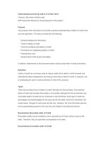
Diagnosis
Labor is a clinical diagnosis characterized by regular, painful uterine con-
tractions that increase in frequency and intensity are associated with progressive
cervical effacement or dilatation. More specifically, it is associated with a change
in the myometrial contractility pattern from irregular ‘‘contractures’’ (long-
lasting, low-frequency activity) to regular ‘‘contractions’’ (high-intensity, high-
frequency activity) [1]. It is important to note that uterine contractions alone
in the absence of cervical change are not sufficient to make the diagnosis. A
bloody mucous discharge (‘‘show’’) may precede the onset of labor by several
0889-8545/05/$ – see front matter D 2005 Elsevier Inc. All rights reserved.
doi:10.1016/j.ogc.2005.01.001 obgyn.theclinics.com
Dr. Liao is a Berlex-NICHD Scholar of the Reproductive Scientist Development Program
supported by NIH grant #5K12HD00849 and the Berlex Foundation.
* Corresponding author.
E-mail address: (E.R. Norwitz).
Obstet Gynecol Clin N Am
32 (2005) 145 – 164
days but is not a prerequisite for the diagnosis. In normal labor at term, there
seems to be a time -depend ent relationship between these elements: the
biochemical connective tissue changes in the cervix usually precede uterine
contractions, which, in turn, precede cervical dilatation. The fetal membranes
typically rupture during the course of labor. Occasionally, however, the mem-
branes may rupture with leakage of amniotic fluid before the onset of labor.
The onset of labor
Labor at term may best be regarded physiologically as an event initiated by
the removal of the inhibitory effects of pregnancy on the myometrium rather than
as an active process governed by uterine stimulants [1]. Fo r example, in vitro
studies have shown that quiescent myometrium obtained from term uteri and
placed in an isotonic solution contract vigorously and spontaneously without
added stimuli [2,3]. In vivo, however, it is likely that both mechanisms are im-
portant [4].
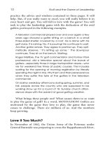
For the purposes of considering how uterine activity is regulated during the
latter part of pregnancy and labor, four distinct physiologic phases are de-
scribed (Fig. 1) [4]. During pregnancy, the uterus is maintained in a state of
functional quiescence (Phase 0) through the integrated action of one or more of
a series of inhibitors, including progesterone, prostacyclin, relaxin, nitric oxide,
parathyroid hormone-related peptide, calcitonin gene-related peptide, adrenome-
dullin, and vasoactive intestinal peptide. Before term, the uterus undergoes a
process of activation (Phase 1) and stimulation (Phase 2). Activation is brought
about in response to one or more uterotropins (such as estrogen) with increased
expression of a series of contraction-associated proteins (including myometrial
receptors for prostaglandins and oxytocin), functional activation of select ion
Fig. 1. Regulation of uterine activity during pregnancy and labor. (Adapted from Challis JRG,
Gibb W. Control of parturition. Prenat Neonat Med 1996;1:283; Taylor and Francis Ltd. http://
www.tandf.co.uk/journals; with permission.)
liao et al146
channels, and an increase in connexin-43 (a key component of gap junctions).
After activation, the ‘‘primed’’ uterus can be acted upon by uterotonins, such as
oxytocin and the stimulatory prostaglandins (E
2
and F
2a
), and stimulated to
contract. Because no single factor has been shown to be primarily responsible, it
is more accurate to refer to factors that promote rather than initiate the onset of
labor. Phase 3 events (uterine involution) occur after delivery and are mediated
primarily by oxytocin and possibly thrombin.
The endocrine control of labor
Considerable evidence suggests that the fetus is in control of the timing of
labor. Around the time of Hippocrates, it was believed that the reason the fetus
presented head first was so that it could kick its legs up against the fundus of
the uterus and propel itself through the birth canal. Although we have moved
away from this simple and mechanical concept of labor, the idea that the fetus
plays a central role in the initiation of labor remains and has been supported by
experimental data in other viviparous mammalian species [5,6]. Cross-breeding
experiments with horses and donkeys in the 1950s, for example, demonstrated a
gestational length intermediate between those of the parent species, which
suggested a critical role for the fetal genotype in determining the onset of labor
and the duration of gestation [7]. In domestic ruminants, such as sheep and cows,
the mechanism by which the fetus triggers labor at term has been elucidated
elegantly and involves glucocorticoid-mediated activation of a placental enzyme,
17a-hydroxylase/17,20-lyase, which catalyzes the conversion of progesterone to
estradiol-17b. This switch in the progesterone:estrogen ratio leads to uterine
prostaglandin production and labor [6,8,9]. Similarly, secretion of surfactant
protein-A from the lungs into the amniotic fluid at the end of pregnancy has been
shown to be important for the initiation of labor in a murine model [10].
Unfortunately, there is as yet insufficient evidence to suggest that any of these
factors are critical for the onset of labor in humans. For example, the human
placenta does not contain glucocorticoid-inducible 17a-hydroxylase/17,20-lyase
enzyme [7]. The slow progress in our understanding of the biochemical events
involved in the process of labor in the human reflects in large part the difficulty
in extrapolating from the endocrine control mechanisms in various animal models
to the paracrine/autocrine nature of parturition in women—processes that in
humans preclude direct investigation.
Although the precise signal varies, the final common pathway toward labor
seems to be activation of the fetal hypothalamic-pituitary-adrenal axis and is
probably common in all viviparous species. In humans, activation of the fetal
hypothalamic-pituitary-adrenal axis results in the release of C-19 steroid (dehy-
droepiandrostenedione), which serves as an essential precursor for placental
estrogen (estriol) production [11,12]. Administration of this estrogen precursor—
but not estrogen itself—is capable of inducing preterm labor in pregnant rhesus
monkeys [13]. Infusion of an aromatase inhibitor, 4-hydroxyandrostenedione,
normal labor: mechanism and duration 147
blocks this effect [14], which demonstrates that conversion of this precursor to
estrogen at the level of the fetoplacental unit is critical for the onset of labor.
Regardless of whether the signal for labor begins with the mother or the fetus,
the final common pathway for labor ends in the maternal tissues of the uterus
and is characterized by the development of regular phasic uterine contractions.
As in other smooth muscles, myometrial contractions are mediated throu gh the
ATP-dependent binding of myosin to actin. In contrast to vascular smooth
muscle, however, myometr ial cells have a sparse innervation that is further
reduced during pregnancy [15]. The regulation of the contractile mechanism of
the uterus is largely humoral and depends on intrinsic factors within myometrial
cells [4]. The transition of the uterus from a quiescent entity to a dynamic, con-
tractile one comes through the recruitment and communication of myometrial
cells through gap junctions (Fig. 2). An increase in gap junctions allows for
action potentials to be propagated between adjacent myometrial cells [15],
thereby establishing electrical synchr ony within the myometrium and allowing
for effective coordination of contractions [7,16]. A key component of gap
junctions, mRN A for connexin-43, has been shown to increase with the onset of
labor [17].
It is likely that a ‘‘parturition cascade’’ exists in humans (Fig. 3) that is
responsible, at term, for the removal of mechanisms that maintain uterine
quiescence and the recruitment of factors that act to promote uterine activity. In
such a model, pathways in the fetus, placenta, and mother are interconnected
at many levels and require sequential recruitment, which allows for a level of
redundancy that can, by design, prevent a single derangement from preventing
or prematurely activating the cascade [4,18]. A comprehensive analysis of the
individual paracrine/autocrine pathways implicated in the process of labor has
been reviewed in detailed elsewhere [1,18–20]. In brief, labor is a multifactorial
physiologic event that involves an inte grated set of changes within the maternal
tissues of the uterus (ie, myometrium, decidua, and uterine cervix) that occur
Fig. 2. Electron micrograph of gap junction between myometrial cells. (From Buhimschi CS, et al.
Forces of labor. Fetal and Maternal Medicine Review 2003;14(4):273–307; with permission.)
liao et al148
16-OH DHEAS from fetal adrenal
FETUS PLACENTA / FETAL MEMBRANES MOTHER
17α hydroxylase/
17,20-desmolase
dehydroandrostenedione
? -
ve
feedback
loop
Hypothalamus
Anterior
pituitary
from
fetal
zone of
adrenal
gland
from
definitive
adrenal
cortex
prepares
fetal organ
systems for
delivery
Fetal
liver
+
ve
feedback
loop
CRH
?
ACTH
DHEAS
CORTISOL
cortisol
cortisone
11β -HSD
progesterone
estrone
17β−estradiol
ESTRIOL
placental OT
PGs
placental CRH
membrane phospholipids
16-OH DHEAS to placenta / fetal membranes
cholesterol
5
-pregnenolone
17α -hydroxy-
pregnenolone
estrone
17β -estradiol
estriol
(16-OH estradiol)
16 - hydroxylase
17-oxido-
reductase
4 _
androstenedione
placental
sulfatase
cortisol
PLA
2
AA
PGE
2
(PGF
2α
)
PGEM
(PGFM)
15-OH
PGDH
COX-2
placental
vasodilation
LABOR
inhibited by
progestrone
acting through
glucocorticoid
receptors
+
+
-
+
+
+
+
+
+
Hypothalamus
Posterior
pituitary
OT
+
+
aromatase
uterus
decidual
PGF
2α
PG receptors
OT receptors
gap junctions
OT
utero-
placental
PGE
2
+
+
17α hydroxylase/
17,20-desmolase
3β -HSD
Adrenal
gland
+
SROM
Fig. 3. Proposed ‘‘parturition cascade’’ of paracrine/autocrine hormones responsible for uterine
contractions in spontaneous labor. (Modified from Norwitz ER, Robinson JN, Repke JT. The initiation
of parturition: a comparative analysis across species. Curr Probl Obstet Gynecol Fertil 1999;22:4;
with permission.)
normal labor: mechanism and duration 149
gradually over a period of days to weeks. Such changes include, but are not
limited to, an increase in prostaglandin synthesis and release within the uterus,
an increase in myometrial gap junction formation, and upregulation of
myometrial oxytocin receptors. When the myometrium and cervix have been
prepared appropriately, endocrine or paracrine/autocrine factors from the
fetoplacental unit bring about a switch in the pattern of myometrial activity
from contractures to contractions (uterine stimulation). The fetus may coordinate
this switch in myometrial activity throu gh its influence on placental steroid
hormone production, through mechanical distention (stretch) of the uterus, and
through secretion of neurohypophyseal hormones and other stimulators of
prostaglandin synthesis.
Mechanics of normal labor
Uterine contractions have two major functions: to dilate the cervix and to push
the fetus through the birth canal. The fetus is not merely the passive recipient of
these forces, however. The ability of the fetus to negotiate the pelvis successfully
depends on the complex interaction of three variables: the powe rs, the passenger,
and the passage.
Powers
Powers refer to the force generated by the uterine musculature during
contractions. It is generally believed that the more optimal the powers, the more
likely a successful outcome. No data exist to support this statement, however. The
features used to describe contractions are frequency, intensity, and duration. It
should be noted that the frequency of contractions does not necessarily reflect the
force of contraction.
As with other types of muscle contractions, action potentials must be
generated and propagated to yield effective contractions in a process known
as electromechanical coupling [15,17]. The generation of action potentials of
+12 to +25 mV from a normal resting potential of À65 to À 80 mV in pregnant
myometrial cells relies on the rapid shifts of ions through membrane ion channels
[21,22], the most important of which seem to be calcium and potassium channels
[23–26]. Autonomous pacemaker cells in the uterus that have a higher resting
potential than other muscle cells can initiate action potentials spontaneously [27].
Action potentials in the uterus occur in bursts, and the strength of contractions
relies on their frequency and duration. This, in turn, determines the number of
myometrial cells recruited for action. In this way, the electrical activity is
translated in mechanical forces exerted on the contents of the uterus in a
synchronous fashion (Fig. 4). The strength of contractions depends on the stage
of labor, with early labor contractions having a peak intensity from +25 to
+30 mm Hg, which increases to +60 to +65 mm Hg in the second stage of labor
liao et al150
[16]. Other variables that may influence the strength of the contractions include
parity, the condition of the cervix, exogenous oxytocin administration, and pain
medication (including epidural analge sia).
Uterine activity can be assessed qualitatively by simple observation of the
mother and palpat ion of the fundus of the uterus through the abdomen or by
external tocodynamometry. External tocodynamometry is noninvasive and
requires little expertise to measure and interpret. It measures uterine contraction
indirectly through changes in the shape of the abdominal wall and, as such,
cannot accurately determine basal intrauterine tone. Uterine activity also can be
measured quantitatively by direct measurement of intrauterine pressure via
internal manometry or pressure transducers. Placement of an intrauterine pressure
catheter allows for objective measurement of uterine activity. It is invasive, can
only be performed after ruptu re of the fetal membranes, and has been associated
with uterine injury (perforation) and an increased incidence of intrauterine
infection, however. Montevideo units (calculated by multiplying the average peak
strength of contractions in mm Hg by the number of contractions in 10 minutes)
is the most widely used calculation for measuring the strength of uterine
contractions [28]. This formula does not take into account uterine wall tension
[29] or the duration of contractions, howe ver [28]. For these reasons, some
investigators have p roposed using an integrated formula that uses the area under
Fig. 4. Uterine electrical activity recorded from two distinct sites S1 and S2, noninvasively from the
abdominal surface. During active labor, electrical bursts become synchronous with uterine pressure
elevations, as measured by an intrauterine pressure catheter. (From Buhimschi CS, et al. Forces of
labor. Fetal and Maternal Medicine Review 2003;14(4):273–307; with permission.)
normal labor: mechanism and duration 151
the contraction curve [30,31]. No evidence exists that one method is significantly
better than another [32].
Despite technologic improvements, the criteria for adequate uterine activity
during labor are unclear. Classically, the occurrence of three to five contractions
in 10 minutes has been used to define adequate labor and is seen in approximately
95% of women in spontaneous labor at term [4]. Using an internal pressure
monitor, adequate labor is generally defined as 200 to 250 Montevideo units [28].
In one retrospective series, 91% of women in spontaneous active labor achieved
contractile activity more than 200 Montevideo units, and 40% reached
300 Montevideo units [33]. It is important to understand, however, that although
achieving this level of uterine contractility makes a clinician more confident of
a successful labor, it is no guarantee of a successful vaginal delivery. Adequate
contractions in the face of other unfavorable factors (such as malposition) still
may lead to cephalopelvic disproportion and a need for cesarean delivery [4].
Passenger
The passenger is the fetus. Several fetal variables may infl uence the course of
normal labor and delivery.
Fetal size. Fetal macrosomia, which is defined by the American College of
Obstetricians and Gynecologists as an estimated fetal weight (not birth
weight) more than or equal to 4500 g [34], is associated with an increased
risk of cesarean delivery because of cephalopelvic disproportion. Assess-
ment of estimated fetal weight can be made either by clinical examination
(Leopold’s maneuvers) or ultrasound, although both approaches are subject
to significant errors (approximately 15%–20% at term).
Lie. Fetal lie refers to the long axis of the fetus relative to the longitudi-
nal axis of the uterus and can be longitudinal, transverse, or oblique. For a
single gestation, a vaginal delivery should be attempted only if the lie
is longitudinal.
Presentation. Fetal presentation refers to the fetal part that directly overlies
the pelvic inlet. With a longitudinal lie, presentation is usually cephalic
(vertex), breech, or shoulder. When more than one fetal part presents at
the pelvic inlet, the term ‘‘compound presentation’’ is used. Rarely, the
umbilical cord may present at the inlet, which is known as a funic pre-
sentation. Approximately 5% of singleton pregnancies at term have a
malpresentation in labor.
Attitude. Fetal attitude describes the degree of flexion or extension of the
fetal head in relation to the fetal spine. Adequate flexion (chin to ch est) is
necessary to achieve the smallest possible presenting diameter in a cephalic
presentation. Deflexion in the early stages of labor may be corrected by the
architecture of the pelvic floor and uterine contractions.
Position. Fetal position refers to the relationship of a nominated site of the
fetal presenting part to a denominating location on the maternal pelvis
liao et al152
(Fig. 5). For example, in a ceph alic presentation, the fetal site used for
reference is typically the occiput (eg, right occiput anterior). In a breech
presentation, the sacrum is used as the designated fetal site (eg, right sacrum
anterior). Any fetal position that is not right occipu t, occiput anterior, or left
occiput anterior is referred to as a malposition.
Station. Fetal station refers to how far the leading bony edge of the
presenting part of the fetus has descended into the maternal pelvis relative to
the ischial spines. It is typically assessed clinically by bimanual examina-
tion. An older arbitrary system (À3 to +3, with 0 being at the level of the
ischial spines) has been replaced with a more recent classification designed
to quantify the distance from the ischial spines (À3 to +5 cm).
Number of fetuses.
Presence of fetal anomalies. Anomalies may obstruct delivery (eg, sacro-
coccygeal teratoma).
Fig. 5. Fetal presentations and positions in labor. LOA, left occiput anterior; LOT, left occi-
put transverse; LOP, left occiput posterior; OA, occiput anterior; OP, occiput posterior; ROA,
right occiput anterior; ROT, right occiput transverse; ROP, right occiput posterior. (Adapted from
Norwitz ER, Robinson J, Repke JT. The initiation and management of labor. In: Seifer DB, Samuels P,
Kniss DA, editors. The physiologic basis of gynecology and obstetrics. Baltimore: Lippincott Wil-
liams & Wilkins; 2000. p. 422; with permission.)
normal labor: mechanism and duration 153
Passage
The passage through which the fetus must pass during normal labor and
delivery consists of the bony pelvis and the soft tissues of the birth canal
(ie, cervix, pelvic floor musculature), both of which offer varying degrees of
resistance to fetal expulsion.
The bony pelvis is comprised of the greater and lesser pelvis and is divided
by the pelvic brim. Its anatomic boundaries are made up of the sacral promon-
tory, the anterior ala of the sacrum, the arcuate line of the ilium, the pectineal line
of the pubis, and the symphysis pubis. The true pelvis can be divided into planes
that must be navigated by the fetus during labor, including the pelvi c inlet,
midcavity, and outlet. The female pelvis is classically described as having one
of four shapes: gynecoid, anthropoid, android, and platypoid. This classification
was designed to separate the more favorable configurations for successful vaginal
delivery (ie, gynecoid, anthropoid) from the less favorable ones [35]. In practice,
however, the shape of the female pelvis reflects a continuum rather than strict
adherence to one of these four categories, and the classification has not been
shown to predict consistently the success of vaginal delivery. For these reasons,
this classification is of littl e clinical use. The bony pelvis is assessed by
pelvimetry (ie, quantitative measurement of pelvic capacity), which can be
performed clinically [4] or via imaging studies (radiography, CT, MRI) [36–39].
Imaging techniques have defined average and critical limit values for the various
parameters of the bony pelvis [37,38]. Such measurements are of limited clinical
value, however, because they are not able to predict consistently women at risk
for cephalopelvic disproportion [40]. Radiographic and CT studies of unclear
clinical use are generally avoided in pregnancy because of the theoretic risks to
the fetus of ionizing radiation [41]. For these reasons, pelvimetry has been
replaced, in large part, by clinical trial of the pelvis (a ‘‘trial of labor’’).
The soft tissues of the birth canal (ie, cervix, pelvic floor musculature) also
provide resistance to the progress of labor and, as such, are important variables
that allow for successful vaginal delivery. For several weeks before delivery, the
connective tissues of the cervix undergo biochemical changes in preparation for
labor, including alterations in water, collagen, elastin, and proteoglycan
composition. These changes result in changes to the physical properties of
elasticity, plasticity, and tensile strength. Our understanding of the factors
responsible for cervical effacement and dila tion in labor remains unclear. Some
investigators have suggested that the primary factors leading to cervical dilatation
are the traction forces of the myometrial contractions, whereas others argue that
the pressure of the fetal head is the most important determinant. The widely held
belief that amniotomy (artificial rupture of the forebag) increases the pressure of
fetal head on the cervix has been disputed by recent studies that have measured
pressure objectively between the fetal head and the cervix before and after
amniotomy [42]. Taken together, these data suggest that both factors may be
important [16]. Other facto rs also may be involved. For example, studies in
animals [43–46] and humans [43–45,47] have shown that nitric oxide may be
liao et al154
an important mediator of uterine quiescence and cervical compe tence before
labor, whereas this same agent acting through the cyclic guanosine mono-
phosphate signal transduction pathway in labor may promote uterine contractility
and cervical effacement.
In the second stage of labor, the musculature of the pelvic floor is the main
source of soft-tissue resistance to fetal descent and delivery. These muscles are
believed to play an important role in facilitating rotation and flexion of the fetal
head as it passes through the birth canal. For example, internal rotation is known
to occur when the fetal head descends to the level of the pelvic floor, resulting
in 95% of vertex infants delivering in the most favorable (occiput anterior)
position [48]. Interference with this process by, for example, relaxation of the
pelvic floor musculature with the use of early epidural analgesia may be asso-
ciated with an increased likelihood of fetal malposition [49].
Stages and duration of normal labor
Although labor is a continuous process, it traditionally has been divided into
three stages to facilitate study and assist in clinical management.
First stage
The first stage refers to the interval between the onset of labor and full cervical
dilatation. It has been subdivided into three phases [50–53] according to the rates
of cervical dilatation (Fig. 6):
1. Latent phase. The latent phase refers to the period between the onset of
labor and the point at which a change in the slope of the rate of cervical
dilatation is noted [50–52]. It is characterized by slow cervical dilatation
and is of variable duration.
2. Active phase. This phase is associated with a faster rate of cervical
dilatation and usually begins at approximately 2 to 4 cm dilatation [50–53].
The active phase is broken down further into an acceleration phase, a phase
of maximum slope, and a deceleration phase, but these subdivisions are
rarely used currently.
3. Descent phase. Descent of the fetus usually coincides with the second stage
of labor. A distinct descent phase was included in the original descriptions
[50–52]. The existence of a separate descent phase is not universally
accepted, however.
The characteristics of the labor curve do not differ among ethnic or racial
groups [51,52,54], but there are significant differences between the labor curves
of nulliparous and multiparous women [51,52,54]. In classic studies, Friedman
[50–52] determined the average duration for each stage of labor in these two
groups of parturients and calculated the maximum duration of each stage, de-
normal labor: mechanism and duration 155
fined as two standard deviations from the mean (Table 1). For example, the
minimum rate of cervical dilatation of 1.2 cm/h for a nulliparous patient
represents two standard deviations below the mean rate of cervical dilatation
for nulliparas, not the average rate of dilatation among these women (which is
3 cm/h). By comparing a parturient’s rate of cervical dilatation with the normal
profile described by Friedman, it is possible to detect abnormal labor patterns and
identify pregnancies at risk for adverse events. This task can be facilitated by use
of a partogram [55], which is a graphic representation of the labor curve against
which a patient’s progress in labor is plotted. In this way, abnormal labor patterns
can be identified easily and appropriate measures taken.
Second stage
The second stage of labor refers to the interval between full cervical
dilatation (10 cm) and delivery of the infant. It is characterized by descent of
the presenting part through the maternal pelvis a nd culminates with expulsion of
the fetus. Indications that the second stage has started are an increase in bloody
show, maternal desire to bear down with each contraction, a feeling of pressure
on the rectum accompanied by the desire to defecate, and onset of nausea and
vomiting. The mother typically assumes a more active role in the second stage
Fig. 6. Cervical dilation curve for nulliparous labor. (Data from Friedman EA. Labor: clinical
evaluation and management. 2nd edition. Norwalk (CT): Appleton-Century-Crofts; 1978.)
liao et al156
than the first stage because she pushes or bears down to aid descent of the fetus .
In the presence of a reassuring fetal heart rate, it is desirable for a nulliparous
patient without regional anesthesia to push for as long as 2 hours (3 hours with
regional anesthesia) before resorting to interventions to facilitate delivery
[56]. For a multiparous woman, the recommendation is 1 hour and 2 hours,
respectively [56]. If there is continued progress and no evidence of mater-
nal or fetal compr omise, however, longer times are not associated with in-
creased morbidity.
Third stage
The third stage of labor refers to the time from delivery of the baby to
separation and expulsion of the placenta and fetal membranes. The three classic
signs of placental separation are (1) lengthening of the umbilical cord, (2) a gush
of blood from the vagina, which signifies separation of the placenta from the
uterine wall, and (3) a change in the shape of the uterine fundus from discoid to
globular, with elevation of the fundal height. The major complication associated
with this period is hemorrhage, which remains an important cause of maternal
morbidity and mortality. Average blood loss at delivery is generally estimated to
be 500 mL. Obstetric care providers should be alert to excessive blood loss
and should be prepared to intervene as required. There are no uniform criteria for
the normal length of the third stage of labor. Retention of the placenta for longer
than 30 minutes at term is a commonly used endpoint for intervention even in the
Table 1
Progression of spontaneous labor at term
Parameter Mean 5
th
percentile
Nulliparas
Total duration of labor (hours) 10.1 h 25.8 h
Stage of labor
Duration of the first stage (hours) 9.7 h 24.7 h
Duration of the second stage (minutes) 33.0 min 117.5 min
Duration of latent phase (hours) 6.4 h 20.6 h
Rate of cervical dilatation during active phase (cm/h) 3.0 cm/h 1.2 cm/h
Duration of the third stage (minutes) 5.0 min 30.0 min
Multiparas
Total duration of labor (hours) 6.2 h 19.5 h
Stage of labor
Duration of the first stage (hours) 8.0 h 18.8 h
Duration of the second stage (minutes) 8.5 min 46.5 min
Duration of latent phase (hours) 4.8 h 13.6 h
Rate of cervical dilatation during active phase (cm/h) 5.7 cm/h 1.5 cm/h
Duration of the third stage (minutes) 5.0 min 30.0 min
Data from Norwitz ER, Robinson JN, Repke JT. Labor and delivery. In: Gabbe SG, Niebyl JR,
Simpson JL, editors. Obstetrics: normal and problem pregnancies. 4th edition. New York: Churchill-
Livingstone; 2001. p. 353–400; with data from Friedman EA. Labor: clinical evaluation and
management. 2nd edition. Norwalk (CT): Appleton-Century-Crofts; 1978.
normal labor: mechanism and duration 157
Fig. 7. The cardinal movements of labor. (From Norwitz ER, Robinson JN, Repke JT. Labor and
delivery. In: Gabbe SG, Niebyl JR, Simpson JL, editors. Obstetrics: normal and problem pregnancies.
4th edition. New York: Churchill-Livingstone; 2001. p. 353–400; with permission.)
liao et al158
absence of active hemorrhage. The World Health Organization defines a retained
placenta as one that has not been expelled by 60 minutes after delivery [57].
Cardinal movements in labor
The cardinal movements of labor refer to changes in the position of the fetal
head during its passage through the birth canal. Because of asymmetry in the
shape of the fetal head and the maternal bony pelvis, such rotations are required
if the fetus is to negotiate the birth canal successfully. These seven discrete
movements are engagement, descent, flexion, internal rotation, extension,
external rotation or restitution, and expulsion (Fig. 7).
Engagement. Engagement refers to the passage of the widest diameter of
the fetal presen ting part to a level below the plane of the pelvic inlet. In
the cephalic presentation with a well-flexed head, the largest transverse
diameter of the fetal head is the biparietal diameter (9.5 cm). In the breech,
the widest diameter is the bitrochanteric diameter. Engagement can be
confirmed clinically by palpation of the presenting part abdominally (when
only two fifths of the head can be palpated abdominally) or vaginally (with
confirmation of station at or below the ischial spines). Engagement is an
important clinical milestone in the progress of labor, because it demonstrates
that the bony pelvis is adequate to allow passage of the fetal head. For
multiparous women, engagement may occur at any time after 36 weeks. In
primipara, however, failure of engagement to take place by 36 weeks is
often an early sign of cephalopelvic disproportion [4].
Descent. Descent refers to the downward passage of the presenting part
through the pelvis. Descent of the fetus is not a steady, continuous process.
The greatest rate of descent occurs during the deceleration phase of the first
stage and during the second stage of labor.
Flexion. Flexion of the fetal head occurs passively as the head descends
because of the shape of the bony pelvis and the resistance of the soft tissues
of the pelvic floor. Although flexion of the fetal head onto the chest is
present to some degree in most fetuses antepartum, complete flexion usually
only occurs during the course of labor. With the head completely flexed, the
fetus presents the smallest diameter of its head (suboc cipito-bregmatic
diameter), which allows optimal passage through the pelvis.
Internal rotation. Internal rotation is the rotation of the presenting part from
its original position (usually transverse with regard to the birth canal) to the
anteroposterior position as it passes through the pelvis. This change
typically results in the fetal occiput rotating toward the symphysis pubis
as it descends, which leads to the widest axis of the fetal head lining up with
the widest axis of the pelvic passage. The curvature of the maternal sacrum
causes the fetal head to descend in an asynclitic fashion at first, but it
typically corrects. As with flexion, internal rotation is a passive movement
normal labor: mechanism and duration 159
that results from the shape of the pelvis and the resistance of the pelvic
floor musculature
Extension. Extension occurs once the fetus has descended to the level of
the introitus. This descent brings the base of the occipu t into contact with the
inferior margin of the symphysis pubis. At this point, the birth canal curves
upward. The fetal head is delivered by extension and rotates around the
symphysis pubis. The forces responsible for this motion are the downward
force exerted on the fetus by uterine contractions and maternal expul-
sive efforts along with the upward forces exerted by the muscles of the
pelvic floor.
External rotation (restitution). After the fetal head deflexes (extends), it
rotates to the correct anatomic position in relation to the fetal torso; left or
right rotation depends on the orientation of the fetus. This is again a passive
movement that results from a release of the forces exerted on the fetal head
by the maternal bony pelvis and its musculature, and it is mediated by the
basal tone of the fetal musculature.
Expulsion. Expulsion refers to delivery of the body of the fetus. After
delivery of the head and exter nal rotation, further descent brings the anterior
shoulder to the level of the symphysis pubis. The anterior shoulder rotates
under the symphysis pubis, after which the rest of the body usually delivers
without difficulty.
Maternal pushing in labor
The cardinal movements are largely the result of uterine contractions and the
passive action of the pelvic musculature and soft tissues of the descending fetal
head. Obstetric practice in the United States often dictates that the parturient
begin to bear down (push) in concert with each contraction when the cervix
attains full dilation (10 cm), even if she does not feel the urgency to do so.
Despite the widespread implementation of this practice, it is not clear whether
it facilitates or speeds delivery [58,59]. Women with spinal cord injuries and
quadriplegia who are unable to push voluntarily are able to deliver vaginally
without difficulty. Recent studies suggest that most of the increased intrauterine
pressure in the second stage of labor results from uterine contractions, with only a
small contribution from maternal expulsive efforts even under optimal conditions
[30]. Several factors may influence maternal pushing performance, including
body mass index [30], fetal weight [30] , myometrial thickness [30], maternal
position [60], and oxytocin augmentation [61] (but not parity [61]).
The timing of maternal pushing is also debated. Several recent randomized
prospective studies have questioned the practice of encouraging pushing at the
beginning of the second stage and have suggested that pushing be delayed for
1 to 2 hours to allow the presenting fetal part to descend [62–64]. As an example,
a large (n = 1862), randomized, multicenter study documented that delayed
pushing for 1 hour was an effective means of reducing ‘‘difficult deliveries’’ in
liao et al160
nulliparous women (relative risk (RR), 0.79; 95% confidence interval (CI),
0.66–0.95) [63]. The greatest effect was on midpelvic operative vagina l deliver-
ies (RR, 0.72; 95% CI, 0.55–0.93). Delayed pushing predictably increased
the duration of the second stage (by 54 minutes) and resulted in lower umbilical
cord blood pH, but no difference was detected in overall neonatal morbidity.
Summary
Labor is a physiologic and continuous process. The factors responsible for
the onset and maintenance of normal labor at term are poorly understood and
continue to be under active investigation. Although data exist to describe the
average duration of labor, there is also a great deal of biologic variability. An
improved understanding of the causes and mechanisms of labor will improve
the ability of clinicians to distinguish normal from abnormal labor and to inter-
vene in a timely and effective fashion to ensure a favorable outcome while
moving toward a more individualized approach to each woman’s labor.
References
[1] Norwitz ER, Robinson JN, Challis JR. The control of labor. N Engl J Med 1999;341(9):
660 – 6.
[2] Lopez Bernal A, Rivera J, Europe-Finner GN, et al. Parturition: activation of stimulatory
pathways or loss of uterine quiescence? Adv Exp Med Biol 1995;395:435 – 51.
[3] Garrioch DB. The effect of indomethacin on spontaneous activity in the isolated human
myometrium and on the response to oxytocin and prostaglandin. Br J Obstet Gynaecol 1978;
85(1):47 –52.
[4] Norwitz ER, Robinson JN, Repke JT. Labor and delivery. In: Gabbe SG, Niebyl JR, Simpson JL,
et al, editors. Obstetrics: normal and problem pregnancies. 4
th
edition. New York7 Churchill-
Livingstone; 2001. p. 353 – 400.
[5] Nathanielsz PW, Giussani DA, Wu WX. Stimulation of the switch in myometrial activity
from contractures to contractions in the pregnant sheep and nonhuman primate. Equine Vet J
Suppl 1997;24:83– 8.
[6] Liggins GC, Fairclough RJ, Grieves SA, et al. The mechanism of initiation of parturition in the
ewe. Recent Prog Horm Res 1973;29:111–59.
[7] Liggins GC. Initiation of labour. Biol Neonate 1989;55(6):366 – 75.
[8] Thorburn GD, Hollingworth SA, Hooper SB. The trigger for parturition in sheep: fetal
hypothalamus or placenta? J Dev Physiol 1991;15(2):71 – 9.
[9] Matthews SG, Challis JRG. Regulation of the hypothalamo-pituitary-adreno-cortical axis in
fetal sheep. Trends Endocrinol Metab 1996;7(7):239 –46.
[10] Condon JC, Jeyasuria P, Faust JM, et al. Surfactant protein secreted by the maturing mouse
fetal lung acts as a hormone that signals the initiation of parturition. Proc Natl Acad Sci
U S A 2004;101(14):4978–83.
[11] Challis JR. Characteristics of parturition. In: Creasy RKRR, editor. Maternal-fetal medicine:
principles and practice. Philadelphia7 WB Saunders Co.; 1994. p. 482.
[12] Madden JD, Gant NF, MacDonald PC. Study of the kinetics of conversion of maternal plasma
dehydroisoandrosterone sulfate to 16 alpha-hydroxydehydroisoandrosterone sulfate, estradiol,
and estriol. Am J Obstet Gynecol 1978;132(4):392 – 5.
normal labor: mechanism and duration 161
[13] Mecenas CA, Giussani DA, Owiny JR, et al. Production of premature delivery in pregnant
rhesus monkeys by androstenedione infusion. Nat Med 1996;2(4):443–8.
[14] Nathanielsz PW, Jenkins SL, Tame JD, et al. Local paracrine effects of estradiol are central to
parturition in the rhesus monkey. Nat Med 1998;4(4):456–9.
[15] Wolfs GM, van Leeuwen M. Electromyographic observations on the human uterus during labour.
Acta Obstet Gynecol Scand Suppl 1979;90:1 – 61.
[16] Buhimschi C, Buhimschi IA, Malinow AM, et al. The forces of labour. Fetal and Maternal
Medicine Review 2003;14(4):273–307.
[17] Garfield RE, Sims S, Daniel EE. Gap junctions: their presence and necessity in myometrium
during parturition. Science 1977;198(4320):958–60.
[18] Myers DA, Nathanielsz PW. Biologic basis of term and preterm labor. Clin Perinatol
1993;20(1):9–28.
[19] Honnebier MB, Nathanielsz PW. Primate parturition and the role of the maternal circadian
system. Eur J Obstet Gynecol Reprod Biol 1994;55(3):193 – 203.
[20] Nathanielsz PW. Comparative studies on the initiation of labor. Eur J Obstet Gynecol Reprod
Biol 1998;78(2):127 –32.
[21] Kumar D, Barnes AC. Studies in human myometrium during pregnancy. II. Resting membrane
potential and comparative electrolyte levels. Am J Obstet Gynecol 1961;82:736 – 41.
[22] Kuriyama H, Csapo A. A study of the parturient uterus with the microelectrode technique.
Endocrinology 1961;68:1010–25.
[23] Wray S, Jones K, Kupittayanant S, et al. Calcium signaling and uterine contractility. J Soc
Gynecol Investig 2003;10(5):252–64.
[24] Carvajal JA, Thompson LP, Weiner CP. Chorion-induced myometrial relaxation is mediated
by large-conductance Ca2 + -activated K + channel opening in the guinea pig. Am J Obstet
Gynecol 2003;188(1):84–91.
[25] Woodcock NA, Taylor CW, Thornton S. Effect of an oxytocin receptor antagonist and rho
kinase inhibitor on the [Ca + +]i sensitivity of human myometrium. Am J Obstet Gynecol
2004;190(1):222–8.
[26] Papandreou L, Chasiotis G, Seferiadis K, et al. Calcium levels during the initiation of labor.
Eur J Obstet Gynecol Reprod Biol 2004;115(1):17–22.
[27] Kao CY. Long-term observations of spontaneous electrical activity of the uterine smooth muscle.
Am J Physiol 1959;196(2):343 – 50.
[28] Caldeyro-Barcia RPJ. Physiology of the uterine contraction. Clin Obstet Gynecol 1960;3:
386 – 408.
[29] Csapo A. Force of labour. In: Iffy LKH, editor. Principles and practice of obstetrics and
perinatology. New York7 Wiley; 1981. p. 761–99.
[30] Buhimschi CS, Buhimschi IA, Malinow AM, et al. Pushing in labor: performance and not
endurance. Am J Obstet Gynecol 2002;186(6):1339–44.
[31] Buhimschi CS, Buhimschi IA, Malinow AM, et al. Effects of sublingual nitroglycerin on
human uterine contractility during the active phase of labor. Am J Obstet Gynecol 2002;
187(1):235 – 8.
[32] Chua S, Kurup A, Arulkumaran S, et al. Augmentation of labor: does internal tocography
result in better obstetric outcome than external tocography? Obstet Gynecol 1990;76(2):
164 – 7.
[33] Hauth JC, Hankins GD, Gilstrap III LC, et al. Uterine contraction pressures with oxytocin
induction/augmentation. Obstet Gynecol 1986;68(3):305–9.
[34] American College of Obstetricians and Gynecologists. Fetal macrosomia. Practice bulletin
No. 22. Washington, DC7 American College of Obstetricians and Gynecologists; 2000.
[35] Caldwell WE, Moloy HC. Anatomical variations in the female pelvis and their effect in labor
with a suggested classification. Am J Obstet Gynecol 1933;26:479 – 505.
[36] Raman S, Samuel D, Suresh K. A comparative study of X-ray pelvimetry and CT pelvimetry.
Aust N Z J Obstet Gynaecol 1991;31(3):217 – 20.
[37] O’Brien WF, Cefalo RC. Evaluation of X-ray pelvimetry and abnormal labor. Clin Obstet
Gynecol 1982;25(1):157–64.
liao et al162
[38] Joyce DN, Giwa-Osagie F, Stevenson GW. Role of pelvimetry in active management of labour.
BMJ 1975;4(5995):505–7.
[39] van Loon AJ, Mantingh A, Serlier EK, et al. Randomised controlled trial of magnetic-resonance
pelvimetry in breech presentation at term. Lancet 1997;350(9094):1799–804.
[40] Pattinson RC. Pelvimetry for fetal cephalic presentations at term. Cochrane Database Syst Rev
2000;2:CD000161.
[41] Morris CW, Heggie JC, Acton CM. Computed tomography pelvimetry: accuracy and radiation
dose compared with conventional pelvimetry. Australas Radiol 1993;37(2):186–91.
[42] Manabe Y, Sagawa N. Changes in the mechanical forces of cervical distention before and after
rupture of the membranes. Am J Obstet Gynecol 1983;147(6):667 – 71.
[43] Vaisanen-Tommiska M, Nuutila M, Ylikorkala O. Cervical nitric oxide release in women
postterm. Obstet Gynecol 2004;103(4):657–62.
[44] Okawa T, Vedernikov YP, Saade GR, et al. Effect of nitric oxide on contractions of uterine
and cervical tissues from pregnant rats. Gynecol Endocrinol 2004;18(4):186 – 93.
[45] Chen DC, Ku CH, Huang YC, et al. Urinary nitric oxide metabolite changes in spontaneous
and induced onset active labor. Acta Obstet Gynecol Scand 2004;83(7):641–6.
[46] Buhimschi IA, Yallampalli C, Buhimschi CS, et al. Distinct regulation of nitric oxide and
cyclic guanosine monophosphate production by steroid hormones in the rat uterus. Mol Hum
Reprod 2000;6(5):404–14.
[47] Buhimschi I, Yallampalli C, Dong YL, et al. Involvement of a nitric oxide-cyclic guanosine
monophosphate pathway in control of human uterine contractility during pregnancy. Am J
Obstet Gynecol 1995;172(5):1577–84.
[48] Cunningham F, Gant NF, Leveno KJ, et al. Williams obstetrics. 21
st
edition. New York7
McGraw-Hill; 2001.
[49] Ponkey SE, Cohen AP, Heffner LJ, et al. Persistent fetal occiput posterior position: obstetric
outcomes. Obstet Gynecol 2003;101(5 Pt 1):915 –20.
[50] Friedman E. The graphic analysis of labor. Am J Obstet Gynecol 1954;68(6):1568 – 75.
[51] Friedman EA. Primigravid labor: a graphicostatistical analysis. Obstet Gynecol 1955;6(6):
567 – 89.
[52] Friedman EA. Labor in multiparas: a graphicostatistical analysis. Obstet Gynecol 1956;8(6):
691 – 703.
[53] Peisner DB, Rosen MG. Transition from latent to active labor. Obstet Gynecol 1986;68(4):
448 – 51.
[54] Duignan NM, Studd JW, Hughes AO. Characteristics of normal labour in different racial groups.
Br J Obstet Gynaecol 1975;82(8):593 – 601.
[55] Studd J. Partograms and nomograms of cervical dilatation in management of primigravid labour.
BMJ 1973;4(5890):451–5.
[56] American College of Obstetricians and Gynecologists. Dystocia and augmentation of labor.
Practice bulletin No. 49. Washington, DC7 American College of Obstetricians and Gynecolo-
gists; 2003.
[57] The prevention and management of postpartum haemorrhage: report of a technical working group.
Geneva7 World Health Organization/Maternal and Child Health and Family Planning; 1990.
[58] Mayberry LJ, Hammer R, Kelly C, et al. Use of delayed pushing with epidural anesthesia:
findings from a randomized, controlled trial. J Perinatol 1999;19(1):26 – 30.
[59] Thomson AM. Pushing techniques in the second stage of labour. J Adv Nurs 1993;18(2):
171 – 7.
[60] Buhimschi CS, Buhimschi IA, Malinow A, et al. Use of McRoberts’ position during delivery
and increase in pushing efficiency. Lancet 2001;358(9280):470–1.
[61] Gurewitsch ED, Diament P, Fong J, et al. The labor curve of the grand multipara: does prog-
ress of labor continue to improve with additional childbearing? Am J Obstet Gynecol 2002;
186(6):1331–8.
[62] Vause S, Congdon HM, Thornton JG. Immediate and delayed pushing in the second stage
of labour for nulliparous women with epidural analgesia: a randomised controlled trial. Br J
Obstet Gynaecol 1998;105(2):186–8.
normal labor: mechanism and duration 163
[63] Fraser WD, Marcoux S, Krauss I, et al. Multicenter, randomized, controlled trial of delayed
pushing for nulliparous women in the second stage of labor with continuous epidural analgesia:
the PEOPLE (Pushing Early or Pushing Late with Epidural) Study Group. Am J Obstet Gynecol
2000;182(5):1165–72.
[64] Petrou S, Coyle D, Fraser WD. Cost-effectiveness of a delayed pushing policy for patients
with epidural anesthesia: the PEOPLE (Pushing Early or Pushing Late with Epidural) Study
Group. Am J Obstet Gynecol 2000;182(5):1158 – 64.
liao et al164
Labor with Abnormal Presentation and Position
Michael L. Stitely, MD
a,
*
, Robert B. Gherman, MD
b
a
Department of Obstetrics and Gynecology, West Virginia University School of Medicine,
1 Medical Center Drive, PO Box 9186, Morgantown, WV 26506-9186, USA
b
Department of Obstetrics and Gynecology, Division of Maternal-Fetal Medicine,
Washington Adventist Hospital, Takoma Park, MD, USA
The fetus delivers in the cephalic presentation in approximately 97% of
deliveries. Abnormal presentation—usually the breech presentation—complicates
the remaining 3% of deliveries.
Breech presentation
There is considerable controversy concerning the optimal route of delivery for
a fetus that presents in the breech position. A full discussion of this issue
is beyond the scope of this article. Diagnosis and management options are
discussed, however.
Etiology
The prevalence of breech presentation depends on gestational age. Scheer
and Nubar [1] described the fetal presentation sonographically at various ges-
tational ages. They found that at 21 to 24 weeks’ gestation, 33.3% of fetuses were
in the breech position. By contrast, only 6.7% of fetuses were in the breech
position at 37 to 40 weeks’ gestation. Other risk factors for breech presentation
include multiparity, previous breech delivery, polyhydramnios, fetal anomalies,
and uterine anomalies.
0889-8545/05/$ – see front matter D 2005 Elsevier Inc. All rights reserved.
doi:10.1016/j.ogc.2004.12.005 obgyn.theclinics.com
* Corresponding author.
E-mail address: (M.L. Stitely).
Obstet Gynecol Clin N Am
32 (2005) 165 – 179
Diagnosis
The diagnosis of breech presentation can be made reliably using a com-
bination of abdominal palpation and vaginal examination. The first Leopold
maneuver detects the fetal head at the fundal aspect of the uterus. Vaginal
examination and palpation reveal either the ischial tuberosities and sacrum or—in
footling breech presentations—the lower extremities. When the cervix is dilated
and the membranes are ruptured, the fetal anus may be identified on examination.
Ultrasound can be used to confirm the presen tation, classify the type of
breech presentation, assess the estimated fetal weight, and identify gross fetal
anomalies. Complete breech presentations have both hips flexed with one or both
knees flexed. Incomplete breech presentations have one or both hips extended.
Frank breech presentations have both hips flexed and both knees extended.
Management
Patients should be offered external cephalic version when breech presentation
is diagnosed in late pregnancy. The Cochrane Database of Systematic Reviews
addressed the issue of external cephalic version of breech presentation at term [2].
Six randomized trials were included in the review. External cephalic version
at term significantly reduced the incidence of noncephalic births (Relative risk
0.42, 95% confidence interval 0.35–0.5) and cesarean delivery (Relative risk
0.52, 95% confidence interval 0.39–0.71) without a signifi cant effect on peri-
natal mortality.
Technique
External cephalic version can be performed with either one or two operators.
The procedure should be performed in a setting in which the fetus can be
monitored and an immediate cesarean delivery can be performed if necessary.
Contraindications include third-trimester bleeding, oligohydramnios, ruptured
membranes, severe fetal anomalies, and the usual contraindications to vaginal
birth (ie, placenta previa, prior classical cesarean delivery, vasa previa).
Results of a reactive non–stress test should be obtained before the procedure,
and the patient should undergo counseling for informed consent before the
procedure. Ultrasound should be performed to confirm the breech presentation
and assess the amniotic fluid volume. Administration of beta-mimetic tocolytics
may be beneficial [3].
The patient should be tilted laterally to prevent supine hypotension. First
the fetal breech is elevated out of the maternal pelvis. The version is then per-
formed by attempting to turn the fetus into a forward roll. If attempts at inducing
a forward roll motion are unsuccessful, the opposite direction may be attempted.
The amount of force exerted is gauged by the patient’s pain tolerance. The use
of spinal or epidural analgesia is controversial. Some trials have shown benefit
[4,5], whereas others have not [6]. After the version attempt, the fetus should be
stitely & gherman166