Tài liệu AACN Essentials of Critical Care Nursing—Pocket Handbook doc
Bạn đang xem bản rút gọn của tài liệu. Xem và tải ngay bản đầy đủ của tài liệu tại đây (2.63 MB, 208 trang )
AACN Essentials of Critical Care
Nursing—Pocket Handbook
Notice
Medicine is an ever-changing science. As new research and clinical experience broaden our knowledge, changes in treatment and drug therapy are
required. The editor and publisher of this work have checked with sources believed to be reliable in their efforts to provide information that is
complete and generally in accord with the standards accepted at the time of publication. However, in view of the possibility of human error or
changes in medical sciences, neither the editors nor the publisher nor any other party who has been involved in the preparation or publication of
this work warrants that the information contained herein is in every respect accurate or complete, and they disclaim all responsibility for any errors
or omissions or for the results obtained from use of the information contained in this work. Readers are encouraged to confirm the information
contained herein with other sources. For example and in particular, readers are advised to check the product information sheet included in the
package of each drug they plan to administer to be certain that the information contained in this work is accurate and that changes have not been
made in the recommended dose or in the contraindications for administration. This recommendation is of particular importance in connection with
new or infrequently used drugs.
AACN Essentials of Critical Care Nursing
Pocket Handbook
Marianne Chulay, RN, PhD, FAAN
Consultant, Critical Care Nursing and Clinical Research
Gainesville, Florida
Suzanne M. Burns RN, MSN, RRT, ACNP, CCRN, FAAN, FCCM, FAANP
Professor of Nursing, Acute and Specialty Care
School of Nursing
Advanced Practice Nurse Level 2, Director Professional Nursing Staff Organization Research Program
University of Virginia Health System
Charlottesville, Virginia
New York Chicago San Francisco Lisbon London Madrid Mexico City Milan
New Delhi San Juan Seoul Singapore Sydney Toronto
Second Edition
Copyright © 2010, 2006 by The McGraw-Hill Companies, Inc. All rights reserved. Except as permitted under the United States Copyright Act of 1976,
no part of this publication may be reproduced or distributed in any form or by any means, or stored in a database or retrieval system, without the prior
written permission of the publisher.
ISBN: 978-0-07-170273-7
MHID: 0-07-170273-3
The material in this eBook also appears in the print version of this title: ISBN: 978-0-07-166408-0, MHID: 0-07-166408-4.
All trademarks are trademarks of their respective owners. Rather than put a trademark symbol after every occurrence of a trademarked name, we use
names in an editorial fashion only, and to the benefi t of the trademark owner, with no intention of infringement of the trademark. Where such designa-
tions appear in this book, they have been printed with initial caps.
McGraw-Hill eBooks are available at special quantity discounts to use as premiums and sales promotions, or for use in corporate training programs.
To contact a representative please e-mail us at
TERMS OF USE
This is a copyrighted work and The McGraw-Hill Companies, Inc. (“McGrawHill”) and its licensors reserve all rights in and to the work. Use of this
work is subject to these terms. Except as permitted under the Copyright Act of 1976 and the right to store and retrieve one copy of the work, you may
not decompile, disassemble, reverse engineer, reproduce, modify, create derivative works based upon, transmit, distribute, disseminate, sell, publish or
sublicense the work or any part of it without McGraw-Hill’s prior consent. You may use the work for your own noncommercial and personal use; any
other use of the work is strictly prohibited. Your right to use the work may be terminated if you fail to comply with these terms.
THE WORK IS PROVIDED “AS IS.” McGRAW-HILL AND ITS LICENSORS MAKE NO GUARANTEES OR WARRANTIES AS TO THE
ACCURACY, ADEQUACY OR COMPLETENESS OF OR RESULTS TO BE OBTAINED FROM USING THE WORK, INCLUDING ANY
INFORMATION THAT CAN BE ACCESSED THROUGH THE WORK VIA HYPERLINK OR OTHERWISE, AND EXPRESSLY DISCLAIM
ANY WARRANTY, EXPRESS OR IMPLIED, INCLUDING BUT NOT LIMITED TO IMPLIED WARRANTIES OF MERCHANTABILITY OR
FITNESS FOR A PARTICULAR PURPOSE. McGraw-Hill and its licensors do not warrant or guarantee that the functions contained in the work will
meet your requirements or that its operation will be uninterrupted or error free. Neither McGraw-Hill nor its licensors shall be liable to you or anyone
else for any inaccuracy, error or omission, regardless of cause, in the work or for any damages resulting therefrom. McGraw-Hill has no responsibil-
ity for the content of any information accessed through the work. Under no circumstances shall McGraw-Hill and/or its licensors be liable for any
indirect, incidental, special, punitive, consequential or similar damages that result from the use of or inability to use the work, even if any of them has
been advised of the possibility of such damages. This limitation of liability shall apply to any claim or cause whatsoever whether such claim or cause
arises in contract, tort or otherwise.
Contributors
Earnest Alexander, PharmD, FCCM
Manager, Clinical Pharmacy Services
Tampa General Hospital
Clinical Assistant Professor
University of Florida and Florida
A&M University
Tampa, Florida
Suzanne M. Burns, RN, MSN, RRT, ACNP,
CCRN, FAAN, FCCM, FAANP
Professor of Nursing, Acute and Specialty
Care
Advanced Practice Nuse Level 2, Director
Professional Nursing Staff Organization
Research Program
School of Nursing
University of Virginia Health System
Charlottesville, Virginia
Marianne Chulay, RN, PhD, FAAN
Consultant, Critical Care Nursing and
Clinical Research
Gainesville, Florida
v
vi
Carol Jacobson, RN, MN
Director, Quality Education Services
Seattle, Washington
Barbara Leeper, MN, RN, CCRN
Clinical Nurse Specialist, Cardiovascular
Services
Baylor University Medical Center
Dallas, Texas
Dea Mahanes, RN, MSN, CCRN, CNRN,
CCNS
APN1, Nerancy Neuro-ICU
University of Virginia Health System
Charlottesville, Virginia
Leanna R. Miller, RN, MN, CCRN, CEN, NP
Educator for Trauma, Neuro, Flight
Vanderbilt University Medical Center
Nashville, Tennessee
Maureen Seckel, RN, APN, ACNS, BC
Clinical Nurse Specialist, Medical
Pulmonary Critical Care
Christiana Care Health System
Newark, Delaware
Robert E. St. John, MSN, RN, RRT
Marketing Manager
Covidien Imaging &
Pharmaceutical Solutions
Hazelwood, Missouri
Mary Fran Tracy, PhD, RN, CCRN, CCNS
Critical Care CNS
Fairview—University Medical Center
Minneapolis, Minnesota
vii
Preface / xi
Dedication / xii
Section 1. Normal Values 1
1.1 Normal Values Table / 2
Section 2. Assessment 7
2.1 Summary of Prearrival and Admission Quick
Check Assessments / 8
2.2 Summary of Comprehensive Admission
Assessment Requirements / 9
2.3 Suggested Questions for Review of Past History
Categorized by Body System / 10
2.4 Ongoing Assessment Template / 12
2.5 Identification of Symptom Characteristics / 13
2.6 Chest Pain Assessment / 14
2.7 Pain Assessment Tools Commonly Used
in Critically Ill Patients / 15
2.8 CAM-ICU Worksheet / 16
2.9 Glasgow Coma Scale / 18
2.10 Sensory Dermatomes / 19
2.11 Edema Rating Scale / 21
2.12 Peripheral Pulse Rating Scale / 21
2.13 Physiologic Effects of Aging / 22
Section 3. ECG Concepts 23
3.1 ECG Lead Placement for a Three-Wire
System / 25
3.2 ECG Lead Placement for a Five-Wire System / 27
3.3 Twelve-Lead ECG Placement / 28
3.4
Right Side ECG Chest Lead
Placement
/ 29
Contents
viii
3.5 Waves, Complexes, and Intervals / 30
3.6 Heart Rate Determination / 31
3.7 Heart Rate Determination Using the
Electrocardiogram Large Boxes / 32
3.8 Recommended Leads for Continuous ECG
Monitoring / 33
3.9 Advantages of Common Monitoring Leads / 34
3.10 Evidence-Based Practice: Bedside Cardiac
Monitoring for Arrhythmia Detection / 35
3.11 Evidence-Based Practice: ST-Segment
Monitoring / 36
3.12 Cardiac Rhythms, ECG Characteristics,
and Treatment Guide / 37
3.13 Guidelines for Management of Atrial Fibrillation
and Atrial Flutter (Class I Recommendations
Only) / 61
3.14 Guidelines for Management of Supraventricular Ar-
rhythmias (Class I Recommendations Only) / 64
3.15 Guidelines for Management of Ventricular
Arrhythmias (Class I Recommendations
Only) / 67
3.16 Normal 12-Lead ECG Waves / 69
3.17 Normal ST Segment and T Waves / 70
3.18 Zones of Myocardial Ischemia, Injury, and
Infarction with Associated ECG Changes / 71
3.19 ECG Patterns Associated with Myocardial
Ischemia / 72
3.20 ECG Patterns Associated with Acute Myocardial
Injury / 73
3.21 ECG Changes Associated with Myocardial
Infarction / 74
3.22 Typical Plasma Profiles / 75
3.23 Clinical Presentation of Myocardial Ischemia
and Infarction / 76
3.24 Evidence-Based Practice: Acute Coronary
Syndrome ST-Elevation MI and
Non–ST-Elevation MI / 78
3.25 Summary of Causes of Axis Deviations / 79
3.26 ECG Clues for Differentiating Aberration from
Ventricular Ectopy / 80
3.27 Pacemaker Codes / 81
3.28 Dual-Chamber Pacing Modes / 82
Section 4. Cardiovascular Concepts 83
4.1 Intra-Aortic Balloon Pump Frequency of 1:2 / 85
4.2 Intra-Aortic Balloon Pump Frequency of 1:1 / 86
4.3 Inaccurate Intra-Aortic Balloon Pump Timing / 87
4.4 Advanced Cardiovascular Life Support (ACLS)
Pulseless Arrest Algorithm / 89
4.5 Advanced Cardiovascular Life Support (ACLS)
Bradycardia Algorithm / 92
4.6 Advanced Cardiovascular Life Support (ACLS)
Tachycardia Algorithm / 94
4.7 Problems Encountered with Arterial Catheters / 96
4.8 Inaccurate Arterial Pressure Measurements / 98
4.9 Pulmonary Artery Port Functions / 100
4.10 Leveling of the PA Catheter / 101
4.11 Referencing and Zeroing the Hemodynamic
Monitoring System / 102
4.12 Assessing Damping Concepts from Square
Wave Test / 103
4.13 Pressure Waveforms Observed during
Pulmonary Artery Catheter Insertion / 106
ix
4.14 Pulmonary Artery Waveform and
Components / 108
4.15 Effect of a Mechanical Ventilator Breath on PA
Waveform / 109
4.16 Reading End Expiration Before a Spontaneous
Breath / 110
4.17 Evidence-Based Practice: Pulmonary Artery Pres-
sure Measurement / 111
4.18 Problems Encountered with Pulmonary Artery
Catheters / 112
4.19 Inaccurate Pulmonary Artery Pressure
Measurements / 118
4.20 Troubleshooting Problems with Thermodilution
Cardiac Output Measurements / 121
4.21 Common Inotropic Therapies in Treating
Abnormal Hemodynamics / 125
4.22 Common Preload Reducers for Abnormal
Hemodynamics / 125
4.23 Common Afterload Reducing Agents / 126
Section 5. Respiratory Concepts 127
5.1 Normal Chest X-Ray / 128
5.2 Mediastinal Structures Visible on a Chest
X-Ray / 129
5.3 Chest X-Ray of COPD / 130
5.4 Chest X-Ray of Pneumothorax / 131
5.5 Chest X-Ray of Right Lower Lobe Pneumonia / 132
5.6 Chest X-Ray Showing Carina and Right
Bronchus / 133
5.7 Chest X-Ray with PA Catheter, ET Tube, and
Chest Tube / 134
5.8 Acid-Base Abnormalities / 135
5.9 Indications for Mechanical Ventilation / 136
5.10 Pulmonary Specific Wean Criteria
Thresholds / 137
5.11 Burns’ Wean Assessment Program (BWAP) / 138
5.12 Algorithm for Management of Ventilator Alarms
and/ or Development of Acute Respiratory
Distress / 140
5.13 Algorithm to Correct Hypoxaemia in an Acute
COPD Patient / 141
Section 6. Neurologic Concepts 143
6.1 Glasgow Coma Scale / 144
6.2 Cranial Nerve Function / 145
6.3 Circle of Willis / 146
6.4 Incomplete Spinal Cord Injury Syndromes / 147
6.5 Spinal Cord Injury–Functional Goals for Specific
Levels of Complete Injury / 148
6.6 Intracranial Pressure Monitoring Systems / 152
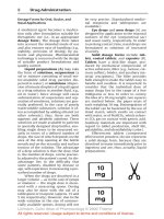
Section 7. Pharmacology Tables 153
7.1 Intravenous Medication Administration
Guidelines / 154
7.2 Neuromuscular Blocking Agents / 179
7.3 Vasoactive Agents / 182
7.4 Antiarrhythmic Agents / 185
7.5 Therapeutic Drug Monitoring / 191
7.6 Tips for Calculating IV Medication Infusion
Rates / 194
This page intentionally left blank
xi
Given the complexity of critical care practice today, it’s
impossible for even experienced clinicians to remember
all the information required to give safe and effective care
to critically ill patients. Clinicians frequently need to use a
variety of clinical resources to verify drug information,
normal laboratory and physiologic values, ECG and he-
modynamic monitoring information, emergency algo-
rithms, and other essential facts of patient management.
To save time and avoid frustration, clinicians often
create their own “pocket guides” by cutting and past-
ing together information from a variety of sources so
they always have a quick reference source available.
The
AACN Essentials of Critical Care Nursing Pocket
Handbook
is designed to provide busy clinicians with
an easy to use resource that can, literally, be kept in
their pockets. The pocket handbook contains selected
tables and figures from the textbook,
AACN Essentials
of Critical Care Nursing,
and includes items that clini-
cians are most likely to need at their fingertips:
• Critical care drug tables (common vasoactive drugs,
neuromuscular blocking agents, antiarrhythmics, IV
medication guidelines)
• Normal values table for laboratory tests and physio-
logic parameters
• Lists of assessment components
• Cardiac rhythms: ECG characteristics and treatment
guides including sample rhythm strips
• 12-lead ECG changes in acute myocardial ischemia
and infarct
• Troubleshooting guides for hemodynamic monitoring
equipment
• Indications for mechanical ventilation
• Weaning assessment tool
• Chest x-ray interpretation
We hope this pocket book will, indeed, be placed in your
pocket and assist you in making a difference in the lives
of the patients and families you encounter.
Marianne Chulay
Suzi Burns
Preface
xii
To our critical care nursing colleagues around the world whose wonderful work and efforts
ensure the safe passage of patients through the critical care environment.
1
Normal Values
Section
᭤
1.1 Normal Values Table / 2
NORMAL VALUES
1.1
᭤
Normal Values Table
Abbreviation Definition Normal Value Formula
BSA Body surface area Meters squared (m
2
) Value obtained from a nomogram based on height and weight
C(a Ϫ v)O
2
Arteriovenous oxygen content 4-6 mL/100 mL C(a Ϫ v)O
2
(mL/100 mL or vol %) ϭ CaO
2
Ϫ CvO
2
difference
CaO
2
Arterial oxygen content Will vary with hemoglobin CaO
2
(mL O
2
/100 mL blood or vol %) ϭ
concentration and Pa
O
2
on (Hb ϫ 1.39) SaO
2
ϩ (PaO
2
ϫ 0.0031)
air from 19-20 mL/100 mL
CI Cardiac index 2.5-3.0 L/min/m
2
CI (L/min/m
2
) ϭ
CK Creatinine kinase Ͻ150 mcg/L
CK-MB Creatinine kinase MB band Ͻ10 ng/mL or Ͻ3% of total
CO Cardiac output 4-6 L/min CO ϭ Stroke volume ϫ heart rate
CvO
2
Mixed venous oxygen content Will vary with CaO
2
, cardiac
output, and O
2
consumption
from 14-15 mL/100 mL
CVP Central venous pressure 2-8 mm Hg
dp/dt First time derivative of left 13-14 seconds
ventricular pressure
EDC Effective dynamic compliance 35-45 mL/cm H
2
O women EDC (mL/cm H
2
O) ϭ
40-50 mL/cm H
2
O men
EDV End-diastolic volume 50-90 mL
EF Ejection fraction 70% Ejection fraction ϭ
SV
EDV
tidal volume (mL)
peak airway pressure (cm H
2
O)
cardiac output (L/min)
body surface area (m
2
)
2
1.1
᭤
Normal Values Table (
continued
)
Abbreviation Definition Normal Value Formula
FRC Functional residual capacity 2400 mL
HR Heart rate 60-90 beats/min
IF
Inspiratory force
75-100 cm H
2
O
LVSW Left ventricular stroke work 8-10 g/m/m
2
LVSW ϭ SI ϫ MAP ϫ 0.0144
MAP Mean systemic arterial pressure Ͼ 70 mm Hg Map estimate ϭ
O
2
availability Oxygen availability 550-650 mL/min/m
2
O
2
availability (mL/min/m
2
) ϭ CI ϫ CaO
2
ϫ 10
O
2
extraction ratio Oxygen extraction ratio 0.25 O
2
extraction ratio ϭ
P(A Ϫ a)
O
2
Alveolar-arterial oxygen gradient 25-65 mm Hg at FiO
2
ϭ 1.0 P(A Ϫ a)O
2
(mm Hg) ϭ PAO
2
Ϫ PaO
2
P(A Ϫ a)o
2
Mean partial pressure of oxygen 104 mm Hg
in alveolus
P(A Ϫ a)co
2
Partial pressure of carbon dioxide 40 mm Hg
in alveolus
Pac
O
2
Partial pressure of carbon 35-45 mm Hg
dioxide in arterial blood
PAD Pulmonary artery diastolic 5-12 mm Hg
C(a Ϫ v)O
2
CaO
2
(Systolic ϩ 2 Diastolic)
3
3
4
1.1
᭤
Normal Values Table (
continued
)
Abbreviation Definition Normal Value Formula
Pa
O
2
Partial pressure of oxygen in Will vary with patient’s age
arterial blood and the Fi
O
2
. On room air:
80-95 mm Hg. On 100%
O
2
: 640 mm Hg
PAS Pulmonary artery 54 systolic 16-24 mm Hg
pressure
PCWP Mean pulmonary capillary wedge 5-12 mm Hg
pressure
Pv
CO
2
Partial pressure of carbon 41-51 mm Hg
dioxide in mixed venous blood
Pv
O
2
Partial pressure of oxygen in Will vary with the FiO
2
,
mixed venous blood cardiac output, and oxygen
consumption from
35-40 mm Hg
PVR Pulmonary vascular resistance 120-200 dynes/s/cm
5
PVR 5 ϭ (dynes/s/cm
5
) ϭ
1.5-2.5 mm Hg
Q
S/QT Right-to-left shunt (percentage 5%-8% QS/QT(%) ϭϫ100
of cardiac output flowing past
nonventilated alveoli or the Valid only when arterial blood is 100% saturated
equivalent
0.0031 ϫ P(A Ϫ a)O
2
C(a Ϫ v)O
2
ϩ (0.0031 ϫ P[A Ϫ a]O
2
)
(PA [mm Hg] Ϫ PCWP [mm Hg]) ϫ 79.9
cardiac output (L/min)
5
1.1
᭤
Normal Values Table (
continued
)
Abbreviation Definition Normal Value Formula
R or RQ Respiratory quotient 0.8 RQ ϭ
RVSW Right ventricular stroke work 51-61 g/m/m
2
RVSW ϭ SI ϫ MPAP ϫ 0.0144
SaO
2
Percentage of oxyhemoglobin 96%-100% (air)
saturation of arterial blood
SI Stroke index 35-50 mL/m
2
SI (mL/min/m
2
) ϭ
SV Stroke volume 50-100 mL/beat SV (mL/beat) ϭ
SvO
2
Percentage of oxyhemoglobin 70-80% (air)
saturation of mixed venous
blood
SVR Systemic vascular resistance 900-1200 dynes/s/cm
5
SVR (TPR) (dynes/s/cm
5
) ϭ
10-15 mm Hg
(mm Hg ϫ 80 ϭ dynes/s/cm
5
)
Troponin I Troponin I Ͻ0.4 ng/mL
Troponin T Troponin T Ͻ0.1 ng/mL
(MAP [mm Hg] Ϫ CVP [mm Hg]) ϫ 79.9
cardiac output (L/min)
cardiac output (mL)
heart rate
stroke volume
body surface area
VCO
2
VO
2
6
1.1
᭤
Normal Values Table (
continued
)
Abbreviation Definition Normal Value Formula
VC Vital capacity 65-75 mL/kg
V
CO
2
Carbon dioxide production 192 mL/min
V
D
Dead space 150 mL V
D
/V
T
ϭ
V
D
/V
T
Dead space to tidal volume ratio 0.25-0.40
VO
2
Oxygen consumption 115-165 mL/min/m
2
O
2
extraction ratio ϭ
V
T
Tidal volume 6-8 mL/kg
Adapted from: Hall J, Schmidt G, Wood L.
Principles of critical care. 3rd ed.
New York: McGraw Hill, 2005; cover tables I-IV.
C(a Ϫ v)O
2
CaO
2
PaCO
2
Ϫ PEco
2
Paco
2
2
Assessment
Section
᭤
2.1 Summary of Prearrival and
Admission Quick Check
Assessments / 8
᭤
2.2 Summary of Comprehensive
Admission Assessment
Requirements / 9
᭤
2.3 Suggested Questions for Review of
Past History Categorized by Body
System / 10
᭤
2.4 Ongoing Assessment Template / 12
᭤
2.5
Identification of Symptom
Characteristics / 13
᭤
2.6 Chest Pain Assessment / 14
᭤
2.7 Pain Assessment Tools Commonly
Used in Critically Ill Patients / 15
᭤
2.8 CAM-ICU Worksheet / 16
᭤
2.9
Glasgow Coma Scale / 18
᭤
2.10 Sensory Dermatomes / 19
᭤
2.11 Edema Rating Scale / 21
᭤
2.12 Peripheral Pulse Rating Scale / 21
᭤
2.13 Physiologic Effects of Aging / 22
ASSESSMENT
8
2.1
᭤
Summary of Prearrival and Admission Quick Check Assessments
Prearrival Assessment
• Abbreviated report on patient (age, gender, chief complaint, diagnosis,
pertinent history, physiologic status, invasive devices,
equipment, and status of laboratory/diagnostic tests)
• Complete room setup, including verification of proper
equipment functioning
Admission Quick Check Assessment
• General appearance (consciousness)
•
A
irway:
Patency
Position of artificial airway (if present)
•
B
reathing:
Quantity and quality of respirations (rate, depth, pattern,
symmetry, effort, use of accessory muscles)
Breath sounds
Presence of spontaneous breathing
•
C
irculation and
C
erebral Perfusion:
ECG (rate, rhythm, and presence of ectopy)
Blood pressure
Peripheral pulses and capillary refill
Skin, color, temperature, moisture
Presence of bleeding
Level of consciousness, responsiveness
•
C
hief Complaint:
Primary body system
Associated symptoms
•
D
rugs and
D
iagnostic Tests:
Drugs prior to admission (prescribed, over-the-counter, illicit)
Current medications
Review diagnostic test results
•
E
quipment:
Patency of vascular and drainage systems
Appropriate functioning and labeling of all equipment connected to patient
• Allergies
9
2.2
᭤
Summary of Comprehensive Admission Assessment Requirements
Past Medical History
• Medical conditions, surgical procedures
• Psychiatric/emotional problems
• Hospitalizations
• Medications (prescription, over-the-counter, illicit drugs) and
time of last medication dose
• Allergies
• Review of body systems (see Table 1-7)
Social History
• Age, gender
• Ethnic origin
• Height, weight
• Highest educational level completed
• Occupation
• Marital status
• Primary family members/significant others
• Religious affiliation
• Advance Directive and Durable Power of Attorney for Health Care
• Substance use (alcohol, drugs, caffeine, tobacco)
• Domestic Abuse or Vulnerable Adult Screen
Psychosocial Assessment
• General communication
• Coping styles
• Anxiety and stress
• Expectations of critical care unit
• Current stresses
• Family needs
Spirituality
• Faith/spiritual preference
• Healing practices
Physical Assessment
• Nervous system
• Cardiovascular system
• Respiratory system
• Renal system
• Gastrointestinal system
• Endocrine, hematologic, and immune systems
• Integumentary system
10
2.3
᭤
Suggested Questions for Review of Past History Categorized by Body System
Body System History Questions
Nervous • Have you ever had a seizure?
• Have you ever fainted, blacked out, or had
delirium tremens (DTs)?
• Do you ever have numbness, tingling, or
weakness in any part of your body?
• Do you have any difficulty with your hearing,
vision,or speech?
• Has your daily activity level changed due to your
present condition?
• Do you require any assistive devices such
as canes?
Cardiovascular • Have you experienced any heart problems
or disease such as heart attacks or strokes?
• Do you have any problems with extreme fatigue?
• Do you have an irregular heart rhythm?
• Do you have high blood pressure?
• Do you have a pacemaker or an implanted
defibrillator?
Respiratory • Do you ever experience shortness of breath?
• Do you have any pain associated with breathing?
• Do you have a persistent cough? Is it productive?
• Have you had any exposure to environmental
agents that might affect the lungs?
• Do you have sleep apnea?
Body System History Questions
Renal • Have you had any change in frequency of
urination?
• Do you have any burning, pain, discharge,
or difficulty when you urinate?
• Have you had blood in your urine?
Gastrointestinal • Has there been any recent weight loss or gain?
• Have you had any change in appetite?
• Do you have any problems with nausea
or vomiting?
• How often do you have a bowel movement and
has there been a change in the normal
pattern? Do you have blood in your stools?
• Do you have dentures?
• Do you have any food allergies?
Integumentary • Do you have any problems with your skin?
Endocrine • Do you have any problems with bleeding?
Hematologic • Do you have problems with chronic infections?
Immunologic • Have you recently been exposed to a contagious
illness?
11
Body System History Questions
Psychosocial • Do you have any physical conditions which make
communication difficult (hearing loss, visual
disturbances, language barriers, etc)?
• How do you best learn? Do you need information
repeated several times and/or require information
in advance of teaching sessions?
• What are the ways you cope with stress, crises, or pain?
• Who are the important people in your “family” or
network? Who do you want to make decisions with you,
or for you?
• Have you had any previous experiences with critical illness?
• Have you ever been abused?
• Have you ever experienced trouble with anxiety, irritability,
being confused, mood swings, or suicide attempts?
• What are the cultural practices, religious influences,
and values that are important to the family?
• What are family members’ perceptions and expectations
of the critical care staff and the setting?
2.3
᭤
Suggested Questions for Review of Past History Categorized by Body System (
continued
)
Body System History Questions
Spiritual • What is your faith or spiritual preference?
• What practices help you heal or deal with
stress?
• Would you like to see a chaplain, priest,
or other type of healer?
12
2.4
᭤
Ongoing Assessment Template
Body System Assessment Parameters
Nervous • LOC
• Pupils
• Motor strength of extremities
Cardiovascular • Blood pressure
• Heart rate and rhythm
• Heart sounds
• Capillary refill
• Peripheral pulses
• Patency of IVs
• Verification of IV solutions
and medications
• Hemodynamic pressures and waveforms
• Cardiac output data
Respiratory • Respiratory rate and rhythm
• Breath sounds
• Color and amount of secretions
• Noninvasive technology information (eg, pulse
oximetry, end-tidal CO
2
)
• Mechanical ventilatory parameters
• Arterial and venous blood gases
Renal • Intake and output
• Color and amount of urinary output
• BUN/creatinine values
Body System Assessment Parameters
Gastrointestinal • Bowel sounds
• Contour of abdomen
• Position of drainage tubes
• Color and amount of secretions
• Bilirubin and albumin values
Endocrine, • Fluid balance
hematologic, • Electrolyte and glucose values
and • CBC and coagulation values
immunologic • Temperature
• WBC with differential count
Integumentary • Color and temperature of skin
• Intactness of skin
• Areas of redness
Pain/discomfort • Assessed in each system
• Response to interventions
Psychosocial • Mental status and behavioral responses
• Reaction to critical illness experience
(eg, stress, anxiety, coping, mood)
• Presence of cognitive impairments (dementia,
delirium), depression, or demoralization
• Family functioning and needs
• Ability to communicate needs and participate in care
• Sleep patterns