Predictors of angle widening after laser iridotomy in Chinese patients with primary angleclosure suspect using ultrasound biomicroscopy
Bạn đang xem bản rút gọn của tài liệu. Xem và tải ngay bản đầy đủ của tài liệu tại đây (863.79 KB, 9 trang )
Int J Ophthalmol, Vol. 15, No. 2, Feb.18, 2022
www.ijo.cn
Tel: 8629-82245172 8629-82210956 Email:
·Clinical Research·
Predictors of angle widening after laser iridotomy in
Chinese patients with primary angle-closure suspect
using ultrasound biomicroscopy
Xue-Ting Pei, Shu-Hua Wang, Xia Sun, Hong Chen, Bing-Song Wang, Shu-Ning Li, Tao Wang
Beijing Tongren Eye Center, Beijing Tongren Hospital, Capital
Medical University, Beijing Ophthalmology and Visual
Science Key Laboratory, Beijing 100730, China
Correspondence to: Tao Wang. Beijing Tongren Eye Center,
Beijing Tongren Hospital, Capital Medical University,
Beijing Ophthalmology and Visual Science Key Laboratory,
Dongjiaominxiang No.1, Dongcheng District, Beijing 100730,
China.
Received: 2021-02-17
Accepted: 2021-09-23
basal iris insertion are associated with less angle widening
after LPI. Quadrants with iris angulation as well as a flatter
iris configuration predict a smaller angle change after LPI.
● KEYWORDS: laser peripheral iridotomy; angle opening
distance; ultrasound biomicroscopy; iris convexity; iris
angulation
DOI:10.18240/ijo.2022.02.07
Citation: Pei XT, Wang SH, Sun X, Chen H, Wang BS, Li SN, Wang T.
Predictors of angle widening after laser iridotomy in Chinese patients
Abstract
● AIM: To assess the predictive value of baseline
parameters of ultrasound biomicroscopy (UBM) for angle
widening after prophylactic laser peripheral iridotomy (LPI)
in patients with primary angle-closure suspect (PACS).
● METHODS: Angle-opening distance (AOD), trabecular iris
angle (TIA), iris thickness, trabecular-ciliary process angle,
and trabecular-ciliary process distance were measured
using UBM performed before and two weeks after LPI. Iris
convexity (IC), iris insertion, angulation, and ciliary body
(CB) size and position were graded. Uni- and multivariate
regression analyses were used to determine factors
predicting the change in AOD (ΔAOD500, calculated as an
angle width change before and after LPI) in all quadrants
and in subgroup quadrants based on IC.
● RESULTS: In 94 eyes of 94 patients with PACS, LPI led to
angle widening with increases in AOD500 and TIA (P<0.01).
Multivariable regression analysis showed that IC (P<0.001),
CB position (P=0.007) and iris insertion (P=0.049) were
significantly predictive for ΔAOD500. All quadrants were
categorized into extreme IC (27.8%), moderate IC (62.3%),
and absent IC (9.9%) subgroups. The AOD500 increased
by 220% and no other predictive factor was found in the
extreme IC quadrants. The AOD500 increased by 55%, and
baseline iris angulation was predictive for smaller changes
in ΔAOD500 in the moderate IC quadrants.
● CONCLUSION: In PACS patients, quadrants with greater
iris bowing predict substantial angle widening after LPI.
Quadrants with a flatter iris, anteriorly positioned CB, and
with primary angle-closure suspect using ultrasound biomicroscopy.
Int J Ophthalmol 2022;15(2):233-241
INTRODUCTION
rimary angle-closure glaucoma (PACG) is a main
cause of bilateral irreversible blindness worldwide.
Approximately 23.36 million people aged 40-80y had PACG
worldwide in 2020, and the number of the case is estimated
to reach 32.04 million in 2040[1]. Asian patients account for
approximately 77% of worldwide angle-closure glaucoma
cases[1-2]. Angle-closure glaucoma includes three categories:
primary angle-closure suspect (PACS) with narrow angles
that are predisposed to angle closure, primary angle-closure
(PAC) with occluded angle and trabecular obstruction
without glaucomatous optic disc damage, and PACG with
glaucomatous optic neuropathy[3-4]. The commonly known
pathological mechanism for angle-closure glaucoma is
pupillary block (PB), which prevents aqueous flow and
thus results in anterior bowing of the peripheral iris[5]. Laser
peripheral iridotomy (LPI) has long been the first-line standard
intervention for angle closure based on the elimination of
PB, and can be used for treating symptomatic cases and as
prophylactic treatment in PACS patients[6]. However, PACS
patients appear to gain less benefit from prophylactic LPI for
two reasons. First, the conversion rate from PACS to angleclosure disease is very low[7]. Second, after LPI, persistent angle
closure still exists in many eyes[8-10], which develop peripheral
anterior synechia[11]. Under this condition, non-PB mechanisms
as well as the PB mechanism often simultaneously contribute
P
233
Morphologic changes following iridotomy
to angle closure, including thick peripheral iris, anteriorly
positioned ciliary body (CB), and plateau iris[6,12-14]. Thus, the
use of LPI as prophylactic treatment for narrow angle is being
considered[15].
Anterior segment optical coherence tomography (AS-OCT)
parameters are commonly used to assess predictive parameters
for enlargement of the anterior chamber angle following LPI. It
has been reported that increased postoperative angle widening
is correlated with a shorter baseline angle-opening distance
(AOD) and axial length as well as a greater baseline anterior
chamber depth (ACD), iris curvature, and lens vault [16-17].
These parameters mainly reflect factors associated with PB.
However, AS-OCT imaging has some disadvantages such as
limited resolution and difficulty in identifying the location of
the ciliary processes and iris angulation. In addition, images of
only nasal and temporal quadrants are obtained by AS-OCT,
and thus, the analysis of superior and inferior quadrants is not
possible.
The noninvasive ultrasound biomicroscopy (UBM) can be
used to view structures behind the iris[18]. A cross-sectional
view of the anterior chamber angle anatomy and the relative
position of the iris and CB can be achieved by radially oriented
scanning through the limbus. UBM can provide insight
into the underlying mechanism of PAC diseases and aid the
identification of risk factors for a progressive narrow angle
after LPI. In the present study, UBM images of four quadrants
were used to quantitatively measure and qualitatively describe
the anterior segment morphology in PACS patients before
and after LPI. The morphological parameters were analyzed
to study the possible predictive factors for angle widening
as the LPI outcome. Moreover, we categorized all quadrants
according to the configuration of iris convexity (IC) to analyze
the effect of LPI and investigated the predictive factors for
each subgroup.
SUBJECTS AND METHODS
Ethical Approval The study was approved by the Ethics
Committee Board of Beijing Tongren Hospital and conducted
in accordance with the tenets of the Declaration of Helsinki.
All patients provided written informed consent ahead of
participation.
Patients This retrospective study was performed at the Eye
Center of Beijing Tongren Hospital. The medical records
of consecutive patients who visited the glaucoma clinic
between January 2019 and September 2020 were reviewed.
The inclusion criteria were as follows: 1) age >50y; 2) PACS
diagnosis and UBM examination; 3) treatment with LPI.
Patients were excluded if they had secondary angle closure,
previous attack of acute angle closure, cataract (visual acuity,
worse than 20/40), a history of any eye injuries (intraocular
surgery or penetrating eye injury), or used topical or systemic
234
medications that could affect the anterior chamber angle. All
participants underwent complete ophthalmic examinations,
including a review of their medical history, measurement
of best corrected visual acuity, slit-lamp biomicroscopy,
intraocular pressure measurements with Goldmann applanation
tonometry, gonioscopy, funduscopic examination with a
90-diopter lens, stereoscopic optic disc photography, visual
field test, and UBM. The visual field test was analyzed using
a Humphrey Visual Field Analyzer II (Carl Zeiss Meditec,
Dublin, California, USA) with the standard Swedish interactive
threshold algorithm in a 24-2 test pattern.
PACS was defined as follows: 1) having 180° of the posterior
trabecular meshwork, which was not visualized on the basis
of static gonioscopic examination; 2) intraocular pressure
<21 mm Hg; 3) no peripheral anterior synechiae (abnormal
adhesions of the iris to the angle by more than half a clock hour
in width); 4) glaucomatous optic neuropathy [a vertical cup-todisc (C/D) ratio > 0.7, C/D asymmetry > 0.2, focal notching, or
visual field changes compatible with glaucoma]. Only one eye
was chosen randomly in patients with two eligible eyes and
included in the analysis.
Gonioscopy Slit lamp gonioscopy was performed using a
Goldmann-type, one-mirror lens (Ocular OSMG, Bellevue
WA, USA). Gonioscopic examinations were conducted by
an experienced observer (Pei XT). Indentation gonioscopy
was used to identify the cause of angle closure (apposition or
peripheral anterior synechiae). Appositional angle closure was
verified by gonioscopy for all patients.
Laser Peripheral Iridotomy LPI was performed after topical
application of 2% pilocarpine for pupil constriction (Zhenrui;
Bausch and Lomb Freda, Shandong, China) and proparacaine
(0.5%) for anesthesia (Alcaine; Alcon, Fort Worth, TX, USA).
A neodymium-yttrium-aluminum-garnet laser was set at
variable energy levels between 6 and 8 mJ (1-10 shots). One
opening was created using an Abraham lens, and a crypt was
selected in the peripheral iris when possible. UBM was used to
confirm iridotomies. Prednisolone eye drops (4 times daily for
3d) was applied following the intervention.
All cases were examined with UBM before and 2wk after
LPI. UBM examinations were performed with a UBM Model
MD-300L instrument (MEDA Co., Ltd., Tianjin, China). After
topical application of proparacaine (0.5%) in both eyes, an
eyecup filled with sterile normal saline was used as a coupling
agent. Images were taken under the same lighting conditions
(3.25 cd/m 2) by the same experienced operator. Under a
sufficient lighting condition, eyes were examined in axial
section, and the probe was kept perpendicular to the cornealscleral surface. Images were obtained from the superior, nasal,
inferior and temporal quadrants as well as the center of the
pupil.
Int J Ophthalmol, Vol. 15, No. 2, Feb.18, 2022
www.ijo.cn
Tel: 8629-82245172 8629-82210956 Email:
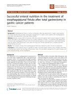
Image Analysis Image J 1.51 software (Wayne Rasband,
NIH, Rockville, MD, USA) was used for analyzing all images
(Figure 1). The scleral spur (SS) was located based on the
difference in the tissue density between the collagen fibers of
the SS and the longitudinal muscle of the CB.
The following quantitative anterior segment parameters
were measured (Figure 1). Pupil diameter was defined as the
distance between pupillary margins. ACD was measured by
the distance between the corneal endothelium and the anterior
surface of the lens. AOD500 was calculated as the distance
from the corneal endothelium to the anterior iris perpendicular
to a line drawn along the trabecular meshwork at 500 μm from
the SS. Trabecular iris angle (TIA) was measured with the SS
as the apex and the corneal endothelium and superior surface
of the iris as the arms of the angle. IT750 was defined as the
thickness of the iris thickness 750 μm from the SS. Trabecularciliary process angle (TCA) was measured with the SS as the
apex and the corneal endothelium and superior surface of the
ciliary process as the arms of the angle. Trabecular-ciliary process
distance (TCPD) was measured as the perpendicular length of the
line extending from the corneal endothelium 500 μm from the
SS through the posterior surface of iris to the ciliary process.
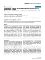
The following qualitative parameters were assessed according
to standard UBM photographs (Figure 2). IC (absent/moderate/
extreme) was graded by the curvature of the posterior surface
of the iris. Iris insertion (basal/middle/apical) was graded based
on the location of the iris insertion into the CB. Iris angulation
(none/mild/pronounced) was identified based on the change of
the iris at the insertion point into the CB. CB size was defined
as the greatest distance between the apex of the CB and base,
in reference to the limbal cornea thickness (small, less than
limbal corneal thickness; medium, greater than the limbal
corneal distance by <2-fold; and large, greater than the limbal
corneal thicknesses by ≥2-fold). CB position was categorized
as neutral or anteriorly positioned on the basis of the direction
of the axis of the CB processes.
Fifteen eyes (60 quadrants) were randomly selected for
assessment of intra-examiner reproducibility. The quantitative
parameters were measured repeatedly by the same observer.
Qualitative parameters were assessed independently by two
glaucoma specialists (Pei XT and Sun X). If the specialists had
different opinions, a third experienced examiner (Wang SH)
made the final decision.
Statistical Analysis Statistical analyses were performed using
SPSS version 20.0 (SPSS Inc., Chicago, Illinois, USA). Intraexaminer repeatability for UBM parameters was assessed by
intraclass correlation coefficients. The paired Student’s t-test
was performed to analyze the differences in the parameters
before and after LPI. Covariance was used for subgroup
differences in the parameters with pupil diameter as a
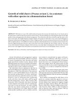
Figure 1 Quantitative parameters measured on ultrasound
biomicroscopic images A circle with 500 μm in radius was drawn
using the SS (O) as the center. The points of intersection were at
the back of the cornea (A) and the anterior surface of the CB (C).
The AOD was measured on a line perpendicular to the plane of the
trabecular surface 500 μm anterior to the SS and extended to meet
the surface of the iris (B); the TCPD was a line measured between A
and C. For iris thickness, a circle with 750 μm in radius was drawn
using the SS (O) as the center, and the iris thickness was the distance
from the intersection point (E) on the anterior surface of the iris to the
intersection point (F) on the posterior surface. The TIA was the angle
AOB; the TCA was the angle AOC.
covariate. The Chi-square test was used to compare categorical
variables of qualitative assessment.
Linear regression adjusted for PD was performed to assess the
association between baseline UBM parameters and changes
in AOD500 (ΔAOD500) defined as the difference between
AOD500 after LPI and AOD500 before LPI. Predictors
of angle widening was determined by using multivariable
forward stepwise linear regression algorithms. Variables with
a probability value ≤0.10 on univariate analysis were included
in the multivariate analysis. P values <0.05 were considered
statistically significant.
RESULTS
A total of 94 eyes of 94 Chinese patients with PACS (65
females and 29 males) were included in the study. The mean
patient age was 61.5±7.8y (range, 50-72y). The intra-examiner
intraclass coefficient values for the UBM parameters were
between 0.875 and 0.927.
The mean pupil diameter was 3.3±0.7 mm before LPI and
3.2±0.8 mm after LPI (P=0.312). The mean ACD was
2.10±0.43 mm before LPI and 2.11±0.39 mm after LPI
(P=0.165). The pupil diameter and ACD before and after LPI
were not significantly different.
Table 1 summarizes the UBM parameters in the four quadrants
before and after LPI. There were significant differences in
angle width in the four quadrants before and after LPI. The
parameters for the anterior chamber angle width increased
significantly after LPI. For all quadrants, the mean AOD500
increased by 100% from 0.10±0.07 mm before LPI to
0.20±0.10 mm after LPI (P<0.01). The mean TIA increased
235
Morphologic changes following iridotomy
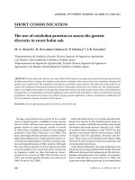
Figure 2 Standard photographs were used to assess UBM results A: Absent IC; B: Moderate IC; C: Extreme IC. D: Basal iris insertion;
E: Middle iris insertion; F: Apical iris insertion; G: No iris angulation; H: Mild iris angulation; I: Pronounced iris angulation; J: Small CB; K:
Medium CB; L: Large CB; M: Neutral CB position; N: Anterior CB position.
significantly from 9°±7° before LPI to 19°±10° after LPI
(P<0.01). However, the angle width decreased or remain
unchanged in 37.4% of quadrants.
Linear regression analyses showed that greater IC and shorter
IT750 predicted greater angle widening following LPI,
which was defined as an increase in AOD500 after LPI
(ΔAOD500>0) in each quadrant (P<0.05). Multivariable
stepwise regression analysis (overall model adjusted R2=0.64;
P<0.001) demonstrated that IC and CB position were
predictive for angle changes after LPI. Iris insertion reached
marginal significance as a predictor for angle widening.
Greater angle widening following LPI was correlated with a
greater IC at baseline (P<0.001). A more anteriorly positioned
CB (P=0.007) and a closer basal iris insertion (P=0.049) were
associated with smaller angle widening after LPI (Table 2).
Linear regression analyses adjusted for sex and intraocular
pressure as covariates were further performed, and the
236
results were similar to the analysis without adjusted sex and
intraocular pressure as covariates.
All quadrants were subcategorized according to IC, as the
extreme IC group (104 quadrants, 27.8%), moderate IC group
(235 quadrants, 62.3%), and absent IC group (37 quadrants,
9.9%). Table 3 summarizes the UBM parameters in the three
groups before and after LPI. Angle widening was significantly
different among the groups. Compared with pre-LPI values, the
AOD500 was increased significantly by 220% after LPI in the
extreme IC quadrants and increased significantly by 55% in the
moderate IC quadrants. No statistically significant difference
in AOD500 before and after LPI was noted in the absent IC
group. The TCA in the moderate IC group was significantly
narrower than that in the extreme IC group.
Linear regression analyses and multivariable stepwise
regression analysis were performed in each subgroup.
Univariate linear regression analysis showed that no anatomic
Int J Ophthalmol, Vol. 15, No. 2, Feb.18, 2022
www.ijo.cn
Tel: 8629-82245172 8629-82210956 Email:
Table 1 Quantitative measurement and qualitative grading of UBM images in four quadrants before and after LPI
Parameters
Quadrant
Superior
Nasal
Inferior
Temporal
AOD500 (mm)
Pre-LPI
0.03±0.04
0.13±0.07
0.09±0.06
0.14±0.06
Post-LPI
0.16±0.08
0.23±0.09
0.20±0.11
0.26±0.13
<0.001
<0.001
<0.001
<0.001
P
TIA (degrees)
Pre-LPI
4.0±3.8
10.3±4.9
9.0±6.2
11.4±8.3
Post-LPI
15.0±8.3
18.7±10.9
17.0±9.3
20.0±11.8
0.005
0.019
0.003
0.018
P
IT750 (mm)
Pre-LPI
0.31±0.06
0.36±0.05
0.35±0.04
0.35±0.06
Post-LPI
0.35±0.05
0.38±0.06
0.39±0.05
0.33±0.04
0.036
0.142
0.025
0.376
P
TCA (degrees)
Pre-LPI
40.4±11.1
57.3±11.8
55.3±14.1
61.4±17.5
Post-LPI
53.0±15.4
57.0±10.9
59.7±23.2
57.3±12.1
0.078
0.938
0.620
0.284
P
TCPD500 (mm)
Pre-LPI
0.43±0.10
0.59±0.10
0.57±0.13
0.62±0.17
Post-LPI
0.53±0.13
0.58±0.08
0.58±0.15
0.59±0.11
0.121
0.525
0.888
0.262
Pre-LPI
6/58/30
10/58/26
8/51/35
13/66/15
Post-LPI
94/0/0
94/0/0
94/0/0
94/0/0
Pre-LPI
74/16/4
67/20/7
66/23/5
54/28/12
Post-LPI
81/13/0
72/15/7
73/16/5
57/27/10
0.081
0.309
0.514
0.282
60/31/3
47/44/3
56/36/2
36/52/6
35/39/20
28/50/16
28/45/21
12/46/36
37/57
25/69
45/49
40/54
P
Iris convexity (absent/moderate/extreme)
Iris angulation (none/mild/pronounced)
P
Iris insertion (basal/middle/apical)
Pre-LPI
CB relative size (small/medium/large)
Pre-LPI
CB position (neutral/anterior)
Pre-LPI
AOD500: Angle-opening distance at 500 μm from scleral spur; LPI: Laser peripheral iridotomy; TIA: Trabecular iris angle; IT750: Iris thickness
at 750 μm from scleral spur; TCA: Trabecular-ciliary process angle; TCPD500: Trabecular-ciliary process distance at 500 μm from scleral spur;
CB: Ciliary body.
factors were significantly associated with ΔAOD500 in the
extreme IC group (Table 4). Iris angulation was found to be a
predictor for ΔAOD500 in the moderate IC group (P=0.029). A
greater iris angulation before LPI was associated with a smaller
change in the angle width following LPI (Table 5). Linear
regression analyses adjusted for sex and intraocular pressure as
covariates were further performed, and the results were similar
to the analysis without adjusted sex and intraocular pressure as
covariates.
DISCUSSION
The present study found that prophylactic LPI treatment
increased the anterior chamber angle width in Chinese patients
with PACS. We assessed the potential predictive parameters
for the change in the angle measured by UBM. The angle
widening observed in each quadrant was associated with three
baseline factors: IC, CB position, and iris insertion. As IC was
the key factor that affected the effect of LPI, we categorized all
quadrants according to the IC grading. Extreme IC quadrants
were associated with the best outcomes from LPI, and no
baseline parameters were significantly associated with angle
widening. LPI reduced angle width in moderate IC quadrants
compared with extreme quadrants, and the angle width change
237
Morphologic changes following iridotomy
Table 2 Uni- and multivariate linear regression analyses of the association between baseline UBM parameters and ΔAOD500 after LPI
in all quadrants
Parameters
SE
Univariate B coefficient (95%CI)
P
PreAOD500
0.541
-0.653 (-1.778, 0.473)
0.241
PreTIA
0.006
0.004 (-0.007, 0.016)
0.787
PreIT750
0.354
0.037 (-0.700, 0.774)
0.010
PreTCA
0.004
-0.001 (-0.008, 0.007)
0.849
PreTCPD
0.395
0.242 (-0.580, 1.064)
0.547
SE
Multivariate B coefficient (95%CI)
P
0.023
0.125 (0.077, 0.173)
0.000
Iris convexity
0.044
0.103 (0.012, 0.193)
0.000
Iris angulation
0.045
-0.056 (-0.149, 0.037)
0.227
Iris insertion
0.039
0.065 (-0.001, 0.132)
0.053
0.028
0.058 (0.000, 0.117)
0.049
CB position
0.018
-0.061 (-0.153, 0.033)
0.002
0.041
-0.121 (-0.206, -0.036)
0.007
CB size
0.022
-0.014 (-0.061, 0.033)
0.537
SE: Spherical equivalent; AOD500: Angle-opening distance at 500 μm from scleral spur; TIA: Trabecular iris angle; IT750: Iris thickness at 750 μm
from scleral spur; TCA: Trabecular-ciliary process angle; TCPD500: Trabecular-ciliary process distance at 500 μm from scleral spur; CB: Ciliary
body.
Table 3 Comparison of quantitative measurements and qualitative grading of UBM parameters before and after LPI in three iris
convexity grading subgroups
Parameters
AOD500 (mm)
Pre-LPI
Post-LPI
P
TIA (degrees)
Pre-LPI
Post-LPI
P
IT750 (mm)
Pre-LPI
Post-LPI
P
TCA (degrees)
Pre-LPI
Post-LPI
P
TCPD500 (mm)
Pre-LPI
Post-LPI
P
Iris angulation (none/mild/pronounced)
Pre-LPI
Post-LPI
P
Iris insertion (basal/middle/apical)
Pre-LPI
CB relative size (small/medium/large)
Pre-LPI
CB position (neutral/anterior)
Pre-LPI
Extreme IC (n=104)
Subgroups
Moderate IC (n=235)
Absent IC (n=37)
0.10±0.08
0.32±0.11
<0.001
0.09±0.06
0.14±0.09
0.005
0.16±0.13
0.20±0.12
0.552
0.305
0.003
9.1±6.6
29.8±11.2
<0.001
8.4±6.3
12.3±7.1
0.008
15.6±10.5
18.6±11.8
0.707
0.220
<0.001
0.30±0.04
0.33±0.06
0.057
0.36±0.05
0.38±0.04
0.037
0.41±0.04
0.40±0.05
0.392
0.004
0.038
60.5±17.2
70.5±11.8
0.046
46.8±12.1
48.7±11.4
0.447
64.6±16.0
61.3±13.3
0.233
0.032
<0.001
0.61±0.17
0.66±0.09
0.448
0.49±0.10
0.50±0.12
0.758
0.65±0.14
0.60±0.11
0.162
0.055
<0.001
78/26/0
91/13/0
0.085
168/51/16
177/46/12
0.680
15/9/13
15/12/10
0.764
<0.001
<0.001
70/34/0
122/104/9
9/24/4
<0.001
39/51/14
64/105/66
0/24/13
0.003
49/55
84/150
15/22
0.278
P (intergroup)
IC: Iris convexity; AOD500: Angle-opening distance at 500 μm from scleral spur; LPI: Laser peripheral iridotomy; TIA: Trabecular iris angle;
IT750: Iris thickness at 750 μm from scleral spur; TCA: Trabecular-ciliary process angle; TCPD500: Trabecular-ciliary process distance
at 500 μm from scleral spur; CB: Ciliary body.
238
Int J Ophthalmol, Vol. 15, No. 2, Feb.18, 2022
www.ijo.cn
Tel: 8629-82245172 8629-82210956 Email:
Table 4 Uni- and multivariate linear regression analyses of the associations between baseline UBM
parameters and ΔAOD500 after LPI in the extreme IC quadrants
Parameters
SE
Univariate B coefficient (95%CI)
P
PreAOD500
2.055
0.184 (0.095, 0.273)
0.943
PreTIA
0.024
-0.026 (-0.049, -0.003)
0.616
PreIT750
3.438
-1.844 (-2.645, -1.032)
0.687
PreTCA
0.032
-0.037 (-0.097, 0.068)
0.453
PreTCPD
4.849
7.958 (3.469, 11.395)
0.348
Iris angulation
0.301
-0.557 (-0.976, -0.203)
0.315
Iris insertion
0.264
0.220 (0.002, 0.462)
0.558
CB position
0.821
0.578 (0.390, 0.836)
0.609
CB Size
0.212
-0.074 (-0.134, 0.037)
0.787
SE: Spherical equivalent; AOD500: Angle-opening distance at 500 μm from scleral spur; TIA: Trabecular iris
angle; IT750: Iris thickness at 750 μm from scleral spur; TCA: Trabecular-ciliary process angle; TCPD500:
Trabecular-ciliary process distance at 500 μm from scleral spur; CB: Ciliary body.
Table 5 Uni- and multivariate linear regression analyses of the associations between baseline UBM parameters and ΔAOD500 after LPI
in moderate IC quadrants
Parameters
SE
Univariate B coefficient (95%CI)
P
PreAOD500
0.492
-0.411 (-1.524, 0.702)
0.425
PreTIA
0.005
0.004 (0.008, 0.015)
0.499
PreIT750
0.272
0.392 (-0.224, 1.008)
0.184
PreTCA
0.003
0.002 (-0.004, 0.008)
0.396
PreTCPD
0.289
-0.288 (-0.942, 0.367)
0.346
Iris angulation
0.033
-0.080 (-0.157, -0.009)
0.029
Iris insertion
0.033
0.046 (-0.029, 0.120)
0.198
CB position
0.035
-0.059 (-0.138, 0.020)
0.124
CB size
0.019
0.018 (-0.026, 0.062)
0.378
SE
Multivariate B coefficient (95%)
P
0.033
-0.080 (-0.150, -0.009)
0.029
SE: Spherical equivalent; AOD500: Angle-opening distance at 500 μm from scleral spur; TIA: Trabecular iris angle; IT750: Iris thickness
at 750 μm from scleral spur; TCA: Trabecular-ciliary process angle; TCPD500: Trabecular-ciliary process distance at 500 μm from scleral spur;
CB: Ciliary body.
was significantly associated with baseline iris angulation. The
angle width remained unchanged in the absent IC quadrants.
Our results are consistent with previous research showing that
the peripheral anterior chamber width increases following
LPI[16,19-22]. Our data showed LPI led to an immediate increase
in the anterior chamber angle width in Chinese PACS patients.
The mean AOD500 increased significantly by 100% (from
0.10 mm before LPI to 0.20 mm after LPI). In previous studies
of eyes without PAS, the quantitative angle-width parameters
increased after LPI, and the mean width changes varied from
54.7% to 135% based on the UBM or AS-OCT results[16,19].
However, in the present study, the AOD500 remained
unchanged or decreased in one-third of all quadrants after
LPI. He et al[22] found that after LPI, about 60% of the eyes in
Chinese PACS patients still had appositional closure detected
by UBM. Meduri et al[16] reported that approximately 70%
of angles opened in more than two quadrants, whereas 50%
opened in all four quadrants after LPI in patients with PAC and
PACG, especially in South Indian patients with PACS.
The outcomes of LPI differed in distinct anatomical quadrants.
Our results showed that IC is the key predictive determinant
associated with the outcome of LPI, which is used to remove
the PB. The increase in angle width after LPI was associated
with the degree of IC at baseline, suggesting that the greater
preoperative PB would be associated with larger peripheral
anterior chamber angle width after LPI. Therefore, IC can
reflect the severity of PB, and iris bowing predicts the degree
of relative PB. Consistent with our findings, previous studies
reported that LPI-induced angle widening is correlated with
less iris curvature measured by AS-OCT[16-17,20,23] and greater
baseline IC[17,23].
Multiple pathogenic mechanisms contribute to PAC, including
PB and non-PB mechanisms. A previous Chinese study
found that PB contributes to 38% of angle closure, and
combined non-PB and PB mechanisms contribute to 54% of
angle closure[14]. Non-PB factors, which contribute to angle
239
Morphologic changes following iridotomy
crowding, include a thick peripheral iris, an anteriorly located
peripheral iris, an anteriorly positioned CB, and a plateau iris.
The degree of preoperative non-PB was negatively correlated
with the peripheral anterior chamber widening after LPI.
Anterior positioned CB is one of the most important nonPB mechanisms. Our results showed baseline CB position
was predictive for angle changes after LPI. Multivariate
regression showed that a qualitative anteriorly positioned
CB was associated with reduced angle widening after LPI.
The anterior position of the CB has been extensively proven
to be a predisposing factor for angle closure, especially in
Chinese patients[24-25]. Anteriorly positioned CB occurred more
frequently in closed angles than in opened angles after LPI in
Chinese patients with PACS[22]. In a study of 73 Korean PAC
and PACG patients, using UBM and AS-OCT, Kwon et al[26]
found that a narrower TCA showed less effect on IOP lowering
as an outcome of LPI.
In this study, we found that iris insertion was correlated
with angle widening after LPI, suggesting that iris insertion
is a predictive parameter for angle widening after LPI. LPI
was associated with less angle widening in patients with a
peripheral iris in closer proximity to the angle. It has been
reported that eyes with basal iris insertion are prone to have
angle closure than those without iris insertion[27]. However, Yun
et al[28] found basal iris insertion did not affect angle widening
after LPI based on AS-OCT images from nasal and temporal
quadrants. Using UBM images from four quadrants, we found
that iris insertion was marginally predictive and thus included
in the predictive model.
Univariate linear regression analysis showed that a thinner
iris thickness was correlated with greater angle widening.
However, multivariate linear regression analysis did not
identify iris thickness as an independent predictor for angle
widening after LPI. Other non-PB factors, such as CB size
and iris angulation, may contribute to angle closure PAC, but
we did not find they were predictive for angle widening after
LPI. As a key confounding factor, IC may affect other factors.
Mizoguchi et al[13] reported that lower baseline measurements
of iris thickness are associated with greater iris curvature.
Kwon et al[26] reported that a flatter IC is associated with
narrower TCA. Here, we categorized quadrants according to
the configuration of IC to control the IC factor, and estimated
the predictive factors for LPI outcome.
In the extreme IC group, which accounted for 30% of all
quadrants, the mean AOD500 increased significantly by 220%
(from 0.10 mm before LPI to 0.32 mm after LPI treatment).
However, no predictive factor was found to be associated with
angle widening in extreme IC quadrants. In the moderate IC
quadrants, which accounted for approximately 70% of quadrants,
the mean AOD500 increased by 55% (from 0.09 mm before
240
LPI to 0.14 mm after LPI treatment). Regression analysis showed
that greater baseline iris angulation was correlated with less
angle widening in the moderate IC group. The angle width
remained unchanged in the absent IC quadrant group.
Plateau iris occurs in about 30% of Asian PACG eyes with
a patent LPI[29-30]. Iris angulation is one of key features for
plateau iris configuration. It is speculated that plateau iris
configuration occurred after LPI in the moderate IC group with
iris angulation, a flatter IC, and a narrower TCA. In the present
study, iris angulation was found to be the only significant
predictor in the quadrants with moderate IC.
This study has some limitations such as the small sample
size, especially in the AIC group. The inadequate sample
size may reduce the power to identify significant differences
between the groups. However, our analysis was mainly based
on quadrants. The inclusion of four quadrants increased the
sample size for data analysis. In addition, the classification of
the qualitative assessment system was arbitrarily selected in
this study. Difficulty in identifying some features may affect
the classification results. Since pupil diameter can influence
anterior segment parameter measurement due to light and
fixation, pupil diameter was selected as a covariate for
analysis. Finally, UBM images were not acquired in the dark.
Anterior chamber angles are inclined to close in the dark; thus,
dark UBM acquisition may improve our understanding of the
mechanisms of angle closure.
In conclusion, the present study showed the effect of enlarging
the anterior chamber angle and identified three predictive
factors for greater enlargement of the anterior chamber angle,
including IC, neutral positioned CB, and iris insertion at
baseline. Quadrants with extreme IC exhibited substantial
anterior chamber angle enlargement after LPI, but no
predictive factors were identified. Quadrants with moderate
IC showed mild angle widening after LPI, and iris angulation
was found to be a predictor factor for a smaller change in the
anterior chamber angle. Our findings show the angle-widening
benefit of prophylactic LPI and may help guide treatment
planning in PACS patients.
ACKNOWLEDGEMENTS
Conflicts of Interest: Pei XT, None; Wang SH, None; Sun X,
None; Chen H, None; Wang BS, None; Li SN, None; Wang
T, None.
REFERENCES
1 Tham YC, Li X, Wong TY, Quigley HA, Aung T, Cheng CY. Global
prevalence of glaucoma and projections of glaucoma burden through
2040: a systematic review and meta-analysis. Ophthalmology
2014;121(11):2081-2090.
2 Chan EW, Li X, Tham YC, Liao JM, Wong TY, Aung T, Cheng
CY. Glaucoma in Asia: regional prevalence variations and future
projections. Br J Ophthalmol 2016;100(1):78-85.
Int J Ophthalmol, Vol. 15, No. 2, Feb.18, 2022
www.ijo.cn
Tel: 8629-82245172 8629-82210956 Email:
3 Foster PJ, Buhrmann R, Quigley HA, Johnson GJ. The definition and
classification of glaucoma in prevalence surveys. Br J Ophthalmol
2002;86(2):238-242.
4 Marchini G, Chemello F, Berzaghi D, Zampieri A. New findings in the
diagnosis and treatment of primary angle-closure glaucoma. Prog Brain
Res 2015;221:191-212.
5 Wang WJ, Song HF, Liu ZC. Computational study on the biomechanics
of pupil block phenomenon. Biomed Res Int 2019;2019:4820167.
6 Sun XH, Dai Y, Chen YH, Yu DY, Cringle SJ, Chen JY, Kong XM,
Wang XL, Jiang CH. Primary angle closure glaucoma: what we know
and what we don’t know. Prog Retin Eye Res 2017;57:26-45.
prophylactic laser peripheral iridotomy. Invest Ophthalmol Vis Sci
2013;54(5):3763-3770.
18 Kirchhoff A, Stachs O, Guthoff R. Three-dimensional ultrasound
findings of the posterior iris region. Graefes Arch Clin Exp Ophthalmol
2001;239(12):968-971.
19 Ma XY, Zhu D, Zou J, Zhang WJ, Cao YL. Comparison of ultrasound
biomicroscopy and spectral-domain anterior segment optical coherence
tomography in evaluation of anterior segment after laser peripheral
iridotomy. Int J Ophthalmol 2016;9(3):417-423.
20 Moghimi S, Bijani F, Chen R, Yasseri M, He MG, Lin SC, Weinreb
RN. Anterior segment dimensions following laser iridotomy in acute
7 He MG, Jiang YZ, Huang SS, Chang DS, Munoz B, Aung T, Foster
primary angle closure and fellow eyes. Am J Ophthalmol 2018;186:59-68.
PJ, Friedman DS. Laser peripheral iridotomy for the prevention of
21 Theinert C, Wiedemann P, Unterlauft JD. Laser peripheral
angle closure: a single-centre, randomised controlled trial. Lancet
iridotomy changes anterior chamber architecture. Eur J Ophthalmol
2019;393(10181):1609-1618.
2017;27(1):49-54.
8 Mou DP, Liang YB, Fan SJ, Peng Y, Wang NL, Thomas R. Progression
22 He MG, Friedman DS, Ge J, Huang WY, Jin CJ, Cai XY, Khaw PT,
rate to primary angle closure following laser peripheral iridotomy in
Foster PJ. Laser peripheral iridotomy in eyes with narrow drainage
primary angle-closure suspects: a randomised study. Int J Ophthalmol
angles: ultrasound biomicroscopy outcomes. The Liwan Eye Study.
2021;14(8):1179-1184.
Ophthalmology 2007;114(8):1513-1519.
9 Gupta R, Kumar R, Chauhan L. Anterior chamber morphology changes
23 Huang GF, Gonzalez E, Lee R, Osmonavic S, Leeungurasatien T, He
in eyes with narrow angles by Scheimpflug imaging: pilocarpine versus
MG, Lin SC. Anatomic predictors for anterior chamber angle opening
laser peripheral iridotomy. Int Ophthalmol 2021;41(6):2099-2108.
after laser peripheral iridotomy in narrow angle eyes. Curr Eye Res
10 Chan PP, Tang FY, Leung DY, Lam TC, Baig N, Tham CC. Ten-
2012;37(7):575-582.
year clinical outcomes of acute primary angle closure randomized to
24 He N, Wu LL, Qi M, He MG, Lin S, Wang X, Yang F, Fan X.
receive early phacoemulsification versus laser peripheral iridotomy. J
Comparison of ciliary body anatomy between American caucasians
Glaucoma 2021;30(4):332-339.
and ethnic Chinese using ultrasound biomicroscopy. Curr Eye Res
11 Qiu L, Yan YJ, Wu LL. Appositional angle closure and conversion of
primary angle closure into glaucoma after laser peripheral iridotomy.
Br J Ophthalmol 2020;104(3):386-391.
12 Chen SY, Wu LL. Effect of anatomic features of ciliary body on
primary angle closure. Zhonghua Yan Ke Za Zhi 2018;54(9):716-720.
2016;41(4):485-491.
25 Henzan IM, Tomidokoro A, Uejo C, Sakai H, Sawaguchi S, Iwase A,
Araie M. Comparison of ultrasound biomicroscopic configurations
among primary angle closure, its suspects, and nonoccludable angles:
the kumejima study. Am J Ophthalmol 2011;151(6):1065-1073.e1.
13 Mizoguchi T, Ozaki M, Wakiyama H, Ogino N. Peripheral iris
26 Kwon J, Sung KR, Han S, Moon YJ, Shin JW. Subclassification
thickness and association with iridotrabecular contact after laser
of primary angle closure using anterior segment optical coherence
peripheral iridotomy in patients with primary angle-closure and
tomography and ultrasound biomicroscopic parameters. Ophthalmology
primary angle-closure glaucoma. Clin Ophthalmol 2014;8:517-522.
2017;124(7):1039-1047.
14 Wang N, Ouyang J, Zhou W, Lai M, Ye T, Zeng M, Chen J. Multiple
27 Ku JY, Nongpiur ME, Park J, Narayanaswamy AK, Perera SA, Tun
patterns of angle closure mechanisms in primary angle closure
TA, Kumar RS, Baskaran M, Aung T. Qualitative evaluation of the iris
glaucoma in Chinese. Zhonghua Yan Ke Za Zhi 2000;36(1):
and ciliary body by ultrasound biomicroscopy in subjects with angle
46-51,5,6.
closure. J Glaucoma 2014;23(9):583-588.
15 Tanner L, Gazzard G, Nolan WP, Foster PJ. Has the EAGLE landed
28 Yun SC, Hong JW, Sung KR, Lee JY. Effects of laser peripheral
for the use of clear lens extraction in angle-closure glaucoma? And
iridotomy in subgroups of primary angle closure based on iris insertion.
how should primary angle-closure suspects be treated? Eye (Lond)
J Ophthalmol 2015;2015:581719.
2020;34(1):40-50.
29 Kumar RS, Tantisevi V, Wong MH, Laohapojanart K, Chansanti O,
16 Meduri E, Gillmann K, Bravetti GE, Niegowski LJ, Mermoud
Quek DT, Koh VT, MohanRam LS, Lee KY, Rojanapongpun P, Aung
A, Weinreb RN, Mansouri K. Iridocorneal angle assessment after
T. Plateau iris in Asian subjects with primary angle closure glaucoma.
laser iridotomy with swept-source optical coherence tomography. J
Arch Ophthalmol 2009;127(10):1269-1272.
Glaucoma 2020;29(11):1030-1035.
17 Lee RY, Kasuga T, Cui QN, Huang GF, He MG, Lin SC. Association
between baseline angle width and induced angle opening following
30 Nongpiur ME, Verma S, Tun TA, Wong TT, Perera SA, Aung T.
Plateau iris and severity of primary angle closure glaucoma. Am J
Ophthalmol 2020;220:1-8.
241