Tài liệu Báo cáo khoa học: Brain angiogenesis in developmental and pathological processes: regulation, molecular and cellular communication at the neurovascular interface pdf
Bạn đang xem bản rút gọn của tài liệu. Xem và tải ngay bản đầy đủ của tài liệu tại đây (439.42 KB, 14 trang )
MINIREVIEW
Brain angiogenesis in developmental and pathological
processes: regulation, molecular and cellular
communication at the neurovascular interface
Hye Shin Lee
1,2
, Jiyeon Han
1,2
, Hyun-Jeong Bai
1,2
and Kyu-Won Kim
1,2,3
1 Neurovascular Coordination Research Center, College of Pharmacy, Seoul National University, Korea
2 Research Institute of Pharmaceutical Science, Seoul National University, Korea
3 Department of Molecular Medicine and Biopharmaceutical Sciences, Seoul National University, Korea
Development of the brain vasculature
Blood vessels form via two distinct processes: vasculo-
genesis and angiogenesis. Vasculogenesis involves the
proliferation and differentiation of mesoderm-derived
angioblasts into endothelial cells [1]. Before the heart
even begins to beat, the primary vascular plexus is
formed throughout the body by vasculogenesis [2]. The
extracerebral vascular plexus is established by vasculo-
genesis within the brain vasculature [2]. Early in
embryogenesis, angioblasts invade the head region and
form the perineural vascular plexus, which ultimately
covers the entire neural tube [3]. After the primary vas-
cular plexus is formed by vasculogenesis, a more com-
plex vascular network is established via angiogenesis
(i.e. the production of vessel branches from pre-exist-
ing vessels). Indeed, the vascular network of the brain
is predominantly formed by angiogenesis. During this
Keywords
astrocyte; barriergenesis; blood–brain
barrier; brain angiogenesis; endothelial cell;
neuron; neurovascular interface; pericyte;
perivascular macrophage; smooth muscle
cell
Correspondence
K W. Kim, Neurovascular Coordination
Research Center, College of Pharmacy,
Seoul National University, Seoul 151-742,
Korea
Fax: +82 2 885 1827
Tel: +82 2 880 6988
E-mail:
(Received 19 February 2009, revised 6 May
2009, accepted 10 June 2009)
doi:10.1111/j.1742-4658.2009.07174.x
The vascular network of the brain is formed by the invasion of vascular
sprouts from the pia mater toward the ventricles. Following angiogenesis
of the primary vascular network, brain vessels experience a maturation pro-
cess known as barriergenesis, in which the blood–brain barrier is formed.
In this minireview, we discuss the processes of brain angiogenesis and bar-
riergenesis, as well as the molecular and cellular mechanisms underlying
brain vessel formation. At the molecular level, angiogenesis and barriergen-
esis occur via the coordinated action of oxygen-responsive molecules (e.g.
hypoxia-inducible factor and Src-suppressed C kinase substrate ⁄ AKAP12)
and soluble factors (e.g. vascular endothelial growth factor and angiopoie-
tin-1), as well as axon guidance molecules and neurotrophic factors. At the
cellular level, we focus on neurovascular cells, such as pericytes, astrocytes,
vascular smooth muscle cells, neurons and brain macrophages. Each cell
type plays a unique role, and works with other types to maintain environ-
mental homeostasis and to respond to certain stimuli. Taken together, this
minireview emphasizes the importance of the coordinated action of mole-
cules and cells at the neurovascular interface, with regards to the regulation
of angiogenesis and barriergenesis.
Abbreviations
Ang-1, angiopoietin-1; AQP4, aquaporin4; BBB, blood–brain barrier; BDNF, brain-derived neurotrophic factor; CNS, central nervous system;
HIF, hypoxia-inducible factor; NGF, nerve growth factor; NT, neurotrophins; SEMA, semaphorin; SSeCKS, Src-suppressed C kinase
substrate; TGF, transforming growth factor; VEGF, vascular endothelial growth factor; VEGFR, vascular endothelial growth factor receptor;
vSMC, vascular smooth muscle cell.
4622 FEBS Journal 276 (2009) 4622–4635 ª 2009 The Authors Journal compilation ª 2009 FEBS
process, vascular sprouts from the pia mater invade
the brain and extend toward the ventricles [4]. Like
other vascular networks, brain vessels undergo forma-
tion, stabilization, branching, pruning and specializa-
tion. In brief, the nascent vasculatures formed by
vasculogenesis and angiogenesis are stabilized via the
recruitment of mural cells and generation of the extra-
cellular matrix. The nascent vasculatures are then fine-
tuned in response to environmental cues from
neighboring cells [5]. Finally, vessels acquire features
suitable for the function of each respective organ.
Brain vessels have extremely specialized characteris-
tics that allow them to form the blood–brain barrier
(BBB). The concept of the BBB was first suggested
more than 100 years ago when Paul Ehrlich discovered
that dyes injected into the vascular system did not pen-
etrate brain tissues but were easily absorbed by periph-
eral tissues [3]. The BBB consists of interendothelial
junctions and a specialized transporter system. The
impermeability of the BBB results from the physical
barrier between adjacent endothelial cells lining the mi-
crovessel wall. Brain endothelial cells acquire their
property as a physical barrier from three different
junctions (tight, adherens and possibly gap junctions)
[6,7]. Tight junctions consist of three integral mem-
brane proteins known as occludin, claudins and junc-
tional adhesion molecules. The extracellular domains
of these proteins form homophilic adhesions with
those of neighboring cells, and their cytoplasmic com-
ponents are linked to accessory proteins (e.g. zonula
occludens proteins and cingulin), forging a connection
to the actin cytoskeletons of endothelial cells [8]. Occlu-
din is an 65 kDa phosphoprotein that regulates
paracellular permeability [9]. Claudins are 22 kDa
phosphoproteins that are thought to help maintain
high transendothelial electrical resistance. Three types
of claudins (claudin1 ⁄ 3, claudin5 and claudin12) are
found in the BBB [10]. In addition, junctional adhesion
molecule-1, -2 and -3 are present in the BBB and are
thought to form part of the tight junction structure.
However, the function of these proteins with regards
to the BBB remains unclear. Adherens junctions are
formed at the intersections of membrane protein cad-
herins. Cadherins form a complex with the beta- and
gamma-catenins in their cytoplamic tails. As with tight
junctions, adherens junctions are linked to the actin
cytoskeleton via the binding of beta- and gamma-cate-
nin to alpha-catenin [8]. Gap junctions have been iden-
tified as BBB components [7]; however, their role in
the function of the BBB remains unclear.
The physical barrier resulting from tight, adherens
and gap junctions enhances transcellular, rather than
paracellular, transport when the brain parenchyma
and blood exchange factors across the vessel wall.
Because the physical barrier primarily functions to
protect the brain from toxins in the blood, a special-
ized transport system is needed to absorb essential
molecules and release substances from the brain.
Nutrients are typically transported from the blood
to the brain via a carrier-mediated transport system.
Because glucose is one of the brain’s primary energy
sources, the Glut-1 transporter is of principal impor-
tance to the BBB [11]. The Glut-1 transporter is asym-
metrically distributed, with a greater abundance found
at the abluminal side than at the luminal membrane.
This distribution ensures that the proper level of glu-
cose is supplied to the brain by preventing the accumu-
lation of glucose in the interstitial fluid [12,13].
Essential amino acids, nucleosides and vitamins also
use carrier systems. For example, the L1 system trans-
ports large neutral amino acids, whereas the y+ sys-
tem transports cationic amino acids and the CNT2
adenosine transporter serves as a carrier for nucleo-
sides [13]. In addition to carrier-mediated transporters,
the BBB endothelium has a receptor-mediated trans-
porter system used by proteins, such as insulin, trans-
ferrin and leptin, to cross the BBB [13].
Molecular basis of brain angiogenesis
and barriergenesis
Hypoxia and the hypoxia-inducible factor system
As an embryo develops and its structure increases in
complexity, the simple diffusion of oxygen and nutri-
ents is no longer adequate for survival [14] and a hyp-
oxic gradient forms inside the body to signal for the
formation of new vessels. In particular, hypoxia-induc-
ible factor-1 (HIF-1) regulates the transcription of
various angiogenic factors [e.g. vascular endothelial
growth factor (VEGF) and erythropoietin) and plays
an important role in vascular development [15]. The
HIF-1 protein is a dimeric transcription factor com-
posed of a and b subunits. The HIF-1a subunit is
induced by low levels of oxygen, whereas the HIF-1 b
(ARNT) subunit remains stable. Previous studies have
shown that HIF-1b deficiency results in embryonic
lethality at around days E9.5 and E10.5, with severe
defects in vessel formation, especially in the yolk sac
[16]. Similarly, HIF-1a deficiency results in vascular
defects at a similar stage of development and neuronal
defects (e.g. failure of neural tube closing and abnor-
mal ventricle formation) have also been detected [17].
Moreover, the selective mutation of HIF-1a in neuro-
nal cells decreases vessel density in the brain, because
of enhanced apoptosis [18]. These findings highlight
H. S. Lee et al. Regulation of angiogenesis and barriergenesis
FEBS Journal 276 (2009) 4622–4635 ª 2009 The Authors Journal compilation ª 2009 FEBS 4623
the importance of HIF-1a in the development of brain
vasculature and other tissues.
Reoxygenation and the SSeCKS/AKAP12 system
during barriergenesis
Hypoxic signals are no longer needed once they induce
new vessel formation in areas lacking oxygen and
nutrients. New vessels then undergo maturation steps
suitable for their environment. Vessel maturation in
the brain involves the acquisition of specialized fea-
tures, including the BBB.
In an attempt to connect the missing link between
angiogenesis and barriergenesis, Lee et al. [19] identified
the Src-suppressed C kinase substrate (SSeCKS) protein
(also known as AKAP12 or gravin in humans), which
is upregulated by changes in oxygen tension during
reoxygenation after hypoxic insult. In cultured primary
astrocytes, overexpression of SSeCKS reduced VEGF
expression and induced angiopoietin-1 (Ang-1), thereby
promoting the expression of tight junction proteins and
strengthening the bonds between brain endothelial cells
[19]. Recent studies indicate that SSeCKS ⁄ AKAP12
downregulates HIF-1a expression by enhancing interac-
tions with von Hippel-Lindau tumor suppressor protein
(pVHL) and prolyl hydroxylase domain 2 (PHD2) [20].
These findings strongly suggest that SSeCKS may trig-
ger the transition from angiogenesis to barriergenesis.
Extracellular factors regulating angiogenesis and
barriergenesis
VEGF
Within the brain, formation of the primary vascular
plexus is largely dependent on VEGF signaling. The
interaction between VEGF and the vascular endothe-
lial growth factor receptor (VEGFR) is thought to
promote the differentiation of angioblasts into endo-
thelial cells. Previous studies have shown that null
mutations in VEGFR2 (Flk-1) lead to defects in
hemangioblast and endothelial cells, resulting in
embryonic lethality at around day E9 [21]. Further-
more, VEGF transcripts have been detected at the
periventricular matrix zone and the VEGFR has been
identified in migrating endothelial cells, suggesting that
VEGF signaling contributes to the migration of vessels
from the pia mater to the periventricular region
[22,23]. In the healthy brain, VEGF is downregulated
to maintain the balance between pro- and anti-angio-
genesis. However, during the course of pathological
conditions such as ischemia and tumor growth, VEGF
contributes to break the BBB and promote endothelial
permeability and vascular sprouting.
Six homologs of VEGF have been identified to date
(VEGF-A, VEGF-B, VEGF-C, VEGF-D, VEGF-E and
placenta growth factor). These homologs each play dis-
tinct roles in angiogenesis. For example, VEGF-C is
mainly involved in lymphangiogenesis, whereas VEGF-
D may contribute to tumor angiogenesis [2,24]. Interest-
ingly, VEGF-B, which works with VEGF-A to respond
to brain injury, probably helps maintain the BBB [25].
A number of growth factors and cytokines are known
to regulate VEGF expression. For example, epidermal
growth factor, platelet-derived growth factor, basic
fibroblast growth factor and tumor necrosis factor-a
upregulate VEGF expression in glioma cells [26,27].
Angiopoietin
Angiopoietin has also been identified as a potent
angiogenic factor during embryonic vessel develop-
ment. Ang-1 deficiency leads to embryonic vascular
defects in the central nervous system (CNS) and many
other parts of the body, because of an inappropriate
association of the extracellular matrix and supporting
cells [28]. Knockout mice deficient in Ang-1, tyrosine
kinase with immunoglobulin-like and EGF-like
domains (Tie)-1 and Tie-2 receptors experienced vascu-
lar defects at a relatively later stage than did VEGF
null mutant mice [29,30]. These findings indicate that
the Ang-1–Tie system may function during vessel mat-
uration and stabilization, rather than during vessel
sprouting. Although Ang-1-overexpressing transgenic
mice experienced increased vascularization, Ang-1 also
increases the tightness of BBB endothelial cells and
reduces vessel permeability [19]. Despite the contro-
versy surrounding the role of Ang-1 as either an angio-
genic factor or a maturation factor, recent studies
clearly show that Ang-1 is a prominent regulator of
vascular development. In addition to its role in vascu-
lar maturation, angiopoietin seems to play an impor-
tant role in the maintenance of BBB homeostasis.
Previous studies have shown that Ang-1 mRNA levels
decrease in conditions that induce BBB breakdown,
such as middle cerebral artery occlusion, whereas the
expression of Ang-2, an endogenous antagonist of
Ang-1, increases [31]. Moreover, when mice with ische-
mic lesions that had been induced by middle cerebral
artery occlusion or VEGF application were treated
with Ang-1, they experienced reduced cerebral vessel
leakage and smaller ischemic lesions [32,33]. Although
Ang-1 is known to play a major role in BBB formation
and homeostasis, it is still unclear which cell type is
the major source of Ang-1. Indeed, astrocytes-condi-
tioned medium contains Ang-1 and has a role in BBB
tightness [19]. Furthermore, another report suggests
Regulation of angiogenesis and barriergenesis H. S. Lee et al.
4624 FEBS Journal 276 (2009) 4622–4635 ª 2009 The Authors Journal compilation ª 2009 FEBS
that Ang-1 secreted from pericytes mediates the expres-
sion of tight junction proteins [34].
Transforming growth factor-b
Transforming growth factor (TGF)-b is a prototypic
member of the large TGF-b superfamily, which con-
tains 30 different factors, including TGF-b, acti-
vin, nodal and bone morphogenic proteins. TGF-bs
are involved in a wide range of biological functions,
including cell growth, differentiation, embryogenesis
and morphogenesis [35]. TGF-b has five isoforms
(TGF-b1, 2, 3, 4 and 5), however, only TGF-b1is
known to alter BBB integrity. In an in vitro model
of the BBB, TGF-b1 treatment reduced permeability
[36]. As with Ang-1, TGF-b1 is thought to contri-
bute to BBB permeability by mediating cellular com-
munication between the endothelium and pericytes or
astrocytes. Pericyte- and astrocyte-derived soluble
factors seem to contribute to BBB organization, and
TGF-b1 may function as a major mediator of this
communication [37,38].
Wnts
In addition to the classical angiogenic regulators dis-
cussed above, Wnt family growth factors have
recently been highlighted as key molecules for CNS
angiogenesis and barriergenesis. Wnts are a large
family of growth factors crucial for a variety of bio-
logical processes; in particular, their functions are
well established in CNS development, for example,
they control dorsal–ventral, anterior–posterial pattern-
ing of CNS tissues, dendrite morphogenesis and
synaptogenesis (for a review, see reference [39]).
According to recent reports, b-catenin, an effecter
molecule of canonical Wnt pathway, is expressed in
the developing CNS vasculature and has critical roles
in embryonic vascular development [40–42]. Interest-
ingly, Wnt ⁄ b-catenin signaling is not only responsible
for angiogenesis, but also regulates barriergenesis.
Conditional knockout of b-catenin in endothelial cells
results in a reduction in CNS vessels, vascular hemor-
rhage and malformation [40]. At the same time, it
impairs Glut-1 expression and claudin-3-mediated
endothelial tightness, reflecting the importance of this
pathway for BBB induction [40,41]. Various types of
Wnt ligands exist in neural tissues to transmit signals
to the perineural endothelium. Wnt7a and Wnt7b are
expressed in ventral–lateral spinal cord, whereas
Wnt1, Wnt3 and Wnt3a are located in dorsal part of
the spinal cord [40,42].
Neurogenic factors involved in angiogenesis and
barriergenesis
Vessels and nerves are located in close proximity to
each other and not only share anatomical similarity,
but also constantly coordinate to form a proper net-
work. Neurovascular coordination requires the sharing
of major signaling pathways involved in pathfinding,
growth, migration and differentiation. In fact, a num-
ber of factors known in CNS development, including
axon guidance molecules and neurotrophins, also func-
tion as regulators of vascular systems (Table 1). Con-
versely, many well-known pro- and anti-angiogenic
factors, including VEGF and Ang-1, are also responsi-
ble for the development and function of the nervous
system (Table 2) [43]. Here, we focus on the molecular
factors specific to nerves and vessels, especially neuro-
genic factors that affect vessel formation and differen-
tiation.
Table 1. Neurogenic factors affecting the vascular system. BDNF, brain-derived neurotrophic factor; NGF, nerve growth factor; NT, neurotro-
phins; SEMA, semaphorin.
Molecules Receptors Effects References
Axon guidance cues
Ephrin-B2 EphB4 Arterial–venous specification, Mural cell recruitment, Lymphatic vessel development,
Tumor angiogenesis
[45]
SEMA3A Neurophilin 1 (NP1) Inhibit angiogenesis [46]
SEMA3F Neurophilin 2 (NP2) Inhibit angiogenesis [46]
SEMA4D Plexin B1 (PLEXB1) Induce tumor angiogenesis [46]
Slit-2 Robo-4 and Robo-1 Repulsion or attraction of endothelial cell migration, tumor angiogenesis [47]
Netrin-1 UNC5B, NeogeninA2b Vascular pathfinding, inhibit or induce angiogenesis [87]
Neurotrophins
NGF TrkA Promote endothelial cell proliferation and migration, induce angiogenesis [48]
BDNF TrkB Cardiac vessel development, chemotactic for hematopoietic precursor cells [53]
NT-3 TrkC Inhibit proliferation of cerebral endothelial cells [88]
H. S. Lee et al. Regulation of angiogenesis and barriergenesis
FEBS Journal 276 (2009) 4622–4635 ª 2009 The Authors Journal compilation ª 2009 FEBS 4625
Axon guidance cues
Recently, it has become widely accepted that four
major axon guidance cues (ephrins, semaphorins, slits
and netrins) are responsible for vascular patterning
[44]. In particular, ephrin B2 contributes to arterial–
venous specification, mural cell recruitment, lymphatic
vessel development and tumor angiogenesis via its
receptor, EphB4 [45]. Semaphorins may perform two
functions, with regards to angiogenesis. Class 3 sem-
aphorins (SEMA), such as SEMA3A and SEMA3F,
inhibit angiogenesis via competition with VEGF for
their common receptor, neuropilin. By contrast,
SEMA4D functions as a pro-angiogenic factor that
induces tumor angiogenesis [46]. Slit2 and Netrin 1
also participate in vessel development and tumor
angiogenesis via their receptor UNC5B, as well as the
ROBO4 receptor for slit and the NeogeninA2b recep-
tor for Netrin [47].
Neurotrophins
Neurotrophins (NTs) are well-known trophic factors
involved in neuronal proliferation, survival and path-
finding. The NT family consists of four members [i.e.
nerve growth factor (NGF), brain-derived neuro-
trophic factor (BDNF), NT-3 and NT-4]. Besides their
classical functions on neuronal cells, a growing body
of evidence suggests that NTs play other roles in
non-neuronal tissues, especially blood vessels.
First and foremost, NGF is known to trigger endo-
thelial cell proliferation and migration in vitro, and
induces angiogenesis in in vivo angiogenic assays (e.g.
the rat corneal assay and the chick embryo chorioa-
llantoic membrane assay) [48]. Furthermore, the angio-
genic activity of NGF was also implicated in a
hind-limb ischemia model, in which NGF markedly
increased arteriole length and density [49]. The role of
NGF in angiogenesis is, in part, because of cross-talk
with VEGF signaling. In neuronal and adipose tissues,
NGF elevated VEGF expression and consequently
stimulated angiogenesis [50,51]. Similarly, BDNF is
also induced by ischemic conditions and overexpres-
sion of this factor promotes the revascularization of
ischemic tissues. During development, BDNF seems to
play an important role in cardiac vascular formation,
because BDNF deficiency impairs the survival of endo-
thelial cells and contributes to vascular hemorrhage in
cardiac vessels [52]. By contrast, BDNF overexpression
increases capillary density within the heart [52,53].
Cellular basis of brain angiogenesis
and barriergenesis
As discussed earlier, angiogenesis and barriergenesis of
the brain vasculature occur via the complex coordi-
nation of various molecules. The molecular dynamics
of this process involve several different types of cells
surrounding the brain vessels. These cells shape the
vascular environment, with each cell type playing a
unique role in the development and function of brain
vessels. Hence, we now discuss the major types of cells
in the vascular environment, including pericytes, astro-
cytes, vascular smooth muscle cells, neurons and brain
macrophages (Fig. 1).
Pericytes
Pericytes are vascular mural cells belonging to the vas-
cular smooth muscle cell (vSMC) lineage. Although
these cells were discovered more than 100 years ago,
pericytes seldom attracted interest because they were
merely considered mural cells that supported endothe-
lial cells. Recent studies have established that pericytes
not only provide physical support to endothelial cells,
but also play critical roles in vessel functioning. Most
importantly, pericytes and endothelial cells share a
basement membrane, enabling them to communicate
directly. In fact, pericytes form focal contacts with
endothelial cells at sites known as peg–socket contacts.
At these contacts, pericytes are connected to endothe-
lial cells through tight, gap and adherence junctions
(Fig. 1) [54]. Pericyte coverage varies among different
types of vessels. The pericyte ⁄ endothelial cell ratio
ranges from 1 : 100 in skeletal muscle to 1 : 1 in the
retina. In general, vessels in the CNS exhibit the high-
est pericyte coverage, highlighting the importance of
pericytes in the formation and maintenance of CNS
vasculature [13,54].
During embryonic angiogenesis, pericyte recruitment
is the first event to stabilize the primary vascular
Table 2. Angiogenic regulatory factors affecting nervous system.
Ang-1, angiopoietin-1; FGF, fibroblast growth factor; IGF, insulin-like
growth factor; VEGF, vascular endothelial growth factor.
Molecules Receptors Effects References
VEGF Flk-1, Nrp1 Axonal growth,
neuron survival,
Neurogenesis
[43]
FGF-2 FGFR-1,
FGFR-2
Neurogenesis,
neuroprotection,
NSC proliferation
[43]
IGF-1 IGF-1R Neurogenesis [89]
Ang-1 Tie-2 Neuroprotection [90]
TSP-1 ⁄ 2 CD47 ⁄ IAP?
LRP1?
Synaptogenesis [91]
Regulation of angiogenesis and barriergenesis H. S. Lee et al.
4626 FEBS Journal 276 (2009) 4622–4635 ª 2009 The Authors Journal compilation ª 2009 FEBS
plexus. During this process, PDGF signaling plays an
important role. The PDGF-B protein is expressed in
sprouting endothelial cells, and its receptor PDGFR-b
is expressed in pericyte precursors. Genetic ablation of
PDGF-B or PDGFR-b results in the loss of pericytes
and severe defects in the brain and heart, leading to
vascular leakage and edema [55–57]. Ligand–receptor
signaling between two types of cells may mediate cellu-
lar communication and, in turn, form the proper struc-
ture of mature vessels. After pericytes are recruited,
the contact between pericyte precursors and endothe-
lial cells signals for the production of TGF-b, which
consequently induces the differentiation of pericyte
precursors to mature pericytes [57]. The opposite situa-
tion also seems possible, in which pericytes induce and
guide vessel sprouting. In the developing human brain,
migrating pericytes are found in front of growing ves-
sels and pericyte-driven angiogenesis participates in the
organization of growing vessels [58].
Another question that arises is: why are pericytes
abundant in the brain vasculature? Brain pericytes
may perform specialized roles involved in the develop-
ment and maintenance of brain vessels. First and fore-
most, pericytes are thought to enhance BBB integrity.
Generally, in vitro models of the BBB involve the
co-culturing of endothelial cells and astrocytes. How-
ever, when pericytes are added to the co-culture, endo-
thelial cells reorganized into stable, capillary-like
structures [59]. Furthermore, pericytes play a protec-
tive role in hypoxia-induced disruption of the BBB
[60]. Ang-1, a key factor regulating barriergenesis, also
contributes to pericyte-induced BBB formation; in fact,
pericyte-derived Ang-1 induces occludin expression in
cultured brain endothelial cells [34].
Pericytes are sometimes confused with vSMCs,
because a specific marker capable of distinguishing
pericytes from vSMCs has not yet been developed.
However, it seems clear that the mural cells located in
the brain microvessels are pericytes. Like vSMCs in
other parts of the body, pericytes are able to regulate
vessel diameter and blood flow. One of the observa-
tions supporting this idea is that pericytes express a
contractile protein known as a-smooth muscle actin.
In addition, some in vitro studies have directly demon-
strated the contractile activity of pericytes. Further-
more, several kinds of molecules have been identified
A
C
N
MG
vSMC
E
C
n
e
m
uL
AA
A
H
2
O
H
2
O
culGeso
Synapse
PC
Peg- socket
junctional
co
mpl
e
x
PM
BM
AC
Endf eet
N
Blood vessel
c
u
lGe
so
A Adenosine
Amino acid
AQP4
Neurotransmitter
Adherence junction
Gap junction
Tight junction
AA
ulGt1
1L
2T
N
C
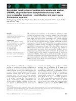
Fig. 1. Cellular communication at the neurovascular interface. The neurovascular unit consists of neurons (N), endothelial cells (EC) and other
types of cells located in the neurovascular unit, i.e. astrocytes (AC), pericytes (PC), vascular smooth muscle cells (vSMC), microglia (MG) and
perivascular macrophages (PM). Endothelial cells form a blood–brain barrier characterized by tight, adherence and gap junctions, as well as a
specialized transporter system (i.e. consisting of Glut-1, L1 and CNT2). Pericytes share basement membranes with blood vessels and directly
contact endothelial cells via peg–socket junction complexes. Astrocytes stretch their endfeet toward blood vessels and neuronal synapses to
integrate neuronal activity with the vascular response. Note that astrocytic endfeet contain water channel AQP4 proteins to regulate water
homeostasis. The immune cells of the CNS (i.e. microglia, macrophages and pericytes) participate in the brain’s immune response.
H. S. Lee et al. Regulation of angiogenesis and barriergenesis
FEBS Journal 276 (2009) 4622–4635 ª 2009 The Authors Journal compilation ª 2009 FEBS 4627
to induce the constriction or dilation of pericytes. In
particular, a-2 adrenergic agonists, histamine, angio-
tensin II and endothelin-1 contribute to vasoconstric-
tion, whereas b-2 adrenergic agonists and nitric oxide
contribute to vasodilation [61].
Another interesting function of brain pericytes is
their macrophage-like activity. Indeed, CNS pericytes
possess a number of macrophage-like features, includ-
ing the capacity to absorb soluble molecules delivered
into the blood or cerebrospinal fluid, the presence of
macrophage-specific class III major histocompatibility
complex proteins and phagocytotic activity [61].
Although it is not clear whether pericytes function as
macrophages in vivo, growing evidence supports the
involvement of pericytes in CNS immunity.
Astrocytes
Astrocytes are the most abundant cells in the brain. In
the past, astrocytes were considered ‘glue’ that provided
physical support for neurons. However, recent studies
suggest that astrocytes play active roles in various brain
functions. In particular, astrocytes function as adult
neural stem cells, participate in the formation and mod-
ulation of synapses, and in the process of ‘gliotransmis-
sion,’ which generates and distributes excitatory
chemical signals in a neuron-coordinated manner [62].
Astrocytes are also interesting with regards to brain vas-
culature, because they regulate the formation and main-
tenance of BBB, modulate neurovascular coupling and
maintain several parts of brain homeostasis. In this
minireview, we focus on the active functions of astro-
cytes in regards to brain vasculature. Anatomically,
most astrocytes have stellate shapes containing multiple
processes. These cells expand toward neurons and ves-
sels. The ends of the cells, so-called endfeet, contact the
vessel wall and form large compartments that enclose
most blood vessels of the brain (Figs 1, 2A). Thus, one
astrocyte can contact several synapses, in addition to
blood vessels, making it possible to integrate signals
generated from both neurons and vessels. Consequently,
astrocytes are believed to function as key mediators of
neurovascular coordination.
Roles in BBB formation and maintenance
Vessel sprouting is completed before birth, whereas
astrocyte differentiation occurs during the late embry-
onic and early postnatal periods. Because of the dis-
cordance in developmental timing, it seems difficult for
astrocytes to modulate developmental angiogenesis.
Rather, astrocytes may play a role in barriergenesis.
The period of astrocyte differentiation coincides with
that of BBB formation. Differentiating astrocytes may
extend their processes to the vessel wall, thereby send-
ing signals to acquire BBB properties. When cultured
astrocytes were transplanted into a rat anterior eye
chamber or a chick chorioallantoic membrane (where
vessels are leaky), the vessels acquired BBB properties
[63]. It is also clear that if brain endothelial cells are
cultured in vitro, they loose certain BBB characteris-
tics, such as high TEER, the membrane localization of
tight junction proteins and transporter expression;
however, most BBB characteristics can be regained via
co-culture with astrocytes or treatment with astrocyte-
conditioned medium [38,64,65]. Moreover, most mole-
cules that regulate barriergenesis are astrocyte-derived
factors, which include SSeCKS, angiopoietin and
TGF-b [19,38]. Although this issue remains controver-
sial, it seems clear that, at least in vitro, astrocytes help
endothelial cells obtain BBB characteristics.
Regulation of vascular tone in response to neuronal
activities
One of the most surprising features of the brain is its abil-
ity to control blood flow in response to neuronal activi-
ties, a process known as functional hyperemia. Cerebral
blood flow rate and vessel diameter are not fixed in spe-
cific regions, but depend on the local demand for oxygen
and nutrients by synaptic transmission and neuronal fir-
ing. Imaging techniques, such as functional MRIs, have
determined that neuronal activity and cerebral blood
flow regulation are precisely coupled [44]. However, the
exact mechanisms underlying this phenomenon are not
fully understood and may depend on the anatomical
position of astrocytes. As previously mentioned, astro-
cytes make contact with, and possibly integrate and
deliver signals between, neurons and vessels.
To understand the role of astrocytes in cerebral
blood flow regulation, we focus on the Ca
2+
ion. Vari-
ous neurotransmitters generated from synaptic activi-
ties increase intracellular Ca
2+
levels in astrocytes [66].
For example, glutamate, an excitatory neurotransmit-
ter, stimulates astrocytes via the metabotropic mGluR
receptor, and activated mGluR consequently triggers a
Ca
2+
increase [67]. Local increases in Ca
2+
concentra-
tion diffuse throughout the entire body of astrocytes,
including the endfeet. The Ca
2+
signals released by as-
trocytes subsequently alter vascular tone and promote
either vasoconstriction [68] or vasodilation [67]. In
astrocytes, vasoconstriction and vasodilation both
require arachidonic acid, but the next step is different.
The conversion of arachidonic acid to 20-hydroxyeico-
satetraenoic acid occurs during vasoconstriction, and
arachidonic acid is converted to prostaglandin E
2
or
Regulation of angiogenesis and barriergenesis H. S. Lee et al.
4628 FEBS Journal 276 (2009) 4622–4635 ª 2009 The Authors Journal compilation ª 2009 FEBS
epoxyeicosatrienoic acid during vasodilation [66]. In
the latter event, the COX enzyme might play a role in
prostaglandin E
2
production, and the nitric oxide-
sensitive CYP450 enzyme may contribute to epoxyei-
cosatrienoic acid conversion [66,67]. The level of nitric
oxide seems to influence the type of vasomotor
response, because nitric oxide produced by neighboring
cells diffuses to astrocytes and arterioles, then results
in vessel dilation.
Maintenance of water and ion homeostasis in the brain
One of the classic functions of astrocytes involves scav-
enging for neurotoxic metabolites, including neuro-
transmitters and ions generated from neuronal
activities. Astrocytes take up neurotransmitters, which,
in turn, terminate signal transmission and prevent the
accumulation of toxic levels of neurotransmitters [69].
In this role, glutamate is the best-known neurotransmit-
ter. Indeed, astrocytes possess glutamate transporters
such as GLT1 and GLAST1 at their end processes [70].
The imported glutamate is converted to glutamine and
subsequently released into the extracellular space.
Because glutamine is not neurotoxic, neurons can take
it up again and recycle it for further neurotransmission
[69]. Similar to neurotransmitters, astrocytes also regu-
late ion homeostasis of the brain. For example, neuro-
nal activity causes an increase in extracellular K
+
content, which leads to an influx of K
+
into astrocytes.
The clearance of neurotransmitters and ions is accom-
panied by the movement of water, which is buffered by
astrocytes. The increased Na
+
concentration caused by
glutamate transport, and increased intracellular Ca
2+
levels from mGluR activation, lead to water uptake and
slight swelling of the astrocytes [10,67]. The astrocytic
foot processes that surround blood vessels have a high
density of aquaporin4 (AQP4), a water channel, which
transports water bidirectionally between the blood and
the brain. Astrocytes secrete water into the perivascular
space via AQP4, thereafter maintaining water homeo-
stasis in the brain environment (Fig. 1) [69]. During
pathogenesis, AQP4 is likely responsible for the forma-
tion and clearance of brain edema. Interestingly, AQP4
plays opposite roles in cytotoxic and vasogenic edema
(Fig. 2B). Deletion of AQP4 worsens vasogenic edema
and prevents water elimination, whereas AQP4 null
mutants protect against cytotoxic edema by reducing
the flow of water into the brain [71].
vSMCs
vSMCs are myocytes that mediate vasoconstriction
and vasodilation. The thickness of the vSMC layer dif-
fers according to the size of the vessels. In the brain,
pial arteries invade the brain parenchyma and reduce
the width of arterioles, then form deep branches that
become small capillaries. As vessels become smaller,
the smooth muscle layer becomes thinner and, in turn,
disappears and is replaced by pericytes at the capillary
level [72]. Therefore, brain vSMCs may help regulate
vascular tone, especially at the arteriole level. At the
forefront of functional hyperemia, vSMCs receive
vasoactive signals from astrocyte endfeet or perivascu-
lar nerves, which in turn alter the vascular tone by reg-
ulating myofilaments [66]. In addition to their roles in
functional hyperemia, parenchymal arterioles exhibit
vasomotion activity (i.e. a rhythmic oscillation of ves-
sel diameter) in the absence of disease. This activity
coincides with an oscillation in the intracellular Ca
2+
concentration of vSMCs. Interestingly, neuronal acti-
vation, followed by changes in the intracellular Ca
2+
levels of astrocytes, prevents vasomotion and promotes
local changes in vascular tone [72,73].
Neurons
Neurons are major participants in brain function.
However, with regards to blood vessels, neurons are
anatomically distant from blood vessels, with the
exception of neural stem cells and perivascular nerves.
As we previously discussed, neurons and vessels are
functionally coordinated by sharing factors for neuro-
genesis and angiogenesis, as well as arising functional
hyperemia by mediating astrocytes. Although the
majority of mature neurons and blood vessels are not
located within a proximal distance, neural stem cells
lie close to blood vessels and even make direct con-
tact with specialized regions [74]. It is now widely
accepted that our brain contains neural stem cells
throughout our entire lifespan, and that these stem
cells are located at certain regions (i.e. the subgranu-
lar zone of the hippocampus and the subventricular
zone of the cerebral cortex) known as the stem cell
niche. Anatomical analyses of the stem cell niche
have suggested that the environment surrounding neu-
ral stem cells, especially the vascular environment, is
important for maintenance and differentiation. Inter-
estingly, the stem cell niche has high angiogenic
potential, with part of the proliferating cell popula-
tion composed of endothelial precursor cells [75].
These findings indicate that angiogenesis and neuro-
genesis share common signals and blood vessels con-
tribute to neural stem cell behavior by generating
environmental cues. The direct effects of endothelial
cells on neural stem cells were demonstrated via an
in vitro co-culture system. When neural stem cells
H. S. Lee et al. Regulation of angiogenesis and barriergenesis
FEBS Journal 276 (2009) 4622–4635 ª 2009 The Authors Journal compilation ª 2009 FEBS 4629
were co-cultured with endothelial cells, they exhibited
greater self-renewal activity followed by extensive neu-
rogenesis [76]. These findings suggest that the vascular
environment of the neural stem cell may contribute
to the maintenance and proper differentiation of the
stem cell population under certain conditions, with
the help of soluble factors.
Perivascular nerves participate in functional hyper-
emia. These nerves originate from the peripheral ner-
vous system (or interneurons of the CNS) and extend
their terminals toward cerebral blood vessels located
<1lm away (Fig. 1) [77]. In general, perivascular
nerves transfer neuronal activity to blood vessels by
releasing vasoactive modulators [77].
Brain macrophages
Brain macrophages contribute to brain immunity.
Guillemin & Brew [78] defined systemic macrophages
located in the CNS (e.g. microglia, perivascular macro-
phages and pericytes) as ‘brain macrophages’. The
ontogeny of brain macrophages is quite controversial;
however, the most acceptable hypothesis supports a
monocyte origin and, to a lesser extent, a mesenchymal
progenitor origin [78]. According to the monocyte
hypothesis, during embryogenesis, and even in adults,
migrating monocytes enter the brain via blood vessels
and then differentiate into certain types of brain mac-
rophages depending on the environmental signals [79].
wodkaer
B
A
B
DC
nB
f
o
BB
Leukocyte infiltration
Vascular hemorrhage
H
2
O
H
2
O
H
2
O
ursiDpnoitfo T JMB &
att
eDh
cm
etn
a
fos
ortc
y
t
e
A(C)e ndfeet
osaVegcinedeam
y
Cto
t
o
x
ic e
d
e
ma
Vucsaalr rutpure
Vucsaalmeh
ror
r
he
g
a
go
r
ciMilM(a
G
)acitvai
t
no
&c
ykotiene
s
aele
r
Le
c
ok
u
y
t
e( CL
)
i
n
ifltartino
E damenoitamrof
Blood vessel
Blood vessel
Blood vessel
Blood vessel
BM
RBC
RBC
IgG
MG
N
N
N
AC
AC
CK
EC
BM
TJ
AC
BM
MG
LC
Fig. 2. Neurovascular dysfunction. A number of brain disorders can disrupt homeostasis of the neurovascular unit. (A) Degradation of junc-
tions in the blood–brain barrier (BBB) disrupts neurovascular interactions. (B) Brain edema is a clinically important symptom induced by
increased extracelluar fluid, which results from the increased permeability of brain capillary endothelial cells (i.e. vasogenic edema) or swell-
ing of cellular elements of the brain with interstitial fluid (i.e. cytotoxic edema). (C) Autoimmune disorders and viral infection contribute to
abnormal immune responses, which promote microglia activation and leukocyte infiltration. The activated immune cells release self-targeted
antibodies and enzymes that cause cellular damage at the neurovascular interface. (D) Hemorrhage, induced by events such as brain trauma
and stroke, is one of the most common abnormalities of brain vessels. N, neuron; MG, microglia; AC, astrocyte; EC, endothelial cell; TJ,
tight junction; BM, basement membrane; LC, leukocyte; Ck, cytokine; RBC, red blood cell.
Regulation of angiogenesis and barriergenesis H. S. Lee et al.
4630 FEBS Journal 276 (2009) 4622–4635 ª 2009 The Authors Journal compilation ª 2009 FEBS
Perivascular macrophages are located within astrocyte
endfeet (Fig. 1) and may belong to a similar lineage of
blood-derived macrophages, rather than to resident
microglia [78]. It has also been suggested that blood-
derived macrophages enhance the tightness of the
BBB, based on studies of an in vitro co-culture system
[80]. As we discussed previously, pericytes play a role
in the brain immune response and are thus included in
brain macrophages. Microglia are the major type of
immune cell found in brain parenchyma. Because brain
macrophages fundamentally mediate the immune
response, these cells play crucial roles in pathological,
rather than physiological, conditions. However, some
experiments have demonstrated the involvement of
these cells in BBB integrity. During the resting state,
microglia acquire different shapes and functions, and
continuously survey their microenvironment [81].
When they sense brain damage, microglia begin to
modify their behavior and acquire an ameboid form.
This, in turn, alters their antigen presentation and
stimulates the release of cytokines, thereby instigating
subsequent immune responses by recruiting leukocytes
from blood to the brain parenchyma (Fig. 2C) [13].
The transmigration of leukocytes through the BBB
occurs via both the paracellular and transcellular path-
ways. Leukocyte and endothelial cell interactions are
necessary for extravasation, a process during which
rolling leukocytes dock to the luminal membrane of
endothelial cells via interactions between selectins,
chemokines and integrins. After docking to the vessel
wall, leukocytes extend their processes toward interen-
dothelial junctions to search for abluminal chemokine
cues. Chemokine–chemokine receptor interactions
encourage leukocytes to migrate to the perivascular
space. Some leukocytes are retained at the perivascular
space, whereas others keep migrating toward brain
parenchyma across the glia limitance [82]. In this pro-
cess, leukocytes migrate through the extracellular
matrix with the help of matrix metalloproteinase [82].
The consequent event of brain macrophages is one
of the most important defense mechanisms used by the
brain. However, this phenomenon sometimes leads to
neuroinflammatory disorders, such as multiple sclerosis
[83] and neuro-AIDS [84]. The inflammatory response
resulting from the activation of microglia and leuko-
cyte infiltration affects normal cells, which, in turn,
causes neuronal dysfunction.
Cell–cell interaction in the
neurovascular unit
As discussed, all types of cells in the brain have
unique roles and coordinate with each other to main-
tain brain homeostasis and enable proper reactions to
environmental stimuli. From this point of view, neuro-
vascular research has been moving toward an inte-
grated theory that defines the entire cellular and
molecular population, anatomically and functionally,
as a single ‘unit’. In particular, the concept of a
‘neurovascular unit’ that encompasses neurons, vessels
and other types of cells located at their interface
has become one of the main themes of brain science
[43,44]. Researchers targeting the neurovascular unit
have uncovered clues that may help treat several
kinds of brain disease. Indeed, a number of brain
disorders (e.g. neurodegenerative diseases, such as
stroke, Alzheimer’s disease and Parkinson’s disease;
neuroimmune diseases, such as multiple sclerosis and
neuro-AIDS; and many types of brain tumors) are
accompanied by vascular dysfunction, which presents
as BBB disruption, edema formation, leukocyte infil-
tration and vascular hemorrhage (Fig. 2). Under cer-
tain pathological conditions, cells at the neurovascular
interface have protective roles while also accelerating
pathologic progression. For example, astrocytes pro-
tect the brain from toxic materials via their high buf-
fering capacity; however, if astrocytes reach the state
of reactive gliosis, they are also capable of releasing
cytokines that disrupt the BBB and elicit inflammatory
responses [85]. In addition, the slight edema of astro-
cytes helps maintain the water balance in the brain,
but pathologic cytotoxic edema is a main cause of
increased intracranial pressure [86]. To overcome such
neurodiseases, it may be necessary to understand the
precise mechanisms underlying cellular communication
within the neurovascular unit. However, the in vitro
systems used for studying cellular communication (e.g.
the co-culture system or treatment with conditioned
medium) have inevitable limitations and cannot
entirely reproduce the in vivo environment. For exam-
ple, co-cultures of astrocytes and endothelial cells elim-
inate the effects of the basement membrane and
differences in the luminal–abluminal polarity of the
endothelium. To bridge the gap between in vitro condi-
tions and the actual environment, experiments should
incorporate improved in vivo imaging techniques, con-
struct clear marker systems and develop proper animal
models for certain brain diseases.
Acknowledgements
This work was supported by the Korea Science and
Engineering Foundation (KOSEF) grant funded by the
Ministry of Education, Science & Technology (MEST)
through the Creative Research Initiatives Program
(Grant R16-2004-001-01001-0, 2008).
H. S. Lee et al. Regulation of angiogenesis and barriergenesis
FEBS Journal 276 (2009) 4622–4635 ª 2009 The Authors Journal compilation ª 2009 FEBS 4631
References
1 Zadeh G & Guha A (2003) Angiogenesis in nervous
system disorders. Neurosurgery 53, 1362–1374; discus-
sion 1374–1366.
2 Harrigan MR (2003) Angiogenic factors in the central
nervous system. Neurosurgery 53, 639–660; discussion
660–661.
3 Risau W & Wolburg H (1990) Development of the
blood–brain barrier. Trends Neurosci 13, 174.
4 Greenberg DA & Jin K (2005) From angiogenesis to
neuropathology. Nature 438, 954–959.
5 Jain RK (2003) Molecular regulation of vessel matura-
tion. Nat Med 9, 685–693.
6 Hawkins BT & Davis TP (2005) The blood–brain bar-
rier ⁄ neurovascular unit in health and disease. Pharma-
col Rev 57, 173–185.
7 Nagasawa K, Chiba H, Fujita H, Kojima T, Saito T,
Endo T & Sawada N (2006) Possible involvement of
gap junctions in the barrier function of tight junctions
of brain and lung endothelial cells. J Cell Physiol 208,
123–132.
8 Ballabh P, Braun A & Nedergaard M (2004)
The blood–brain barrier: an overview: structure,
regulation, and clinical implications. Neurobiol Dis 16,
1–13.
9 Hirase T, Staddon JM, Saitou M, Ando-Akatsuka Y,
Itoh M, Furuse M, Fujimoto K, Tsukita S & Rubin LL
(1997) Occludin as a possible determinant of tight junc-
tion permeability in endothelial cells. J Cell Sci 110
(Pt 14), 1603–1613.
10 Abbott NJ, Ronnback L & Hansson E (2006) Astro-
cyte–endothelial interactions at the blood–brain barrier.
Nat Rev Neurosci 7, 41–53.
11 Qutub AA & Hunt CA (2005) Glucose transport to the
brain: a systems model. Brain Res Brain Res Rev 49,
595–617.
12 Simpson IA, Carruthers A & Vannucci SJ (2007) Sup-
ply and demand in cerebral energy metabolism: the role
of nutrient transporters. J Cereb Blood Flow Metab 27,
1766–1791.
13 Zlokovic BV (2008) The blood–brain barrier in health
and chronic neurodegenerative disorders. Neuron 57,
178–201.
14 Maltepe E & Simon MC (1998) Oxygen, genes, and
development: an analysis of the role of hypoxic gene
regulation during murine vascular development. J Mol
Med 76, 391–401.
15 Hickey MM & Simon MC (2006) Regulation of angio-
genesis by hypoxia and hypoxia-inducible factors. Curr
Top Dev Biol 76, 217–257.
16 Maltepe E, Schmidt JV, Baunoch D, Bradfield CA &
Simon MC (1997) Abnormal angiogenesis and
responses to glucose and oxygen deprivation in mice
lacking the protein ARNT. Nature 386, 403–407.
17 Ryan HE, Lo J & Johnson RS (1998) HIF-1 alpha is
required for solid tumor formation and embryonic vas-
cularization. EMBO J 17, 3005–3015.
18 Tomita S, Ueno M, Sakamoto M, Kitahama Y,
Ueki M, Maekawa N, Sakamoto H, Gassmann M,
Kageyama R, Ueda N
et al. (2003) Defective brain
development in mice lacking the HIF-1alpha gene in
neural cells. Mol Cell Biol 23, 6739–6749.
19 Lee SW, Kim WJ, Choi YK, Song HS, Son MJ,
Gelman IH, Kim YJ & Kim KW (2003) SSeCKS
regulates angiogenesis and tight junction formation in
the blood–brain barrier. Nat Med 9, 900–906.
20 Choi YK, Kim JH, Kim WJ, Lee HY, Park JA,
Lee SW, Yoon DK, Kim HH, Chung H, Yu YS et al.
(2007) AKAP12 regulates human blood–retinal barrier
formation by downregulation of hypoxia-inducible
factor-1alpha. J Neurosci 27, 4472–4481.
21 Shalaby F, Rossant J, Yamaguchi TP, Gertsenstein M,
Wu XF, Breitman ML & Schuh AC (1995) Failure of
blood-island formation and vasculogenesis in Flk-1-
deficient mice. Nature 376, 62–66.
22 Millauer B, Wizigmann-Voos S, Schnurch H, Martinez
R, Moller NP, Risau W & Ullrich A (1993) High affin-
ity VEGF binding and developmental expression sug-
gest Flk-1 as a major regulator of vasculogenesis and
angiogenesis. Cell 72, 835–846.
23 Breier G, Clauss M & Risau W (1995) Coordinate expres-
sion of vascular endothelial growth factor receptor-1 (flt-1)
and its ligand suggests a paracrine regulation of murine
vascular development. Dev Dyn 204, 228–239.
24 Debinski W, Slagle-Webb B, Achen MG, Stacker SA,
Tulchinsky E, Gillespie GY & Gibo DM (2001) VEGF-
D is an X-linked ⁄ AP-1 regulated putative onco-angiogen
in human glioblastoma multiforme. Mol Med 7, 598–608.
25 Nag S, Eskandarian MR, Davis J & Eubanks JH
(2002) Differential expression of vascular endothelial
growth factor-A (VEGF-A) and VEGF-B after brain
injury. J Neuropathol Exp Neurol 61, 778–788.
26 Tsai JC, Goldman CK & Gillespie GY (1995) Vascular
endothelial growth factor in human glioma cell lines:
induced secretion by EGF, PDGF-BB, and bFGF.
J Neurosurg 82, 864–873.
27 Ryuto M, Ono M, Izumi H, Yoshida S, Weich HA,
Kohno K & Kuwano M (1996) Induction of vascular
endothelial growth factor by tumor necrosis factor
alpha in human glioma cells. Possible roles of SP-1.
J Biol Chem 271, 28220–28228.
28 Suri C, Jones PF, Patan S, Bartunkova S, Maisonpierre
PC, Davis S, Sato TN & Yancopoulos GD (1996)
Requisite role of angiopoietin-1, a ligand for the TIE2
receptor, during embryonic angiogenesis. Cell 87,
1171–1180.
29 Sato TN, Tozawa Y, Deutsch U, Wolburg-Buchholz K,
Fujiwara Y, Gendron-Maguire M, Gridley T, Wolburg
H, Risau W & Qin Y (1995) Distinct roles of the
Regulation of angiogenesis and barriergenesis H. S. Lee et al.
4632 FEBS Journal 276 (2009) 4622–4635 ª 2009 The Authors Journal compilation ª 2009 FEBS
receptor tyrosine kinases Tie-1 and Tie-2 in blood vessel
formation. Nature 376, 70–74.
30 Dumont DJ, Gradwohl G, Fong GH, Puri MC,
Gertsenstein M, Auerbach A & Breitman ML (1994)
Dominant-negative and targeted null mutations in the
endothelial receptor tyrosine kinase, tek, reveal a critical
role in vasculogenesis of the embryo. Genes Dev 8 ,
1897–1909.
31 Nourhaghighi N, Teichert-Kuliszewska K, Davis J,
Stewart DJ & Nag S (2003) Altered expression of
angiopoietins during blood–brain barrier breakdown
and angiogenesis. Lab Invest 83, 1211–1222.
32 Zhang ZG, Zhang L, Croll SD & Chopp M (2002)
Angiopoietin-1 reduces cerebral blood vessel leakage
and ischemic lesion volume after focal cerebral embolic
ischemia in mice. Neuroscience 113, 683–687.
33 Valable S, Montaner J, Bellail A, Berezowski V, Brillault
J, Cecchelli R, Divoux D, Mackenzie ET, Bernaudin M,
Roussel S et al. (2005) VEGF-induced BBB permeability
is associated with an MMP-9 activity increase in cerebral
ischemia: both effects decreased by Ang-1. J Cereb Blood
Flow Metab 25, 1491–1504.
34 Hori S, Ohtsuki S, Hosoya K, Nakashima E & Terasaki
T (2004) A pericyte-derived angiopoietin-1 multimeric
complex induces occludin gene expression in brain
capillary endothelial cells through Tie-2 activation
in vitro. J Neurochem 89, 503–513.
35 Yun C, Mendelson J, Blake T, Mishra L & Mishra B
(2008) TGF-beta signaling in neuronal stem cells. Dis
Markers 24, 251–255.
36 Dohgu S, Yamauchi A, Takata F, Naito M, Tsuruo T,
Higuchi S, Sawada Y & Kataoka Y (2004) Transform-
ing growth factor-beta1 upregulates the tight junction
and P-glycoprotein of brain microvascular endothelial
cells. Cell Mol Neurobiol 24, 491–497.
37 Takata F, Dohgu S, Yamauchi A, Sumi N, Nakagawa
S, Naito M, Tsuruo T, Shuto H & Kataoka Y (2007)
Inhibition of transforming growth factor-beta produc-
tion in brain pericytes contributes to cyclosporin
A-induced dysfunction of the blood–brain barrier. Cell
Mol Neurobiol 27, 317–328.
38 Garcia CM, Darland DC, Massingham LJ &
D’Amore PA (2004) Endothelial cell–astrocyte interac-
tions and TGF beta are required for induction of
blood–neural barrier properties. Brain Res Dev Brain
Res 152, 25–38.
39 Ciani L & Salinas PC (2005) WNTs in the vertebrate
nervous system: from patterning to neuronal connectiv-
ity. Nat Rev Neurosci 6, 351–362.
40 Daneman R, Agalliu D, Zhou L, Kuhnert F, Kuo CJ
& Barres BA (2009) Wnt/beta-catenin signaling is
required for CNS, but not non-CNS, angiogenesis. Proc
Natl Acad Sci USA 106, 641–646.
41 Liebner S, Corada M, Bangsow T, Babbage J, Taddei A,
Czupalla CJ, Reis M, Felici A, Wolburg H, Fruttiger M
et al. (2008) Wnt/beta-catenin signaling controls
development of the blood-brain barrier. J Cell Biol 183,
409–417.
42 Stenman JM, Rajagopal J, Carroll TJ, Ishibashi M,
McMahon J & McMahon AP (2008) Canonical Wnt
signaling regulates organ-specific assembly and differen-
tiation of CNS vasculature. Science 322, 1247–1250.
43 Park JA, Choi KS, Kim SY & Kim KW (2003) Coordi-
nated interaction of the vascular and nervous systems:
from molecule- to cell-based approaches. Biochem
Biophys Res Commun 311, 247–253.
44 Lok J, Gupta P, Guo S, Kim WJ, Whalen MJ, van
Leyen K & Lo EH (2007) Cell–cell signaling in the
neurovascular unit. Neurochem Res 32, 2032–2045.
45 Kuijper S, Turner CJ & Adams RH (2007) Regulation
of angiogenesis by Eph–ephrin interactions. Trends
Cardiovasc Med 17, 145–151.
46 Neufeld G & Kessler O (2008) The semaphorins: versa-
tile regulators of tumour progression and tumour angio-
genesis. Nat Rev Cancer 8, 632–645.
47 Klagsbrun M & Eichmann A (2005) A role for axon
guidance receptors and ligands in blood vessel develop-
ment and tumor angiogenesis. Cytokine Growth Factor
Rev 16, 535–548.
48 Nico B, Mangieri D, Benagiano V, Crivellato E &
Ribatti D (2008) Nerve growth factor as an angiogenic
factor. Microvasc Res 75, 135–141.
49 Turrini P, Gaetano C, Antonelli A, Capogrossi MC &
Aloe L (2002) Nerve growth factor induces angiogenic
activity in a mouse model of hindlimb ischemia. Neuro-
sci Lett 323, 109–112.
50 Calza L, Giardino L, Giuliani A, Aloe L & Levi-
Montalcini R (2001) Nerve growth factor control of
neuronal expression of angiogenetic and vasoactive
factors. Proc Natl Acad Sci USA 98, 4160–4165.
51 Hansen-Algenstaedt N, Algenstaedt P, Schaefer C,
Hamann A, Wolfram L, Cingoz G, Kilic N, Schwarzloh
B, Schroeder M, Joscheck C et al. (2006) Neural driven
angiogenesis by overexpression of nerve growth factor.
Histochem Cell Biol 125, 637–649.
52 Donovan MJ, Lin MI, Wiegn P, Ringstedt T, Kraemer
R, Hahn R, Wang S, Ibanez CF, Rafii S & Hempstead
BL (2000) Brain derived neurotrophic factor is an endo-
thelial cell survival factor required for intramyocardial
vessel stabilization. Development 127, 4531–4540.
53 Kermani P & Hempstead B (2007) Brain-derived neuro-
trophic factor: a newly described mediator of angiogen-
esis. Trends Cardiovasc Med 17, 140–143.
54 Armulik A, Abramsson A & Betsholtz C (2005) Endo-
thelial ⁄ pericyte interactions. Circ Res 97, 512–523.
55 Hellstrom M, Kalen M, Lindahl P, Abramsson A &
Betsholtz C (1999) Role of PDGF-B and PDGFR-beta
in recruitment of vascular smooth muscle cells and peri-
cytes during embryonic blood vessel formation in the
mouse. Development 126, 3047–3055.
H. S. Lee et al. Regulation of angiogenesis and barriergenesis
FEBS Journal 276 (2009) 4622–4635 ª 2009 The Authors Journal compilation ª 2009 FEBS 4633
56 Lindahl P, Johansson BR, Leveen P & Betsholtz C
(1997) Pericyte loss and microaneurysm formation in
PDGF-B-deficient mice. Science 277, 242–245.
57 Bergers G & Song S (2005) The role of pericytes in
blood-vessel formation and maintenance. Neuro Oncol
7, 452–464.
58 Virgintino D, Girolamo F, Errede M, Capobianco C,
Robertson D, Stallcup WB, Perris R & Roncali L
(2007) An intimate interplay between precocious,
migrating pericytes and endothelial cells governs human
fetal brain angiogenesis. Angiogenesis 10, 35–45.
59 Ramsauer M, Krause D & Dermietzel R (2002)
Angiogenesis of the blood–brain barrier in vitro and
the function of cerebral pericytes. FASEB J 16 ,
1274–1276.
60 Hayashi K, Nakao S, Nakaoke R, Nakagawa S,
Kitagawa N & Niwa M (2004) Effects of hypoxia on
endothelial ⁄ pericytic co-culture model of the blood–
brain barrier. Regul Pept 123, 77–83.
61 Rucker HK, Wynder HJ & Thomas WE (2000) Cellular
mechanisms of CNS pericytes. Brain Res Bull 51, 363–
369.
62 Volterra A & Meldolesi J (2005) Astrocytes, from brain
glue to communication elements: the revolution contin-
ues. Nat Rev Neurosci 6, 626–640.
63 Janzer RC & Raff MC (1987) Astrocytes induce blood–
brain barrier properties in endothelial cells. Nature 325,
253–257.
64 Rubin LL, Hall DE, Porter S, Barbu K, Cannon C,
Horner HC, Janatpour M, Liaw CW, Manning K,
Morales J et al. (1991) A cell culture model of the
blood–brain barrier. J Cell Biol 115, 1725–1735.
65 Sobue K, Yamamoto N, Yoneda K, Hodgson ME,
Yamashiro K, Tsuruoka N, Tsuda T, Katsuya H,
Miura Y, Asai K et al. (1999) Induction of blood–brain
barrier properties in immortalized bovine brain
endothelial cells by astrocytic factors. Neurosci Res 35,
155–164.
66 Gordon GR, Mulligan SJ & MacVicar BA (2007)
Astrocyte control of the cerebrovasculature. Glia 55,
1214–1221.
67 Zonta M, Angulo MC, Gobbo S, Rosengarten B,
Hossmann KA, Pozzan T & Carmignoto G (2003)
Neuron-to-astrocyte signaling is central to the dynamic
control of brain microcirculation. Nat Neurosci 6, 43–50.
68 Mulligan SJ & MacVicar BA (2004) Calcium transients
in astrocyte endfeet cause cerebrovascular constrictions.
Nature 431, 195–199.
69 Simard M & Nedergaard M (2004) The neurobiology of
glia in the context of water and ion homeostasis. Neuro-
science 129, 877–896.
70 Rothstein JD, Dykes-Hoberg M, Pardo CA, Bristol
LA, Jin L, Kuncl RW, Kanai Y, Hediger MA, Wang
Y, Schielke JP et al. (1996) Knockout of glutamate
transporters reveals a major role for astroglial transport
in excitotoxicity and clearance of glutamate. Neuron 16,
675–686.
71 Papadopoulos MC & Verkman AS (2007) Aquaporin-4
and brain edema. Pediatr Nephrol
22, 778–784.
72 Girouard H & Iadecola C (2006) Neurovascular
coupling in the normal brain and in hypertension, stroke,
and Alzheimer disease. J Appl Physiol 100, 328–335.
73 Nilsson H & Aalkjaer C (2003) Vasomotion: mecha-
nisms and physiological importance. Mol Interv 3,
79–89.
74 Tavazoie M, Van der Veken L, Silva-Vargas V, Louis-
saint M, Colonna L, Zaidi B, Garcia-Verdugo JM &
Doetsch F (2008) A specialized vascular niche for adult
neural stem cells. Cell Stem Cell 3, 279–288.
75 Palmer TD, Willhoite AR & Gage FH (2000) Vascular
niche for adult hippocampal neurogenesis. J Comp
Neurol 425 , 479–494.
76 Shen Q, Goderie SK, Jin L, Karanth N, Sun Y,
Abramova N, Vincent P, Pumiglia K & Temple S
(2004) Endothelial cells stimulate self-renewal and
expand neurogenesis of neural stem cells. Science 304,
1338–1340.
77 Hamel E (2006) Perivascular nerves and the regulation
of cerebrovascular tone. J Appl Physiol 100, 1059–1064.
78 Guillemin GJ & Brew BJ (2004) Microglia, macrophages,
perivascular macrophages, and pericytes: a review of
function and identification. J Leukoc Biol 75, 388–397.
79 Jordan FL & Thomas WE (1988) Brain macrophages:
questions of origin and interrelationship. Brain Res 472,
165–178.
80 Zenker D, Begley D, Bratzke H, Rubsamen-Waigmann
H & von Briesen H (2003) Human blood-derived
macrophages enhance barrier function of cultured
primary bovine and human brain capillary endothelial
cells. J Physiol 551, 1023–1032.
81 Nimmerjahn A, Kirchhoff F & Helmchen F (2005)
Resting microglial cells are highly dynamic surveillants
of brain parenchyma in vivo. Science 308, 1314–1318.
82 Man S, Ubogu EE & Ransohoff RM (2007) Inflamma-
tory cell migration into the central nervous system: a
few new twists on an old tale. Brain Pathol 17, 243–250.
83 Minagar A & Alexander JS (2003) Blood–brain barrier
disruption in multiple sclerosis. Mult Scler 9, 540–549.
84 Buckner CM, Luers AJ, Calderon TM, Eugenin EA &
Berman JW (2006) Neuroimmunity and the blood–brain
barrier: molecular regulation of leukocyte transmigration
and viral entry into the nervous system with a focus on
neuroAIDS. J Neuroimmune Pharmacol 1, 160–181.
85 Pekny M & Nilsson M (2005) Astrocyte activation and
reactive gliosis. Glia 50, 427–434.
86 Rosenblum WI (2007) Cytotoxic edema: monitoring its
magnitude and contribution to brain swelling. J Neuro-
pathol Exp Neurol 66, 771–778.
87 Freitas C, Larrivee B & Eichmann A (2008) Netrins and
UNC5 receptors in angiogenesis. Angiogenesis 11, 23–29.
Regulation of angiogenesis and barriergenesis H. S. Lee et al.
4634 FEBS Journal 276 (2009) 4622–4635 ª 2009 The Authors Journal compilation ª 2009 FEBS
88 Takeo C, Nakamura S, Tanaka T, Uchida D, Noguchi
Y, Nagao T, Saito Y & Tatsuno I (2003) Rat cerebral
endothelial cells express trk C and are regulated by
neurotrophins-3. Biochem Biophys Res Commun 305,
400–406.
89 Anderson MF, Aberg MA, Nilsson M & Eriksson PS
(2002) Insulin-like growth factor-I and neurogenesis in
the adult mammalian brain. Brain Res Dev Brain Res
134, 115–122.
90 Valable S, Bellail A, Lesne S, Liot G, Mackenzie ET,
Vivien D, Bernaudin M & Petit E (2003) Angio-
poietin-1-induced PI3-kinase activation prevents neuro-
nal apoptosis. FASEB J 17, 443–445.
91 Christopherson KS, Ullian EM, Stokes CC, Mullowney
CE, Hell JW, Agah A, Lawler J, Mosher DF, Bornstein
P & Barres BA (2005) Thrombospondins are astrocyte-
secreted proteins that promote CNS synaptogenesis.
Cell 120, 421–433.
H. S. Lee et al. Regulation of angiogenesis and barriergenesis
FEBS Journal 276 (2009) 4622–4635 ª 2009 The Authors Journal compilation ª 2009 FEBS 4635