Tài liệu Báo cáo khoa học: Plant–pathogen interactions: what is proteomics telling us? doc
Bạn đang xem bản rút gọn của tài liệu. Xem và tải ngay bản đầy đủ của tài liệu tại đây (234.09 KB, 16 trang )
REVIEW ARTICLE
Plant–pathogen interactions: what is proteomics
telling us?
Angela Mehta
1
, Ana C. M. Brasileiro
1
, Djair S. L. Souza
1,2,
*, Eduardo Romano
1,
*,
Magno
´
lia A. Campos
3,
*, Maria F. Grossi-de-Sa
´
1,
*, Marı
´
lia S. Silva
4,
*, Octa
´
vio L. Franco
5,6,
*,
Rodrigo R. Fragoso
4,
*, Rosangela Bevitori
7,
* and Thales L. Rocha
1,
*
1 Embrapa Recursos Gene
´
ticos e Biotecnologia, Brası
´
lia, Brazil
2 Departamento de Biologia Celular, Universidade de Brası
´
lia, Brazil
3 Universidade Federal de Lavras, Brazil
4 Embrapa Cerrados, Planaltina, Brazil
5 Centro de Ana
´
lises Proteo
ˆ
micas e Bioquı
´
micas, Po
´
s-Graduac¸a˜o em Cie
ˆ
ncias Genomicas e Biotecnologia, Universidade Cato
´
lica de Brası
´
lia,
Brazil
6 Departamento de Biologia, Universidade Federal de Juiz de Fora, Brazil
7 Embrapa Arroz e Feija˜o, Goia
ˆ
nia, Brazil
Introduction
Plant–pathogen interactions have been studied exten-
sively over the years from both the plant and pathogen
viewpoints. An understanding of how plants and
pathogens recognize each other and differentiate to
establish either a successful or an unsuccessful relation-
ship is crucial in this field of investigation. Looking at
Keywords
bacteria; defence proteins; functional
genomics; fungi; mass spectrometry;
nematode; pathogenicity proteins;
proteomics; two-dimensional
electrophoresis; virus
Correspondence
A. Mehta, Embrapa Recursos Gene
´
ticos e
Biotecnologia, PBI, PqEB Av. W 5 Norte
Final, CEP 70770-900 Brası
´
lia, DF, Brazil
Fax: +55 61 3340 3658
Tel: +55 61 3448 4901
E-mail:
*These authors contributed equally to this
work
(Received 27 Mar 2008, revised 22 May
2008, accepted 29 May 2008)
doi:10.1111/j.1742-4658.2008.06528.x
Over the years, several studies have been performed to analyse plant–patho-
gen interactions. Recently, functional genomic strategies, including proteo-
mics and transcriptomics, have contributed to the effort of defining gene
and protein function and expression profiles. Using these ‘omic’
approaches, pathogenicity- and defence-related genes and proteins
expressed during phytopathogen infections have been identified and enor-
mous datasets have been accumulated. However, the understanding of
molecular plant–pathogen interactions is still an intriguing area of investi-
gation. Proteomics has dramatically evolved in the pursuit of large-scale
functional assignment of candidate proteins and, by using this approach,
several proteins expressed during phytopathogenic interactions have been
identified. In this review, we highlight the proteins expressed during plant–
virus, plant–bacterium, plant–fungus and plant–nematode interactions
reported in proteomic studies, and discuss these findings considering the
advantages and limitations of current proteomic tools.
Abbreviations
1DE ⁄ 2DE, one- ⁄ two-dimensional electrophoresis; AHL, N-acyl homoserine lactone; Avr, avirulence; CWDE, cell wall-degrading enzyme; EST,
expressed sequence tag; GST, glutathione S-transferase; MDL, mandelonitrile lyase; OPG, osmoregulated periplasmic glucan; OsPR-10, rice
pathogenesis-related protein class 10; PBZ1, probenazole-inducible protein; PMMoV-S, pepper mild mottle tobamovirus Spanish strain S;
PPV, plum pox potyvirus; PR, pathogenesis-related; Prx, peroxiredoxin; RLK, receptor-like protein kinase; RYMV, rice yellow mottle
sobemovirus; SOD, superoxide dismutase; TLP, thaumatin-like protein; TMV, tobacco mosaic tobamovirus; TTSS, type III secretion system.
FEBS Journal 275 (2008) 3731–3746 ª 2008 The Authors Journal compilation ª 2008 FEBS 3731
the defence mechanisms in plants, the recognition and
signalling events that occur in plant cells in response
to microorganism challenge need to be extremely
rapid, reliable and specific, and are part of the strategy
evolved by plants to survive attacks. The intracellular
sensitive perception of pathogens and the recognition
of pathogen-associated molecular patterns, such as
lipopolysaccharides and flagellin, lead to the activation
of the plant basal defence (or resistance), which is the
first defence response, and trigger a generic mechanism
consisting of plant cell wall thickening, papilla deposi-
tion, apoplast acidification and signal transduction and
transcription of defence genes [1]. This generic basal
defence mechanism has been observed in several
incompatible plant–microorganism interactions, and is
believed to corroborate the observation that most
plants are resistant to invasion by the majority of
pathogens. Therefore, successful pathogens must
evolve mechanisms to interfere with or suppress basal
defence to colonize the host and develop disease.
Superimposed on the basal defence, some plant vari-
eties express resistance proteins that guard against this
interference and trigger a specific, genetically defined
hypersensitive response and subsequent programmed
cell death. The function of the hypersensitive response
is to contain the pathogen, and it is typified by various
biochemical perturbations, known as generic plant
responses, including changes in ion fluxes, lipid hyper-
peroxidation, protein phosphorylation, nitric oxide
generation and a burst of reactive oxygen species and
antimicrobial compounds. This rapid incompatibility
response effectively puts an end to pathogen invasion
and prevents further disease development [1].
With regard to plant pathogens, the capacity to over-
come plant defence, by protecting themselves from the
oxidative stress activated by the plant in response to
pathogen perception, is of extreme importance. There-
fore, pathogens induce several genes, such as catalases
and superoxide dismutase (SOD), which are responsible
for the inactivation of H
2
O
2
and O
2
)
. The importance
of secretion pathways for pathogenicity has also been
well established. Effector proteins expressed by the
pathogen are predicted to collaborate in the suppression
of basal resistance through the modification of specific
host proteins. The secretion of extracellular enzymes,
such as pectin esterases, polygalacturonases, xylanases,
pectato lyases and cellulases, is another essential process
for colonization and pathogenicity [2].
With the increase in genomic and postgenomic stud-
ies, a large amount of information is available, and
advances have been achieved in the understanding of
defence mechanisms in plants, as well as the patho-
genicity strategies employed by microbial pathogens.
At present, the functional assignment of given proteins
is considered to be the main challenge in postgenomic
studies. Transcriptional changes do not reflect the
complete cellular regulatory mechanism, as post-trans-
criptional processes which alter the amount of active
protein, such as synthesis, degradation, processing and
post-translational modification, are not taken into
account. Thus, complementary approaches, such as
proteome-based expression profiling, are needed to
obtain a full picture of the regulatory elements. More-
over, several studies have revealed that the levels of
mRNA do not necessarily predict the levels of the cor-
responding proteins in the cell [3]. The different stabili-
ties of mRNAs and different efficiencies in translation
can affect the generation of new proteins. Once
formed, proteins also differ significantly in their stabil-
ity and turnover rate, which makes proteomic investi-
gation even more important.
Proteomics, or the analysis of the protein comple-
ment of the genome, provides experimental continuity
between genome sequence information and the protein
profile in a specific tissue, cell or cellular compartment
during standard growth or different treatment condi-
tions. Although the genome defines potential contribu-
tions to cellular function, the expressed proteome
represents actual contributions. Moreover, by using
proteomic approaches, differences in the abundance of
proteins actually present at the time of sampling can
be distinguished and different forms of the same pro-
tein can be resolved. The analysis of proteomes from
organisms has been performed extensively by exploring
the high resolution of two-dimensional electrophoresis
(2DE) coupled with MS. These data, when comple-
mented by de novo sequencing, allow the unequivocal
identification of proteins involved in different biologi-
cal functions. The proteomic approach is a fundamen-
tal method by which we can obtain an understanding
and identification of the functions of proteins
expressed in a given condition.
In this review, we highlight the proteins expressed
during plant–virus, plant–bacterium, plant–fungus and
plant–nematode interactions reported in proteomic
studies, and discuss these findings considering the
advantages and limitations of current proteomic tools.
Plant–virus interactions
For the success of plant infection, viruses must first be
transmitted either mechanically or by a vector (transmis-
sion), replicate in plant cells (replication), subsequently
move through plasmodesmata to neighbouring cells
(cell-to-cell movement) and, finally, attain the vascular
tissue to circulate systemically through the phloem to
Plant–pathogen interactions: proteomics A. Mehta et al.
3732 FEBS Journal 275 (2008) 3731–3746 ª 2008 The Authors Journal compilation ª 2008 FEBS
the sink tissues of the host (vascular movement). After
being unloaded from the phloem, viruses establish
systemic infection through new cycles of replication and
cell-to-cell ⁄ vascular movement. In both compatible
(susceptible host) and incompatible (resistant host)
interactions, viruses use plant host proteins to complete
the steps of the infection process and suffer the influ-
ences of plant host proteins as a counteraction against
the infection. The genes that encode these proteins have
been studied extensively in numerous host–virus
systems, mainly using transcriptional analysis [4].
Recently, 2DE and subsequent MALDI-TOF MS
have been performed to analyse the induced expression
of nuclear proteins in Capsicum annuum cv. Bugang
(hot pepper) infected by tobacco mosaic tobamovirus
(TMV) [5]. C. annuum cv. Bugang is hypersensitive
response resistant against TMV-P
0
and susceptible to
TMV-P
1.2
strains. A hypothetical protein and five
annotated nuclear proteins (Table 1) were identified in
hot pepper infected by TMV-P
0
, including four
defence-related proteins [14-3-3 protein (regulator of
proteins involved in response to biotic stresses), 26S
proteasome subunit (RPN7) (postulated to be involved
in programmed cell death), mRNA-binding protein
(may interact with viral RNA or interfere with plant
RNA metabolism) and Rab11 GTPase (responsible
for membrane trafficking ⁄ recycling and endocytosis ⁄
exocytosis)] and a ubiquitin extension protein.
Diaz-Vivancos et al. [6] used proteomic approaches
to study the changes in enzymatic activity and protein
expression in the antioxidative system within the leaf
apoplast of Prunus persica cv. GS305 (peach) on plum
pox potyvirus (PPV) infection. PPV infection provoked
oxidative stress in peach leaf apoplast by increasing
the antioxidant enzymatic activities and H
2
O
2
con-
tents. 2DE of apoplastic fluids from peach leaves
infected with PPV, and subsequent MALDI-TOF MS
analyses, revealed the identification of four proteins of
the 22 analysed: one thaumatin-like and three mandelo-
nitrile lyases (MDLs) (Table 1). Thaumatins are pro-
teins involved in the plant response against fungal
infection, and may equally be expressed in peach as a
response to PPV infection [6]. MDLs are flavoproteins
involved in the catabolism of (R)-amygdaline; however,
to define their role in the peach plant–PPV interaction,
further investigations must be performed.
Another study on plant–virus interaction was per-
formed by Rahoutei et al. [7,8]. These authors demon-
strated that the pepper mild mottle tobamovirus
Spanish strain S (PMMoV-S) inhibits photosystem II
electron transport, disturbing the oxygen-evolving
complex, composed of the three proteins PsbP, PsbO
and PsbQ, present within plant thylakoid membranes.
PMMoV-S infection results in a lower expression of
PsbP and PsbQ in the susceptible host Nicotiana benth-
amiana Domin (tobacco) relative to that in healthy
Table 1. Proteins expressed in plant–virus interactions and identified in plants using proteomic approaches.
Protein
Studied
organism Pathogen
Accession
no.
a
Reference
26S proteasome subunit RPN7 C. annuum TMV-P
0
DQ975456 [5]
mRNA-binding protein C. annuum TMV-P
0
DQ991047 [5]
Rab11 GTPase C. annuum TMV-P
0
DQ975457 [5]
Ubiquitin extension protein C. annuum TMV-P
0
DQ975458 [5]
14-3-3 protein C. annuum TMV-P
0
DQ991045 [5]
Thaumatin-like protein Prunus persica PPV AAM00215 [6]
R-(+)mandelonitrile lyase
isoform MDL5 precursor
Prunus serotina PPV AAC61982 [6]
R-(+)mandelonitrile lyase
isoform MDL4 precursor
Pr. serotina PPV AAD02266 [6]
Mandelonitrile lyase Pr. serotina PPV CAA51194 [6]
PsbO (N. benthamiana isoform I) Pisum sativum PMMoV-S P14226 [9]
PsbO (N. benthamiana isoform II) N. tabacum PMMoV-S Q40459 [9]
PsbO (N benthamiana isoforms III, IV) Lycopersicon
esculentum
PMMoV-S P23322 [9]
PsbP (N. benthamiana isoforms A, B, C) N. tabacum PMMoV-S CAA39039 [9]
PsbP (N. benthamiana isoform D) N. tabacum PMMoV-S CAA44292 [9]
Phenylalanine ammonia-lyase O. sativa RYMV P14717 [11]
Mitochondrial chaperonin-60 O. sativa RYMV Q8H903 [11]
Aldolase C-1 O. sativa RYMV Q42476 [11]
a
Accession number from the organism of origin.
A. Mehta et al. Plant–pathogen interactions: proteomics
FEBS Journal 275 (2008) 3731–3746 ª 2008 The Authors Journal compilation ª 2008 FEBS 3733
control plants. In N. benthamiana Domin–PMMoV-S
interaction analysis, Perez-Bueno et al. [9] revealed, by
2DE immunoblotting and N-terminal sequencing of
proteins from the thylakoid membranes, that there are
four isoforms of PsbO and four isoforms of PsbP in
N. benthamiana Domin (Table 1). These authors also
showed that the expression of the four isoforms of
PsbP decreases considerably in relation to PsbO pro-
teins as the infection progresses. The fact that damage
to the activity of the oxygen-evolving complex in
virus-infected plants results in higher viral accumula-
tion in the host may indicate the participation of PsbO
in a basal resistance mechanism against viruses and in
plant counteraction against the deleterious effects of
viruses on photosynthetic activity [10].
Proteomic analysis was also performed to study the
compatible interaction between Oryza sativa (rice) and
rice yellow mottle sobemovirus (RYMV) [11]. This
analysis led to the identification of a phenylalanine
ammonia-lyase, a mitochondrial chaperonin-60 and an
aldolase C (Table 1), but the role of these proteins
during RYMV infection of rice remains to be deter-
mined. In another analysis of the same interaction,
Brizard et al. [12] investigated RYMV–rice (susceptible
O. sativa indica IR64) protein complexes (formed
in vivo or in vitro) to identify plant proteins putatively
involved in the virus–host interactions. SDS-PAGE
analysis, followed by nano-LC-MS ⁄ MS, revealed the
presence of 223 different proteins that fitted into three
functional categories. In the metabolism category, a
large number of enzymes involved in glycolysis, malate
and citrate cycles were found, probably recruited by
RYMV for the production of energy to support viral
replication [12]. In the defence category, proteins
involved in the generation and detoxification of reac-
tive oxygen species were identified, presumably to
maintain an oxido-reduction environment compatible
with viral replication [12]. In the protein synthesis cate-
gory, proteins involved in translation, elongation fac-
tors, chaperones, protein-disulfide isomerases and
proteins involved in protein turnover with the 20S pro-
teasome were observed [12]. Again these proteins may
be recruited by RYMV to optimize the efficiency of
viral infectivity [12]. Finally, in a recent proteomic
study, the interaction of tomato fruits (Lycopersi-
con esculentum) with TMV was analysed. Of the 16
proteins identified, there were several pathogenesis-
related (PR) proteins and antioxidant enzymes found
to be expressed as a probable part of the plant resis-
tance mechanism against viral infection [13].
Although proteomic approaches have shown the
participation of several plant proteins (mentioned
above) in virus replication, the involvement of plant
factors in viral movement has never been demonstrated
through proteomics. As viral movement in plants is
tissue specific and involves various cell types which are
difficult to isolate, such as leaf parenchyma (where
cell-to-cell movement occurs) and phloem (where vas-
cular movement occurs), the performance of proteomic
assays of each separate tissue is hampered.
Plant–bacterium interactions
Bacteria rely on diverse secretion pathways in order to
overcome plant defences and to establish successful
colonization of the host plant. Five secretion systems
(types I–V) have been reported in bacteria, which are
distinguished by their constituent proteins [14]. The
main secretion system used by pathogenic bacteria dur-
ing infection is the type III secretion system (TTSS),
which is involved in some of the most devastating dis-
eases in animals and plants (for a review, see [15]).
This system enables bacteria to directly inject proteins,
called effectors or virulence factors, into the host cell
and subvert cellular processes. TTSS is essential for
pathogenicity and is conserved amongst Gram-negative
bacteria; however, the proteins exported by this system
are more variable [16,17]. The best-studied TTSS effec-
tors are designated avirulence (Avr) proteins, which
have been reported in several plant pathogens [18–21].
Other effectors have also been identified in different
phytopathogenic bacterial species, including Xanthomo-
nas outer protein (Xop) in Xanthomonas [22], Hrp
outer protein (Hop) in Pseudomonas [23] and Pseudo-
monas outer protein (Pop) (based on a previous genus
designation) in Ralstonia [24].
Another important system for bacterial pathogenic-
ity is the type II secretion system, which is involved in
the secretion of extracellular enzymes, toxins and viru-
lence factors. Striking differences in the number and
combinations of these enzymes in different pathogens
are expected to be found.
Most of the data currently available on pathogenicity
mechanisms in bacteria have been obtained by genomic
studies. Few studies have employed the proteomic
approach, which aims to identify the bacterial proteins
putatively involved in pathogenicity. Mehta and Rosato
[25] reported the analysis of Xanthomonas axono-
podis pv. citri cultivated in the presence of the host
Citrus sinensis leaf extract, and identified differentially
expressed proteins, including a sulfate-binding protein,
by NH
2
terminal sequencing (Table 2). The authors
suggested that the induction of this enzyme may have
been caused by the amino acids or different sugars
present in the leaf extract. Tahara et al. [26] analysed
the expressed proteins of X. axonopodis pv. passiflorae
Plant–pathogen interactions: proteomics A. Mehta et al.
3734 FEBS Journal 275 (2008) 3731–3746 ª 2008 The Authors Journal compilation ª 2008 FEBS
during the interaction with the host Passiflorae edulis
leaf extract, and identified an inorganic pyrophospha-
tase and an outer membrane protein upregulated in the
presence of leaf extract, also by NH
2
terminal sequenc-
ing. It was proposed that the outer membrane protein
identified may have an important role in pathogenicity
[26].
Plant extracts have also been used as a stress condi-
tion in the analysis of outer membrane proteins of the
soft rot pathogen Dickeya dadantii (syn. Erwinia chry-
santhemi) by 2DE and MALDI-TOF MS analyses [27].
Several proteins were identified, such as the porin
OmpA, involved in binding to specific host cell recep-
tor molecules [27], HrcC, a member of the PulD ⁄ pIV
superfamily of proteins that function in outer mem-
brane translocation of type II and type III secretion
pathways [28], and the oligogalacturonate-0 specific
porins KdgM and KdgN [27].
The E. chrysanthemi proteome was further analysed
by comparing E. chrysanthemi wild-type and osmoreg-
ulated periplasmic glucan (OPG)-defective mutant
cells, which show a loss of virulence, by 2DE. Several
proteins differentially expressed in the mutant cells,
essential for cellular processes such as protein folding
and degradation and carbohydrate metabolism, were
identified [29]. The authors concluded that E. chrysant-
hemi responds to OPG deficiency by activating cellular
processes that protect the cell against environmental
stresses, which suggests that the opgG strain is
impaired in the perception of its environment [29].
In a 2DE-mediated proteomic study of Xylella fastidi-
osa, the causal agent of citrus variegated chlorosis, it
was observed that X. fastidiosa did not produce signifi-
cant changes in heat shock protein expression when
compared with X. axonopodis pv. citri [30]. However, it
was found that X. fastidiosa constitutively expressed
several stress-inducible proteins, such as HspA and
GroeS, which were induced in X. citri under stress con-
ditions. The authors suggested that the constitutive
expression of these proteins may help X. fastidiosa cope
with sudden environmental changes and stresses.
Secretome analysis is a primary field of study of
bacterial pathogenicity, which may reveal new virulence
proteins. As a result of the high importance of secreted
proteins in the bacterial infection process, the E. chry-
santhemi secretome was analysed and revealed an
upregulation of several pectate lyases expressed in the
presence of leaf extract of Chrysanthemum [31]. These
enzymes play a crucial role in E. chrysanthemi infec-
tion, and the occurrence of several isoforms may
Table 2. Proteins identified in phytopathogenic bacteria using proteomic approaches.
Protein Studied organism Plant ⁄ condition
Accession
no.
a
Reference
Sulfate-binding protein X. axonopodis pv. citri Citrus sinensis (leaf extract) PO2906 [25]
Inorganic pyrophosphatase X. axonopodis pv. passiflorae Passiflorae edulis (leaf extract) AAM38285.1 [26]
Outer membrane protein X. axonopodis pv. passiflorae Pa. edulis (leaf extract) AAM38389.1 [26]
Outer membrane
protein A (OmpA)
Dickeya dadantii
(syn. E. chrysanthemi)
Saintpaulia ionantha
(leaf extract)
18822 [27]
Type III secretory pathway,
porin component (HrcC)
D. dadantii (syn. E. chrysanthemi) Sa. ionantha (leaf extract) 20864 [27]
Oligogalacturonate
specific porin (KdgN)
D. dadantii (syn. E. chrysanthemi) Sa. ionantha (leaf extract) 15523 [27]
Oligogalacturonate
specific porin (KdgM)
D. dadantii (syn. E. chrysanthemi) Sa. ionantha (leaf extract) 19629 [27]
Polygalacturonase X (pehX) E. chrysanthemi Chrysanthemum leaves
(leaf extract)
14958 [31]
Avr-like protein E. chrysanthemi Chrysanthemum leaves
(leaf extract)
19143 [31]
Metalloprotease A E. chrysanthemi Chrysanthemum leaves
(leaf extract)
20373 [31]
Cellulase E. chrysanthemi Chrysanthemum leaves
(leaf extract)
18772 [31]
OmpA-related protein X. campestris pv. campestris Culture media AAM42288 [32]
Cellulase X. campestris pv. campestris Culture media AAM42791 [32]
Superoxide dismutase X. campestris pv. campestris Culture media AAM41557 [32]
Arabinogalactan
endo-1,4-b-galactosidase
X. campestris pv. campestris Culture media AAM42894 [32]
GroEL (60 kDa chaperonin) X. campestris pv. campestris Culture media AAM39839 [32]
a
Accession number from the organism of origin.
A. Mehta et al. Plant–pathogen interactions: proteomics
FEBS Journal 275 (2008) 3731–3746 ª 2008 The Authors Journal compilation ª 2008 FEBS 3735
permit pathogenicity to a variety of different condi-
tions and hosts [31]. A polygalacturonase X, which is
another cell wall-degrading enzyme (CWDE), was also
identified using MALDI-TOF analysis [31]. Similarly,
several secreted proteins involved in various functions
were identified in the Xanthomonas secretome [32],
including outer membrane proteins, proteins involved
in trace element acquisition, degrading enzymes, meta-
bolic enzymes, proteins involved in maintenance and
folding, and proteins with other functions (Table 2).
Other proteomic studies have reported global protein
expression and reference maps of important bacterial
plant pathogens, including X. fastidiosa [33] and Agro-
bacterium tumefaciens [34]; however, proteomic studies
of the direct interaction of these pathogens with the
plant or plant extracts are still at an initial stage.
With regard to plant defence responses, direct evi-
dence of the involvement of target proteins has also
been provided by proteomic studies. Although few, the
reports outlined below clearly show the importance of
proteomic approaches, which can aid significantly in
the understanding of plant–bacterium interactions.
Jones et al. [3], in the same study, analysed the proteo-
mic and transcriptomic profiles of Arabidopsis thaliana
leaves during early responses (1–6 h postinoculation)
to the challenge by Pseudomonas syringae pv. tomato.
They compared the proteomic changes in A. thaliana
in response to the P. syringae pv. tomato highly viru-
lent strain DC3000, which results in successful parasit-
ism, a DC3000 hrp mutant, which induces basal
resistance, and a transconjugant of DC3000 expressing
avrRpm1, which triggers a gene-for-gene-based resis-
tance. Two subsets of proteins, which consistently
showed clear differences in abundance after various
challenges and time intervals, were glutathione S-trans-
ferases (GSTs) and peroxiredoxins (Prxs). Both of
these groups of antioxidant enzymes were considered
to have probable significant roles in the regulation
Table 3. Proteins expressed in plant–bacterium interactions and identified in plants using proteomic approaches.
Protein
Studied
organism Pathogen
Accession
no.
a
Reference
Glutathione S-transferase A. thaliana P. syringae At2g47730
At4g02520
At1g02930
At1g02920
[3,35]
Peroxiredoxin A. thaliana P. syringae At5g06290
At3g52960
At3g11630
[3,35]
Peroxiredoxin, chloroplast O. sativa X. oryzae pv. oryzae AM039889 [36]
Glyceraldehyde 3-phosphate
dehydrogenase
O. sativa X. oryzae pv. oryzae S33872 [36]
Triosephosphate isomerase, cytosolic
(EC 5.3.1.1)
O. sativa X. oryzae pv. oryzae P46226 [36]
Thaumatin-like protein O. sativa X. oryzae pv. oryzae P31110 [36]
Superoxide dismutase O. sativa X. oryzae pv. oryzae S29146 [36]
Alcohol dehydrogenase 1 O. sativa X. oryzae pv. oryzae CAA34363 [37]
Quinone reductase O. sativa X. oryzae pv. oryzae NP_916411 [37]
Prohibitin O. sativa X. oryzae pv. oryzae NP_916591 [37]
Hypersensitive-induced response O. sativa X. oryzae pv. oryzae AAK54610 [37]
Ascorbate peroxidase O. sativa X. oryzae pv. oryzae XP_470658 [37]
Zinc finger and C2 domain protein-like O. sativa X. oryzae pv. oryzae XP_478243 [37]
Low molecular weight heat shock protein O. sativa X. oryzae pv. oryzae NP_912354 [37]
Universal Stress Protein O. sativa X. oryzae pv. oryzae AAP53941 [37]
Remorin 1 Lycopersicon
hirsutum
Clavibacter michiganensis ssp.
michiganensis
4731573 [38]
Phospholipid hydroperoxide
glutathione peroxidase
L. hirsutum Cl. michiganensis ssp.
michiganensis
31872080 [38]
Pathogenesis-related 3
(endochitinase precursor)
L. hirsutum Cl. michiganensis ssp.
michiganensis
Q05540 [38]
Glutathione S-transferase L. hirsutum Cl. michiganensis ssp.
michiganensis
TC116034 [38]
Ascorbate peroxidase L. hirsutum Cl. michiganensis ssp.
michiganensis
6066418 [38]
a
Accession number from the organism of origin.
Plant–pathogen interactions: proteomics A. Mehta et al.
3736 FEBS Journal 275 (2008) 3731–3746 ª 2008 The Authors Journal compilation ª 2008 FEBS
of redox conditions within infected tissue (Table 3).
These results were further related to changes in the
expression profiles for the corresponding GST and Prx
genes, identified by Affymetrix GeneChip analysis. In
general, a good correlation was observed between
changes obtained at the transcript and protein levels
for the Prx family, but not for the GST family. Only
for the PrxB protein was the decrease observed in the
spot intensity following pathogen challenge clearly
related to transcriptional suppression. These observa-
tions were used to highlight the complexity of compar-
ative proteomics and transcriptomics, even when
derived from the same inoculation system.
As a follow-up study, the same group [35] examined
the global proteomic profile in three subcellular frac-
tions (soluble protein, chloroplast- and mitochondria-
enriched) of A. thaliana responding to the same three
P. syringae pv. tomato DC3000 strains. This was the
first report to associate post-translational events (1–6 h
postinoculation) occurring before significant transcrip-
tional reprogramming. In total, 73 differential spots rep-
resenting 52 unique proteins were successfully identified,
and were representative of two major functional groups:
defence-related antioxidants and metabolic enzymes.
The results show that several chloroplast systems are
modified during all aspects of the defence response.
Components of the Calvin–Benson cycle are rapidly
altered during basal defence, and some of these changes
are reversed by type III effectors. Photosystem II has
emerged as a target of resistance signalling. Mitochon-
drial porins appear to be modified early in basal defence,
with specific alterations to other components in response
to AvrRpm1. Finally, the interplay between redox status
and glycolysis, with probable links to lipid signalling
[through glyceraldehyde 3-phosphate dehydrogenase,
some GSTs, lipase and NADH: quinone oxidoreductase
(NQR)], may coordinate communication between
organelles. Significant changes to photosystem II and to
mitochondrial porins seem to occur early in basal
defence. Rapid communication between organelles and
the regulation of primary metabolism through redox-
mediated signalling are supported by these results.
To investigate the role of defence-responsive proteins
in the rice–Xanthomonas oryzae pv. oryzae interaction,
Mahmood et al. [36] applied a proteomic approach.
Cytosolic and membrane proteins were fractionated
from the rice leaf blades 3 days postinoculation with
incompatible and compatible X. oryzae pv. oryzae
races. From 366 proteins analysed by 2DE, 20 were
differentially expressed in response to bacterial inocu-
lation (Table 3). Analyses clearly revealed that four
defence-related proteins [PR-5, probenazole-inducible
protein (PBZ1), SOD and Prx] were induced for both
compatible and incompatible X. oryzae pv. oryzae
races, wherein PR-5 and PBZ1 were more rapid and
showed higher induction in incompatible interactions
and in the presence of jasmonic acid.
Studying the same rice–X. oryzae pv. oryzae inter-
action, Chen et al. [37] analysed proteins from rice
plasma membrane to study the early defence responses
involved in XA21-mediated resistance. XA21 is a rice
receptor kinase, predicted to perceive the X. oryzae
pv. oryzae signal at the cell surface, leading to the
‘gene-for-gene’ resistance response. They observed a
total of 20 proteins differentially regulated by pathogen
challenge at 12 and 24 h postinoculation, and identified
at least eight putative plasma membrane-associated and
two non-plasma membrane-associated proteins
(Table 2) with potential functions in rice defence.
Proteins from the wild tomato species Lycopers-
icon hirsutum that are regulated in response to the causal
agent of bacterial canker (Clavibacter michiganen-
sis ssp. michiganensis) were identified by comparing two
partially resistant lines and a susceptible control line in a
time course (72 and 144 h postinoculation) experiment
[38]. Using 2DE and ESI-MS ⁄ MS, 26 differentially reg-
ulated tomato proteins were identified, 12 of which were
directly related to defence and stress responses
(Table 3).
Proteomic analysis was also used to detect the
responses of the model legume Medicago truncatula to
the pathogenic bacterium Pseudomonas aeruginosa in
the presence of known bacterial quorum-sensing
signals, such as N-acyl homoserine lactone (AHL) [39].
The fast and reliable detection of bacterial AHL
signals by plant hosts is essential to make appropriate
responses to the pathogen. Therefore, M. truncatula is
able to detect very low concentrations of AHL from
P. aeruginosa, and responds in a global manner by sig-
nificant changes in the accumulation of 154 proteins,
21 of which are related to defence and stress responses.
As phosphorylation plays a central role in the
initiation of the plant response to bacterial signals,
phosphoproteomics (large-scale analysis of phospho-
proteins) is a powerful strategy to better understand
the events that occur rapidly in the host after bacterial
perception [40]. Although it has been shown that the
phosphorylation pathway of proteins changes rapidly
after signal perception, relatively few of these phospho-
proteins have been identified in plant species. By using
a phosphoproteome approach, early changes in pro-
teins potentially phosphorylated during the bacterial
defence response have been described, and include
dehydrin, chaperone, heat shock protein and glucanase
[41,42]. The phosphorylation of these proteins is prob-
ably part of the early basal plant defence response.
A. Mehta et al. Plant–pathogen interactions: proteomics
FEBS Journal 275 (2008) 3731–3746 ª 2008 The Authors Journal compilation ª 2008 FEBS 3737
Plant–fungus interactions
Considerable advances have been achieved in the last
10 years in the identification of the determinants of
plant–fungus interactions. Currently, more than 25
fungal genomes have been elucidated, including human
and plant pathogens, such as Aspergillus fumigatus and
Magnaporthe grisea, respectively (ad.
mit.edu/annotation/fgi/). A key challenge in modern
fungal biology is to analyse the expression, function
and regulation of the entire set of proteins encoded by
the revealed fungal genomes.
When pathogenic fungi start the infection process,
secreted and intracellular proteins are up- or downreg-
ulated, improving the predation ability of fungi
[43,44]. In this field, several proteomic studies have
been carried out in order to understand fungal patho-
genicity. These include pioneering studies, aimed at an
understanding of the dimorphic transition from bud-
ding to filamentous growth [45] as well as appresso-
rium construction [46]. Appressorium formation is
believed to be an important event in the establishment
of a successful interaction between the pathogen
Phytophtora infestans and its host plant potato [46].
Although most spots were not identified, some pro-
teins involved in amino acid biosynthesis, including
methionine and threonine synthases, were obtained
(Table 4).
Proteomic analyses have also been used to study
wheat leaf rust, caused by the fungus Puccinia triticina
[47]. Rust diseases cause a significant annual decrease
in the yield of cereal crops worldwide [48]. In order to
better understand this problem at the molecular level,
the proteomes of both host and pathogen were evalu-
ated during disease development. A susceptible line of
wheat infected with a virulent race of leaf rust was
compared with mock-inoculated wheat using 2DE
(with isoelectric focusing, pH 4–8) and MS analysis
[47]. The fungus differentially expressed 22 different
proteins during pathogen infection, including proteins
with known and hypothetical functions.
Another approach, which has been frequently
employed for the study of fungal proteins, involves the
analysis of the exoproteome, also known as the secre-
tome [49]. In this context, Fusarium graminearum ,a
devastating pathogen of wheat, maize and other cere-
als, was grown on hop (Humulus lupulus) cell walls.
Using 1DE and 2DE, followed by MS analyses, 84
fungal secreted proteins were identified [49]. Amongst
the identified proteins were cellulases, glucano-
syltransferases, endoglucanases, phospholipases,
proteinases and chitinases (Table 4). It was observed
that 45% of the proteins observed in F. graminearum
grown in the presence of hop cells were strictly
involved in cell wall degradation and indirectly related
to carbon and nitrogen absorption. When this same
fungus was grown in a medium containing glucose,
however, the enzyme patterns were totally different,
showing that fungi are capable of regulating their
secretion according to the presence of substrate [49].
A cell wall proteome was also proposed for Phytoph-
thora ramorum, the causal agent of sudden oak death
[50]. This study showed an inventory of cell wall-asso-
ciated proteins based on MS sequence analysis. Seven-
teen proteins were identified, all of which were
authentic secretory proteins. Functional classification
based on homology searches revealed six putative muc-
ins, five putative glycoside hydrolases, two transgluta-
minases, one annexin-like protein and one Kazal-type
protease inhibitor [50], clearly suggesting that cell wall
proteins are also important for fungal pathogenicity
(Table 4).
Another fungal exoproteome was analysed in order
to gain a more thorough understanding of the phy-
topathogenic fungus Sclerotinia sclerotiorum [51].
Extracted secreted proteins collected from liquid
culture were separated using 2DE and annotated
following ESI-Q-TOF MS ⁄ MS. Fifty-two secreted
proteins were identified by MALDI-MS ⁄ MS peptide
sequencing, and many of the annotated secreted
proteins were cell wall-degrading enzymes that had
been identified previously as pathogenicity or
virulence factors of S. sclerotiorum. However, one of
the identified proteins, a-l-arabinofuranosidase,
which is involved in the virulence process of
S. sclerotiorum, was not detected by EST studies,
clearly demonstrating the merit of performing prote-
ome-level research [51].
With regard to plant responses, although only a few
proteomic studies have focused on plant–pathogen
interactions, the plant–fungus association has been the
most studied using this approach. In such studies, sev-
eral proteins involved in diverse biological processes,
including defence and stress responses, signal trans-
duction, photosynthesis, electron transport and meta-
bolism, have been found. Some examples reporting
these proteins are mentioned below.
The Ma. grisea–rice interaction is a model system
for understanding plant disease because of its great
economic importance, and also because of the genetic
and molecular genetic tractability of the fungus [52].
What makes this an important system is that both
genomes have been sequenced and a rice proteome
database is available ( />RPD/main.html). A pioneering study on rice proteo-
mics was performed to analyse the protein profile after
Plant–pathogen interactions: proteomics A. Mehta et al.
3738 FEBS Journal 275 (2008) 3731–3746 ª 2008 The Authors Journal compilation ª 2008 FEBS
Ma. grisea infection, and was conducted using infected
leaf blades fertilized with various levels of nitrogen
[53]. Rice plants grown with high levels of nitrogen
nutrient are more susceptible to infection by the blast
fungus [54]. Although this study failed to establish any
correlation between nitrogen application and disease
resistance, leaf proteins revealed some minor changes
when plants grown under different levels of nitrogen
were compared [55]. Twelve proteins, including the rice
thaumatin-like protein (TLP) (PR-5), were identi-
fied with accumulation changes at different levels of
nitrogen.
Another study of the same interaction was per-
formed by Kim et al. [56] using rice suspension-
cultured cells. Twelve proteins from six different genes
were identified, including the rice pathogenesis-related
protein class 10 (OsPR-10), isoflavone reductase-like
protein (PBZ1), glucosidase and putative receptor-like
protein kinase (RLK), which had not been reported
previously in suspension-cultured rice cells (Table 5).
The authors followed with another proteome study
using rice leaves, where they identified eight proteins
newly induced or with increased expression [57]. The
identified proteins belonged to several groups of PR
proteins, and included two RLKs, two b-1,3-glucanases
(Glu1, Glu2), TLP, peroxidase (POX 22.3), PBZ1 and
OsPR-10 (Table 5). Although the proteins identified by
Kim et al. [56,57] are most probably involved in the
plant response to fungal attack and plant resis-
tance ⁄ susceptibility, the purpose and function of each
was not investigated in these preliminary and explor-
atory studies.
Another rice–fungus interaction study reported
recently was that of sheath blight, caused by the fun-
gus Rhizoctonia solani. Lee et al. [58] investigated rice
sheath leaves after infection with this fungus, and the
Table 4. Proteins identified in phytopathogenic fungi using proteomic approaches.
Protein Studied organism Plant ⁄ condition Accession no.
a
Reference
Methionine synthase
(Pi-met1) gene
Phytophtora infestans Solanum tuberosum NP_660391 [46]
Threonine synthase Ph. infestans So. tuberosum 8439546 [46]
Chitinase F. graminearum Humulus lupulus – [49]
Serine proteinase F. graminearum Hu. lupulus – [49]
Leucine aminopeptidase F. graminearum Hu. lupulus – [49]
Lipases F. graminearum Hu. lupulus – [49]
Pectate lyase F. graminearum Hu. lupulus – [49]
a-Arabinofuranidase F. graminearum Hu. lupulus – [49]
Ceramidase F. graminearum Hu. lupulus – [49]
Chitin deacetylase F. graminearum Hu. lupulus – [49]
b-Glucosidase F. graminearum Hu. lupulus – [49]
Polygalacturonidase F. graminearum Hu. lupulus – [49]
Trypsin F. graminearum Hu. lupulus – [49]
Aspartyl proteinase F. graminearum Hu. lupulus – [49]
Xyloglucanase F. graminearum Hu. lupulus – [49]
Carboxypeptidase F. graminearum Hu. lupulus – [49]
a-Amylase F. graminearum Hu. lupulus v [49]
Mucin Ph. ramorum Oak 73547 [50]
Glucanase Ph. ramorum Oak 74257a
74257b
72319
83680
[50]
Transglutaminases Ph. ramorum Oak 53744
83169
[50]
Exopolygalacturonase S. sclerotiorum Culture media gi32454433
gi1483221
gi2196886
[51]
Cellobiohydrolase 1 catalytic
domain
S. sclerotiorum Culture media gi20986705 [51]
Acid protease S. sclerotiorum Culture media gi6984107 [51]
Aspartic proteinase precursor:
aspartyl proteinase
S. sclerotiorum Culture media gi12002205 [51]
a
Accession number from the organism of origin.
A. Mehta et al. Plant–pathogen interactions: proteomics
FEBS Journal 275 (2008) 3731–3746 ª 2008 The Authors Journal compilation ª 2008 FEBS 3739
results revealed six proteins whose relative abundance
varied significantly in the resistant and susceptible
lines, and 11 additional proteins which were identified
in abundance in the response of the resistant line only.
These proteins have been reported previously to be
involved in antifungal activity, signal transduction,
energy metabolism, photosynthesis, protein folding
and degradation, and antioxidation (Table 5), indicat-
ing a common pathway for both stress and non-stress
plant functions.
Many other efforts have focused on the plant
response to fungal attack. Fusarium head blight,
caused mainly by F. graminearum, is one of the most
destructive diseases of wheat, and the interaction
between them has been investigated [59]. Zhou et al.
[59] found 33 plant proteins which were expressed in
response to F. graminearum in wheat spikes (Table 5).
These proteins were divided into two groups, each
related to defence response or metabolism. The
authors suggested that several of these proteins were
Table 5. Proteins expressed in plant–fungus interactions and identified in plants using proteomic approaches.
Protein Studied organism Pathogen Accession no.
a
Reference
Peroxidases (PR-9) O. sativa
O. sativa
Triticum aestivum
Tomato
A. thaliana
Ma. grisea
Rhizoctonia solani
F. graminearum
F. oxysporum
Fusarium elicitor
AAC49818
gi32879781
AAL08496
–
At1g07890
[57]
[58]
[59]
[62]
[75]
b-1,3-Glucanases (PR-2) O. sativa
O. sativa
T. aestivum
Zea mays
Tomato
Ma. grisea
R. solani
F. graminearum
F. verticillioides
F. oxysporum
BBA77783
gi4884530
AAD28734
–
AAA03617
[57]
[58]
[59]
[61]
[62]
Thaumatin-like protein (PR-5) O. sativa
O. sativa
T. aestivum
Tomato
Ma. grisea
Ma. grisea
F. graminearum
F. oxysporum
–
T04165
CAA66278
AAM23272
[53]
[57]
[59]
[62]
Chitinase (PR-3) O. sativa
T. aestivum
Tomato
R. solani
F. graminearum
F. oxysporum
gi55168113
BAB82472
CAA78845
[58]
[59]
[62]
Glutathione S-transferase T. aestivum
Z. mays
A. thaliana
F. graminearum
F. verticillioides
Fusarium elicitor
CAC94005
2288968
At1g02930
[59]
[61]
[75]
Glyceraldehyde 3-phosphate
dehydrogenase
O. sativa
T. aestivum
Z. mays
R. solani
F. graminearum
F. verticillioides
gi166702
XP493811
Q09054
[58]
[59]
[61]
Pathogenesis-related class 10 O. sativa
O. sativa
M. truncatula
Ma. grisea
Ma. grisea
Aphanomuces
euteiches
T14817
AF416604
P93333
[56]
[57]
[60]
Fructose-bisphosphate aldolase Z. mays
A. thaliana
F. verticillioides
Fungal elicitor
P08440
At3g52930
[61]
[75]
Probenazole-induced protein O. sativa
O. sativa
Ma. grisea
Ma. grisea
T02973
T02973
[56]
[57]
Adenosine kinase Z. mays F. verticillioides AJ012281 [61]
Superoxide dismutase (Cu–Zn) Z. mays F. verticillioides P23346 [61]
Glutamate dehydrogenase T. aestivum F. graminearum AAB51596 [59]
Thioredoxin T. aestivum F. graminearum CAA06735 [59]
Disease-resistance-response
protein pi 49
M. truncatula Aphanomuces
euteiches
PI4710 [60]
20S proteasome b unit O. sativa R. solani gi50933089 [58]
Chaperonin 60 b percursor O. sativa R. solani gi34897924 [58]
Receptor-like protein kinase O. sativa Ma. grisea [56]
O. sativa Ma. grisea AAL87185 [57]
14-3-3-like protein O. sativa R. solani gi7271253 [58]
a
Accession number from the organism of origin.
Plant–pathogen interactions: proteomics A. Mehta et al.
3740 FEBS Journal 275 (2008) 3731–3746 ª 2008 The Authors Journal compilation ª 2008 FEBS
directly involved in mounting the plant defence against
infection by protecting against the oxidative burst
inside the plant cell. Such a burst can be caused in
plant cells by invading fungus.
Although most reports have focused on the leaf pro-
teome, some studies have also analysed other tissues
and organs. Using 2DE, the root protein profiles of
M. truncatula were analysed after Aphanomyces eutei-
ches pathogen infection during a time course experi-
ment [60]. The majority of the induced proteins
belonged to the PR-10 family, whereas others corre-
sponded to putative cell wall proteins and enzymes of
the phenylpropanoid–isoflavonoid pathway (Table 5).
Another study focused on Zea mays embryos in
response to the fungus Fusarium verticillioides [61]. The
proteins identified included PR proteins, antioxidant
enzymes and proteins involved in protein synthesis,
folding and stabilization.
Another interesting study was performed to investi-
gate the molecular details of the interaction between
the xylem-colonizing plant-pathogenic fungus Fusarium
oxysporum and tomato [62]. The composition of the
xylem sap proteome of infected tomato plants was
investigated and compared with that of healthy plants.
Two-dimensional gel separation and MS identified 33
different proteins. Sixteen tomato proteins were found
in the xylem sap for the first time. Amongst these
proteins were peroxidases, chitinases, polygalacturon-
ase and a subtilisin-like protease. It should be noted
that these induced proteins are involved in cell wall,
cell structure and antioxidant protection.
Plant–nematode interactions
Plants are continuously attacked by phytonematodes,
which cause severe damage in susceptible agricultural
crops, resulting in extensive economic losses worldwide
[63]. Some of the most harmful plant-parasitic nema-
todes include the obligate sedentary endoparasites
Meloidogyne spp., Heterodera spp. and Globodera spp.
[63]. These organisms invade plant roots as juvenile
larvae (J2) and, after three moults, develop into adult
forms that reproduce in repeated cycles. This leads to
severe modifications in the root system, which cause
significant reductions in nutrient and water uptake and
plant death [64].
In recent years, several nematode expressed sequence
tag (EST) libraries have been constructed, mainly to
identify parasitic nematode-specific genes, and approxi-
mately 100 000 ESTs have been sequenced from Meloi-
dogyne, Globodera and Heterodera species (http://
www.nematode.net). Despite the large number of
ESTs, only a few of these genes are known to be
involved in parasitism, although many of the tran-
scripts are differentially expressed during parasitic
stages [65–68]. Proteomic approaches have also
contributed to the identification of candidates for the
phytonematode parasitome, although to a lesser extent
[69–71]. Some of these identified nematode proteins are
highlighted in Table 6, and are involved in feeding site
and cell wall degradation.
Despite the few proteomic studies, 2DE allied to MS
is a powerful and rapid strategy to generate peptide
sequence tags that can be linked to ESTs in silico. These
peptides can be further used to design primers in order
to obtain full-length gene sequences, contributing to
parasitic genome projects [72]. In spite of the large
amount of experimental and in silico evidence, few stud-
ies have aimed to determine the real importance of these
sequences in plant–nematode interactions. In addition,
EST libraries obtained by the micro-aspiration of
cytoplasmic material from the oesophageal glands of
Table 6. Proteins expressed in plant-parasitic nematode species identified by proteomic approaches.
Protein Studied organism Accession no.
a
Reference
b-1,4-endoglucanase 2 precursor H. schachtii AJ299387 [69]
No known homologue H. schachtii – [69]
Calreticulin precursor Ml. incognita – [70]
Tropomyosin Ml. incognita – [70]
Myosin regulatory light chain 2 Ml. incognita – [70]
ATP synthase b chain Ml. incognita – [70]
Chaperonin protein HSP-60 Ml. arenaria–
Ml. javanica–Meloidogyne sp.
AAA28077 [71]
Actin protein 4, isoform c Ml. arenaria–
Ml. javanica–Meloidogyne sp.
Q8I9k0 [71]
Translation initiation factor eIF-4A Ml. incognita S26281 [71]
Enolase Ml. incognita Q8MU59 [71]
a
Accession number from the organism of origin.
A. Mehta et al. Plant–pathogen interactions: proteomics
FEBS Journal 275 (2008) 3731–3746 ª 2008 The Authors Journal compilation ª 2008 FEBS 3741
Meloidogyne incognita and Heterodera glycines reveal
that the majority of the genes expressed in these salivary
glands encode proteins with unknown function
(Ml. incognita, 89%; H. glycines, 72%) [66,67].
Considering the other side of the plant–nematode
interaction, some plants have evolved protective mecha-
nisms to prevent nematode attraction, penetration,
migration, feeding site formation, nourishment by diges-
tion, reproduction and survival. Several resistance genes
have been isolated in various plants [73]; however, stud-
ies on the proteome of the plant–nematode interaction
are at an early stage. In a recent study, three proteins
expressed in response to nematode infection have been
reported using the proteomic approach, including a
chitinase and a PR protein in Coffea canephora and a
quinone reductase 2 in Gossipium hirsutum [74].
Understanding plant–pathogen
interactions in the light of proteomic
studies
In this review, we have presented the recent proteomic
studies performed to better understand plant–virus,
plant–bacterium, plant–fungus and plant–nematode
interactions. Taken together, the data available reveal
that several proteins are commonly expressed in
diverse pathosystems (Fig. 1).
In the case of pathogens, several of the proteins
involved in pathogenicity are secretion proteins, which
were observed in bacteria, fungi and nematodes, and
were mainly identified by secretomic studies. These
proteins include proteases, cellulases and pectate
lyases, which are important CWDEs, crucial for
host plant colonization (Fig. 1). These results clearly
show the importance of secretomic studies when
searching for pathogenicity proteins. In addition to
these well-known enzymes, other proteins, such as
SODs and oxidases, have also been reported in the
different pathogens, and are associated with protection
against the oxidative stress response by the plant on
infection.
A similar scenario was observed with regard to
defence-related proteins in plants. The most reported
defence-related proteins are PR proteins, including
thaumatins, glucanases, peroxidases and chitinases,
observed in several pathosystems described here
(Fig. 1). The involvement of these proteins in plant
defence has been well established; however, their direct
role in resistance enhancement still needs to be demon-
strated. The general biotic stress response represents
another class of regulated proteins, which include
GST, SOD and heat shock proteins, also commonly
identified in several plant–pathogen proteomic studies
described in this review (Fig. 1).
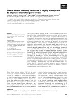
Fig. 1. Overview of plant–pathogen interactions and insights into proteomic studies of the proteins involved in these processes. Plants pos-
sess receptors that can activate basal resistance, mediated by pathogen-associated molecular patterns (PAMPs) or cell wall-degrading
enzymes (CWDEs), which may result in a compatible or incompatible interaction. In both interactions, several defence-related and biotic
stress-responsive proteins are induced. Suppression of plant defences by pathogen effectors leads to susceptibility in host plants. Some
host plants express resistance (R) proteins, which guard against this interference and trigger a specific resistance, referred to as the hyper-
sensitive response (HR). Proteomic studies of plant–pathogen interactions have revealed several pathogen and plant proteins expressed in
different pathosystems. These proteins, identified using proteomic tools, are highlighted in blue (pathogen) and red (plant) in the different
stages of the interaction.
Plant–pathogen interactions: proteomics A. Mehta et al.
3742 FEBS Journal 275 (2008) 3731–3746 ª 2008 The Authors Journal compilation ª 2008 FEBS
Although several proteins expressed during plant–
pathogen interactions have been highlighted, most are
well known and are mainly involved in the conflict
between the pathogen and the plant to suppress or
induce, respectively, the basal plant defence mecha-
nism. The results that emerge from most proteomic
analyses are of extreme importance for the validation
of the expression of the genes identified by genomic or
transcriptomic studies. However, a small amount of
novel information has been obtained, and can be
explained by the fact that key proteins are expressed in
low abundance, and are therefore not detected by cur-
rent proteomic tools. Indeed, only the most abundant
proteins are detected in two-dimensional gels and suc-
cessfully identified by MS. Another major problem
faced in proteomic analyses is protein identification by
peptide mass fingerprinting. Unequivocal identification
is usually obtained only when the genome sequence or
a large amount of sequence data are available in public
databases. When analysing poorly studied organisms,
identification must be performed by de novo seque-
ncing, which requires more sophisticated equipment,
not readily available, especially in developing countries.
Therefore, a gap appears to exist in the bioinformatics
pipeline for the proteomics of organisms with incom-
plete sequenced genomes. These technical limitations in
proteomic studies need to be overcome in order to
advance our knowledge on protein expression during
plant–pathogen interactions. Nevertheless, proteomic
tools are rapidly improving and new methods and
equipment are being developed. We believe that future
proteomic studies, coupled with functional validation
analysis, may provide new insights into disease resis-
tance and pathogenicity.
Another important aspect to be considered when
performing proteomic analyses is the follow-up study
of the identified proteins, which should be performed
in order to correctly assign protein function. The
multiple roles of proteins are a significant barrier to
progress in the unambiguous identification of proteins
involved in processes such as plant–pathogen inter-
actions. Moreover, a frequent result found in proteomic
studies is the large amount of proteins obtained with
unknown function. It is important to further investi-
gate these proteins, which may present new biological
functions and may play important roles in the
processes under investigation.
The examples reviewed here demonstrate the
complex cellular network that exists in different
plant–pathogen interactions. Overall, the use of
proteomic studies, allied to functional validation
analyses, can provide fascinating contributions to the
understanding of complex mechanisms, such as
plant–pathogen interactions. The first step in the
understanding of disease resistance is currently being
met with the identification of the proteins expressed
during plant–pathogen interactions. The next step
will be to determine which proteins confer pathoge-
nicity and disease resistance, and the mechanisms by
which they do so.
Acknowledgements
We wish to thank Dr Gilbert Engler for critical evalu-
ation of the manuscript and English revision.
References
1 Alfano JR & Collmer A (2004) Type III secretion sys-
tem effector proteins: double agents in bacterial disease
and plant defence. Annu Rev Phytol 42, 385–414.
2 Van Sluys MA, Monteiro-Vitorello CB, Camargo LEA,
Menck CFM, Da Silva ACR, Ferro JA, Oliveira MC,
Setubal JC, Kitajima JP & Simpson AJ (2002) Compar-
ative genomic analysis of plant-associated bacteria.
Annu Rev Phytol 40, 169–189.
3 Jones AME, Thomas V, Truman B, Lilley K, Mansfield
J & Grant M (2004) Specific changes in the Arabidopsis
proteome in response to bacterial challenge: differentiat-
ing basal and R-gene mediated resistance. Phytochemis-
try 65, 1805–1816.
4 Whitham SA, Yang C & Goodin MM (2006) Global
impact: elucidating plant responses to viral infection.
Mol Plant–Microbe Interact 19, 1207–1215.
5 Lee BJ, Kwon SJ, Kim SK, Kim KJ, Park CJ, Kim YJ,
Park OK & Paek KH (2006) Functional study of hot
pepper 26S proteasome subunit RPN7 induced by
tobacco mosaic virus from nuclear proteome analysis.
Biochem Biophys Res Commun 351, 405–411.
6 Diaz-Vivancos P, Rubio M, Mesonero V, Periago PM,
Barcelo AR, Martinez-Gomez P & Hernandez JA
(2006) The apoplastic antioxidant system in Prunus:
response to long-term plum pox virus infection. J Exp
Bot 57, 3813–3824.
7 Rahoutei J, Baro
´
n M, Garcı
´
a-Luque I, Droppa M,
Neme
´
nyi A & Horvath G (1999) Effect of tobamovirus
infection on the thermoluminescence characteristics of
chloroplast from infected plants. Z Naturforsch Teil C
54, 634–639.
8 Rahoutei J, Garcia-Luque I & Baron M (2000) Inhibi-
tion of photosynthesis by viral infection: effect on PSII
structure and function. Physiol Plant 110, 286–292.
9 Perez-Bueno ML, Rahoutei J, Sajnani C, Garcia-Luque
I & Baron M (2004) Proteomic analysis of the oxygen-
evolving complex of photosystem II under biotic stress:
studies on Nicotiana benthamiana infected with tobamo-
viruses. Proteomics 4, 418–425.
A. Mehta et al. Plant–pathogen interactions: proteomics
FEBS Journal 275 (2008) 3731–3746 ª 2008 The Authors Journal compilation ª 2008 FEBS 3743
10 Abbink TE, Peart JR, Mos TN, Baulcombe DC, Bol
JF & Linthorst HJ (2002) Silencing of a gene encoding
a protein component of the oxygen-evolving complex of
photosystem II enhances virus replication in plants.
Virology 295, 307–319.
11 Delalande F, Carapito C, Brizard JP, Brugidou C &
Van Dorsselaer A (2005) Multigenic families and proteo-
mics: extended protein characterization as a tool for
paralog gene identification. Proteomics 5, 450–460.
12 Brizard JP, Carapito C, Delalande F, Van Dorsselaer A
& Brugidou C (2006) Proteome analysis of plant–virus
interactome: comprehensive data for virus multiplication
inside their hosts. Mol Cell Proteomics 5, 2279–2297.
13 Casado-Vela J, Selles S & Martinez RB (2006) Proteo-
mic analysis of tobacco mosaic virus-infected tomato
(Lycopersicon esculentum M.) fruits and detection of
viral coat protein. Proteomics 6(Suppl. 1), S196–S206.
14 Lee VT & Schneewind O (2001) Protein secretion and
the pathogenesis of bacterial infections. Genes Dev 15,
1725–1752.
15 Pu
¨
hler A, Arlat M, Becker A, Go
¨
ttfert M, Morrissey JP
& O’Gara F (2004) What can bacterial genome research
teach us about bacteria–plant interactions? Curr Opin
Plant Biol 7, 137–147.
16 Galan JE & Collmer A (1999) Type III secretion
machines: bacterial devices for protein delivery into host
cells. Science 284 , 1322–1328.
17 Cornelis GR & Van Gijsegem F (2000) Assembly and
function of type III secretory systems. Annu Rev Micro-
biol 54, 735–774.
18 Keen NT (1990) Gene-for-gene complementarity in
plant–pathogen interactions. Annu Rev Genet 24, 447–
463.
19 Staskawicz BJ, Dahlbeck D & Keen NT (1984) Cloned
avirulence gene of Pseudomonas syringae pv. glycinea
determines race-specific incompatibility on Glycine max
(L.) Merr. Proc Natl Acad Sci USA 81, 6024–6028.
20 Lahaye T & Bonas U (2001) Molecular secrets of bacte-
rial type III effector proteins. Trends Plant Sci 6, 479–
485.
21 Schechter LM, Roberts KA, Jamir Y, Alfano JR &
Collmer A (2004) Pseudomonas syringae type III
secretion system targeting signals and novel effectors
studied with a Cya translocation reporter. J Bacteriol
186, 543–555.
22 Noel L, Thieme F, Nennstiel D & Bonas U (2001)
cDNA-AFLP analysis unravels a genome-wide hrpG-
regulon in the plant pathogen Xanthomonas campestris
pv. vesicatoria. Mol Microbiol 41, 1271–1281.
23 Alfano JR & Collmer A (1997) The type III (Hrp)
secretion pathway of plant pathogenic bacteria: traffick-
ing harpins, Avr proteins, and death. J Bacteriol 179,
5655–5662.
24 Arlat M, Van Gijsegem F, Huet JC, Pernollet JC &
Boucher CA (1994) PopA1, a protein which induces a
hypersensitivity-like response on specific Petunia geno-
types, is secreted via the Hrp pathway of Pseudomonas
solanacearum. EMBO J 13, 543–553.
25 Mehta A & Rosato YB (2001) Differentially expressed
proteins in the interaction of Xanthomonas axonopodis
pv. citri with leaf extract of the host plant. Proteomics
1, 1111–1118.
26 Tahara ST, Mehta A & Rosato YB (2003) Proteins
induced by Xanthomonas axonopodis pv. passiflorae with
leaf extract of the host plant (Passiflorae edulis). Proteo-
mics 3, 95–102.
27 Babujee L, Venkatesh B, Yamazaki A & Tsuyumu S
(2007) Proteomic analysis of the carbonate insoluble
outer membrane fraction of the soft-rot pathogen Dic-
keya dadantii (syn. Erwinia chrysanthemi) strain 3937.
J Proteome Res 6, 62–69.
28 Russel M (1994) Mutants at conserved positions in
gene IV, a gene required for assembly and secretion of
filamentous phages. Mol Microbiol 14, 357–369.
29 Bouchart F, Delangle A, Lemoine J, Bohin JP & Lac-
roix JM (2007) Proteomic analysis of a non-virulent
mutant of the phytopathogenic bacterium Erwinia chry-
santhemi deficient in osmoregulated periplasmic glucans:
change in protein expression is not restricted to the
envelope, but affects general metabolism. Microbiology
153, 760–767.
30 Martins D, Astua-Monge G, Coletta-Filho HD, Winck
FV, Baldasso PA, de Oliveira BM, Marangoni S, Mach-
ado MA, Novello JC & Smolka MB (2007) Absence of
classical heat shock response in the citrus pathogen
Xylella fastidiosa. Curr Microbiol 54, 119–123.
31 Kazemi-Pour N, Condemine G & Hugouvieux-Cotte-
Pattat N (2004) The secretome of the plant pathogenic
bacterium Erwinia chrysanthemi. Proteomics 4, 3177–
3186.
32 Watt SA, Wilke A, Patschkowski T & Niehaus K
(2005) Comprehensive analysis of the extracellular pro-
teins from Xanthomonas campestris pv. campestris B100.
Proteomics 5, 153–167.
33 Smolka MB, Martins D, Winck FV, Santoro CE,
Castellari RR, Ferrari F, Brum IJ, Galembeck E,
Della Coletta Filho H, Machado MA et al. (2003)
Proteome analysis of the plant pathogen Xylella fas-
tidiosa reveals major cellular and extracellular proteins
and a peculiar codon bias distribution. Proteomics 3,
224–237.
34 Rosen R, Sacher A, Shechter N, Becher D, Buttner K,
Biran D, Hecker M & Ron EZ (2004) Two-dimensional
reference map of Agrobacterium tumefaciens proteins.
Proteomics 4, 1061–1073.
35 Jones AME, Thomas V, Bennett MH, Mansfield J &
Grant M (2006) Modifications to the Arabidopsis
defence proteome occur prior to significant transcrip-
tional change in response to inoculation with Pseudomo-
nas syringae. Plant Physiol 142, 1603–1620.
Plant–pathogen interactions: proteomics A. Mehta et al.
3744 FEBS Journal 275 (2008) 3731–3746 ª 2008 The Authors Journal compilation ª 2008 FEBS
36 Mahmood T, Jan A, Kakishima M & Komatsu S
(2006) Proteomic analysis of bacterial-blight defence-
responsive proteins in rice leaf blades. Proteomics 6,
6053–6065.
37 Chen F, Yuan Y, Li Q & He Z (2007) Proteomic analy-
sis of rice plasma membrane reveals proteins involved
in early defence response to bacterial blight. Proteomics
7, 1529–1539.
38 Coaker GL, Willard B, Kinter M, Stockinger EJ &
Francis DM (2004) Proteomic analysis of resistance
mediated by Rcm 2.0 and Rcm 5.1, two loci controlling
resistance to bacterial canker of tomato. Mol Plant–
Microbe Interact 17, 1019–1028.
39 Mathesius U, Mulders S, Gao M, Teplitski M, Caet-
ano-Anolles G, Rolfe BG & Bauer WD (2003) Exten-
sive and specific responses of a eukaryote to bacterial
quorum-sensing signals. Proc Natl Acad Sci USA 100,
1444–1449.
40 Xing T, Ouellet TR & Miki BL (2002) Towards geno-
mic and proteomic studies of protein phosphorylation
in plant–pathogen interactions. Trends Plant Sci 7 , 224–
230.
41 Peck SC, Nu
¨
hse TS, Hess D, Iglesias A, Meins F &
Boller T (2001) Directed proteomics identifies a plant-
specific protein rapidly phosphorylated in response to
bacterial and fungal elicitors. Plant Cell 13, 1467–1475.
42 Jones AME, Bennett MH, Mansfield JW & Grant M
(2006) Analysis of the defence phosphoproteome of
Arabidopsis thaliana using differential mass tagging.
Proteomics 6, 4155–4165.
43 Murad AM, Laumann RA, Lima Tde A, Sarmento RB,
Noronha EF, Rocha TL, Valadares-Inglis MC &
Franco OL (2006) Screening of entomopathogenic
Metarhizium anisopliae isolates and proteomic analysis
of secretion synthesized in response to cowpea weevil
(Callosobruchus maculatus) exoskeleton. Comp Biochem
Physiol C Toxicol Pharmacol 142, 365–370.
44 Murad AM, Laumann RA, Mehta A, Noronha EF &
Franco OL (2007) Screening and secretomic analysis of
entomopathogenic Beauveria bassiana isolates in
response to cowpea weevil (Callosobruchus maculatus)
exoskeleton. Comp Biochem Physiol C Toxicol Pharma-
col 145, 333–338.
45 Bohmer M, Colby T, Bohmer C, Brautigam A, Schmidt
J & Bolker M (2007) Proteomic analysis of dimorphic
transition in the phytopathogenic fungus Ustilago may-
dis. Proteomics 7, 675–685.
46 Grenville-Briggs LJ, Avrova AO, Bruce CR, Williams
A, Whisson SC, Birch PR & van West P (2005) Ele-
vated amino acid biosynthesis in Phytophthora infestans
during appressorium formation and potato infection.
Fungal Genet Biol 42, 244–256.
47 Rampitsch C, Bykova NV, McCallum B, Beimcik E &
Ens W (2006) Analysis of the wheat and Puccinia triticina
(leaf rust) proteomes during a susceptible host–patho-
gen interaction. Proteomics 6, 1897–1907.
48 Webb CA & Fellers JP (2006) Cereal rust fungi genom-
ics and the pursuit of virulence and avirulence factors.
FEMS Microbiol Lett 264, 1–7.
49 Phalip V, Delalande F, Carapito C, Goubet F, Hatsch
D, Leize-Wagner E, Dupree P, Dorsselaer AV & Jeltsch
JM (2005) Diversity of the exoproteome of Fusarium
graminearum grown on plant cell wall. Curr Genet
48,
366–379.
50 Meijer HJ, van de Vondervoort PJ, Yin QY, de Koster
CG, Klis FM, Govers F & de Groot PW (2006) Identifi-
cation of cell wall-associated proteins from Phytophthora
ramorum. Mol Plant–Microbe Interact 19, 1348–1358.
51 Yajima W & Kav NN (2006) The proteome of the phy-
topathogenic fungus Sclerotinia sclerotiorum. Proteomics
6, 5995–6007.
52 Talbot NJ (2003) On the trail of a cereal killer: explor-
ing the biology of Magnaporthe grisea. Annu Rev Micro-
biol 57, 177–202.
53 Konishi H, Ishiguro K & Komatsu S (2001) A proteo-
mics approach towards understanding blast fungus
infection of rice grown under different levels of nitrogen
fertilization. Proteomics 1, 1162–1171.
54 Long DH, Lee FN & TeBeest DO (2000) Effect of
nitrogen fertilization on disease progress of rice blast on
susceptible and resistant cultivars. Plant Dis 84, 403–
409.
55 Rakwal R & Agrawal GK (2003) Rice proteomics: cur-
rent status and future perspectives. Electrophoresis 24,
3378–3389.
56 Kim ST, Cho KS, Yu S, Kim SG, Hong JC, Han CD,
Bae DW, Nam MH & Kang KY (2003) Proteomic
analysis of differentially expressed proteins induced by
rice blast fungus and elicitor in suspension-cultured rice
cells. Proteomics 3, 2368–2378.
57 Kim ST, Kim SG, Hwang DH, Kang SY, Kim HJ, Lee
BH, Lee JJ & Kang KY (2004) Proteomic analysis of
pathogen-responsive proteins from rice leaves induced
by rice blast fungus, Magnaporthe grisea. Proteomics 4,
3569–3578.
58 Lee J, Bricker TM, Lefevre M, Pinson SRM & Oard
JH (2006) Proteomic and genetic approaches to identify
defence-related proteins in rice challenged with the fun-
gal pathogen Rhizoctonia solani. Mol Plant Pathol 7,
405–416.
59 Zhou W, Eudes F & Laroche A (2006) Identification of
differentially regulated proteins in response to a com-
patible interaction between the pathogen Fusarium
graminearum and its host, Triticum aestivum. Proteomics
6, 4599–4609.
60 Colditz F, Nyamsuren O, Niehaus K, Eubel H, Braun
HP & Krajinski F (2004) Proteomic approach: identifi-
cation of Medicago truncatula proteins induced in roots
A. Mehta et al. Plant–pathogen interactions: proteomics
FEBS Journal 275 (2008) 3731–3746 ª 2008 The Authors Journal compilation ª 2008 FEBS 3745
after infection with the pathogenic oomycete Aphanomy-
ces euteiches. Plant Mol Biol 55, 109–120.
61 Campo S, Carrascal M, Coca M, Abian J & San Seg-
undo B (2004) The defence response of germinating
maize embryos against fungal infection: a proteomics
approach. Proteomics 4, 383–396.
62 Houterman PM, Speijer D, Dekker HL, De Koster CG,
Cornelissen BJC & Rep M (2007) The mixed xylem sap
proteome of Fusarium oxysporum-infected tomato
plants. Mol Plant Pathol 8, 215–221.
63 Chitwood DJ (2003) Research on plant-parasitic nema-
tode biology conducted by the United States Depart-
ment of Agriculture-Agricultural Research Service. Pest
Manag Sci 59, 748–753.
64 Curtis RH (2007) Plant parasitic nematode proteins and
the host–parasite interaction. Brief Funct Genomic Pro-
teomic 6, 50–58.
65 Smant G, Stokkermans JP, Yan Y, de Boer JM,
Baum TJ, Wang X, Hussey RS, Gommers FJ,
Henrissat B, Davis EL et al. (1998) Endogenous
cellulases in animals: isolation of beta–1,4–endoglucan-
ase genes from two species of plant-parasitic
cyst nematodes. Proc Natl Acad Sci USA 95, 4906–
4911.
66 Gao B, Allen R, Maier T, Davis EL, Baum TJ & Hus-
sey RS (2003) The parasitome of the phytonematode
Heterodera glycines. Mol Plant–Microbe Interact 16,
720–726.
67 Huang G, Gao B, Maier T, Allen R, Davis EL, Baum
TJ & Hussey RS (2003) A profile of putative parasitism
genes expressed in the esophageal gland cells of the
root-knot nematode Meloidogyne incognita. Mol Plant–
Microbe Interact 16, 376–381.
68 Tytgat T, Vercauteren I, Vanholme B, De Meutter J,
Vanhoutte I, Gheysen G, Borgonie G, Coomans A &
Gheysen G (2005) An SXP ⁄ RAL–2 protein produced
by the subventral pharyngeal glands in the plant para-
sitic root-knot nematode Meloidogyne incognita. Parasi-
tol Res 95, 50–54.
69 De Meutter J, Vanholme B, Bauw G, Tytgat T, Ghey-
sen G & Gheysen G (2001) Preparation and sequencing
of secreted proteins from the pharyngeal glands of the
plant parasitic nematode Heterodera schachtii. Mol
Plant Pathol 2, 297–301.
70 Jaubert S, Ledger TN, Laffaire JB, Piotte C, Abad P &
Rosso MN (2002) Direct identification of stylet secreted
proteins from root-knot nematodes by a proteomic
approach. Mol Biochem Parasitol 121, 205–211.
71 Calvo E, Flores-Romero P, Lopez JA & Navas A
(2005) Identification of proteins expressing differences
among isolates of Meloidogyne spp. (Nematoda: Melo-
idogynidae) by nano-liquid chromatography coupled
to ion-trap mass spectrometry. J Proteome Res 4,
1017–1021.
72 Ashton PD, Curwen RS & Wilson RA (2001) Linking
proteome and genome: how to identify parasite pro-
teins. Trends Parasitol 17, 198–202.
73 Williamson VM & Kumar A (2006) Nematode resis-
tance in plants: the battle underground. Trends Genet
22, 396–403.
74 Mehta A, Magalha
˜
es BS, Souza DSL, Vasconcelos
EAR, Silva LP, Grossi-de-Sa
´
MF, Franco OL, da
Costa PHA & Rocha TL (2008) Rooteomics: the chal-
lenge of discovering plant defence-related proteins in
roots. Curr Prot Pep Sci 9, 108–116.
75 Chivasa S, Hamilton JM, Pringle RS, Ndimba BK,
Simon WJ, Lindsey K & Slabas AR (2006) Proteomic
analysis of differentially expressed proteins in fungal
elicitor-treated Arabidopsis cell cultures. J Exp Bot 57,
1553–1562.
Plant–pathogen interactions: proteomics A. Mehta et al.
3746 FEBS Journal 275 (2008) 3731–3746 ª 2008 The Authors Journal compilation ª 2008 FEBS