Tài liệu Báo cáo khoa học: Octaketide-producing type III polyketide synthase from Hypericum perforatum is expressed in dark glands accumulating hypericins pdf
Bạn đang xem bản rút gọn của tài liệu. Xem và tải ngay bản đầy đủ của tài liệu tại đây (686.02 KB, 14 trang )
Octaketide-producing type III polyketide synthase from
Hypericum perforatum is expressed in dark glands
accumulating hypericins
Katja Karppinen
1
, Juho Hokkanen
2,
*, Sampo Mattila
2
, Peter Neubauer
3
and Anja Hohtola
1
1 Department of Biology, University of Oulu, Finland
2 Department of Chemistry, University of Oulu, Finland
3 Department of Process and Environmental Engineering, University of Oulu, Finland
Hypericum perforatum L., St John’s wort, is a medici-
nal plant that is widely utilized for the treatment of
mild to moderate depression [1,2]. Hypericins, a
group of red-pigmented naphthodianthrones including
hypericin and pseudohypericin, as well as their
intimate precursors protohypericin and proto-
pseudohypericin (Fig. 1), are considered the principal
agents in the range of biological activities reported for
H. perforatum [3–5]. Hypericins, together with other
bioactive compounds of the crude plant extract, such
as hyperforins and flavonoids, have been found to con-
tribute to the antidepressant activity of the plant [1–3].
Hypericin is the most potent natural photosensitizer
described to date, and its photodynamic activities
allow hypericin to also act as an antiviral and antitu-
moral agent [3,6–8]. In H. perforatum, hypericins have
been suggested to act as the plant’s defence against
insects [9].
H. perforatum is characterized by the presence of
dark glands in the aerial parts of the plant [10–13].
Keywords
dark glands; hypericin; octaketide synthase;
St. John’s wort (Hypericum perforatum L.);
type III polyketide synthase
Correspondence
K. Karppinen, Department of Biology,
University of Oulu, PO Box 3000, FIN-90014
Oulu, Finland
Fax: +358 8 553 1061
Tel: +358 8 553 1544
E-mail: katja.karppinen@oulu.fi
*Present address
Novamass Ltd, Oulu, Finland
Database
Nucleotide sequence data have been
submitted to the DDBJ ⁄ EMBL ⁄ GenBank
databases under the accession number
EU635882
(Received 26 April 2008, revised 18 June
2008, accepted 27 June 2008)
doi:10.1111/j.1742-4658.2008.06576.x
Hypericins are biologically active constituents of Hypericum perforatum
(St John’s wort). It is likely that emodin anthrone, an anthraquinone
precursor of hypericins, is biosynthesized via the polyketide pathway by
type III polyketide synthase (PKS). A PKS from H. perforatum, HpPKS2,
was investigated for its possible involvement in the biosynthesis of hyperic-
ins. Phylogenetic tree analysis revealed that HpPKS2 groups with function-
ally divergent non-chalcone-producing plant-specific type III PKSs, but it
is not particularly closely related to any of the currently known type III
PKSs. A recombinant HpPKS2 expressed in Escherichia coli resulted in an
enzyme of 43 kDa. The purified enzyme catalysed the condensation of
acetyl-CoA with two to seven malonyl-CoA to yield tri- to octaketide prod-
ucts, including octaketides SEK4 and SEK4b, as well as heptaketide aloe-
sone. Although HpPKS2 was found to have octaketide synthase activity,
production of emodin anthrone, a supposed octaketide precursor of
hypericins, was not detected. The enzyme also accepted isobutyryl-CoA,
benzoyl-CoA and hexanoyl-CoA as starter substrates producing a variety
of tri- to heptaketide products. In situ RNA hybridization localized the
HpPKS2 transcripts in H. perforatum leaf margins, flower petals and
stamens, specifically in multicellular dark glands accumulating hypericins.
Based on our results, HpPKS2 may have a role in the biosynthesis of
hypericins in H. perforatum but some additional factors are possibly
required for the production of emodin anthrone in vivo.
Abbreviations
CHS, chalcone synthase; DIG, digoxigenin; IPTG, isopropyl thio-b-
D-galactoside; OKS, octaketide synthase; PCS, pentaketide chromone
synthase; PKS, polyketide synthase; STS, stilbene synthase.
FEBS Journal 275 (2008) 4329–4342 ª 2008 The Authors Journal compilation ª 2008 FEBS 4329
Dark glands appear as black or dark red multicellular
nodules that occur near the leaf margins, stems, flower
petals and stamens [10–12,14]. Hypericum species with
dark glands are known to produce hypericins [15]. The
correlation between the concentrations of hypericins
and the existence of dark glands in H. perforatum tis-
sues has shown the presence of these red pigments in
the glands [12,16,17]. Hypericin has also been shown
to accumulate in red glands in the sepals in H. elodes
[15]. It has been proposed that hypericins not only
accumulate, but are also biosynthesized in the dark
glands [12,18]. The localization of hypericins in the
nodular structures is considered to have evolved as a
mechanism for the plant to avoid the potential auto-
toxicity of these compounds [19].
The biosynthesis of hypericins is currently poorly
understood, but the polyketide pathway is likely to
play a central role [12,20]. Plant-specific type III poly-
ketide synthases (PKSs) are involved in the biosynthe-
sis of a large variety of plant secondary metabolites,
including chalcones, stilbenes, benzophenones, acri-
dones, phloroglucinols and benzalacetone derivatives
[21]. The enzymes catalyse the formation of complex
natural products by condensing various CoA-thioesters
with malonyl-CoA in a reaction sequence that closely
parallels fatty acid biosynthesis [22]. The functional
diversity of simple homodimeric type III PKSs is
derived from small differences in the active site that
influence the substrate specificities, the number of con-
densation reactions and the mechanisms of cyclization
reactions [22,23]. In some cases, the reaction inter-
mediates are also modified by interaction with other
enzymes [23]. The type III PKS involved in the biosyn-
thesis of hypericins has been suggested to condensate
one molecule of acetyl-CoA with seven molecules of
malonyl-CoA to form an octaketide chain that subse-
quently undergoes cyclizations and decarboxylation,
leading to the formation of emodin anthrone (Fig. 1),
a precursor of hypericins [3,12,20]. However, there are
no reports on the characterization of the type III PKS
with octaketide synthase (OKS) activity which is
responsible for the formation of emodin anthrone. The
final stages of hypericin biosynthesis are conducted by
the gene product of hyp-1, encoding for the phenolic
coupling protein that catalyses the oxidative dimeriza-
tion of emodin anthrone to hypericin [3,20].
To date, four different PKS family genes have been
cloned from the genus Hypericum. Chalcone synthase
(CHS) and benzophenone synthase have been cloned
from both H. androsaemum [24] and H. perforatum
[25]. In addition, in a recent study, we described the
cloning of two previously uncharacterized cDNAs
from H. perforatum encoding for PKSs, designated as
HpPKS1 and HpPKS2 [25]. Expression of HpPKS2
was found to correlate with the concentration of hyp-
ericins in H. perforatum tissues and HpPKS2 is thus a
candidate gene for the biosynthesis of hypericins [25].
In this study, the role of H. perforatum HpPKS2 is
investigated in more detail. We describe the functional
characterization of HpPKS2 and the exact localization
of HpPKS2 transcripts in H. perforatum leaves and
flower buds using in situ RNA hybridization. HpPKS2
was found to be an OKS and is specifically expressed in
the dark glands accumulating hypericins. Thus, the
results imply that HpPKS2 may have a role in the bio-
synthesis of hypericins in H. perforatum. The failure of
HpPKS2 to catalyse the formation of emodin anthrone
in vitro, but produce other octaketides instead, is dis-
cussed in terms of a possible need for some additional
factors for the production of emodin anthrone.
CoAS
O
Acetyl-CoA
+ 7 × malonyl-CoA
OKS
O
H
OH O OH
O
Emodin
OH
OH OHO
Oxidation
OOO
O
OOO
SEnz
O
SEK4
O
O
OH
O
O
OH
OH
SEK4b
O
OOH
O
O
OH
OH
in vitro
Cyclizations,
decarboxylation
in vivo
OH O
OH
OH
CH
3
R
OH
O
OH
OH
R = CH
3
, Hypericin
R = CH
2
OH, Pseudohypericin
R = CH
3
, Protohypericin
R = CH
2
OH, Protopseudohypericin
OH
O
OH
CH
3
R
OH O OH
OH
O
H
Oxidative
dimerization
3
2
O
Emodin anthrone
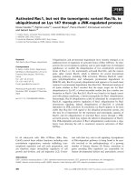
Fig. 1. Putative reaction of OKS involved in the biosynthesis of hypericins in H. perforatum. In vivo OKS is suggested to condensate one ace-
tyl-CoA with seven malonyl-CoA to form an octaketide chain that subsequently undergoes cyclizations and decarboxylation to form emodin
anthrone. It is possible that in vitro OKS affords shunt products SEK4 and SEK4b in the absence of some additional, yet unidentified factors.
Octaketide synthase from Hypericum perforatum K. Karppinen et al.
4330 FEBS Journal 275 (2008) 4329–4342 ª 2008 The Authors Journal compilation ª 2008 FEBS
Results
During amplification of the HpPKS2 coding sequence
from H. perforatum, several cDNA clones of HpPKS2
that differed slightly from one another were encoun-
tered. The deduced amino acid sequences of the clones
shared 99–100% identity. The HpPKS2 clone that was
found to be the most abundant of the different cDNA
clones in H. perforatum was selected for investigation
in this study. The nucleotide sequence of the clone has
been deposited in GenBank under the accession num-
ber EU635882. However, because the HpPKS2 clones
showed such high sequence similarity and thus their
expression in H. perforatum tissues could not be distin-
guished from each other, the general name HpPKS2 is
used in this study.
Phylogenetic analysis
The overall similarity of the deduced amino acid
sequence of HpPKS2 with other type III PKS family
proteins was investigated using a neighbor-joining
tree (Fig. 2). Phylogenetic analysis showed that the
members of the plant-specific type III PKSs grouped
into CHSs and non-CHSs, except stilbene synthases
(STSs) from Fabaceae and Gymnosperms. In these
cases, the STSs were closer to CHSs of the same or
related species than other non-chalcone-forming PKSs.
HpPKS2 grouped with functionally divergent
non-chalcone-forming plant-specific type III PKSs,
including OKS and pentaketide chromone synthase
(PCS) from Aloe arborescens [26,27]. However,
HpPKS2 was positioned on a sub-branch of its own
without any particularly closely related proteins.
Expression of HpPKS2 in Escherichia coli
To study the enzymatic function of HpPKS2 in more
detail, the coding region of the HpPKS2 cDNA was
functionally expressed in Escherichia coli strain M15
[pREP4] with pQE30 vector. When E. coli cells
harbouring the recombinant plasmid were grown at
Oryza sativa CHS (AB000801)
Zea mays CHS (X60205)
Ruta graveolens CHS (AJ297789)
Gerbera hybrida CHS (Z38096)
Arabidopsis thaliana CHS (AF112086)
Vitis vinifera CHS (X75969)
Hypericum androsaemum CHS (AF315345)
Sorbus aucuparia CHS (DQ286037)
Camellia sinensis CHS (D26593)
Petunia hybrida CHS (X04080)
Hydrangea macrophylla CHS (AB011467)
Picea mariana CHS (AF227627)
Pinus sylvestris CHS (X60754)
Pinus sylvestris STS (S50350)
Pueraria lobata CHS (D10223)
Phaseolus vulgaris CHS (X06411)
Pisum sativum CHS (X63333)
Medicago sativa CHS (L02902)
Glycine max CHS (X53958)
Arachis hypogaea STS (L00952)
Vitis vinifera STS (S63221)
Rheum palmatum BAS (AF326911)
Humulus lupulus VPS (AB015430)
Hydrangea macrophylla CTAS (AB011468)
Hydrangea macrophylla STCS (AF456445)
Ruta graveolens ACS (AJ297788)
Gerbera hybrida 2-PS (Z38097)
Rheum palmatum ALS (AY517486)
Plumbago indica PKS (AB259100)
Phalaenopsis sp. BBS (X79903)
Bromheadia finlaysoniana BBS (AJ131830)
Sorbus aucuparia BIS (DQ286036)
Hypericum androsaemum BPS (AF352395)
Wachendorfia thyrsiflora PKS1 (AY727928)
Ipomoea purpurea CHS-B (U15947)
Ipomoea purpurea CHS-A (U15946)
Aloe arborescens PCS (AY823626)
Aloe arborescens OKS (AY567707)
Hypericum perforatum HpPKS2 (EU635882)
Aspergillus oryzae csyB (AB206759)
Aspergillus oryzae csyA (AB206758)
Fusarium graminearum FG08378.1 (XM_388554)
Magnaporthe grisea MG04643.4 (XM_362198)
Streptomyces griseus RppA (AB018074)
100
100
100
100
100
100
100
99
94
90
57
54
51
87
100
98
100
100
0,1
Fabaceae
Gymnosperms
divergent PKSs
CHSs
plants
fungi
bacteria
Oryza sativa CHS (AB000801)
Zea mays CHS (X60205)
Ruta graveolens CHS (AJ297789)
Gerbera hybrida CHS (Z38096)
Arabidopsis thaliana CHS (AF112086)
Vitis vinifera CHS (X75969)
Hypericum androsaemum CHS (AF315345)
Sorbus aucuparia CHS (DQ286037)
Camellia sinensis CHS (D26593)
Petunia hybrida CHS (X04080)
Hydrangea macrophylla CHS (AB011467)
Picea mariana CHS (AF227627)
Pinus sylvestris CHS (X60754)
Pinus sylvestris STS (S50350)
Pueraria lobata CHS (D10223)
Phaseolus vulgaris CHS (X06411)
Pisum sativum CHS (X63333)
Medicago sativa CHS (L02902)
Glycine max CHS (X53958)
Arachis hypogaea STS (L00952)
Vitis vinifera STS (S63221)
Rheum palmatum BAS (AF326911)
Humulus lupulus VPS (AB015430)
Hydrangea macrophylla CTAS (AB011468)
Hydrangea macrophylla STCS (AF456445)
Ruta graveolens ACS (AJ297788)
Gerbera hybrida 2-PS (Z38097)
Rheum palmatum ALS (AY517486)
Plumbago indica PKS (AB259100)
Phalaenopsis sp. BBS (X79903)
Bromheadia finlaysoniana BBS (AJ131830)
Sorbus aucuparia BIS (DQ286036)
Hypericum androsaemum BPS (AF352395)
Wachendorfia thyrsiflora PKS1 (AY727928)
Ipomoea purpurea CHS-B (U15947)
Ipomoea purpurea CHS-A (U15946)
Aloe arborescens PCS (AY823626)
Aloe arborescens OKS (AY567707)
Hypericum perforatum HpPKS2 (EU635882)
Aspergillus oryzae csyB (AB206759)
Aspergillus oryzae csyA (AB206758)
Fusarium graminearum FG08378.1 (XM_388554)
Magnaporthe grisea MG04643.4 (XM_362198)
Streptomyces griseus RppA (AB018074)
100
100
100
100
100
100
100
99
94
90
57
54
51
87
100
98
100
100
0,1
Fabaceae
Gymnosperms
CHSs
plants
fungi
bacteria
Fabaceae
Gymnosperms
FabaceaeFabaceae
GymnospermsGymnosperms
CHSsCHSs
Functionally
CHSs
plants
fungi
bacteria
plants
fungi
bacteria
Fig. 2. Phylogenetic analysis of type III PKS
enzymes. The tree was constructed using
the neighbor-joining algorithm. The numbers
at the forks are bootstrap values that
indicate the per cent values for obtaining
this particular branching in 1000 replicates;
only values > 50% are shown. The indicated
scale represents 0.1 amino acid substitu-
tions per site. The GenBank accession num-
bers are followed by the names of the
species. ACS, acridone synthase; ALS, aloe-
sone synthase; BAS, benzalacetone syn-
thase; BBS, bibenzyl synthase; BIS,
biphenyl synthase; BPS, benzophenone syn-
thase; CHS, chalcone synthase; CTAS,
4-coumaroyltriacetic acid synthase; OKS,
octaketide synthase; PCS, pentaketide
chromone synthase; 2-PS, 2-pyrone
synthase; STCS, stilbene carboxylate
synthase; STS, stilbene synthase; VPS,
valerophenone synthase.
K. Karppinen et al. Octaketide synthase from Hypericum perforatum
FEBS Journal 275 (2008) 4329–4342 ª 2008 The Authors Journal compilation ª 2008 FEBS 4331
37 °C after induction with isopropyl thio-b-d-galacto-
side (IPTG), all the induced HpPKS2 proteins
became insoluble. Similar phenomena have been
reported previously in the expression of some plant-
specific type III PKSs in E. coli [28,29], and in many
cases, a low temperature has been used to obtain
recombinant PKS in a soluble form [28,30–32]. There-
fore, the culture temperature was lowered to 16 °C
after induction with IPTG. Under these culture condi-
tions, IPTG-induced E. coli cells produced the soluble
HpPKS2 protein, as shown on SDS ⁄ PAGE gel
(Fig. 3). Because the recombinant HpPKS2 protein
contained an additional hexahistidine tag at the
N-terminus, it enabled us to obtain the enzyme with
high purity after purification with Ni-NTA agarose.
After purification, commonly 2.5 mg of pure
recombinant HpPKS2 was obtained from 1 g of
E. coli cell pellet. The purified enzyme gave a band
with a molecular mass of 43 kDa on SDS ⁄ PAGE
gel (Fig. 3).
Enzyme activity of recombinant HpPKS2
The enzymatic activity of the purified recombinant
HpPKS2 was tested for suggested emodin anthrone-
forming activity by using acetyl-CoA as a starter sub-
strate. Some of the products (Fig. 4), determined by
UPLC ⁄ ESIMS, were simple a-pyrones with a linear
keto side chain (A1, A2, A3) showing loss of CO
2
from the parent ion ([M-H-44]
)
) in the negative ioni-
zation mode (Fig. 5A). Other characteristic fragments
at m ⁄ z 125 corresponding to [C
6
H
5
O
3
]
)
(pyrone moi-
ety) and m ⁄ z 167 corresponding to [C
8
H
7
O
4
]
)
were
also detected for some a-pyrones, depending on the
chain length of the particular compound. Octaketides
SEK4 (A4) and SEK4b (A7), as well as heptaketide
aloesone (A9), were also found from incubations. The
proposed fragmentation patterns of SEK4 and SEK4b
in the negative ionization mode are presented in
Fig. 5B,C, respectively. A heptaketide aloesone was
identified based on its UV spectrum and the structure
was confirmed by its fragmentation in the positive
ionization mode (Fig. 5D). HpPKS2 also produced
pentaketide chromone A8 (2,7-dihydroxy-5-methyl-
chromone) and heptaketide chromone A10 [1-(5,7-
dihydroxy-4-oxo-4H-chromen-2-yl)pentane-2,4-dione].
The structure A8 was identified based on its UV
spectrum, exact mass and retention behaviour [26].
Structure A10 was identified based on its exact mass
and fragment ion at m ⁄ z 189 (loss of acyl side chain)
in ESI
)
conditions. Also, heptaketide phenylpyrone
A6 [6-(2,4-dihydroxy-6-(2-oxopropyl)phenyl)-4-hydro-
xy-2H-pyran-2-one] showing loss of CO
2
from the
parent ion ([M-H-44]
)
), but no other fragments under
ESI
)
conditions, was identified. Although HpPKS2
showed the expected OKS activity, emodin anthrone,
a supposed octaketide precursor of hypericins, was
not detected.
Recombinant HpPKS2 was also examined for its
ability to use other CoA-thioesters as starter sub-
strates. It was found that HpPKS2 accepted all tested
starter units (isobutyryl-CoA, benzoyl-CoA and hexa-
noyl-CoA) to produce a variety of tri- to heptaketide
products (Fig. 4), most of which were identified as
a-pyrones with a linear keto side chain (B1, B2, B3,
B4, C1, C2, C3, C5, C7, D1, D2, D4, D6). In
addition, chromones B8 [1-(5,7-dihydroxy-4-oxo-4H-
chromen-2-yl)-5-methylhexane-2,4-dione] and C6 [5,7-
dihydroxy-2-(2-oxo-2-phenylethyl)-4H-chromen-4-one],
phloroglucinols B6 [6-methyl-1-(2,4,6-trihydroxyphe-
nyl)heptane-1,3,5-trione] and D5 [1-(2,4,6-trihydroxy-
phenyl)decane-1,3,5-trione], as well as phenylpyrones
B5 [6-(2,4-dihydroxy-6-(3-methyl-2-oxobutyl)phenyl)
-4-hydroxy-2H-pyran-2-one], C4 [6-(3,5-dihydroxy
biphenyl-2-yl)-4-hydroxy-2H-pyran-2-one] and D3
[6-(2,4-dihydroxy-6-pentylphenyl)-4-hydroxy-2H-pyran-
2-one] were detected. Identification of the compounds
was made based on their similar fragmentation, UV
characteristics and retention behaviour compared with
the corresponding products obtained using acetyl-
CoA as a starter substrate. None of the above-men-
tioned products was found from negative control
reactions that contained heat-denatured enzyme with
corresponding substrates.
1234 M
kDa
116.0
66.2
45.0
35.0
25.0
18.4
14.4
Fig. 3. SDS ⁄ PAGE analysis of recombinant HpPKS2 expressed in
E. coli. (1) Total proteins from E. coli without induction, (2) total pro-
teins from E. coli induced with IPTG, (3) soluble proteins, (4) puri-
fied recombinant HpPKS2 protein, (M) protein molecular mass
marker, with sizes (kDa) indicated at the right.
Octaketide synthase from Hypericum perforatum K. Karppinen et al.
4332 FEBS Journal 275 (2008) 4329–4342 ª 2008 The Authors Journal compilation ª 2008 FEBS
Heptaketides
Hexaketides
Pentaketides
Substrate
Tetraketides
Triketides
OO
OH
OOOO
C2
RT
UPLC
= 1.50 min
m/z 355 [M-H]
-
λ
max
274 nm
OO
OH
OHOH
RT
UPLC
= 1.89 min
m/z 295 [M-H]
-
λ
max
307 nm
C4
OO
OH
O
O
C7
RT
UPLC
= 2.63 min
m/z 271 [M-H]
-
λ
max
316 nm
O
OOH
OH
O
C6
RT
UPLC
= 2.54 min
m/z 295 [M-H]
-
λ
max
242, 285, 340 nm
OO
OH
OOO
C1
RT
UPLC
= 1.25 min
m/z 313 [M-H]
-
λ
max
264 nm
CoAS
O
Benzoyl-CoA
OO
OH
O
C3
RT
UPLC
= 1.77 min
m/z 229 [M-H]
-
λ
max
246, 284 nm
OO
OH
RT
UPLC
= 1.93 min
m/z 187 [M-H]
-
λ
max
219, 233, 318 nm
C5
OO
OH
OHOH
D3
RT
UPLC
= 2.42 min
m/z 289 [M-H]
-
λ
max
298 nm
O
O
O
OH
O
D6
RT
UPLC
= 3.16 min
m/z 265 [M-H]
-
λ
max
281 nm
O
OH
O
OOO
D1
RT
UPLC
= 1.79 min
m/z 307 [M-H]
-
λ
max
261 nm
OHOH
OH
OO
O
D5
RT
UPLC
= 3.13 min
m/z 307 [M-H]
-
λ
max
288 nm
CoAS
O
Hexanoyl-CoA
O
OH
O
O
D2
RT
UPLC
= 2.39 min
m/z 223 [M-H]
-
λ
max
284 nm
OO
OH
D4
RT
UPLC
= 2.54 min
m/z 181 [M-H]
-
λ
max
227, 284 nm
OO
OH
OHOH
O
B5
RT
UPL C
= 1.77 min
m/z 303 [M-H]
-
λ
max
238, 286 nm
OHOH
OH
O
OO
RT
UPL C
= 2.18 min
m/z 279 [M-H]
-
λ
max
238, 287 nm
B6
OO
OH
O
OOO
B2
RT
UPL C
= 1.12 min
m/z 321 [M-H]
-
λ
max
264 nm
OO
OH
O
OO
B1
RT
UPL C
= 0.96 min
m/z 279 [M-H]
-
λ
max
262 nm
OOH
OH O
O
O
B8
RT
UPL C
= 2.89 min
m/z 303 [M-H]
-
λ
max
230, 281, 324, 337, 404 nm
CoAS
O
Isobutyryl-CoA
OO
OH
O
B3
RT
UPL C
= 1.28 min
m/z 195 [M-H]
-
λ
max
284 nm
OO
OH
RT
UPL C
= 1.43 min
m/z 153 [M-H]
-
λ
max
225, 284 nm
B4
Octaketides
O
OOH
O
O
OH
OH
A7
RT
UPL C
= 1.31 min
m/z 317 [M-H]
-
λ
max
230, 280 nm
O
OOH
O
O
OH
OH
A4
RT
UPL C
= 0.84 min
m/z 317 [M-H]
-
λ
max
284 nm
OO
OH
OH
OH
O
A6
RT
UPL C
= 1.17 min
m/z 275 [M-H]
-
λ
max
282 nm
CoAS
O
Acetyl-CoA
OO
OH
OOO
RT
UPL C
= 0.45 min
m/z 251 [M-H]
-
λ
max
270 nm
A1
OO
OH
O
A2
RT
UPL C
= 0.52 min
m/z 167 [M-H]
-
λ
max
285 nm
O
O
OHOH
A8
RT
UPL C
= 1.37 min
m/z 191 [M-H]
-
λ
max
309 nm
RT
UPL C
= 0.62 min
m/z 125 [M-H]
-
λ
max
283 nm
A3
OO
OH
O
O
OH
O
A9
RT
UPL C
= 1.45 min
m/z 231 [M-H]
-
λ
max
243, 251, 292 nm
O
O
OH
OH
OO
A10
RT
UPL C
= 1.54 min
m/z 275 [M-H]
-
λ
max
235, 276 nm
Heptaketides
Hexaketides
Pentaketides
Substrate
Tetraketides
Triketides
Heptaketides
Hexaketides
Pentaketides
Substrate
Tetraketides
Triketides
OO
OH
OOOO
C2
RT
UPLC
= 1.50 min
m/z 355 [M-H]
-
λ
max
274 nm
OO
OH
OHOH
RT
UPLC
= 1.89 min
m/z 295 [M-H]
-
λ
max
307 nm
C4
OO
OH
O
O
C7
RT
UPLC
= 2.63 min
m/z 271 [M-H]
-
λ
max
316 nm
O
OOH
OH
O
C6
RT
UPLC
= 2.54 min
m/z 295 [M-H]
-
λ
max
242, 285, 340 nm
OO
OH
OOO
C1
RT
UPLC
= 1.25 min
m/z 313 [M-H]
-
λ
max
264 nm
CoAS
O
Benzoyl-CoA
OO
OH
O
C3
RT
UPLC
= 1.77 min
m/z 229 [M-H]
-
λ
max
246, 284 nm
OO
OH
RT
UPLC
= 1.93 min
m/z 187 [M-H]
-
λ
max
219, 233, 318 nm
C5
OO
OH
OHOH
D3
RT
UPLC
= 2.42 min
m/z 289 [M-H]
-
λ
max
298 nm
O
O
O
OH
O
D6
RT
UPLC
= 3.16 min
m/z 265 [M-H]
-
λ
max
281 nm
O
OH
O
OOO
D1
RT
UPLC
= 1.79 min
m/z 307 [M-H]
-
λ
max
261 nm
OHOH
OH
OO
O
D5
RT
UPLC
= 3.13 min
m/z 307 [M-H]
-
λ
max
288 nm
CoAS
O
Hexanoyl-CoA
O
OH
O
O
D2
RT
UPLC
= 2.39 min
m/z 223 [M-H]
-
λ
max
284 nm
OO
OH
D4
RT
UPLC
= 2.54 min
m/z 181 [M-H]
-
λ
max
227, 284 nm
OO
OH
OHOH
O
B5
RT
UPL C
= 1.77 min
m/z 303 [M-H]
-
λ
max
238, 286 nm
OHOH
OH
O
OO
RT
UPL C
= 2.18 min
m/z 279 [M-H]
-
λ
max
238, 287 nm
B6
OO
OH
O
OOO
B2
RT
UPL C
= 1.12 min
m/z 321 [M-H]
-
λ
max
264 nm
OO
OH
O
OO
B1
RT
UPL C
= 0.96 min
m/z 279 [M-H]
-
λ
max
262 nm
OOH
OH O
O
O
B8
RT
UPL C
= 2.89 min
m/z 303 [M-H]
-
λ
max
230, 281, 324, 337, 404 nm
CoAS
O
Isobutyryl-CoA
OO
OH
O
B3
RT
UPL C
= 1.28 min
m/z 195 [M-H]
-
λ
max
284 nm
OO
OH
RT
UPL C
= 1.43 min
m/z 153 [M-H]
-
λ
max
225, 284 nm
B4
Octaketides
O
OOH
O
O
OH
OH
A7
RT
UPL C
= 1.31 min
m/z 317 [M-H]
-
λ
max
230, 280 nm
O
OOH
O
O
OH
OH
A4
RT
UPL C
= 0.84 min
m/z 317 [M-H]
-
λ
max
284 nm
OO
OH
OH
OH
O
A6
RT
UPL C
= 1.17 min
m/z 275 [M-H]
-
λ
max
282 nm
CoAS
O
Acetyl-CoA
OO
OH
OOO
RT
UPL C
= 0.45 min
m/z 251 [M-H]
-
λ
max
270 nm
A1
OO
OH
O
A2
RT
UPL C
= 0.52 min
m/z 167 [M-H]
-
λ
max
285 nm
O
O
OHOH
A8
RT
UPL C
= 1.37 min
m/z 191 [M-H]
-
λ
max
309 nm
RT
UPL C
= 0.62 min
m/z 125 [M-H]
-
λ
max
283 nm
A3
OO
OH
O
O
OH
O
A9
RT
UPL C
= 1.45 min
m/z 231 [M-H]
-
λ
max
243, 251, 292 nm
O
O
OH
OH
OO
A10
RT
UPL C
= 1.54 min
m/z 275 [M-H]
-
λ
max
235, 276 nm
OO
OH
OOOO
C2
RT
UPLC
= 1.50 min
m/z 355 [M-H]
-
λ
max
274 nm
OO
OH
OHOH
RT
UPLC
= 1.89 min
m/z 295 [M-H]
-
λ
max
307 nm
C4
OO
OH
O
O
C7
RT
UPLC
= 2.63 min
m/z 271 [M-H]
-
λ
max
316 nm
O
OOH
OH
O
C6
RT
UPLC
= 2.54 min
m/z 295 [M-H]
-
λ
max
242, 285, 340 nm
OO
OH
OOO
C1
RT
UPLC
= 1.25 min
m/z 313 [M-H]
-
λ
max
264 nm
CoAS
O
Benzoyl-CoA
OO
OH
O
C3
RT
UPLC
= 1.77 min
m/z 229 [M-H]
-
λ
max
246, 284 nm
OO
OH
RT
UPLC
= 1.93 min
m/z 187 [M-H]
-
λ
max
219, 233, 318 nm
C5
OO
OH
OOOO
C2
RT
UPLC
= 1.50 min
m/z 355 [M-H]
-
λ
max
274 nm
OO
OH
OOOO
C2
RT
UPLC
= 1.50 min
m/z 355 [M-H]
–
λ
max
274 nm
OO
OH
OHOH
RT
UPLC
= 1.89 min
m/z 295 [M-H]
-
λ
max
307 nm
C4
OO
OH
OHOH
RT
UPLC
= 1.89 min
m/z 295 [M-H]
–
λ
max
307 nm
C4
OO
OH
O
O
C7
RT
UPLC
= 2.63 min
m/z 271 [M-H]
-
λ
max
316 nm
OO
OH
O
O
C7
RT
UPLC
= 2.63 min
m/z 271 [M-H]
–
λ
max
316 nm
O
OOH
OH
O
C6
RT
UPLC
= 2.54 min
m/z 295 [M-H]
-
λ
max
242, 285, 340 nm
O
OOH
OH
O
C6
RT
UPLC
= 2.54 min
m/z 295 [M-H]
–
λ
max
242, 285, 340 nm
OO
OH
OOO
C1
RT
UPLC
= 1.25 min
m/z 313 [M-H]
-
λ
max
264 nm
OO
OH
OOO
C1
RT
UPLC
= 1.25 min
m/z 313 [M-H]
–
λ
max
264 nm
CoAS
O
Benzoyl-CoA
CoAS
O
Benzoyl-CoA
OO
OH
O
C3
RT
UPLC
= 1.77 min
m/z 229 [M-H]
-
λ
max
246, 284 nm
OO
OH
O
C3
RT
UPLC
= 1.77 min
m/z 229 [M-H]
–
λ
max
246, 284 nm
OO
OH
RT
UPLC
= 1.93 min
m/z 187 [M-H]
-
λ
max
219, 233, 318 nm
C5
OO
OH
RT
UPLC
= 1.93 min
m/z 187 [M-H]
–
λ
max
219, 233, 318 nm
C5
OO
OH
OHOH
D3
RT
UPLC
= 2.42 min
m/z 289 [M-H]
-
λ
max
298 nm
O
O
O
OH
O
D6
RT
UPLC
= 3.16 min
m/z 265 [M-H]
-
λ
max
281 nm
O
OH
O
OOO
D1
RT
UPLC
= 1.79 min
m/z 307 [M-H]
-
λ
max
261 nm
OHOH
OH
OO
O
D5
RT
UPLC
= 3.13 min
m/z 307 [M-H]
-
λ
max
288 nm
CoAS
O
Hexanoyl-CoA
O
OH
O
O
D2
RT
UPLC
= 2.39 min
m/z 223 [M-H]
-
λ
max
284 nm
OO
OH
D4
RT
UPLC
= 2.54 min
m/z 181 [M-H]
-
λ
max
227, 284 nm
OO
OH
OHOH
D3
RT
UPLC
= 2.42 min
m/z 289 [M-H]
-
λ
max
298 nm
OO
OH
OHOH
D3
RT
UPLC
= 2.42 min
m/z 289 [M-H]
–
λ
max
298 nm
O
O
O
OH
O
D6
RT
UPLC
= 3.16 min
m/z 265 [M-H]
-
λ
max
281 nm
O
O
O
OH
O
D6
RT
UPLC
= 3.16 min
m/z 265 [M-H]
–
λ
max
281 nm
O
OH
O
OOO
D1
RT
UPLC
= 1.79 min
m/z 307 [M-H]
-
λ
max
261 nm
O
OH
O
OOO
D1
RT
UPLC
= 1.79 min
m/z 307 [M-H]
–
λ
max
261 nm
OHOH
OH
OO
O
D5
RT
UPLC
= 3.13 min
m/z 307 [M-H]
-
λ
max
288 nm
OHOH
OH
OO
O
D5
RT
UPLC
= 3.13 min
m/z 307 [M-H]
–
λ
max
288 nm
CoAS
O
Hexanoyl-CoA
CoAS
O
Hexanoyl-CoA
O
OH
O
O
D2
RT
UPLC
= 2.39 min
m/z 223 [M-H]
-
λ
max
284 nm
O
OH
O
O
D2
RT
UPLC
= 2.39 min
m/z 223 [M-H]
–
λ
max
284 nm
OO
OH
D4
RT
UPLC
= 2.54 min
m/z 181 [M-H]
-
λ
max
227, 284 nm
OO
OH
D4
RT
UPLC
= 2.54 min
m/z 181 [M-H]
–
λ
max
227, 284 nm
OO
OH
OHOH
O
B5
RT
UPL C
= 1.77 min
m/z 303 [M-H]
-
λ
max
238, 286 nm
OHOH
OH
O
OO
RT
UPL C
= 2.18 min
m/z 279 [M-H]
-
λ
max
238, 287 nm
B6
OO
OH
O
OOO
B2
RT
UPL C
= 1.12 min
m/z 321 [M-H]
-
λ
max
264 nm
OO
OH
O
OO
B1
RT
UPL C
= 0.96 min
m/z 279 [M-H]
-
λ
max
262 nm
OOH
OH O
O
O
B8
RT
UPL C
= 2.89 min
m/z 303 [M-H]
-
λ
max
230, 281, 324, 337, 404 nm
CoAS
O
Isobutyryl-CoA
OO
OH
O
B3
RT
UPL C
= 1.28 min
m/z 195 [M-H]
-
λ
max
284 nm
OO
OH
RT
UPL C
= 1.43 min
m/z 153 [M-H]
-
λ
max
225, 284 nm
B4
OO
OH
OHOH
O
B5
RT
UPL C
= 1.77 min
m/z 303 [M-H]
-
λ
max
238, 286 nm
OO
OH
OHOH
O
B5
RT
UPL C
= 1.77 min
m/z 303 [M-H]
–
λ
max
238, 286 nm
OHOH
OH
O
OO
RT
UPL C
= 2.18 min
m/z 279 [M-H]
-
λ
max
238, 287 nm
B6
OHOH
OH
O
OO
RT
UPL C
= 2.18 min
m/z 279 [M-H]
–
λ
max
238, 287 nm
B6
OO
OH
O
OOO
B2
RT
UPL C
= 1.12 min
m/z 321 [M-H]
-
λ
max
264 nm
OO
OH
O
OOO
B2
RT
UPL C
= 1.12 min
m/z 321 [M-H]
–
λ
max
264 nm
OO
OH
O
OO
B1
RT
UPL C
= 0.96 min
m/z 279 [M-H]
-
λ
max
262 nm
OO
OH
O
OO
B1
RT
UPL C
= 0.96 min
m/z 279 [M-H]
–
λ
max
262 nm
OOH
OH O
O
O
B8
RT
UPL C
= 2.89 min
m/z 303 [M-H]
-
λ
max
230, 281, 324, 337, 404 nm
OOH
OH O
O
O
B8
RT
UPL C
= 2.89 min
m/z 303 [M-H]
–
λ
max
230, 281, 324, 337, 404 nm
CoAS
O
Isobutyryl-CoA
CoAS
O
Isobutyryl-CoA
OO
OH
O
B3
RT
UPL C
= 1.28 min
m/z 195 [M-H]
-
λ
max
284 nm
OO
OH
O
B3
RT
UPL C
= 1.28 min
m/z 195 [M-H]
–
λ
max
284 nm
OO
OH
RT
UPL C
= 1.43 min
m/z 153 [M-H]
-
λ
max
225, 284 nm
B4
OO
OH
RT
UPL C
= 1.43 min
m/z 153 [M-H]
–
λ
max
225, 284 nm
B4
Octaketides
O
OOH
O
O
OH
OH
A7
RT
UPL C
= 1.31 min
m/z 317 [M-H]
-
λ
max
230, 280 nm
O
OOH
O
O
OH
OH
A4
RT
UPL C
= 0.84 min
m/z 317 [M-H]
-
λ
max
284 nm
OO
OH
OH
OH
O
A6
RT
UPL C
= 1.17 min
m/z 275 [M-H]
-
λ
max
282 nm
CoAS
O
Acetyl-CoA
OO
OH
OOO
RT
UPL C
= 0.45 min
m/z 251 [M-H]
-
λ
max
270 nm
A1
OO
OH
O
A2
RT
UPL C
= 0.52 min
m/z 167 [M-H]
-
λ
max
285 nm
O
O
OHOH
A8
RT
UPL C
= 1.37 min
m/z 191 [M-H]
-
λ
max
309 nm
RT
UPL C
= 0.62 min
m/z 125 [M-H]
-
λ
max
283 nm
A3
OO
OH
O
O
OH
O
A9
RT
UPL C
= 1.45 min
m/z 231 [M-H]
-
λ
max
243, 251, 292 nm
O
O
OH
OH
OO
A10
RT
UPL C
= 1.54 min
m/z 275 [M-H]
-
λ
max
235, 276 nm
Octaketides
O
OOH
O
O
OH
OH
A7
RT
UPL C
= 1.31 min
m/z 317 [M-H]
-
λ
max
230, 280 nm
O
OOH
O
O
OH
OH
A4
RT
UPL C
= 0.84 min
m/z 317 [M-H]
-
λ
max
284 nm
Octaketides
O
OOH
O
O
OH
OH
A7
RT
UPL C
= 1.31 min
m/z 317 [M-H]
-
λ
max
230, 280 nm
O
OOH
O
O
OH
OH
A7
RT
UPL C
= 1.31 min
m/z 317 [M-H]
–
λ
max
230, 280 nm
O
OOH
O
O
OH
OH
A4
RT
UPL C
= 0.84 min
m/z 317 [M-H]
-
λ
max
284 nm
O
OOH
O
O
OH
OH
A4
RT
UPL C
= 0.84 min
m/z 317 [M-H]
–
λ
max
284 nm
OO
OH
OH
OH
O
A6
RT
UPL C
= 1.17 min
m/z 275 [M-H]
-
λ
max
282 nm
CoAS
O
Acetyl-CoA
OO
OH
OOO
RT
UPL C
= 0.45 min
m/z 251 [M-H]
-
λ
max
270 nm
A1
OO
OH
O
A2
RT
UPL C
= 0.52 min
m/z 167 [M-H]
-
λ
max
285 nm
O
O
OHOH
A8
RT
UPL C
= 1.37 min
m/z 191 [M-H]
-
λ
max
309 nm
RT
UPL C
= 0.62 min
m/z 125 [M-H]
-
λ
max
283 nm
A3
OO
OH
O
O
OH
O
A9
RT
UPL C
= 1.45 min
m/z 231 [M-H]
-
λ
max
243, 251, 292 nm
O
O
OH
OH
OO
A10
RT
UPL C
= 1.54 min
m/z 275 [M-H]
-
λ
max
235, 276 nm
OO
OH
OH
OH
O
A6
RT
UPL C
= 1.17 min
m/z 275 [M-H]
-
λ
max
282 nm
OO
OH
OH
OH
O
A6
RT
UPL C
= 1.17 min
m/z 275 [M-H]
–
λ
max
282 nm
CoAS
O
Acetyl-CoA
CoAS
O
Acetyl-CoA
OO
OH
OOO
RT
UPL C
= 0.45 min
m/z 251 [M-H]
-
λ
max
270 nm
A1
OO
OH
OOO
RT
UPL C
= 0.45 min
m/z 251 [M-H]
–
λ
max
270 nm
A1
OO
OH
O
A2
RT
UPL C
= 0.52 min
m/z 167 [M-H]
-
λ
max
285 nm
OO
OH
O
A2
RT
UPL C
= 0.52 min
m/z 167 [M-H]
–
λ
max
285 nm
O
O
OHOH
A8
RT
UPL C
= 1.37 min
m/z 191 [M-H]
-
λ
max
309 nm
O
O
OHOH
A8
RT
UPL C
= 1.37 min
m/z 191 [M-H]
–
λ
max
309 nm
RT
UPL C
= 0.62 min
m/z 125 [M-H]
-
λ
max
283 nm
A3
OO
OH
RT
UPL C
= 0.62 min
m/z 125 [M-H]
–
λ
max
283 nm
A3
OO
OH
OO
OH
O
O
OH
O
A9
RT
UPL C
= 1.45 min
m/z 231 [M-H]
-
λ
max
243, 251, 292 nm
O
O
OH
O
A9
RT
UPL C
= 1.45 min
m/z 231 [M-H]
–
λ
max
243, 251, 292 nm
O
O
OH
OH
OO
A10
RT
UPL C
= 1.54 min
m/z 275 [M-H]
-
λ
max
235, 276 nm
O
O
OH
OH
OO
A10
RT
UPL C
= 1.54 min
m/z 275 [M-H]
–
λ
max
235, 276 nm
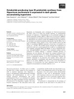
Fig. 4. Structures of enzymatic reaction products of H. perforatum HpPKS2 with different starter substrates. The structures were deter-
mined by UPLC ⁄ ESIMS.
K. Karppinen et al. Octaketide synthase from Hypericum perforatum
FEBS Journal 275 (2008) 4329–4342 ª 2008 The Authors Journal compilation ª 2008 FEBS 4333
Localization of HpPKS2 transcripts in
H. perforatum tissues
In order to obtain more insight into the role of
HpPKS2 in H. perforatum, in situ RNA hybridization
studies were performed. Digoxigenin (DIG)-labelled
HpPKS2 RNA probes were used to hybridize
fixed tissue sections of the leaves and flower buds of
H. perforatum in order to localize exactly the HpPKS2
transcripts in the tissues. After hybridization of the
cross-sections of the leaves with a HpPKS2 RNA anti-
sense probe, a dark blue signal that indicates HpPKS2
expression was clearly observed in the leaf margins
(Fig. 6A). The signal was specifically localized in the
multicellular nodular structures between the lower epi-
dermis and the photosynthetic parenchymal cells of the
H. perforatum leaves. Under test conditions, no signifi-
cant background staining was observed, and the
HpPKS2 probe specificity was confirmed by
the absence of signal in the negative control sections of
the leaves hybridized with HpPKS2 RNA sense probe
(Fig. 6B).
In the hybridized sections of the flower buds, a
strong dark blue signal for HpPKS2 transcripts was
localized in the petals (Fig. 6C) and the stamens
between anthers (Fig. 6E), also restricted to multi-
cellular nodules. The nodules that showed the HpPKS2
expression in flower buds were structurally similar to
those found to contain HpPKS2 transcripts in the leaf
sections. No signal was observed in the corresponding
areas of the negative controls of the flower bud
sections hybridized with HpPKS2 RNA sense probe
(Fig. 6D,F).
Multicellular nodules showing HpPKS2 expression
in both leaves and flower buds consisted of a core of
large cells that was surrounded by one to three flat cell
layers. The HpPKS2 transcripts were found to be pres-
ent in the large cells and also in the some of the inner-
most flat cells of the nodules. In the flower petals of
H. perforatum, two types of multicellular nodules that
share the same anatomical organization in the cross-
sections have been reported previously [10]. Spheroidal
nodules similar to those observed in the leaf margins
are also present in the petal margins, whereas the nod-
ules in the interior parts of the petals are elongated
tubulars [10,12]. In this study, nodules in both the
margins and the interior parts of the petals were found
to contain HpPKS2 transcripts.
Localization of hypericins in H. perforatum
tissues
As reported previously [25], the leaf margins and
flower buds contain the highest amounts of hypericins
in H. perforatum. To see exactly where the red hyperic-
ins are located, unstained cross-sections of the leaves
and flower buds of H. perforatum were observed under
a microscope. The dark red hypericins could be easily
located because they remained in the paraffin sections
and did not disappear until the in situ RNA hybridiza-
tion. Leaf cross-sections showed dark red material in
multicellular nodules in the leaf margins (Fig. 7A).
The nodules were included between the lower
OO
OH
R
OOO
-CO
2
O
OOH
OOH
O
OH
m/z 191
O
OOH
O
O
OH
OH
O
O
OH
O
O
O
OH
-CO
2
-CH
2
O
-CO
2
-CO
2
or
O
OOH
O
O
OH
OH
OH
O
O
OH
OH
OH
OH
OH
O
OOH
O
O
OH
O
OOH
OH
OH
O
O
OH
OO
OH
R
OOO
-CO
2
OO
OH
R
OOO
m/z 125
m/z 167
-CO
2
O
OOH
OOH
O
OH
O
OOH
O
O
OH
OH
O
OOH
OOH
O
OH
m/z 317
m/z 125
O
OOH
O
O
OH
OH
O
O
OH
O
O
O
OH
O
O
OH
O
O
O
OH
m/z 231
m/z 189
-CO
2
-CH
2
O
-CO
2
-CO
2
m/z 317
m/z 273
m/z 229
m/z 287
m/z 243
or
O
OOH
O
O
OH
OH
OH
O
O
OH
OH
OH
OH
OH
O
OOH
O
O
OH
O
OOH
OH
OH
O
O
OH
A
B
C
D
Fig. 5. MS fragmentation patterns of (A) a-pyrones, (B) SEK4 and
(C) SEK4b in the negative ionization mode, and (D) aloesone in the
positive ionization mode. Fragment ions were identified based on
their exact masses.
Octaketide synthase from Hypericum perforatum K. Karppinen et al.
4334 FEBS Journal 275 (2008) 4329–4342 ª 2008 The Authors Journal compilation ª 2008 FEBS
epidermis and the photosynthetic parenchymal cells of
leaves, and they comprised a core of large cells
surrounded by flat cell layers (Fig. 7B). Red material
was present in the large cells and also in the some of
the innermost flat cells of the nodules. Dark red
multicellular nodules of the same structure were also
observed in cross-sections of the flower buds (Fig. 7C).
Smaller red nodules were present in the flower petals
(Fig. 7D), whereas larger ones were found in the
stamens between anthers (Fig. 7E). The red material
AB
CD
EF
Fig. 6. In situ RNA localization of HpPKS2
transcripts in leaves and flower buds of
H. perforatum. Cross-section of (A) leaf, (C)
petal of flower bud and (E) stamen of flower
bud hybridized with DIG-labelled HpPKS2
RNA antisense probe. (B,D,F) Corresponding
sections were hybridized with HpPKS2 RNA
sense probe. Arrows point to multicellular
nodules. Bars = 100 lm.
A
B
C
D
E
Fig. 7. Localization of hypericins in leaves
and flower buds of H. perforatum.
Unstained cross-sections of (A) leaf (B)
showing red pigmented nodules in leaf
margins and (C) flower bud (D) showing red
pigmented nodules in petal and (E) in
stamen. Small arrows point to multicellular
nodules. Bars = 100 lm.
K. Karppinen et al. Octaketide synthase from Hypericum perforatum
FEBS Journal 275 (2008) 4329–4342 ª 2008 The Authors Journal compilation ª 2008 FEBS 4335
was present in the nodules of both the margins and
the interior parts of the flower petals.
Discussion
Despite the fact that hypericins are pharmacologically
important compounds of H. perforatum, a widely used
herbal remedy for the treatment of depression [1,2],
there is little information available about the biosynthe-
sis of these compounds. To date, only one gene has been
cloned and characterized from the biosynthetic route
leading to hypericins. The enzymatic product of hyp-1
has been shown to catalyse the final stages of hypericin
biosynthesis [20]. It has been proposed that type III
PKS would attend to the formation of emodin anthrone,
the initial key reaction step in the biosynthesis of
hypericins [20], but no such activity has been reported.
In this study, the role of a newly found PKS from
H. perforatum, HpPKS2 [25], was investigated for its
possible involvement in the biosynthesis of hypericins.
Phylogenetic analysis showed that the plant-specific
type III PKS family proteins grouped into CHSs and
other functionally divergent PKSs (Fig. 2). The only
exceptions were STSs from Fabaceae and Gymno-
sperms grouping with CHSs from the same or related
species. STSs have been proposed to have evolved
independently from CHSs several times, which explains
their presence in several clusters in the phylogenetic
tree [31–33]. HpPKS2 of H. perforatum grouped
with functionally divergent non-chalcone-forming
plant-specific type III PKSs. The grouping of HpPKS2
with non-CHSs indicates that HpPKS2 is not involved
in the biosynthesis of flavonoids in H. perforatum. The
functionally divergent PKSs include, for example,
OKS and PCS from A. arborescens, which accept
malonyl-CoA or acetyl-CoA as a starter substrate to
produce octaketides (SEK4 and SEK4b) and pentake-
tide chromone (5,7-dihydroxy-2-methyl-chromone),
respectively [26,27]. However, HpPKS2 was not partic-
ularly closely related to any of the currently known
type III PKSs, which indicates that it is a novel
plant-specific type III PKS family protein. We have
previously reported that the deduced amino acid
sequence of HpPKS2 shares only < 52% identity with
previously isolated type III PKSs [25].
HpPKS2 expressed in E. coli resulted in an enzyme
of 43 kDa (Fig. 3). The size coincides with a
predicted molecular mass of 43.1 kDa for HpPKS2,
calculated using bioinformatics tools [25], and with
that of a subunit size typical to plant-specific type III
PKSs. The plant-specific type III PKSs are reported
to be homodimeric proteins with a subunit size of
40–45 kDa [21,23].
Functional characterization of the purified recombi-
nant HpPKS2 revealed the expected OKS activity. But
instead of producing emodin anthrone, an octaketide
precursor of hypericins, the enzyme catalysed the con-
densation of one molecule of acetyl-CoA with seven
molecules of malonyl-CoA to form unnatural octake-
tides SEK4 and SEK4b (Fig. 4). SEK4 and SEK4b,
the longest polyketides known to be produced by
type III PKSs, have also been shown to be the
products of OKS from A. arborescens [26] and
shunt products of minimal type II PKS from
Streptomyces coelicolor [34,35]. The A. arborescens
OKS, along with HpPKS2, is the only enzyme among
unmodified plant-specific type III PKSs that has been
shown to have OKS activity. Because the aloe does
not accumulate SEK4 and SEK4b, the aloe OKS has
been suggested to be involved in the biosynthesis of
anthrones and anthraquinones in the plant and
SEK4 ⁄ SEK4b produced in the absence of additional
tailoring enzymes in vitro [26,36]. The A. arborescens
OKS preferred malonyl-CoA as a starter substrate for
the production of SEK4 and SEK4b. Because SEK4
and SEK4b could not be found from incubations with
starter substrates other than acetyl-CoA in this study,
it is likely that HpPKS2 used only acetyl-CoA as a
starter substrate for production of SEK4 and SEK4b.
HpPKS2 also catalysed the formation of tri- to
heptaketide products, using acetyl-CoA as a starter
substrate (Fig. 4). Triketide and tetraketide pyrones
are often biosynthesized in vitro by PKSs when incu-
bated with acetyl-CoA as a starter substrate [37–39].
Penta- to octaketides are more rare products. Two dif-
ferent pentaketide products have previously been
reported to be produced by unmodified plant-specific
type III PKSs. These are 5,7-dihydroxy-2-methylchro-
mone produced by PCS from A. arborescens [27] and
a-pyrone by PKS from Plumbago indica [39]. The pen-
taketide chromone structure A8 has not previously
been reported to be produced by plant-specific type III
PKS. The hexaketide a-pyrone A1 produced by
HpPKS2 can be classified as a derailment product of
type III PKS. The structure is also the product
of P. indica PKS, along with hexaketide phenylpyrone
[39]. Because the pyrones are not found in P. indica
tissues, it has been suggested that the PKS in vivo
would be involved in the biosynthesis of naphthoqui-
none plumbagin and the pyrones produced in vitro in
the absence of accessory enzymes [39]. Of the three
heptaketides produced by HpPKS2 using acetyl-CoA
as a starter substrate, one was aloesone. Aloesone has
previously been reported as a product of aloesone
synthase of Rheum palmatum [31], a plant known to be
rich with chromones, napthalenes and anthraquinones.
Octaketide synthase from Hypericum perforatum K. Karppinen et al.
4336 FEBS Journal 275 (2008) 4329–4342 ª 2008 The Authors Journal compilation ª 2008 FEBS
Aloesone was also the product of A. arborescens OKS,
along with SEK4 and SEK4b, after a single amino
acid mutation, i.e. replacement of glycine by alanine,
as in the case of aloesone synthase in the Gly207 site
[26]. HpPKS2 has serine in the corresponding site. To
our knowledge, the other two heptaketides produced
by HpPKS2, chromone A10 and phenylpyrone A6,
have not previously been reported as products of
plant-specific type III PKSs. Notably, OKS and PCS
from A. arborescens, PKS from P. indica, aloesone
synthase from R. palmatum and now HpPKS2 from
H. perforatum all share mechanistically related reac-
tions, such as accepting acetyl-CoA ⁄ malonyl-CoA as a
starter substrate, performing high numbers of conden-
sations and two to three cyclization reactions. Because
most type III PKSs perform only one to three exten-
sions and catalyse the formation of one six-membered
ring, it can be assumed that the above-mentioned
PKSs may be involved in the biosynthesis of structur-
ally similar types of compounds in plants.
The acceptance of other, larger starter substrates by
HpPKS2 shows that the enzyme has a broad substrate
acceptance, as reported for other type III PKSs
[22–24,26,27,32,37,39,40]. HpPKS2 accepted both aro-
matic and aliphatic CoA-esters as starter units. By
using isobutyryl-CoA, benzoyl-CoA and hexanoyl-
CoA as starter substrates, HpPKS2 produced tri- to
heptaketide products, mostly pyrones (Fig. 4). In addi-
tion to pyrones, some chromones and phloroglucinols
were also produced. It should be noted that with star-
ter substrates other than acetyl-CoA, HpPKS2 was not
able to produce octaketides but only afforded shorter
products supporting the view that acetyl-CoA could be
the real starter substrate for HpPKS2 in vivo.
To our knowledge, the compounds produced by
HpPKS2 in vitro, which were mostly pyrones, have not
been described as constituents of H. perforatum. Sev-
eral recombinant plant-specific type III PKSs are
known to biosynthesize metabolites, especially pyrones,
that have not been described as being accumulated by
their plants of origin [26,32,37,39,40]. The products
have been found to be typical for in vitro incubations
of type III PKSs with non-physiological substrates,
non-optimal assay conditions and are also suggested to
be produced in the absence of co-operating tailoring
enzymes [23,26,30,32,39]. To date, the only character-
ized example of such a co-operating interaction of
plant-specific type III PKS with tailoring enzyme is the
biosynthesis of 6¢-deoxychalcone [23]. Typically, type I
and type II PKSs consist of many additional subunits,
including ketoreductases, cyclases and aromatases, that
are often needed for the production of specific cyclized
polyketide products [34,35,41–43]. These additional
subunits interact with PKS to stabilize the highly reac-
tive polyketide chain preventing non-specific cycliza-
tions. It is not currently known whether emodin
anthrone biosynthesis requires additional enzymes and
thus it is possible that HpPKS2 failed to produce emo-
din anthrone in this study because of the absence of
additional tailoring enzymes in vitro.
To further s tudy the r ole of HpPKS2 in H. perforatum,
in situ RNA hybridization studies to locate HpPKS2
transcripts were performed. HpPKS2 expression was
found to localize specifically in multicellular nodules in
the leaf margins, flower petals and stamens of H. per-
foratum (Fig. 6). These types of structures present in
the H. perforatum tissues have been described previ-
ously by several authors, and are referred to as dark
glands [10,17,18,44]. In this study, the same nodules
were also found to contain dark red material (Fig. 7).
The red material in the dark glands has previously
been found to consist of hypericins, and their accumu-
lation is shown to be restricted to only the dark glands
in H. perforatum [12,16–18]. The obtained results are
consistent with our previous study in which the expres-
sion of HpPKS2, measured using real-time PCR, was
shown to correlate with the concentrations of hyperic-
ins in different H. perforatum tissues [25]. Recently,
emodin, which is an oxidized derivative of emodin
anthrone (Fig. 1), has also been found to accumulate
at high concentrations in the dark glands of H. perfo-
ratum [12]. The presence of emodin in only the dark
glands in H. perforatum suggests that emodin biosyn-
thesis may take place in the glands [12]. The restriction
of HpPKS2 expression and the presence of both hyper-
icins and emodin specifically in the same cells imply
that HpPKS2 may have a role in the biosynthesis of
hypericins in H. perforatum. The localization of the
HpPKS2 transcripts in the dark glands that accumu-
late hypericins is very similar to the expression pattern
of type III PKS from Humulus lupulus. Valerophenone
synthase from H. lupulus is responsible for the biosyn-
thesis of the phloroglucinol skeleton of hop resin, and
it has been shown to be expressed specifically in secre-
tory structures called ‘lupulin glands’ accumulating the
resin [45].
The HpPKS2 transcripts were found to accumulate
into both the large cells and some of the innermost flat
cells of the multicellular nodules. This indicates that if
HpPKS2 is involved in the biosynthesis of hypericins,
then at least the early phase of biosynthesis, i.e. the
formation of emodin anthrone, may occur in both cell
types. Kornfeld et al. [18] hypothesized that the
biosynthesis of hypericins takes place in the peripheral
flat cells rather than in the large interior cells of the
nodules.
K. Karppinen et al. Octaketide synthase from Hypericum perforatum
FEBS Journal 275 (2008) 4329–4342 ª 2008 The Authors Journal compilation ª 2008 FEBS 4337
Based on these results, H. perforatum HpPKS2 is a
novel plant-specific type III PKS having OKS activity.
Furthermore, our findings show a strong connection
between HpPKS2 expression and the accumulation of
hypericins, indicating that HpPKS2 may have a role in
the initial key reaction step in the biosynthesis of
hypericins in H. perforatum. However, although the
enzyme is capable of carrying out the expected number
of condensation reactions in vitro, it fails in the
cyclization of the produced octaketide chain to emodin
anthrone. The formation of derailment products by
HpPKS2 may mean that the biosynthesis of emodin
anthrone requires some additional, as yet unidentified
factors that are missing in vitro. Recently, several
type III PKSs have been isolated that do not, in vitro,
produce the metabolites that they are expected to cata-
lyse and that are found in their plant of origin. There-
fore, further studies are needed to elucidate the
reasons for these failures to reveal the actual
biosynthesis mechanism of many plant polyketides,
including hypericins.
Experimental procedures
Construction of expression plasmid
cDNA from H. perforatum leaves was prepared as
described previously [25]. The coding region of HpPKS2
was amplified from the cDNA by PCR, using forward
primer 5¢-CATATTG
GGATCCATGGGTTCCCTTGAC-3¢
(the translation start codon is in bold and the BamHI site
is underlined) and reverse primer 5¢-ACGCT
GGTACC
TTAGAGAGGCACACTTCG-3¢ (the translation stop
codon is in bold and the KpnI site is underlined). The PCR
was performed with DyNazymeÔ II DNA polymerase
(Finnzymes, Espoo, Finland). The PCR conditions were
denaturation at 94 °C for 5 min, followed by 40 cycles of
amplification at 94 °C for 1 min, 60 °C for 2 min and
72 °C for 2 min, and a final extension at 72 °C for 10 min.
The amplified PCR product was purified by electrophoresis
on a 1% (w ⁄ v) ethidium bromide-stained agarose gel. The
PCR fragment of the expected size ( 1.2 kb) was excised
from the gel and further purified using MontageÒ DNA
Gel Extraction Kit (Millipore, Bedford, MA, USA). The
purified PCR product was digested with BamHI and KpnI
(Isogen, Bioscience, Maarssen, The Netherlands) and
ligated into the BamHI ⁄ KpnI site of expression vector
pQE30 (Qiagen, Hilden, Germany). Thus, the recombinant
enzyme contains an additional hexahistidine tag at the
N-terminus. The resulting recombinant plasmid pQE30–
HpPKS2 was confirmed by sequencing, using the BigDye
Terminator Cycle Sequencing Kit (Applied Biosystems,
Foster City, CA, USA) and an ABI 310 DNA sequencer
(Model 377; Applied Biosystems).
Expression of recombinant HpPKS2
The recombinant plasmid pQE30–HpPKS2 was transferred
into the E. coli host strain M15 [pREP4] (Qiagen) for pro-
tein expression. E. coli cells harbouring the plasmid were
grown in Luria–Bertani liquid medium in the presence of
ampicillin (100 lgÆmL
)1
) and kanamycin (25 lgÆmL
)1
)at
30 °C until the D
600
of the culture reached 0.6. After the
culture had been cooled on ice, IPTG (Roche, Basel, Swit-
zerland) was added to the culture in a final concentration
of 0.4 mm to induce protein expression. The culture was
incubated further at 16 °C for 20 h.
Enzyme purification
E. coli cells were harvested by centrifugation (4000 g for
20 min) and resuspended in a lysis buffer (50 mm sodium
phosphate buffer, pH 8.0, containing 500 mm NaCl, 10 mm
b-mercaptoethanol, 1% Tween 20 and 20 mm imidazole).
The cells were disrupted using lysozyme (1 mgÆmL
)1
) and
sonication (Type UP50H; Dr Hielscher GmbH, Teltow,
Germany). The lysate was diluted twofold with the same
buffer and centrifuged at 17 000 g for 30 min. The super-
natant was collected for purification of recombinant protein
under native conditions according to the protocol of the
QIAexpressionist [46], using Ni-NTA agarose. Unbound
proteins were washed away with a wash buffer (50 mm
sodium phosphate buffer, pH 7.0, containing 500 mm
NaCl, 10 mm b-mercaptoethanol, 10% glycerol, 1%
Tween 20 and 20 mm imidazole) and the recombinant
protein was eluted with an elution buffer (50 mm sodium
phosphate buffer, pH 7.0, containing 500 mm NaCl, 10 mm
b-mercaptoethanol, 10% glycerol and 250 mm imidazole).
After purification, the protein concentration was deter-
mined according to Bradford [47], using BSA (Sigma, St
Louis, MO, USA) as a standard. The purity of the protein
was verified by SDS ⁄ PAGE, using 12% separation and 3%
stacking gels. The proteins were run along with protein
markers (Fermentas, Vilnius, Lithuania) at 180 V, using a
Mini-Protean II electrophoresis system (Bio-Rad, Hercules,
CA, USA) followed by staining with Coomassie Brilliant
Blue R-250 (Merck, Darmstadt, Germany).
Polyketide synthase assays
Purified recombinant HpPKS2 (100 lg) was mixed with
200 lm starter substrates (acetyl-CoA, isobutyryl-CoA, ben-
zoyl-CoA or hexanoyl-CoA; Sigma) and 300 l m malonyl-
CoA (Sigma). An assay buffer (0.5 m potassium phosphate
buffer, pH 6.8, containing 2.8 mm b-mercaptoethanol and
10 lm dithiothreitol) was then added to 500 lL. For con-
trol reactions, the enzyme was heat-denatured. Incubations
were carried out at 30 °C for 90 min. Reactions were
stopped by adding 50 lL of 20% HCl, and the products
were then extracted twice with 250 lL of ethyl acetate.
Octaketide synthase from Hypericum perforatum K. Karppinen et al.
4338 FEBS Journal 275 (2008) 4329–4342 ª 2008 The Authors Journal compilation ª 2008 FEBS
After evaporation of the solvent with nitrogen flow, the
residue was dissolved in 100 lL of methanol.
Identification of the biosynthetic products by
UPLC ⁄ ESIMS
A Waters ACQUITY UPLC
TM
(Waters, Milford, MA,
USA) system together with Waters ACQUITY UPLC
TM
BEH C18 2.1 · 50 mm column with a particle size of
1.7 lm (Waters) was used to separate the biosynthetic
products. The samples were diluted with 100 lLofUP
grade water (ultra pure, 18.2 MW) prior to injection into
UPLC. The UPLC eluents were 0.1% acetic acid (BDH
Laboratory Supplies, Poole, UK) in UP grade water (A)
and acetonitrile (B) (HPLC grade; Merck). The initial gra-
dient condition was 90% A and 10% B, changing linearly
to 60% B in 4 min followed by 1 min of isocratic elution
and 2 min of equilibration with initial conditions, giving a
total analysis time of 7 min. The eluent flow rate was
0.5 mLÆmin
)1
, and the column temperature was 35 °C;
injection volume was 4 lL. A Waters ACQUITY PDA
detector was used for the measurement of online UV spec-
tra of the biosynthetic products. A range of 210–500 nm
was acquired, and the resolution was set to 1.2 nm.
A Waters LCT Premier
TM
XE time-of-flight mass spec-
trometer (Waters) equipped with lock spray ion source was
used for to detect and identify the biosynthetic products.
Both the negative ion mode (ESI
)
) and positive ion mode
(ESI
+
) were used. The capillary and sample cone voltages
were 2400 and 40 V in the negative ion mode and 2800 and
60 V in the positive ion mode, respectively. The desolvation
and sample cone temperatures were 350 and 150 ° C, respec-
tively. The desolvation gas flow was set to 800 LÆh
)1
. The
aperture 1 voltage was set to 0 V in the negative ion mode
and 30 V in the positive ion mode. Another set of experi-
ments in negative ion mode was done using the aperture 1
voltage of 20 V to obtain more fragmentation. Dynamic
range enhancement and W optics mode were used in all
experiments to obtain the exact masses for all molecular
ions and fragments ions. The lock mass solution was
0.5 lgÆmL
)1
leucine enkephalin in 50% water ⁄ methanol
(v ⁄ v HPLC grade; Merck) in the negative ion mode and
1.5 lgÆmL
)1
leucine enkephalin in 50% water ⁄ methanol
(v ⁄ v) in the positive ion mode. The lock mass solution was
delivered to lock spray inlet, using LCT Premier’s syringe
pump at a flow rate of 10 lLÆmin
)1
, and the reference scan
frequency was set to 40 (analyte to reference scan ratio
39 : 1). Data were acquired and analyzed using masslynx
v. 4.1. Samples and their respective controls (incubations
with heat-denatured enzyme) were compared in order
to find the biosynthetic products. The compounds were
identified based on their UV spectra and exact masses of
molecular and fragment ions (differences between calculated
and measured masses were < 3 mDa) in both ESI
+
and
ESI
)
conditions.
Tissue fixation and embedding
H. perforatum leaves and flower buds were collected from
the botanical garden of the University of Oulu, Finland.
The samples excised from the plants were fixed in 4%
(w ⁄ v) paraformaldehyde and 0.25% (v ⁄ v) glutaraldehyde in
0.1 m sodium phosphate buffer (pH 7.0) overnight at 4 °C.
The samples were rinsed in 0.1 m sodium phosphate buffer
(pH 7.0) and then dehydrated in a graded series of ethanol
up to absolute. The ethanol was replaced by a series of
tert-butanol (25, 50 and 100%, v ⁄ v), after which the sam-
ples were gradually infiltrated with paraffin (Merck). Paraf-
fin-embedded samples were sectioned to a thickness of
8 lm by using a microtome (Microm HM 325, Walldorf,
Germany). The sections were spread on glass slides coated
with 2% (v ⁄ v) 3-aminopropyltriethoxysilane (Sigma) in ace-
tone and dried overnight at 40 °C. Two 20 min incubations
in xylene were used to remove paraffin from the samples.
To localize hypericins, slides were observed without staining
under a light microscope (Nikon Optiphot-2; Nikon Corpo-
ration, Tokyo, Japan). For in situ RNA hybridizations,
samples were rehydrated in a graded ethanol series up to
water.
Preparation of RNA probes for in situ
hybridizations
By using primers 5¢-TGGGATGGATCTACGACCTC-3¢
(forward primer) and 5¢-CGGCTACTCTCGAGCTTGTC-3¢
(reverse primer), a 205-bp fragment from the coding region
of HpPKS2 was amplified from H. perforatum cDNA by
PCR with DyNazymeÔ II DNA polymerase (Finnzymes)
under standard PCR conditions. The PCR fragment was
gel purified using MontageÒ DNA Gel Extraction Kit
(Millipore) and ligated into a pGEM-T Easy vector (Pro-
mega, Madison, WI, USA). DIG-labelled sense and anti-
sense probes were prepared from the linearized plasmid by
in vitro transcription with SP6 or T7 RNA polymerase,
using DIG RNA Labelling Kit according to the manufac-
turer’s instructions (Roche).
In situ RNA hybridization analysis
Before hybridization, rehydrated tissue sections were trea-
ted with proteinase K (1 lgÆmL
)1
in 100 mm Tris ⁄ HCl and
50 mm EDTA, pH 7.5) for 30 min at 37 °C followed by
dehydration in a graded ethanol series up to absolute. The
sections were then air-dried. For the hybridization, an ali-
quot of hybridization mixture (100 lL) was dispersed on
the sections and mounted under coverslips to prevent evap-
oration. The hybridization mixture contained 0.5 lgÆmL
)1
of DIG-labelled RNA antisense or sense probe, 50% (v ⁄ v)
deionized formamide (Sigma), 0.3 m NaCl, 10 mm
Tris ⁄ HCl (pH 7.5), 1 mm EDTA, 1· Denhardt¢s solution
K. Karppinen et al. Octaketide synthase from Hypericum perforatum
FEBS Journal 275 (2008) 4329–4342 ª 2008 The Authors Journal compilation ª 2008 FEBS 4339
(Sigma), 150 lgÆmL
)1
tRNA (Roche), 500 lgÆmL
)1
polyad-
enylic acid (Sigma), 10% (w ⁄ v) dextran sulfate and 0.06 m
dithiothreitol. The hybridization was carried out at 62 °C
for 19 h.
After hybridization, slides were washed in 2· NaCl ⁄ Cit
(1· NaCl ⁄ Cit is 150 mm NaCl and 15 mm sodium citrate,
pH 7.0) at room temperature, in 1· NaCl ⁄ Cit at 37 °C and
0.5· NaCl ⁄ Cit at 37 °C, for 20 min in each. Excess RNA
probes were removed by incubation in a solution that con-
tained 3 lgÆmL
)1
RNase A, 10 mm Tris ⁄ HCl (pH 7.5),
500 mm NaCl and 1 mm EDTA at 37 °C for 60 min. The
slides were then washed four times with the same solution
without RNase A at 37 °C for 15 min and 2· NaCl ⁄ Cit in
room temperature for 30 min.
For immunolocalization of hybridized transcripts, slides
were washed in a NaCl ⁄ Tris buffer (100 mm Tris ⁄ HCl,
pH 7.5, 150 mm NaCl and 0.3% v ⁄ v Triton X-100) for
5 min and blocked with 2% (w ⁄ v) blocking reagent (Roche)
in the NaCl ⁄ Tris buffer for 30 min. A sheep anti-(DIG-AP)
conjugate (Roche) at a 1 : 750 dilution in NaCl ⁄ Tris buffer
was dispensed on the sections and mounted under coverslips.
After incubation for 2 h at room temperature, coverslips
were removed by soaking in the NaCl ⁄ Tris buffer. Unbound
conjugates were washed four times with the NaCl ⁄ Tris buf-
fer for 10 min and with an AP buffer (100 mm Tris ⁄ HCl,
pH 9.5, 100 mm NaCl and 50 mm MgCl
2
) for 5 min. For
colour development, slides were immersed in 5-bromo-4-
chloro-3-indolylphosphate and blue tetrazolium chloride
(Roche) in the AP buffer at room temperature. The develop-
ment time was 17 h for leaf samples and 7 h for flower bud
samples. Slides were washed with water and dehydrated in a
graded series of ethanol up to absolute and then air-dried.
The slides were mounted with immersion oil and covered
with coverslips. In this experiment, DIG-labelled HpPKS2
RNA sense probe was used as a control. RNase-free condi-
tions were maintained throughout the procedure.
Phylogenetic tree construction
In total, 44 amino acid sequences of type III PKS family
proteins including H. perforatum HpPKS2 were aligned
using the clustal w program. The protruding ends of the
sequences were truncated and eventually 396 amino acids,
including gaps, were aligned. A phylogenetic tree was con-
structed using the neighbor-joining method, with the soft-
ware of mega 2.0. The reliability of the tree was measured
by bootstrap analysis with 1000 replicates.
Acknowledgements
The authors would like to thank Novamass Ltd for
providing the equipment to carry out UPLC ⁄ ESIMS
analyses. This research was supported financially by
grants from Oulu University Scholarship Foundation,
Research Foundation of Orion Corporation, and
Jenny and Antti Wihuri Foundation to KK.
References
1 Butterweck V (2003) Mechanism of action of St John’s
wort in depression: what is known? CNS Drugs 17,
539–562.
2 Wurglics M & Schubert-Zsilavecz M (2006) Hyperi-
cum perforatum: a ‘modern’ herbal antidepressant:
pharmacokinetics of active ingredients. Clin Pharmaco-
kinet 45, 449–468.
3 Falk H (1999) From the photosensitizer hypericin to
the photoreceptor stentorin – the chemistry of phen-
anthroperylene quinones. Angew Chem Int Ed 38, 3116–
3136.
4 Kirakosyan A, Hayashi H, Inoue K, Charchoglyan A
& Vardapetyan H (2000) Stimulation of the production
of hypericins by mannan in Hypericum perforatum
shoot cultures. Phytochemistry 53, 345–348.
5 Barnes J, Anderson LA & Phillipson DJ (2001) St
John’s wort (Hypericum perforatum L.): a review of its
chemistry, pharmacology and clinical properties.
J Pharm Pharmacol 53, 583–600.
6 Lopez-Bazzocchi I, Hudson JB & Towers GH (1991)
Antiviral activity of the photoactive plant pigment
hypericin. Photochem Photobiol 54, 95–98.
7 Agostinis P, Vantieghem A, Merlevede W & de Witte
PAM (2002) Hypericin in cancer treatment: more light
on the way. Int J Biochem Cell Biol 34, 221–241.
8 Kubin A, Wierrani F, Burner U, Alth G & Gru
¨
nberger
W (2005) Hypericin – the facts about a controversial
agent. Curr Pharm Des 11, 233–253.
9 Sirvent TM, Krasnoff SB & Gibson DM (2003) Induc-
tion of hypericins and hyperforins in Hypericum perfo-
ratum in response to damage by herbivores. J Chem
Ecol 29, 2667–2681.
10 Curtis JD & Lersten NR (1990) Internal secretory struc-
tures in Hypericum (Clusiaceae): H. perforatum L. and
H. balearicum L. New Phytol 114, 571–580.
11 Fornasiero RB, Bianchi A & Pinetti A (1998) Anatomi-
cal and ultrastuctural observations in Hypericum perfo-
ratum L. leaves. J Herbs Spices Med Plants 5, 21–33.
12 Zobayed SMA, Afreen F, Goto E & Kozai T (2006)
Plant–environment interactions: accumulation of hyperi-
cin in dark glands of Hypericum perforatum. Ann Bot
98, 793–804.
13 Kos
ˇ
uth J, Katkovc
ˇ
inova
´
Z, Olexova
´
P&C
ˇ
ella
´
rova
´
E
(2007) Expression of the
hyp-1 gene in early stages of
development of Hypericum perforatum L. Plant Cell
Rep 26, 211–217.
14 Repc
ˇ
a
´
kM&Ma
´
rtonfi P (1997) The localization of
secondary substances in Hypericum perforatum flower.
Biologia, Bratislava 52, 91–94.
Octaketide synthase from Hypericum perforatum K. Karppinen et al.
4340 FEBS Journal 275 (2008) 4329–4342 ª 2008 The Authors Journal compilation ª 2008 FEBS
15 Piovan A, Filippini R, Can iato R, Borsarini A, Maleci LB
& Cappelletti EM (2004) Detection of hypericins in the
‘red glands’ of Hypericum elodes by ESI-MS ⁄ MS.
Phytochemistry 65, 411–414.
16 Briskin DP & Gawienowski MC (2001) Differential
effects of light and nitrogen on production of hypericins
and leaf glands in Hypericum perforatum. Plant Physiol
Biochem 39, 1075–1081.
17 Pasqua G, Avato P, Monacelli B, Santamaria AR &
Argentieri MP (2003) Metabolites in cell suspension cul-
tures, calli, and in vitro regenerated organs of Hyperi-
cum perforatum cv. Topas. Plant Sci 165, 977–982.
18 Kornfeld A, Kaufman PB, Lu CR, Gibson DM, Bol-
ling SF, Warber SL, Chang SC & Kirakosyan A
(2007) The production of hypericins in two selected
Hypericum perforatum shoot cultures is related to
differences in black gland structure. Plant Physiol
Biochem 45, 24–32.
19 Walker L, Sirvent T, Gibson D & Vance N (2001)
Regional differences in hypericin and pseudohypericin
concentrations and five morphological traits among
Hypericum perforatum plants in the northwestern
United States. Can J Bot 79, 1248–1255.
20 Bais HP, Vepachedu R, Lawrence CB, Stermitz FR &
Vivanco JM (2003) Molecular and biochemical charac-
terization of an enzyme responsible for the formation of
hypericin in St. John’s wort (Hypericum perforatum L.).
J Biol Chem 278, 32413–32422.
21 Schro
¨
der J (1997) A family of plant-specific polyketide
synthases: facts and predictions. Trends Plant Sci 2,
373–378.
22 Austin MB & Noel JP (2003) The chalcone synthase
superfamily of type III polyketide synthases. Nat Prod
Rep 20, 79–110.
23 Schro
¨
der J (2000) The family of chalcone synthase-
related proteins: functional diversity and evolution.
In Evolution of Metabolic Pathways (Romeo JT, Ibra-
him RK, Varin L & De Luca V, eds), pp. 55–89. Perg-
amon, Amsterdam.
24 Liu B, Falkenstein-Paul H, Schmidt W & Beerhues L
(2003) Benzophenone synthase and chalcone synthase
from Hypericum androsaemum cell cultures: cDNA clon-
ing, functional expression, and site-directed mutagenesis
of two polyketide synthases. Plant J 34, 847–855.
25 Karppinen K & Hohtola A (2008) Molecular cloning
and tissue-specific expression of two cDNAs encoding
polyketide synthases from Hypericum perforatum.
J Plant Physiol 165, 1079–1086.
26 Abe I, Oguro S, Utsumi Y, Sano Y & Noguchi H
(2005) Engineered biosynthesis of plant polyketides:
chain length control in an octaketide-producing plant
type III polyketide synthase. J Am Chem Soc 127,
12709–12716.
27 Abe I, Utsumi Y, Oguro S, Morita H, Sano Y &
Noguchi H (2005) A plant type III polyketide synthase
that produces pentaketide chromone. J Am Chem Soc
127, 1362–1363.
28 Akiyama T, Shibuya M, Liu H-M & Ebizuka Y (1999)
p-Coumaroyltriacetic acid synthase, a new homologue
of chalcone synthase, from Hydrangea macrophylla var.
thunbergii. Eur J Biochem 263, 834–839.
29 Pang Y, Shen G, Wu W, Liu X, Lin J, Tan F, Sun X &
Tang K (2005) Characterization and expression of chal-
cone synthase gene from Ginkgo biloba. Plant Sci 168,
1525–1531.
30 Eckermann C, Schro
¨
der G, Eckermann S, Strack D,
Schmidt J, Schneider B & Schro
¨
der J (2003) Stilbene-
carboxylate biosynthesis: a new function in the family
of chalcone synthase-related proteins. Phytochemistry
62, 271–286.
31 Abe I, Utsumi Y, Oguro S & Noguchi H (2004) The
first plant type III polyketide synthase that catalyzes
formation of aromatic heptaketide. FEBS Lett 562,
171–176.
32 Brand S, Ho
¨
lscher D, Schierhorn A, Svatos
ˇ
A, Schro
¨
der
J & Schneider B (2006) A type III polyketide synthase
from Wachendorfia thyrsiflora and its role in diarylhep-
tanoid and phenylphenalenone biosynthesis. Planta 224,
413–428.
33 Tropf S, Lanz T, Rensing SA, Schro
¨
der J & Schro
¨
der
G (1994) Evidence that stilbene synthases have devel-
oped from chalcone synthases several times in the
course of evolution. J Mol Evol 38, 610–618.
34 Fu H, Ebert-Khosla S, Hopwood DA & Khosla C
(1994) Engineered biosynthesis of novel polyketides: dis-
section of the catalytic specificity of the act ketoreduc-
tase. J Am Chem Soc 116, 4166–4170.
35 Fu H, Hopwood DA & Khosla C (1994) Engineered
biosynthesis of novel polyketides: evidence for temporal,
but not regiospecific, control of cyclization of an aro-
matic polyketide precursor. Chem Biol 1, 205–210.
36 Morita H, Mizuuchi Y, Abe T, Kohno T, Noguchi H
& Abe I (2007) Cloning and functional analysis of a
novel aldo-keto reductase from Aloe arborescens. Biol
Pharm Bull 30, 2262–2267.
37 Samappito S, Page J, Schmidt J, De-Eknamkul W &
Kutchan TM (2002) Molecular characterization of root-
specific chalcone synthases from Cassia alata. Planta
216, 64–71.
38 Samappito S, Page JE, Schmidt J, De-Eknamkul W &
Kutchan TM (2003) Aromatic and pyrone polyketides
synthesized by a stilbene synthase from Rheum
tataricum. Phytochemistry 62, 313–323.
39 Springob K, Samappito S, Jindaprasert A, Schmidt J,
Page JE, De-Eknamkul W & Kutchan TM (2007) A
polyketide synthase of Plumbago indica that catalyzes
the formation of hexaketide pyrones. FEBS J 274
,
406–417.
40 Wanibuchi K, Zhang P, Abe T, Morita H, Kohno T,
Chen G, Noguchi H & Abe I (2007) An acridone-
K. Karppinen et al. Octaketide synthase from Hypericum perforatum
FEBS Journal 275 (2008) 4329–4342 ª 2008 The Authors Journal compilation ª 2008 FEBS 4341
producing novel multifunctional type III polyketide syn-
thase from Huperzia serrata. FEBS J 274, 1073–1082.
41 Hertweck C, Xiang L, Kalaitzis JA, Cheng Q, Palzer M
& Moore BS (2004) Context-dependent behavior of the
enterocin iterative polyketide synthase: a new model for
ketoreduction. Chem Biol 11, 461–468.
42 Kalaitzis JA & Moore BS (2004) Heterologous bio-
synthesis of truncated hexaketides derived from the
actinorhodin polyketide synthase. J Nat Prod 67,
1419–1422.
43 Kurosaki F, Mitsuma S & Arisawa M (2002) Activation
of acyl condensation reaction of monomeric 6-hydroxy-
mellein synthase, a multifunctional polyketide biosyn-
thetic enzyme, by free coenzyme A. Phytochemistry 61,
597–604.
44 Onelli E, Rivetta A, Giorgi A, Bignami M, Cocucci M
& Patrignani G (2002) Ultrastructural studies on the
developing secretory nodules of Hypericum perforatum.
Flora 197, 92–102.
45 Okada Y & Ito K (2001) Cloning and analysis of valer-
ophenone synthase gene expressed specifically in lupulin
gland of hop (Humulus lupulus L.). Biosci Biotechnol
Biochem 65, 150–155.
46 QIAGEN (2003) The QIAexpressionist:A Handbook for
High-Level Expression and Purification of 6 · His-
Tagged Proteins. QIAGEN Inc., Valencia, CA.
47 Bradford MM (1976) A rapid and sensitive method for
the quantitation of microgram quantities of protein
utilizing the principle of protein–dye binding. Anal
Biochem 72, 248–254.
Octaketide synthase from Hypericum perforatum K. Karppinen et al.
4342 FEBS Journal 275 (2008) 4329–4342 ª 2008 The Authors Journal compilation ª 2008 FEBS