Tài liệu Báo cáo khoa học: How does hepatitis C virus enter cells? pptx
Bạn đang xem bản rút gọn của tài liệu. Xem và tải ngay bản đầy đủ của tài liệu tại đây (625.88 KB, 15 trang )
REVIEW ARTICLE
How does hepatitis C virus enter cells?
Gundo Diedrich
The World Health Organization estimates that 170
million people, 3% of the world population, are infec-
ted with hepatitis C virus (HCV) [1]. The majority of
those infected (55–85%) fail to clear the virus and
become chronic carriers manifested by the persistent
presence of detectable virus in the serum [2]. The clin-
ical course of chronic hepatitis C is highly variable
ranging from mild hepatitis (inflammation of the liver),
fibrosis (scaring of the liver), cirrhosis (end-stage fibro-
sis) to hepatocellular carcinoma (liver cancer). Liver
damage is not directly caused by the virus, but by the
interplay between the virus and the immune system
that results in the replacement of healthy liver tissue
with fibrous scar tissue. About 20% of patients with
chronic hepatitis C will develop liver cirrhosis within
20 years. Once cirrhosis is established, the rate of he-
patocellular cancer development is 1–4% per year [3].
The standard treatment for chronic HCV infection is
pegylated a-interferon in combination with the nucleo-
side analogue ribavirin. About 55% of patients
respond to the therapy and show a sustained reduction
in viral titer [4]. Few treatment options exist for non-
responders. Ribavarin and a-interferon have general
antiviral properties not specifically related to HCV.
Drugs interfering specifically with HCV RNA replica-
tion or translation and processing of HCV proteins are
not available yet, but a few promising candidates are
in clinical testing [5,6].
Since the discovery of HCV in 1989, the major
bottleneck in HCV research has been the lack of a
robust and reliable cell culture system for the propaga-
tion of the virus, and the absence of a nonprimate ani-
mal model. While cultured liver cells can be infected
with clinical HCV isolates, the process has been ineffi-
cient, transient and not always reproducible [7]. Our
current knowledge about the mechanism of viral cell
entry comes from several different approaches inclu-
ding vaccination of chimpanzees, structural studies of
Keywords
CD81; envelope proteins; exosomes;
hepatitis C virus (HCV); lipoproteins; low
density lipoprotein receptor; scavenger
receptor class B type 1 (SR-BI)
Correspondence
G. Diedrich, diaDexus Inc., 343 Oyster Point
Boulevard, South San Francisco, CA 94080,
USA
Fax: +1 650 2466499
Tel: +1 650 2466481
E-mail:
(Received 27 January 2006, revised 17 May
2006, accepted 13 June 2006)
doi:10.1111/j.1742-4658.2006.05379.x
Hepatitis C virus (HCV) exists in different forms in the circulation of infec-
ted people: lipoprotein bound and lipoprotein free, enveloped and non-
enveloped. Viral particles with the highest infectivity are associated with
lipoproteins, whereas lipoprotein-free virions are poorly infectious. The
detection of HCV’s envelope proteins E1 and E2 in lipoprotein-associated
virions has been challenging. Because lipoproteins are readily endocytosed,
some forms of HCV might utilize their association with lipoproteins rather
than E1 and E2 for cell attachment and internalization. However, vaccin-
ation of chimpanzees with recombinant envelope proteins protected the
animals from hepatitis C infection, suggesting an important role for E1
and E2 in cell entry. It seems possible that different forms of HCV use dif-
ferent receptors to attach to and enter cells. The putative receptors and the
assays used for their validation are discussed in this review.
Abbreviations
ASGPR, asialoglycoprotein receptor; CHO, Chinese hamster ovary; ER, endoplasmic reticulum; HCV, hepatitis C virus; HCVpp, HCV
pseudotyped particles; HCVcc, cell culture-derived HCV particles; HDL, high-density lipoprotein; HSV, herpes simplex virus; LDL, low-density
lipoprotein; MLV, murine leukemia virus; SR-BI, scavenger receptor class B type 1; VLDL, very-low-density lipoprotein; VSV, vesicular
stomatitis virus.
FEBS Journal 273 (2006) 3871–3885 ª 2006 The Author Journal compilation ª 2006 FEBS 3871
clinical isolates, binding studies with recombinant
envelope proteins, and the use of clinical isolates or
recombinant, pseudotyped viruses in infectivity assays.
Results from these different approaches have not
always been consistent and point towards a complex
mechanism for HCV cell entry involving more than
one host protein.
HCV genome and viral proteins
HCV is a single-stranded, positive-sense RNA virus
belonging to the genus Hepacivirus in the Flaviviridae
family. Its genome is 9600 nucleotides in length and
contains a single open reading frame encoding a poly-
protein of 3010 amino acids. Naturally occurring
variants of HCV are classified into six major genotypes
and multiple subtypes. The amino acid sequences of
different genotypes vary by 30%, whereas sequences
of subtypes within a given genotype differ by 5–10%.
Additional variants, known as quasispecies, are present
in infected individuals and are a result of the high
error-rate of the viral RNA polymerase during replica-
tion.
The HCV polyprotein is co- and post-translationally
processed by host and viral proteases into at least 10
mature proteins: Core, E1, E2, p7, NS2, NS3, NS4A,
NS4B, NS5A and NS5B. A ribosomal frame shift dur-
ing the translation of the viral polyprotein can result
in the synthesis of an additional protein termed F or
ARFP (for frame shift and alternative reading frame
protein, respectively), but the functional relevance of
this protein is not known. The structural proteins
include the core, which forms the viral nucleocapsid,
and the envelope proteins E1 and E2. They are cleaved
from the polyprotein by the endoplasmic reticulum
(ER)-resident host enzymes signal peptidase and signal
peptide peptidase. The core protein is mainly found on
the cytosolic side of the ER membrane and on the sur-
face of lipid droplets that bud from the ER membrane
[8]. E1 and E2 are type-I membrane proteins with
extensively glycosylated ectodomains. Both proteins
form a heterodimer and are retained in the ER [9].
The accumulation of the structural proteins on the ER
membrane suggests that the viral capsid and envelope
are formed in this compartment, although direct
experimental evidence is not available. The nonstruc-
tural proteins are NS2, NS3, NS4A, NS4B, NS5A and
NS5B. NS2-3 is an autoprotease, which cleaves the
NS2-NS3 junction. Further proteolytic processing of
the NS3-NS5 region is catalyzed by the NS3 protease
and its cofactor NS4A. In addition to the N-terminal
protease domain, the carboxy-terminal domain of NS3
consists of an RNA helicase and NTPase activity.
NS4A serves as a cofactor for NS3. The functions of
NS4B and NS5A are largely unknown. NS5B is an
RNA polymerase and catalyzes the synthesis of the
viral RNA. Expression of the nonstructural proteins in
the liver cell line Huh7 resulted in the formation of
vesicular membrane structures similar to alterations of
the ER membrane observed in hepatocytes from HCV-
infected liver [10,11]. These structures are thought to
be the viral replication complex.
Physicochemical properties of HCV
Little is known about the structure and morphogenesis
of HCV. Electron microscopy studies of virions isola-
ted from sera of infected patients yielded variable
results with diameters for putative HCV particles ran-
ging from 20 to 100 nm [12–14]. There is evidence that
both enveloped and nonenveloped HCV virions exist
in serum. Virus-like particles were detected by immu-
noelectron microscopy using antibodies against the
viral core and envelope proteins [12,15–17]. It is not
known whether all of the different HCV forms are
infectious or if some of them are noninfectious, defect-
ive viral particles. Structural heterogeneity of HCV
particles is also a result of their variable binding to
serum components such as lipoproteins and immuno-
globulins [18–21]. In many infected sera, HCV RNA
could be quantitatively precipitated with lipoprotein-
specific antibodies [19,22,23]. Removal of lipoproteins
from infected sera by apheresis reduced HCV RNA
levels by 77%, further suggesting that the majority of
viral particles are associated with lipoproteins [24].
Upon separation of infected serum by density centrifu-
gation, HCV RNA was detected in fractions contain-
ing very-low-density lipoprotein (VLDL, d ¼ 0.95–
1.006 gÆmL
)1
), low-density lipoprotein (LDL,
d ¼ 1.006–1.063 gÆmL
)1
), high-density lipoprotein
(HDL, d ¼ 1.063–1.21 gÆmL
)1
) as well as in the lipo-
protein-free fraction. The relative amounts of HCV
RNA in these fractions vary greatly between infected
people. Several factors cause this variability. HCV viri-
ons associated with VLDL are fragile and density cen-
trifugation alters their structure and can partially
destroy these particles [22,25]. The occurrence of HCV
RNA-containing material in the LDL fraction and
fractions of higher density might be, at least in part,
an artifact of the purification procedure. Biological
reasons such as the HCV genotype [23] and lipid meta-
bolism might also influence the extent to which HCV
virions interact with lipoproteins. The binding of im-
munoglobulins to lipoprotein–HCV complexes further
affects the density of these particles [19,23,26]. For
most HCV-positive sera, the majority of HCV RNA
Putative HCV receptors G. Diedrich
3872 FEBS Journal 273 (2006) 3871–3885 ª 2006 The Author Journal compilation ª 2006 FEBS
banded at buoyant densities of about 1.03–1.08 gÆmL
)1
and 1.17–1.25 gÆmL
)1
, which represent densities of
VLDL ⁄ LDL and lipoprotein-free particles, respectively
[12,18–22]. Occasionally, a third population of HCV
RNA-containing material was observed at a medium
density of about 1.13–1.16 gÆmL
)1
[15,27]. Treatment
of HCV RNA-containing material from low density
fractions with strong detergents or chloroform which
remove lipoproteins and the viral envelope shifted the
density of HCV RNA-containing material to buoyant
densities of 1.17–1.25 gÆmL
)1
[20,21,28]. Low concen-
trations of mild detergents shifted the buoyant density
of lipoprotein-associated HCV RNA-containing parti-
cles to 1.11 gÆmL
)1
. These particles lost apolipoprotein
E and some of the associated lipids, but were still
bound to apolipoprotein B and remained enveloped, as
they reacted with antibodies directed against both
envelope proteins [16,22].
HCV RNA was also found to be associated with ex-
osomes in the serum of infected people [29]. Exosomes
are 50–100 nm large vesicles and are formed by many
cells (including hepatocytes) by inward budding of
endosomal membranes. Upon fusion of endosomes
with the plasma membrane, exosomes are released into
the extracellular space. Putative functions of exosomes
are in the elimination of obsolete proteins and in inter-
cellular communication. The nature of the HCV
RNA–exosome complex is not known. It might be
derived from free virions that bind to exosomes in the
circulation (association of two independent particles),
or HCV particles might become integrated into the
center of exosomes during their formation in infected
hepatocytes (formation of a fused virus-exosome parti-
cle). The buoyant densities of exosomes and lipopro-
teins overlap, and it is possible that at least part of the
lipoprotein-associated HCV RNA observed upon den-
sity centrifugation of infected sera is in fact exosome-
associated HCV RNA.
Correlation of infectivity and
lipoprotein association of HCV
Two studies analyzed the correlation between the
buoyant density of HCV RNA-containing material
and infectivity in chimpanzees [20,30]. Bradley et al.
[30] separated infected human serum into five fractions
by density centrifugation and determined the infectious
titer of each fraction by injecting chimpanzees with 10-
fold serial dilutions of the fractions. Almost all infec-
tious particles were contained in the fraction with the
lowest density (< 1.10 gÆmL
)1
). In the second study,
human sera with known infectious titers were separ-
ated by density centrifugation and the distribution of
HCV RNA was determined by RT-PCR [20]. HCV
RNA in highly infectious serum was predominantly
found in fractions with low density (1.06 gÆmL
)1
),
whereas HCV RNA in less infectious plasma was
found at a higher density (1.17 gÆmL
)1
). Both studies
suggest that HCV particles associated with lipoproteins
represent the species with highest infectivity, whereas
lipoprotein-free virions are poorly infectious.
Role of E1 and E2 in viral infection
What is the composition of the virus in lipoprotein-
associated infectious particles? Viral components that
were repeatedly detected in the VLDL ⁄ LDL fractions
of infected sera are HCV RNA and the core protein
suggesting that at least the viral capsid is present
[12,14,17,26,31]. Surprisingly, the detection of the
envelope proteins E1 and E2 within infectious viral
particles has been challenging. Several studies showed
an association between E2 and HCV RNA in infected
sera using either E2-specific antibodies or the E2-bind-
ing protein CD81 as capturing reagent [32–35]. How-
ever, it was not investigated if the captured HCV
RNA was bound to lipoproteins. Three reports provi-
ded evidence that E2 can be part of lipoprotein-associ-
ated HCV particles [16,22,36]. Nielsen et al. [22] used
several different antibodies against E2 and lipoproteins
to precipitate HCV RNA from the VLDL ⁄ LDL frac-
tions of infected serum. Antibodies against lipoproteins
captured >90% of HCV RNA in these fractions,
whereas several anti-E2 antibodies precipitated ¼ 25%
of HCV RNA. The majority of lipoprotein-associated
HCV RNA was not recognized by antibodies against
E2. Others failed to detect E2 at all in HCV RNA-
containing low-density particles [12,26,29]. It remains
puzzling that it has been so difficult to detect envelope
proteins in infectious viral particles. Several scenarios
seem possible: (a) The methods used to detect E1 and
E2 did not have sufficient sensitivity. (b) The epitopes
recognized by the detection reagents were masked, e.g.
by lipoproteins. However, this scenario cannot explain
the failure to detect the envelope protein by western
blotting [26]. (c) As noted above, the viral envelope in
lipoprotein-associated particles might be labile and was
lost during purification of these particles. However,
Bradley et al. [30] demonstrated that viral particles iso-
lated from low-density fractions of sucrose gradients
remained infectious, arguing against major structural
changes or loss of viral components required for infec-
tivity during centrifugation. (d) Alternatively, some of
the lipoprotein-associated viral particles might not be
enveloped. Enzymatic digestion of lipoproteins in
HCV-positive sera made HCV RNA vulnerable to
G. Diedrich Putative HCV receptors
FEBS Journal 273 (2006) 3871–3885 ª 2006 The Author Journal compilation ª 2006 FEBS 3873
ribonucleases [37], whereas viral RNA in enveloped
viruses is usually protected by the envelope and cap-
sid from enzymatic degradation. This result suggests
that lipoprotein-associated virions might have a differ-
ent structural organization than classical enveloped
viruses.
The absence of envelope proteins in lipoprotein-
associated virions would certainly explain the difficul-
ties to detect them. However, as there is no precedent
for an enveloped virus that does not use its envelope
proteins for cell entry, the hypothesis that these parti-
cles exist remains unpopular.
Despite the difficulties in visualizing the envelope
proteins in clinical HCV isolates, functional data sug-
gest that E1 and E2 can be present in infectious parti-
cles. Antibodies specific for E2 block the binding of
HCV from infected serum to human cell lines [38,39].
Vaccination of chimpanzees with recombinant E1 and
E2 either protected the animals from subsequent HCV
infection or enabled them to resolve the infection [40].
Coinjection of HCV and an antiserum against E2 also
protected chimpanzees from infection [41]. These
examples show that antibodies against E1 and E2 can
be generated that block the interaction between HCV
and host cells.
Infectivity assays with HCV particles
In order to validate a cell surface protein as a viral cell
entry receptor, an infectivity assay is required. It
should be shown that (a) a nonpermissive cell line
which does not express this protein is rendered permis-
sive upon expression of the protein; and (b) an anti-
body against the protein, a recombinant form of the
protein or other methods that down-regulate or inacti-
vate the receptor candidate can block viral infection.
Assays to measure HCV infection have used three dif-
ferent types of HCV particles: clinical HCV isolates,
HCV pseudotyped particles (HCVpp), and cell culture-
derived HCV particles (HCVcc). The following section
describes the advantages and disadvantages of these
particles for infectivity assays.
Clinical isolates
The use of clinical isolates in infectivity assays has the
advantage that these particles should closely resemble
the virus as it occurs in infected people, as little or no
manipulation of the infected serum is required to iso-
late the particles. However, HCV from infected sera
infects and replicates in cultured cells only with
very low efficiency and makes the quantification of
infection challenging [7,42]. It has been difficult to
unambiguously distinguish between virus bound to cell
surface receptors and virus having gained access to the
cytoplasm. PCR amplification and in situ hybridization
were used to detect plus-strand HCV RNA associated
with cells. The detection of plus-strand HCV RNA
does not discriminate between bound and internalized
HCV and necessary controls to eliminate cell surface-
bound virus (e.g. low pH wash) were not always per-
formed. Another assay to quantify virus internalization
relies on the uptake of the protein biosynthesis inhib-
itor a-sarcin. a-Sarcin does not enter cells with intact
cell membranes. However, co-entry occurs with inter-
nalization of several animal viruses [43–45]. The inhibi-
tion of protein synthesis therefore correlates with the
infectivity of the viruses. Cells became sensitive to
a-sarcin upon incubation with HCV-infected serum
and it was concluded that this assay could be used to
evaluate the effect of several compounds on HCV
infectivity [46]. Critics may argue that there is no proof
that sensitivity to a-sarcin directly correlates with
HCV entry. Moreover, even if internalization of viri-
ons can be unambiguously demonstrated, the absence
of a robust cell culture system makes it difficult to
prove that the internalized viral genome is in a repli-
cation-competent form. In light of the technical diffi-
culties, experiments measuring infection of cultured
cells with clinical isolates should be interpreted with
caution.
HCV pseudotyped particles (HCVpp)
HCVpp are recombinant viral particles. Their capsids
are derived from a retrovirus that efficiently assembles
in cell culture, such as HIV or murine leukemia virus
(MLV). Instead of displaying HIV or MLV envelope
proteins, they integrate native HCV glycoproteins E1
and E2 into their envelope and therefore should resem-
ble native HCV virions in terms of cell entry pathways
[47–49]. HCVpp do not have a higher infectivity than
native HCV virions, but they are engineered to code
for a reporter protein such as green fluorescence pro-
tein or luciferase. Despite the low infectivity of
HCVpp, the number of infected cells can be deter-
mined by means of highly sensitive fluorescence assays.
For HCVpp with HIV or MLV capsids, both HCV
envelope proteins, E1 and E2, were required for infec-
tivity [47,48]. They preferentially infected hepatocytes
and thus reflect the tropism of HCV. Sera from
patients chronically infected with HCV, but not sera
from healthy donors, were able to neutralize the infec-
tivity of HCVpp further, suggesting that the E1–E2
complex on HCVpp mimics the structure of the envel-
ope proteins in native HCV [48,50,51]. However,
Putative HCV receptors G. Diedrich
3874 FEBS Journal 273 (2006) 3871–3885 ª 2006 The Author Journal compilation ª 2006 FEBS
structural analysis of HCVpp showed that they were
not bound to lipoproteins and therefore lack an
important feature associated with infectivity of clinical
HCV isolates [52]. HCVpp were produced in 293 cells,
which do not synthesize lipoproteins, thus explaining
the lack of lipoprotein association. The production of
HCVpp in VLDL-synthesizing cells such as liver cells
or intestinal cells might lead to the assembly of lipo-
protein-associated HCVpp. However, the inefficient
transduction of these cells and the resulting low
expression levels of E1 and E2 have prevented such an
approach so far. Another potential problem that might
prevent the association of HCVpp with lipoproteins is
that HCVpp assemble at the plasma membrane,
whereas both HCV virions and lipoproteins in infected
liver cells are thought to assemble at the ER mem-
brane [7,10,14,53,54]. It is also possible that the HCV
core protein, which is not present in HCVpp, is
required for lipoprotein association.
Cell culture-derived HCV particles
(HCVcc)
Very recently, three groups developed robust cell cul-
ture systems for the propagation of a HCV strain iso-
lated from a patient with fulminant hepatitis [55–57].
Two groups used the wild-type genome, one group
generated a chimeric clone replacing the core-NS2 gene
region with the corresponding region from another
clone of the same genotype. Hepatoma cells transfect-
ed with the full-length HCV genome produced HCV
particles, which could infect naive hepatoma cells. The
nonstructural protein NS5A was reliably detected in
infected cells by western blotting and immunocyto-
chemistry, thus allowing for the unambiguous identifi-
cation of infected cells. The buoyant densities of the
produced virions differed between the three systems,
probably due to the use of different subclones of the
hepatoma cell lines Huh7 as viral host. In one system,
chimeric virions had a broad density distribution ran-
ging from 1.01 to 1.18 gÆmL
)1
, suggesting an associ-
ation with lipoproteins [56]. Virions with highest
infectivity banded at 1.10 gÆmL
)1
. The majority of par-
ticles banded at densities of 1.14 gÆmL
)1
and above,
but were poorly infectious. Thus, the correlation
observed in chimpanzees between the density of viral
particles and their infectivity was also observed in this
cell culture system. Viral particles produced in the
other two systems were homogenous with densities of
1.10 gÆmL
)1
and 1.16 gÆmL
)1
, respectively [55,57]. Viri-
ons with buoyant densities of 1.16 gÆmL
)1
were used
to infect a chimpanzee [55]. The buoyant density sug-
gests that these virions were not associated with
lipoproteins. The virus was infectious in chimpanzees
and viral RNA was detected in the serum up to
5 weeks postinfection. Thereafter, infection was cleared
without signs of liver inflammation.
The described cell culture systems are an important
breakthrough in HCV research and should enable the
analysis of individual steps of cell entry such as cell
attachment, internalization, and fusion. It is important
to show how representative this HCV strain is and if
the findings apply to other strains. The nucleotide
sequences that set this viral strain apart from others
and allow its propagation in cell culture need to be
identified and will probably lead the way to a more
general cell culture system.
HCV receptor candidates
Despite the difficulties in detecting the envelope pro-
teins in infectious particles, the most common assump-
tion has been that the envelope proteins E1 and E2 are
responsible for viral attachment to cells and subse-
quent cell entry. Consequently, recombinant E1 and
E2 were used to screen for cell-surface receptors with
high affinity to these proteins. Five cell surface pro-
teins were described as potential HCV receptors based
on their affinity to recombinant HCV envelope pro-
teins: CD81, the scavenger receptor class B type I (SR-
BI), L-SIGN, DC-SIGN and the asialoglycoprotein
receptor (ASGPR). Heparan sulfate, a glycosaminogly-
can in the plasma membrane of many cells, also binds
to recombinant E2 with high affinity [58] and blocks
binding of HCV from infected sera to Vero cells [38],
although no binding to E1–E2 heterodimers on
HCVpp was observed [59]. Finally, the LDL receptor
is another receptor candidate based on the finding that
HCV particles in serum associate with lipoproteins and
infectivity correlates with lipoprotein association.
These potential receptors can be grouped into three
categories according to the nature of their interaction
with HCV: CD81 binds directly to amino acids of the
envelope protein E2; L-SIGN, DC-SIGN and ASGPR
bind to carbohydrate residues of E1 or E2; the LDL
receptor probably does not interact directly with any
viral components, but binding is mediated by lipopro-
teins. SR-BI might play a dual role in HCV binding,
i.e. it can directly interact with E2 and it can bind
HCV via lipoproteins.
CD81
CD81 belongs to the family of tetraspanins. It is
expressed in most human tissues with the exception of
red blood cells and platelets. Several functions have
G. Diedrich Putative HCV receptors
FEBS Journal 273 (2006) 3871–3885 ª 2006 The Author Journal compilation ª 2006 FEBS 3875
been attributed to CD81 including cell adhesion, motil-
ity, metastasis and cell activation [60]. CD81 was iden-
tified as a potential HCV receptor by screening a
cDNA expression library with recombinant E2 as a
probe [33]. The interaction between both proteins has
been extensively studied and the binding sites on both
proteins were mapped [61–63]. CD81 has a small and
a large extracellular loop. The large extracellular loop
is sufficient to mediate binding to recombinant E2
[33,65] and is mainly responsible for HCVpp cell entry
[64]. The dissociation constant K
D
between the large
extracellular loop of CD81 and the ectodomain of
E2 is 2nm [65]. CD81 might also facilitate the
release of HCV virions from infected cells by binding
to E2 in the ER and recruiting viral particles into exo-
somes. When expressed in Chinese hamster ovary
(CHO) cells, E1 and E2 were retained in the ER. Co-
expression of human CD81 caused the release of both
envelope proteins into exosomes, which are secreted
from cells [29].
Results from infectivity assays with HCVpp, HCVcc
and clinical isolates relating to CD81 are summarized
in Table 1. CD81 is necessary but not sufficient for cell
entry of HCVpp. The CD81-negative cell line HepG2
was resistant to infection, but became permissive
upon transfection with a CD81 expression construct
[64,66,67,72]. To date, no CD81-negative cell line has
been identified that can be significantly infected with
HCVpp. However, not all CD81-positive cell lines can
be infected [47,64,66]. Antibodies to CD81 inhibited
infection with HCVpp by at least 90% [47,48,68].
Recombinant CD81 caused at least 50% reduction of
infection. CD81-specific siRNA that down-regulated
cell surface expression of CD81 by 70% completely
inhibited infection [64].
Expression of CD81 in host cells is also required for
infectivity of HCVcc. Recombinant CD81 and anti-
bodies to CD81 neutralized infection [55–57]. CD81-
negative HepG2 cells were resistant to infection, but
infectivity was restored in HepG2 cells transfected with
CD81 [56].
In contrast to promoting infectivity of HCVpp and
HCVcc, the role of CD81 in binding and internalizat-
ion of clinical HCV isolates is not as clear. Antibodies
against CD81 or recombinant CD81 had no or only a
marginal effect on the binding and internalization (as
measured by the a-sarcin assay) of HCV from infected
sera to Huh7 cells, HepG2 ⁄ CD81 cells and Molt4 cells
[38,46,68,69]. Overexpression of CD81 in Huh7 cells
enabled binding of HCV particles from infected sera to
these cells, but CD81 by itself was not capable of faci-
litating viral entry. However, if the endocytic activity
of CD81 was increased by fusing the cytoplasmic
domain of the transferrin receptor to CD81, HCV was
internalized and replicated in these cells [36]. This sug-
gests that CD81 requires an endocytotic cofactor in
order to promote HCV cell entry.
SR-BI
SR-BI is primarily expressed in the liver and steroido-
genic tissues. It is a multiligand receptor, binding a
Table 1. Inhibition of cell binding and infection by CD81 antagonists.
Source of virus Reference
Inhibition of infection
Detection method
Cell Antagonist % inhibition
Clinical isolate 38 Huh7 Anti-CD81 (JS81) 0
a
RNA (+) strand by RT-PCR
Huh7 Anti-CD81 (1.3.3.22) 30
a
RNA (+) strand by RT-PCR
68 Huh7 Anti-CD81 (JS81) 20
a
RNA (+) strand by RT-PCR
HepG2 ⁄ CD81 Anti-CD81 (JS81) 0
a
RNA (+) strand by RT-PCR
3T3 ⁄ CD81 Anti-CD81 (JS81) 70
a
RNA (+) strand by RT-PCR
46 Molt4 Recombinant CD81 0 a-Sarcin assay
69 Huh7 Anti-CD81 (JS81) 0–20
a
RNA (+) strand by RT-PCR
HCVpp with
HIV core
64 Huh7 siRNA 100 Fluorescence assay
47 Huh7 Anti-CD81 (5A6) >90 Fluorescence assay
Huh7 Recombinant CD81 100 Fluorescence assay
68 Huh7 Anti-CD81 (JS81) 100 Fluorescence assay
HCVpp with
MLV core
48 Huh7 Anti-CD81 (JS81) 90 Fluorescence assay
Huh7 Recombinant CD81 50 Fluorescence assay
HCVcc 55 Huh7 Anti-CD81 (JS81) >90 Fluorescence assay
56 Huh7.5 Recombinant CD81 80 RNA (+) strand by RT-PCR
57 Huh7.5.1 Anti-CD81 (5A6) >95 RNA (+) strand by RT-PCR
a
Only cell binding was analyzed.
Putative HCV receptors G. Diedrich
3876 FEBS Journal 273 (2006) 3871–3885 ª 2006 The Author Journal compilation ª 2006 FEBS
variety of lipoproteins including HDL, LDL and
VLDL, and proteins such as b-amyloid and maleylated
BSA [70]. SR-BI facilitates the cellular uptake of lipids
from both LDL and HDL, although the underlying
mechanisms are different. Upon binding to SR-BI,
LDL is internalized by receptor-mediated endocytosis
and degraded in lysosomes. This process is similar to,
although less efficient than the LDL-uptake by the
LDL receptor. Binding of HDL to SR-BI does not
lead to lysosomal degradation. Instead, SR-BI selec-
tively extracts the lipids and subsequently releases
lipid-depleted HDL into the extracellular space.
SR-BI was identified as potential HCV receptor by
coprecipitation with recombinant E2 [71]. SR-BI prob-
ably interacts with the hypervariable region 1 (HVR1)
of E2, as recombinant E2 lacking HVR1 did not bind
to SR-BI and antibodies to HVR1 competed with SR-
BI for E2 binding [66,71]. The involvement of SR-BI
in cell entry of HCV particles is summarized in
Table 2. Transfection of 293 cells with SR-BI increased
their susceptibility to infection with HCVpp about 20-
fold. However, the susceptibility of 293 ⁄ SR-BI cells
was still 200- and 20-fold lower than the susceptibil-
ity of the hepatocellular carcinoma cells Huh7 and
HepG2 ⁄ CD81, respectively [66]. The hepatocarcinoma
cell line SK-Hep1, which is CD81-positive and SR-BI-
negative [74], is resistant to HCVpp infection [66]. It
has not been investigated whether ectopic expression
of SR-BI in SK-Hep1 cells restores infectivity. A
polyclonal antiserum against SR-BI inhibited infection
of Huh7 cells with HCVpp by 70% [66,72]. A 90%
down-regulation of SR-BI expression in Huh7 cells by
RNA interference caused a 30–90% inhibition of
HCVpp infection, depending on the HCV genotype
[72,74]. In another study, no siRNA-mediated inhibi-
tion of infection was observed, although SR-BI expres-
sion was down-regulated by 68% [73]. HDL, the
natural ligand of SR-BI, enhanced infectivity of
HCVcc and HCVpp about four-fold and up to nine-
fold, respectively, although it did not act as a carrier
for HCVpp because no association between both parti-
cles was found [73–75]. HDL specifically inhibited
neutralizing antibodies that block the binding of E2 to
CD81, whereas the activity of other neutralizing anti-
bodies was not impaired [74,75]. The stimulating effect
of HDL on infectivity and its inhibiting effect of neut-
ralizing antibodies depended on functionally active
SR-BI, since inhibitors of SR-BI-mediated lipid trans-
fer abrogated the stimulation of infectivity and fully
restored the potency of neutralizing antibodies.
Expression of SR-BI also facilitated binding of HCV
clinical isolates to cells and their subsequent uptake
into the endocytic compartment. SR-BI-transfected
CHO cells bound twice as many virions as parental
CHO cells, and the SR-BI-mediated increase in bind-
ing was completely inhibited by a SR-BI antiserum
Table 2. Inhibition of cell binding and infection by SR-BI antagonists. Additional references for the effect of LDL and VLDL are shown in
Table 3.
Source of virus Reference
Inhibition of infection
Detection method
Cell Antagonist % inhibition
Clinical isolate 78 HepG2 HDL 0 RNA (+) strand by in situ hybridization
38 Vero HDL 0
a
RNA (+) strand by RT-PCR
76 HepG2 HDL 10
a
RNA (+) strand by RT-PCR
HepG2 Anti-SRBI (polyclonal) 20
a
RNA (+) strand by RT-PCR
HepG2 Anti-HCV (polyclonal) 0
a
RNA (+) strand by RT-PCR
HCVpp with
HIV core
47 Huh7 Anti-SRBI (C25) 0 Fluorescence assay
HCVpp with
MLV core
66 Huh7 Anti-SRBI (polyclonal) 70 Fluorescence assay
72 Huh7 siRNA 30–90
b
Fluorescence assay
Huh7 Anti-SRBI (polyclonal) 40–80
b
Fluorescence assay
73 Huh7 siRNA 0 Fluorescence assay
Huh7 HDL 4x increase in infectivity Fluorescence assay
Huh7 LDL 0 Fluorescence assay
74 Huh7 siRNA 80 Fluorescence assay
Huh7 HDL 9x increase in infectivity Fluorescence assay
Huh7 VLDL 0 Fluorescence assay
Huh7 LDL 0 Fluorescence assay
HCVcc 75 Huh7 HDL 4x increase in infectivity Fluorescence assay
a
Only cell binding was analyzed.
b
Depending on E1 ⁄ E2 genotype.
G. Diedrich Putative HCV receptors
FEBS Journal 273 (2006) 3871–3885 ª 2006 The Author Journal compilation ª 2006 FEBS 3877
[76]. Surprisingly, a HCV antiserum, which contained
E1- and E2-specific antibodies and was shown to neut-
ralize infectivity of HCVpp, did not inhibit binding of
clinical isolates to CHO ⁄ SR-BI cells, whereas VLDL
and antibodies to beta-lipoproteins did. Similar results
were obtained with HepG2 cells, although the role of
SR-BI in HCV binding was less pronounced. A SR-BI
antiserum inhibited HCV binding by 20%, whereas the
HCV antiserum did not have any effect. These data
suggest, that clinical isolates can interact with SR-BI
through associated lipoproteins and not through E2.
LDL receptor
Most mammalian cells take up lipoprotein particles
such as LDL from the extracellular space because they
need phospholipids and cholesterol stored in LDL to
build new membranes. LDL binds to the LDL recep-
tor on the plasma membrane of cells and is internal-
ized by receptor-mediated endocytosis. As HCV in
infected sera is associated with LDL and VLDL, the
virus might piggyback on lipoproteins and use their
interaction with the LDL receptor to bind to and enter
cells [18,46,77,78]. It was shown that the removal of
free lipoproteins from serum and cell-bound lipopro-
teins from target cells is a crucial step for the efficient
binding of clinical HCV isolates to hepatoma cell lines
and subsequent infection [26,79]. The viral component
interacting with LDL or VLDL is not known.
Attempts to detect a direct interaction between
LDL ⁄ VLDL and recombinant core protein [80],
recombinant E2 ectodomain [46] and noncovalently
linked E1)E2 heterodimer (which is thought to be the
native conformation) incorporated into liposomes [81]
have failed. Recombinant E1–E2 heterodimers (inclu-
ding their transmembrane domains) interacted with
lipoproteins in the absence of detergents, but this
probably reflects a nonspecific, hydrophobic interac-
tion in a hydrophilic solvent [81]. Both lipoproteins
and HCV assemble in the ER of hepatocytes and intes-
tinal cells. It seems possible that the interaction
between both particles is established during their
assembly [14], but that fully assembled E1–E2 dimers
do not have an affinity for lipoproteins.
Table 3 summarizes the effect of reagents binding to
the LDL receptor on HCV attachment and infectivity.
An anti-LDL receptor antibody inhibited binding
and ⁄ or internalization of HCV from infected sera by
at least 60%, as measured by in situ hybridization or
PCR detection of the HCV RNA plus strand [38,78].
An excess of LDL and VLDL, both natural ligands of
the LDL receptor, inhibited binding and ⁄ or internal-
Table 3. Inhibition of cell binding and infection by LDL receptor antagonists.
Source of virus Reference
Inhibition of infection
Detection method
Cell Antagonist % inhibition
Clinical isolate 78 HepG2 Anti-LDL receptor (C7) 100 RNA (+) strand by in situ hybridization
HepG2 Antiapolipoprotein B ⁄ E 65 RNA (+) strand by in situ hybridization
HepG2 VLDL 100 RNA (+) strand by in situ hybridization
HepG2 LDL 100 RNA (+) strand by in situ hybridization
HepG2 HDL 0 RNA (+) strand by in situ hybridization
38 Vero Anti-LDL receptor (C7) 60
a
RNA (+) strand by RT-PCR
Vero VLDL 80
a
RNA (+) strand by RT-PCR
Vero LDL 80
a
RNA (+) strand by RT-PCR
Vero HDL 0
a
RNA (+) strand by RT-PCR
46 Molt4 LDL 28 a-Sarcin assay
26 PLC VLDL 75 RNA (+) strand by RT-PCR
HepG2 Antiapolipoprotein B ⁄ E 85 RNA (+) strand by RT-PCR
76 HepG2 VLDL 50
a
RNA (+) strand by RT-PCR
HepG2 LDL 20
a
RNA (+) strand by RT-PCR
HepG2 HDL 10
a
RNA (+) strand by RT-PCR
HepG2 Antib-lipoprotein 90
a
RNA (+) strand by RT-PCR
HepG2 Anti-HCV 0
a
RNA (+) strand by RT-PCR
HCVpp with
HIV core
47 Huh7 Anti-LDL receptor 0 Fluorescence assay
HCVpp with
MLV core
48 Huh7 VLDL 20 Fluorescence assay
Huh7 LDL <10 Fluorescence assay
Huh7 Antiapolipoprotein E 50 Fluorescence assay
a
Only cell binding was analyzed.
Putative HCV receptors G. Diedrich
3878 FEBS Journal 273 (2006) 3871–3885 ª 2006 The Author Journal compilation ª 2006 FEBS
ization to the same extent. HDL, which does not
interact with the LDL receptor, had no effect. In
agreement with these results, it was shown that only
HCV RNA-containing particles with buoyant densities
of <1.06 gÆmL
)1
, which corresponds to densities of
VLDL and LDL, could infect cultured cells as meas-
ured by in situ hybridization and by co-entry of
a-sarcin [26,46]. Particles with higher densities corres-
ponding to HDL and lipoprotein-free fractions were
not infectious. These results are in agreement with the
aforementioned infectivity studies in chimpanzees. A
role for the LDL receptor in HCV entry is further sup-
ported by findings that HCV binding to fibroblast and
entry into Molt-4 cells (as measured by the a-sarcin
assay) correlated with the expression level of the LDL
receptor [46,77,78]. Cos7 cells, which do not bind
HCV, gained this property after ectopic expression of
the LDL receptor [77].
Conflicting results were obtained with HCVpp
regarding the role of the LDL receptor. An antibody
against the LDL receptor did not inhibit infectivity of
HCVpp with HIV core [47]. In the MLV system, VLDL
showed a 20% inhibition of infection. This effect was
probably nonspecific, as pseudotyped particles display-
ing the envelope protein of vesicular stomatitis virus
(VSV) were similarly affected by VLDL although VSV
does not use the LDL receptor to enter cells [48]. An
antibody against apolipoprotein E, which is part of
VLDL, neutralized infection by 50%. This neutraliza-
tion was specific for HCVpp, as the antibody did not
neutralize infectivity of VSV-pseudotyped viruses. How-
ever, the sedimentation property in sucrose gradients
suggests that pseudotyped viruses were not associated
with lipoproteins and, therefore, antiapolipoprotein E
antibodies should not affect infectivity [48].
L-SIGN, DC-SIGN and ASGPR
L-SIGN and DC-SIGN were shown to interact with
recombinant E2, HCVpp and clinical HCV isolates
[82–84]. ASPGR binds to recombinant E1 and E2 pro-
duced in insect cells [85]. L-SIGN, DC-SIGN and
ASGPR are C-type (calcium-dependent) lectins and
their binding to HCV is mainly mediated by carbo-
hydrate residues of E1 and E2. In case of ASGPR,
direct interactions with amino acids of E1 and E2 fur-
ther increase the affinity. L-SIGN is largely expressed
on endothelial cells in liver sinusoids, whereas DC-
SIGN is expressed on dendritic cells. Both proteins are
not expressed on hepatocytes, the main target of HCV.
It is therefore unlikely that they function as direct
entry receptors for HCV. However, liver endothelial
cells and Kupffer cells (dendritic cells in the liver) are
localized adjacent to hepatocytes. A possible function
of L-SIGN and DC-SIGN is the capture and transfer
of HCV to hepatocytes, reminiscent of DC-SIGN’s
role in infections with HIV [86–88]. DC-SIGN enhan-
ces infection of T-cells by capturing HIV on dendritic
cells and transferring the virus to T-cells.
ASGPR is most commonly found on liver cells. It
facilitates the clearance of glycoproteins that lack ter-
minal sialic acid residues from the circulation through
receptor-mediated endocytosis [89]. Because insect cells
do not attach sialic acid residues to glycoproteins, the
binding of ASGPR to recombinant E1 and E2 pro-
duced in insect cells might be an artifact. It remains to
be seen if ASGPR can bind to HCV envelope proteins
produced in human cells.
A model for HCV cell entry
The cell entry of HCV has been analyzed using clinical
isolates, HCVpp and HCVcc. The different model sys-
tems predict different requirements for HCV cell entry.
Infectivity assays with HCVpp demonstrate the import-
ance of CD81 and SR-BI , which both bind to envelope
protein E2 [64,66,67,72,74]. CD81 is also required for
cell entry of HCVcc [55–57], whereas the role of SR-BI
has not been analyzed in this system. However, expres-
sion of both proteins is not sufficient for viral entry.
There are several cell lines positive for CD81 and SR-BI
that are nonpermissive for infection with HCVpp
[64,66]. These cells lack at least one protein acting in the
CD81 or SR-BI pathways. A putative entry pathway
involving an interaction of HCV-associated lipoproteins
with lipoprotein receptors cannot be analyzed with cur-
rent HCVpp, because they do not contain lipoproteins.
Binding and infectivity assays with clinical HCV iso-
lates point towards the LDL receptor, rather than
towards CD81, as the main attachment receptor for
HCV. If the a-sarcin assay is indeed an indicator for
viral internalization, the LDL receptor might also
mediate HCV cell entry. SR-BI can also mediate cell
attachment of clinical isolates and their internalization
into endosomes [76]. Rather than being mediated by
E2 (as in the case of the interaction between SR-BI
and HCVpp), this interaction depends on HCV-associ-
ated lipoproteins and is probably very similar to the
interaction of clinical isolates with the LDL receptor.
Other cellular proteins beside the LDL receptor or SR-
BI might be required for the internalization of lipopro-
tein-associated virions, but their identification will be
difficult without an infection assay for clinical isolates.
Such an assay will also be required to demonstrate
that the internalization of virions via lipoprotein recep-
tors can lead to viral replication.
G. Diedrich Putative HCV receptors
FEBS Journal 273 (2006) 3871–3885 ª 2006 The Author Journal compilation ª 2006 FEBS 3879
It seems difficult to merge the results from the dif-
ferent model systems into one mechanism for cell
entry. Potential problems of the model systems, such
as the lack of lipoprotein association of HCVpp and
the difficulty to distinguish between cell attachment
and internalization of clinical HCV isolates, have been
mentioned and might explain the different predictions
for attachment and entry receptors. On the other hand,
HCV is a heterogeneous virus and more than one
entry pathway might exist. It is not uncommon for vir-
uses to use alternative receptors to enter cells. Exam-
ples are HIV and herpes simplex virus (HSV). HIV
usually uses CD4 and either CXCR4 or CCR5 as
receptors to infect cells. Recently, an isolate was identi-
fied that can infect CD8 T-cells which are CD4-negat-
ive [90]. HSV can use different entry receptors
belonging to evolutionary unrelated classes of cell sur-
face molecules such as glycosaminoglycans, the tumor
necrosis alpha receptor family, and the immunoglob-
ulin superfamily [91].
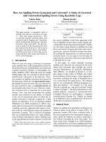
Figure 1 shows a model of HCV cell entry that takes
into account the heterogeneity of the virus and the
results obtained from the different infection assays.
Several forms of HCV have been proposed to exist:
lipoprotein-free enveloped virus, lipoprotein-free non-
enveloped virus, lipoprotein-associated enveloped virus
and lipoprotein-associated nonenveloped virus. These
different forms might use different pathways to infect
cells. HCVpp most likely resembles lipoprotein-free
enveloped viruses. Results from assays with HCVpp
suggest that lipoprotein-free enveloped virions are
infectious and require CD81, SR-BI and an as yet
unidentified protein for infectivity. However, if the cor-
relation between infectivity and lipoprotein association
observed in chimpanzees can be generalized, this form
of the virus only plays a minor role. Its infectivity
in vivo is probably too low to cause a sustained infec-
tion.
Lipoprotein-associated, enveloped viral particles are
probably resembled by HCVcc produced in a recently
described cell culture model [56]. Their infectivity was
dependent on CD81 expression on host cells and inver-
sely correlated with their density, indicating that lipo-
proteins promote infectivity. Lipoprotein receptors
might facilitate the efficient capture of these virions
and transfer them to CD81 or SR-BI in order to initi-
ate fusion of the viral and host cell membranes. At this
point, the entry pathways of enveloped virions with
and without associated lipoproteins would merge.
Without lipoprotein association, the capture of virions
Fig. 1. Model of HCV cell attachment and entry. HCV particles in the circulation can be either enveloped or nonenveloped, and either bound
to or free of lipoproteins. The different forms of HCV might use different receptors for cell attachment and entry. Enveloped virions might
interact with CD81 via envelope proteins E2, whereas the interaction between lipoprotein-associated virions and the LDL receptor might be
independent of the envelope proteins. SR-BI might have a dual role and facilitate binding of enveloped virions via E2, and of lipoprotein-asso-
ciated virions via a lipoprotein-mediated mechanism. Upon endocytosis of lipoprotein-associated enveloped virions, E2 might interact with
CD81 or SR-BI and the entry pathways for enveloped virions with and without associated lipoproteins merge. At least one additional host
protein, which has not yet been identified, is required for cell entry of enveloped virions via the CD81 ⁄ SR-BI pathways. The existence of
nonenveloped, lipoprotein-associated virions and whether they can establish a productive infection is controversial. For simplicity, immuno-
globulins, which can also bind to HCV particles, are not shown.
Putative HCV receptors G. Diedrich
3880 FEBS Journal 273 (2006) 3871–3885 ª 2006 The Author Journal compilation ª 2006 FEBS
would be less efficient, explaining the requirement of
lipoproteins for efficient infection.
Do all lipoprotein-associated virions require CD81
for cell entry? There are at least doubts. Hadlock et al.
[34] cloned several antibodies from a patient’s B-cells
that prevented the binding of CD81 to recombinant
E2 from genotypes 1a, 1b, 2a and 2b. If the patient
had high titers of potentially broadly neutralizing anti-
bodies, why did he continue to exhibit plasma viremia?
The authors speculated that CD81 might not be the
primary receptor for some HCV strains. Alternatively,
the epitopes recognized by the neutralizing antibodies
might not be accessible on HCV particles in the circu-
lation (see below).
Do all HCV particles require an envelope for cell
entry? Again, there are at least doubts. The detection
of both envelope proteins in lipoprotein-associated
virions has been challenging. It will be difficult to
unambiguously demonstrate the existence of lipopro-
tein-associated, nonenveloped HCV particles, as the
failure to detect the envelope can also be the result of
technical problems of the detection methods. However,
there are indications that these particles might exist
[12,22]. Further analysis will be needed to decide whe-
ther nonenveloped, lipoprotein-associated virions exist
and are infectious. How these particles would deliver
their viral genome into the cytoplasm is not known. If
such a cell entry mechanism exists, lipoprotein recep-
tors will probably play an important role.
Electron microscopy studies and separation of viral
particles on density gradients suggest the existence of
lipoprotein-free, nonenveloped virions in infected
serum, but there is no evidence that these particles are
infectious.
The use of lipoproteins for internalization into endo-
cytic vesicles might explain the inefficiency of the
humoral immune response to clear an HCV infection.
Viral epitopes required for the delivery of the viral
genome into the cytoplasm might be covered by lipo-
proteins. If the interaction between lipoproteins and
viral particles is already established during their assem-
bly inside infected cells, then these epitopes will not be
accessible in the circulation to neutralizing antibodies.
Upon internalization of virions via lipoprotein recep-
tors, the environment of endocytic vesicles might
induce a conformational change of the virus–lipopro-
tein complex and expose these epitopes.
Association with exosomes has been suggested as
another means for HCV to enter cells [29,92], but this
hypothesis remains highly speculative. Exosomes con-
tain many host proteins involved in cell adhesion and
membrane fusion. Although experimental evidence is
missing, it is widely believed that exosomes can fuse
with target cells and thus transport cytosolic and mem-
brane components from one cell to another. If HCV
particles are integrated into the center of the exosome
and not just adsorbed to the outside of the membrane
(which remains to be demonstrated), the virus might
use the potentially fusogenic properties of exosomes
for cell entry. This mechanism would be independent
of HCV’s envelope proteins. A similar mechanism has
been proposed for HIV as a low-efficiency pathway for
cell entry [93].
The hypothesis that different forms of HCV particles
use different mechanisms for cell entry is further sup-
ported by sequence analysis of the genome of viral
particles isolated from different tissues. Amino acid
changes in the N-terminal domain of E2 occurred
more frequently in virions isolated from whole plasma
and liver than from lipoprotein-associated virions in
plasma [94]. The N-terminus of E2 in the latter parti-
cles was not subject to any selection pressure from the
immune system and therefore is probably not involved
in receptor binding. In contrast, the majority of viral
particles in plasma and in the liver appear to use that
region of E2 for cell entry. This result further suggests
that viral particles in serum cannot easily switch from
the lipoprotein-associated state to the lipoprotein-free
state and vice versa. It is likely that the interaction
between lipoproteins and virions is established during
viral assembly inside infected cells. It will be important
to learn more about the different forms of HCV and
their correlation with disease progression, to under-
stand why some particles associate with lipoproteins
and others do not, and to identify which cell types the
different forms preferentially infect and replicate in.
Conclusions
Many pieces of the mechanism of HCV cell entry have
been identified in recent years. However, it is unclear
how these pieces fit together. The involvement of sev-
eral proteins in HCV cell entry either points towards a
complex entry pathway including many sequential
steps, or the virus might enter cells through more than
one pathway. Firstly, enveloped HCV might enter cells
through an interaction between the viral envelope pro-
teins and cellular receptors like CD81 and SR-BI.
Second, HCV associated to lipoproteins attaches to
lipoprotein receptors on the plasma membrane and
might gain access to the cytoplasm without utilizing
CD81 and potentially even without involvement of the
viral envelope proteins. The extent to which these
putative entry pathways are used and genetic or envi-
ronmental factors that shift the virus from one path-
way to the other remain difficult to analyze in the
G. Diedrich Putative HCV receptors
FEBS Journal 273 (2006) 3871–3885 ª 2006 The Author Journal compilation ª 2006 FEBS 3881
absence of a general cell culture system for HCV. Such
a system will also be required to analyze which of the
different forms of the virus are able to establish a pro-
ductive infection once they have entered cells. The
developments of pseudotyped viral particles displaying
native HCV envelope proteins and of a cell culture sys-
tem for one viral strain were important steps for the
validation of some receptor candidates. However, these
model systems have several limitations. Pseudotyped
particles produced by current methods do not bind
lipoproteins and thus lack an important feature associ-
ated with HCV infectivity. The current cell culture
model supports the propagation of only one HCV
strain whose properties may or may not be representa-
tive of the majority of HCV strains. Therefore, infec-
tivity assays using these systems might not measure all
of HCV’s properties. Until a general culture system for
the propagation of the majority of clinical HCV iso-
lates will be developed, the pathways for HCV cell
entry remain speculative.
Acknowledgements
The author thanks U. Splittgerber and S. Sauter for
helpful discussions.
References
1 Anonymous (1999) EASL International Consensus
Conference on hepatitis C. Paris, 26–27 February 1999.
Consensus statement. J Hepatol 31, 3–8.
2 Hoofnagle JH (2002) Course and outcome of hepatitis
C. Hepatology 35, S21–S29.
3 Moradpour D, Cerny A, Heim MH & Blum HE (2001)
Hepatitis C: an update. Swiss Med Wkly 131, 291–298.
4 Fried MW (2004) Viral factors affecting the outcome of
therapy for chronic hepatitis C. Rev Gastroenterol
Disord 4, S8–S13.
5 Pawlotsky JM & McHutchison JG (2003) Hepatitis C.
Development of new drugs and clinical trials: promises
and pitfalls. Summary of an AASLD hepatitis single
topic conference, Chicago, IL, February 27-March 1,
2003. Hepatology 39, 554–567.
6 De Francesco R & Migliaccio G (2005) Challenges and
successes in developing new therapies for hepatitis C.
Nature 436, 953–960.
7 Bartenschlager R & Lohmann V (2000) Replication of
hepatitis C virus. J General Virol 81, 1631–1648.
8 Hope RG & McLauchlan J (2000) Sequence motifs
required for lipid droplet association and protein stabi-
lity are unique to the hepatitis C virus core protein.
J General Virol 81, 1913–1925.
9 Voisset C & Dubuisson J (2004) Functional hepatitis C
virus envelope glycoproteins. Biol Cell 96, 413–420.
10 Egger D, Wolk B, Gosert R, Bianchi L, Blum HE,
Moradpour D & Bienz K (2002) Expression of hepatitis
C virus proteins induces distinct membrane alterations
including a candidate viral replication complex. J Virol
76, 5974–5984.
11 Gosert R, Egger D, Lohmann V, Bartenschlager R,
Blum HE, Bienz K & Moradpour D (2003) Identifica-
tion of the hepatitis C virus RNA replication complex
in Huh7 cells harboring subgenomic replicons. J Virol
77, 5487–5492.
12 Petit MA, Lievre M, Peyrol S, De Sequeira S, Berthillon
P, Ruigrok RWH & Trepo C (2005) Enveloped particles
in the serum of chronic hepatitis C patients. Virology
336, 144–153.
13 Roingeard P, Hourioux C, Blanchard E, Brand D &
Ait-Goughoulte M (2004) Hepatitis C virus ultrastruc-
ture and morphogenesis. Biol Cell 96, 103–108.
14 Andre P, Perlemutter G, Budkowska A, Brechot C &
Lotteau V (2005) Hepatitis C virus particles and lipo-
protein metabolism. Semin Liver Dis 25, 93–104.
15 Kaito M, Watanabe S, Tsukiyama-Kohara K, Yamagu-
chi K, Kobayashi Y, Konishi M, Yokoi M, Ishida S,
Suzuki S & Kohara M (1994) Hepatitis C virus particle
detected by immunoelectron microscopic study. J Gen-
eral Virol 75, 1755–1760.
16 Prince AM, Huima-Byron T, Parker TS & Levine DM
(1996) Visualization of hepatitis C virions and putative
defective interfering particles isolated from low-density
lipoproteins. J Viral Hepat 3, 11–17.
17 Maillard P, Krawczynski K, Nitkiewicz J, Bronnert C,
Sidorkiewicz M, Gounon P, Dubuisson J, Faure G,
Crainic R & Budkowska A (2001) Nonenveloped
nucleocapsids of hepatitis C virus in the serum of
infected patients. J Virol 75, 8240–8250.
18 Thomssen R, Bonk S, Propfe C, Heermann KH, Kochel
HG & Uy A (1992) Association of hepatitis C virus in
human sera with beta-lipoprotein. Med Microbiol
Immunol 181, 293–300.
19 Thomssen R, Bonk S & Thiele A (1993) Density hetero-
geneities of hepatitis C virus in human sera due to the
binding of beta-lipoproteins and immunoglobulins. Med
Microbiol Immunol 182, 329–334.
20 Hijikata M, Shimizu YK, Kato H, Iwamoto A, Shih
JW, Alter HJ, Purcell RH & Yoshikura H (1993) Equi-
librium centrifugation studies of hepatitis C virus: evi-
dence for circulating immune complexes. J Virol 67,
1952–1958.
21 Miyamoto H, Okamoto H, Sato K, Tanaka T &
Mishiro S (1992) Extraordinarily low density of hepati-
tis C virus estimated by sucrose density gradient centri-
fugation and the polymerase chain reaction. J General
Virol 73, 715–718.
22 Nielsen SU, Bassendine MF, Burt AD, Martin C,
Pumeechockchai W & Toms GL (2006) Association
between hepatitis C virus and very-low-density
Putative HCV receptors G. Diedrich
3882 FEBS Journal 273 (2006) 3871–3885 ª 2006 The Author Journal compilation ª 2006 FEBS
lipoprotein (VLDL) ⁄ LDL analyzed in iodixanol density
gradients. J Virol 80, 2418–2428.
23 Kono Y, Hayashida K, Tanaka H, Ishibashi H &
Harada M (2003) High-density lipoprotein binding rate
differs greatly between genotypes 1b and 2a ⁄ 2b of hepa-
titis C virus. J Med Virol 70, 42–48.
24 Schettler V, Monazahian M, Wieland E, Ramadori G,
Grunewald RW, Thomssen R & Muller GA (2001)
Reduction of hepatitis C virus load by H.E.L.P LDL
apheresis. Eur J Clin Invest 31, 154–155.
25 Fujita N, Kaito M, Ishida S, Nakagawa N, Ikoma J,
Adachi Y & Watanabe S (2001) Paraformaldehyde pro-
tects of hepatitis C virus particles during ultracentrifua-
tion. J Med Virol 63, 108–116.
26 Andre P, Komurian-Pradel F, Deforges S, Perret M,
Berland JL, Sodoyer M, Pol S, Brechot C, Paranhos-
Baccala G & Lotteau V (2002) Characterization of low-
and very-low-density hepatitis C virus RNA-containing
particles. J Virol 76, 6919–6928.
27 Trestard A, Bacq Y, Buzelay L, Dubois F, Barin F,
Goudeau A & Roingeard P (1998) Ultrastructural and
physicochemical characterization of the hepatitis C virus
recovered from the serum of an agammaglobulinemic
patient. Arch Virol 143, 2241–2245.
28 Kanto T, Hayashi N, Takehara T, Hagiwara H, Mita
E, Naito M, Kasahara A, Fusamoto H & Kamada T
(1994) Buoyant density of hepatitis C virus recovered
from infected hosts: two different features in sucrose
equilibrium density-gradient centrifugation related to
degree of liver inflammation. Hepatology 19, 296–302.
29 Masciopinto F, Giovani C, Campagnoli S, Galli-Stam-
pino L, Colombatto P, Brunetto M, Yen TS, Houghton
M, Pileri P & Abrignani S (2004) Association of hepati-
tis C virus envelope proteins with exosomes. Eur J
Immunol 34, 2834–2842.
30 Bradley D, McCaustland K, Krawczynski K, Spelbring
J, Humphrey C & Cook EH (1991) Hepatitis C virus:
buoyant density of the factor VIII-derived isolate in
sucrose. J Med Virol 34, 206–208.
31 Yasui K, Wakita T, Tsukiyama-Kohara K, Funahashi
SI, Ichikawa M, Kajita T, Moradpour D, Wands JR &
Kohara M (1998) The native form and maturation pro-
cess of hepatitis C virus core protein. J Virol 72, 6048–
6055.
32 Shindo M, Di Bisceglie AM, Akatsuka T, Fong TL,
Scaglione L, Donets M, Hoofnagle JH & Feinstone SM
(1994) The physical state of the negative strand of hepa-
titis C virus RNA in serum of patients with chronic
hepatitis C. Proc Natl Acad USA 91, 8719–8723.
33 Pileri P, Uematsu Y, Campagnoli S, Galli G, Falugi F,
Petracca R, Weiner AJ, Houghton M, Rosa D, Grandi
G et al. (1998) Binding of hepatitis C virus to CD81.
Science 282, 938–941.
34 Hadlock KG, Lanford RE, Perkins S, Rowe J, Yang Q,
Levy S, Pileri P, Abrignani S & Foung SK (2000)
Human monoclonal antibodies that inhibit binding of
hepatitis C virus E2 protein to CD81 and recognize con-
served conformational epitopes. J Virol 74, 10407–
10416.
35 Cerino A, Meola A, Segagni L, Furione M, Marciano
S, Triyatni M, Liang TJ, Nicosia A & Mondelli MU
(2001) Monoclonal antibodies with broad specificity for
hepatitis C virus hypervariable region 1 variants can
recognize viral particles. J Immunol 167, 3878–3886.
36 Tan YJ, Lim SP, Ng P, Goh PY, Lim SG, Tan YH &
Hong W (2003) CD81 engineered with endocytotic sig-
nals mediates HCV cell entry: implications for receptor
usage by HCV in vivo. Virology 308, 250–269.
37 Thomssen R & Bonk S (2002) Virolytic action of lipo-
protein lipase on hepatitis C virus in human sera. Med
Microbiol Immunol 191, 17–24.
38 Germi R, Crance JM, Garin D, Guimet J, Lortat-Jacob
H, Ruigrok RW, Zarski JP & Drouet E (2002) Cellular
glycosaminoglycans and low density lipoprotein receptor
are involved in hepatitis C virus adsorption. J Med
Virol 68, 206–215.
39 Zibert A, Schreier E & Roggendorf M (1995) Antibo-
dies in human sera specific to hypervariable region 1 of
hepatitis C virus can block viral attachment. Virology
208, 653–661.
40 Choo QL, Kuo G, Ralston R, Weiner A, Chien D, Van
Nest G, Han J, Berger K, Thudium K, Kuo C et al.
(1994) Vaccination of chimpanzees against infection by
the hepatitis C virus. Proc Natl Acad Sci USA 91,
1294–1298.
41 Farci P, Shimoda A, Wong D, Cabezon T, De Gioannis
D, Strazzera A, Shimizu Y, Shapiro M, Alter HJ &
Purcell RH (1996) Prevention of hepatitis C virus infec-
tion in chimpanzees by hyperimmune serum against the
hypervariable region 1 of the envelope 2 protein. Proc
Natl Acad Sci USA 93, 15394–15399.
42 Seipp S, Mueller HM, Pfaff E, Stremmel W, Theilmann
L & Goeser T (1997) Establishment of persistent hepati-
tis C virus infection and replication in vitro. J General
Virol 78, 2467–2476.
43 Munoz A, Castrillo JL & Carrasco L (1985) Modifica-
tion of membrane permeability during Semliki Forest
virus infection. Virology 146, 203–212.
44 Liprandi F, Moros Z, Gerder M, Ludert JE, Pujol FH,
Ruiz MC, Michelangeli F, Charpilienne A & Cohen J
(1997) Productive penetration of rotavirus in cultured
cells induces coentry of the translation inhibitor alpha-
sarcin. Virology 237, 430–438.
45 Cuadras MA, Arias CF & Lopez S (1997) Rotaviruses
induce an early membrane permeabilization of MA104
cells and do not require a low intracellular Ca
2+
con-
centration to initiate their replication cycle. J Virol 71,
9065–9074.
46 Wunschmann S, Medh JD, Klinzmann D, Schmidt WN
& Stapleton JT (2000) Characterization of hepatitis C
G. Diedrich Putative HCV receptors
FEBS Journal 273 (2006) 3871–3885 ª 2006 The Author Journal compilation ª 2006 FEBS 3883
virus (HCV) and HCV E2 interactions with CD81 and
the low-density lipoprotein receptor. J Virol 74, 10055–
10062.
47 Hsu M, Zhang J, Flint M, Logvinoff C, Cheng-Mayer
C, Rice CM & McKeating JA (2003) Hepatitis C virus
glycoproteins mediate pH-dependent cell entry of pseu-
dotyped retroviral particles. Proc Natl Acad Sci USA
100, 7271–7276.
48 Bartosch B, Dubuisson J & Cosset FL (2003) Infectious
hepatitis C virus pseudo-particles containing functional
E1–E2 envelope protein complexes. J Exp Med 197,
633–642.
49 Drummer HE, Maerz A & Poumbourios P (2003) Cell
surface expression of functional hepatitis C virus E1
and E2 glycoproteins. FEBS Lett 546, 385–390.
50 Bartosch B, Bukh J, Meunier JC, Granier C, Engle RE,
Blackwelder WC, Emerson SU, Cosset FL & Purcell RH
(2003) In vitro assay for neutralizing antibody to hepati-
tis C virus: evidence for broadly conserved neutralization
epitopes. Proc Natl Acad Sci USA 100, 14199–14204.
51 Logvinoff C, Major ME, Oldach D, Heyward S, Talal
A, Balfe P, Feinstone SM, Alter H, Rice CM &
McKeating JA (2004) Neutralizing antibody response
during acute and chronic hepatitis C virus infection.
Proc Natl Acad Sci USA 101, 10149–10154.
52 Flint M, Logvinoff C, Rice CM & McKeating JA
(2004) Characterization of infectious retroviral pseudo-
type particles bearing hepatitis C virus glycoproteins.
J Virol 78, 6875–6882.
53 Martire G, Viola A, Iodice L, Lotti LV, Gradini R &
Bonatti S (2001) Hepatitis C virus structural proteins
reside in the endoplasmic reticulum as well as in the
intermediate compartment ⁄ cis-Golgi complex region of
stably transfected cells. Virology 280, 176–182.
54 Fisher EA & Ginsberg HN (2002) Complexity in the
secretory pathway: the assembly and secretion of apoli-
poprotein B-containing lipoproteins. J Biol Chem 277,
17377–17380.
55 Wakita T, Pietschmann T, Kato T, Date T, Miyamoto
M, Zhao Z, Murthy K, Habermann A, Krausslich HG,
Mizokami M et al. (2005) Production of infectious
hepatitis C virus in tissue culture from a cloned viral
genome. Nat Med 11, 791–796.
56 Lindenbach BD, Evans MJ, Syder AJ, Wolk B, Telling-
huisen TL, Liu CC, Maruyama T, Hynes RO, Burton
DR, McKeating JA et al. (2005) Complete replication
of hepatitis C virus in cell culture. Science 309, 623–626.
57 Zhong J, Gastaminza P, Cheng G, Kapadia S, Kato T,
Burton DR, Wieland SF, Uprichard SL, Wakita T &
Chisari FV (2005) Robust hepatitis C virus infection in
vitro. Proc Natl Acad Sci USA 102, 9294–9299.
58 Barth H, Schafer C, Adah MI, Zhang F, Linhardt RJ,
Toyoda H, Kinoshita-Toyoda A, Toida T, Van
Kuppevelt TH, Depla E et al. (2003) Cellular binding
of hepatitis C virus envelope glycoprotein E2 requires
cell surface heparan sulfate. J Biol Chem 278, 41003–
41012.
59 Callens N, Ciczora Y, Bartosch B, Vu-Dac N, Cosset
FL, Pawlotsky JM, Penin F & Dubuisson J (2005) Basic
residues in hypervariable region 1 of hepatitis C virus
envelope glycoprotein e2 contribute to virus entry.
J Virol 79, 15331–15341.
60 Levy S, Todd SC & Maecker HT (1998) CD81 (TAPA-
1): a molecule involved in signal transduction and cell
adhesion in the immune system. Annu Rev Immunol 16
,
89–109.
61 Higginbottom A, Quinn ER, Kuo CC, Flint M,
Wilson LH, Bianchi E, Nicosia A, Monk PN,
McKeating JA & Levy S (2000) Identification of
amino acid residues in CD81 critical for interaction
with hepatitis C virus envelope glycoprotein E2.
J Virol 74, 3642–3649.
62 Flint M, Maidens C, Loomis-Price LD, Shotton C,
Dubuisson J, Monk P, Higginbottom A, Levy S &
McKeating JA (1999) Characterization of hepatitis C
virus E2 glycoprotein interaction with a putative cellular
receptor, CD81. J Virol 73, 6235–6244.
63 Yagnik AT, Lahm A, Meola A, Roccasecca RM, Ercole
BB, Nicosia A & Tramontano A (2000) A model for
the hepatitis C virus envelope glycoprotein E2. Proteins
40, 355–366.
64 Zhang J, Randall G, Higginbottom A, Monk P, Rice
CM & McKeating JA (2004) CD81 is required for hepa-
titis C virus glycoprotein-mediated viral infection.
J Virol 78, 1448–1455.
65 Petracca R, Falugi F, Galli G, Norais N, Rosa D, Cam-
pagnoli S, Burgio V, Di Stasio E, Giardina B et al.
(2000) Structure–function analysis of hepatitis C virus
envelope-CD81 binding. J Virol 74, 4824–4830.
66 Bartosch B, Vitelli A, Granier C, Goujon C, Dubuisson
J, Pascale S, Scarselli E, Cortese R, Nicosia A & Cosset
FL (2003) Cell entry of hepatitis C virus requires a set
of co-receptors that include the CD81 tetraspanin and
the SR-BI scavenger receptor. J Biol Chem 278, 41624–
41630.
67 McKeating JA, Zhang LQ, Logvinoff C, Flint M,
Zhang J, Yu L, Butera D, Ho DD, Dustin LB, Rice
CM et al. (2004) Diverse hepatitis C virus glycoproteins
mediate viral infection in a CD81-dependent manner.
J Virol 78, 8496–8505.
68 Cormier EG, Tsamis F, Kajumo F, Durso RJ, Gardner
JP & Dragic T (2004) CD81 is an entry coreceptor for
hepatitis C virus. Proc Natl Acad Sci USA 101, 7270–
7274.
69 Sasaki M, Yamauchi K, Nakanishi T, Kamogawa Y &
Hayashi N (2003) In vitro binding of hepatitis C
virus to CD81-positive and -negative human cell lines.
J Gastroenterol Hepatol 18, 74–79.
70 Rigotti A, Miettinen H & Krieger M (2003) The role of
the high density lipoprotein receptor SR-BI in the lipid
Putative HCV receptors G. Diedrich
3884 FEBS Journal 273 (2006) 3871–3885 ª 2006 The Author Journal compilation ª 2006 FEBS
metabolism of endocrine and other tissues. Endocr Rev
24, 357–387.
71 Scarselli E, Ansuini H, Cerino R, Roccasecca RM,
Acali S, Filocamo G, Traboni C, Nicosia A, Cortese R
& Vitelli A (2002) The human scavenger receptor class
B type I is a novel candidate receptor for the hepatitis
C virus. EMBO J 21, 5017–5025.
72 Lavillette D, Tarr AW, Voisset C, Donot P, Bartosch
B, Bain C, Patel AH, Dubuisson J, Ball JK & Cosset
FL (2005) Characterization of host-range and cell entry
properties of the major genotypes and subtypes of
hepatitis C virus. Hepatology 41, 265–274.
73 Voisset C, Callens N, Blanchard E, Op De Beeck A,
Dubuisson J & Vu-Dac N (2005) High density lipo-
proteins facilitate hepatitis C virus entry through the
scavenger receptor class B type I. J Biol Chem 280,
7793–7799.
74 Bartosch B, Verney G, Dreux M, Donot P, Morice Y,
Penin F, Pawlotsky JM, Lavillette D & Cosset FL
(2005) An interplay between hypervariable region 1 of
the hepatitis C virus E2 glycoprotein, the scavenger
receptor BI, and high-density lipoprotein promotes both
enhancement of infection and protection against neutra-
lizing antibodies. J Virol 79, 8217–8229.
75 Dreux M, Pietschmann T, Granier C, Voisset C,
Ricard-Blum S, Mangeot PE, Keck Z, Foung S, Vu-
Dac N, Dubuisson J et al. (2006) High density lipopro-
tein inhibits hepatitis C virus neutralising antibodies by
stimulating cell entry via activation of the scavenger
receptor BI. J Biol Chem 281, 18285–18295.
76 Maillard P, Huby T, Andreo U, Moreau M, Chapman
J & Budkowska A (2006) The interaction of natural
hepatitis C virus with human scavenger receptor SR-
BI ⁄ Cla1 is mediated by apoB-containing lipoproteins.
FASEB J 20, 735–737.
77 Monazahian M, Bohme I, Bonk S, Koch A, Scholz C,
Grethe S & Thomssen R (1999) Low density lipoprotein
receptor as a candidate receptor for hepatitis C virus.
J Med Virol 57, 223–229.
78 Agnello V, Abel G, Elfahal M, Knight GB & Zhang
QX (1999) Hepatitis C virus and other flaviviridae
viruses enter cells via low density lipoprotein receptor.
Proc Natl Acad Sci USA 96, 12766–12771.
79 Favre D, Berthillon P & Trepo C (2001) Removal of
cell-bound lipoproteins: a crucial step for the efficient
infection of liver cells with hepatitis C virus in vitro.
C R Acad Sci III (324), 1141–1148.
80 Monazahian M, Kippenberger S, Muller A, Seitz H,
Bohme I, Grethe S & Thomssen R (2000) Binding of
human lipoproteins (low, very low, high density lipopro-
teins) to recombinant envelope proteins of hepatitis C
virus. Med Microbiol Immunol 188, 177–184.
81 Lambot M, Fretier S, Op De Beeck A, Quatannens B,
Lestavel S, Clavey V & Dubuisson J (2002) Reconstitu-
tion of hepatitis C virus envelope glycoproteins into
liposomes as a surrogate model to study virus attach-
ment. J Biol Chem 277, 20625–20630.
82 Gardner JP, Durso RJ, Arrigale RR, Donovan GP,
Maddon PJ, Dragic T & Olson WC (2003) L-sign (CD
209L) is a liver-specific capture receptor for hepatitis C
virus. Proc Natl Acad Sci USA 100, 4498–4503.
83 Lozach PY, Lortat-Jacob H, de Lacroix de Lavalette A,
Staropoli I, Foung S, Amara A, Houles C, Fieschi F,
Schwartz O, Virelizier JL et al. (2003) DC-SIGN and
L-SIGN are high affinity binding receptors for hepatitis
C virus glycoprotein E2. J Biol Chem 278, 20358–20366.
84 Pohlmann S, Zhang J, Baribaud F, Chen Z, Leslie GJ,
Lin G, Granelli-Piperno A, Doms RW, Rice CM &
McKeating JA (2003) Hepatitis C virus glycoproteins
interact with DC-SIGN and DC-SIGNR. J Virol 77,
4070–4080.
85 Saunier B, Triyatni M, Ulianich L, Maruvada P, Yen P
& Kohn LD (2003) Role of the asialoglycoprotein
receptor in binding and entry of hepatitis C virus struc-
tural proteins in cultured human hepatocytes. J Virol
77, 546–559.
86 Lozach PY, Amara A, Bartosch B, Virelizier JL, Arenz-
ana-Seisdedos F, Cosset FL & Altmeyer R (2004)
C-type lectins L-SIGN and DC-SIGN capture and
transmit infectious hepatitis C virus pseudotype parti-
cles. J Biol Chem 279, 32035–32045.
87 Cormier EG, Durso RJ, Tsamis F, Boussemart L,
Manix C, Olson WC, Gardner JP & Dragic T (2004)
L-SIGN (CD209L) and DC-SIGN (CD209) mediate
transinfection of liver cells by hepatitis C virus. Proc
Natl Acad Sci USA 101, 14067–14072.
88 Ludwig IS, Lekkerkerker AN, Depla E, Bosman F, Mus-
ters RJ, Depraetere S, van Kooyk Y & Geijtenbeek TB
(2004) Hepatitis C virus targets DC-SIGN and L-SIGN
to escape lysosomal degradation. J Virol 78, 8322–8332.
89 Stockert RJ (1995) The asialoglycoprotein receptor:
relationships between structure, function, and expres-
sion. Physiol Rev 75, 591–609.
90 Saha K, Zhang J, Gupta A, Dave R, Yimen M &
Zerhouni B (2001) Isolation of primary HIV-1 that
target CD8+ T lymphocytes using CD8 as a receptor.
Nat Med 7, 65–72.
91 Spear PG & Longnecker R (2003) Herpesvirus entry: an
update. J Virol 77, 10179–10185.
92 Favre D & Muellhaupt B (2005) Potential cellular
receptors involved in hepatitis C virus entry into cells.
Lipids Health Dis 4,9.
93 Gould SJ, Booth AM & Hildreth JE (2003) The Trojan
exosome hypothesis. Proc Natl Acad Sci USA 100,
10592–10597.
94 Deforges S, Evlashev A, Perret M, Sodoyer M, Pouzol
S, Scoazec JY, Bonnaud B, Diaz O, Paranhos-Baccala
G, Lotteau V et al. (2004) Expression of hepatitis
C virus proteins in epithelial intestinal cells in vivo.
J General Virol 85, 2515–2523.
G. Diedrich Putative HCV receptors
FEBS Journal 273 (2006) 3871–3885 ª 2006 The Author Journal compilation ª 2006 FEBS 3885