Tài liệu Báo cáo khoa học: Cell-free translation systems for protein engineering docx
Bạn đang xem bản rút gọn của tài liệu. Xem và tải ngay bản đầy đủ của tài liệu tại đây (757.33 KB, 8 trang )
MINIREVIEW
Cell-free translation systems for protein engineering
Yoshihiro Shimizu
1
, Yutetsu Kuruma
2
, Bei-Wen Ying
1
, So Umekage
3
and Takuya Ueda
1
1 Department of Medical Genome Sciences, Graduate School of Frontier Sciences, The University of Tokyo, Kashiwanoha, Kashiwa-shi,
Chiba, Japan
2 ‘Enrico Fermi’ Center, Compendio del Viminale, Rome, Italy
3 Division of Bioscience and Biotechnology, Department of Ecological Engineering, Toyohashi University of Technology, Tempaku-cho,
Toyohashi, Aichi, Japan
Introduction
Although noncoding RNAs play significant roles in
cellular function [1,2], especially in higher organisms, it
is proteins that dominate most cellular processes. Pro-
teins are the most abundant cellular components and
are responsible for structural, metabolic and regulatory
functions both inside and outside of cells. Thus, inves-
tigation of proteins and elucidation of the molecular
mechanisms underlying their activities are crucial to
our understanding of life.
Generally, owing to their low cost and high produc-
tivity, proteins are prepared using in vivo gene expres-
sion systems. However, the problems associated with
using living cells for recombinant protein expression
include protein degradation and aggregation, or loss of
template DNA. Furthermore, it requires several labori-
ous experimental steps including DNA cloning in the
vector, DNA transformation in cells, and overexpres-
sion of the desired protein in cells. Thus, there are
limitations associated with using in vivo technology for
protein production.
Cell-free translation represents an alternative to
in vivo expression, and rapid progress is being made in
this field, which is gaining attention for its simplicity
and high degree of controllability. Proteins are pro-
duced only when template DNA or mRNA is added
to the reaction mixture, followed by incubation for
Keywords
cell-free protein synthesis; chaperone;
disulfide bond formation; in vitro selection;
liposome; minimal cell; ribosome display;
translation; unnatural amino acid
Correspondence
T. Ueda, Department of Medical Genome
Sciences, Graduate School of Frontier
Sciences, University of Tokyo, FSB401,
5-1-5, Kashiwanoha, Kashiwa-shi, Chiba
prefecture 277-8562, Japan
Fax: +81 4 7136 3642
Tel: +81 4 7136 3641
E-mail:
(Received 8 May 2006, revised 20 June
2006, accepted 26 June 2006)
doi:10.1111/j.1742-4658.2006.05431.x
Cell-free translation systems have developed significantly over the last two
decades and improvements in yield have resulted in their use for protein
production in the laboratory. These systems have protein engineering appli-
cations, such as the production of proteins containing unnatural amino
acids and development of proteins exhibiting novel functions. Recently, it
has been suggested that cell-free translation systems might be used as the
fundamental basis for cell-like systems. We review recent progress in the
field of cell-free translation systems and describe their use as tools for pro-
tein production and engineering.
Abbreviations
EGFP, enhanced green fluorescent protein; GFP, green fluorescent protein; PDI, protein disulfide isomerase; PURE, protein synthesis using
recombinant elements; scFv, single-chain variable fragment of antibody; Sec, secretory; SR, signal recognition particle receptor; SRP, signal
recognition particle.
FEBS Journal 273 (2006) 4133–4140 ª 2006 The Authors Journal compilation ª 2006 FEBS 4133
several hours. As PCR products can be used, synthes-
ized protein may be obtained rapidly from a small
amount of cDNA. In addition, control can be achieved
easily via modified reaction conditions, such as the
addition of accessory elements or removal of inhibitory
substances. Thus, cell-free translation has the potential
to meet many of the needs of preparatory protein
science, and further improvements will accelerate
exploitation of this technology.
In this article, we focus on the techniques relating
to cell-free translation systems for enhancing the syn-
thesis of biologically active proteins, the creation of
cell-like compartments and the synthesis of artificial
proteins.
Overview
Cell-free translation systems are based on the cellular
ribosomal protein synthesis system. Generally, the sys-
tem is composed of a cell extract (referred to as the
S30 fraction) from Escherichia coli, wheat germ, or
rabbit reticulocytes. These extracts are supernatants
from a 30 000 g centrifugation and contain compo-
nents such as ribosomes, translation factors, amino-
acyl-tRNA synthetases, and tRNAs, which are
required for production of protein. Efficient protein
production may require supplementation of the S30
extract with additional RNA polymerase, as well as
several enzymes for energy regeneration and their sub-
strates (Fig. 1).
The productivity of S30-directed, cell-free translation
systems has improved greatly over the last two dec-
ades. In 1988, the continuous-flow cell-free system [3]
represented the first demonstration that cell-free trans-
lation could be utilized as a tool for producing protein.
This system relied upon a continuous supply of energy
source and amino acids, resulting in a significant
increase in productivity. Although this method was not
used widely due to its complexity and variable repro-
ducibility of yield, the concept resulted in the subse-
quent development of the continuous-exchange
cell-free [4] and the bilayer cell-free systems [5]. Using
these processes, milligram quantities of product were
achieved from a 1 mL reaction. Furthermore, the
developments of the reaction condition such as, opti-
mization of the E. coli system [6,7], improved prepar-
ation of wheat germ cell extract [8] and development
of the energy regeneration system [9], have also contri-
buted the productivity of the system.
An alternative to cell-extract based systems is repre-
sented by protein synthesis using recombinant elements
(PURE) system [10], which comprises individually
purified components of the E. coli translation appar-
atus. This system is currently not well established, yet
as a fully reconstituted system, it may provide a
greater degree of control than the conventional S30-
directed translation processes. Hence, we believe that
further analyses and developments of the system will
improve the system as a strong tool for producing pro-
teins.
Production of biologically active
proteins
In order for the cell-free translation system to produce
biologically active proteins, additional proteins such as
molecular chaperones may be required to ensure cor-
rect folding [11,12]. In E. coli, these chaperones include
the DnaK system (with its cochaperones DnaJ and
GrpE), trigger factor, and the chaperonin GroEL sys-
tem (with its cochaperonin GroES). Even in S30 sys-
tems in which intrinsic chaperones are present in
abundance, molecular chaperones are supplied to reac-
tions in order to increase synthesis of active-state
proteins [13,14]; this practice has been employed suc-
cessfully in the production of luciferase [15] and active
single-chain variable fragment of antibody (scFv) [13].
Similarly, integration of the chaperonin GroEL system
has also been found to assist folding in rabbit reticulo-
cyte lysates [16].
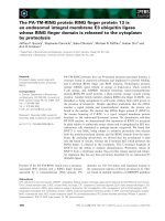
protein synthesis on ribosome
Aminoacylation
amino acid
tRNA
AT P
aminoacyl-tRNA
Transcription
temlate DNA
ATP/GTP/CTP/UTP
RNA polymerase
mRNA
Energy regeneration system
Enzymes
Substrates
(PEP/PK system
CP/CK system etc.)
ATP/GTP
Translation factor
Initiation factor
Elongation factor
Termination factor
Fig. 1. The cell-free protein synthesis system. Efficient protein syn-
thesis requires transcription of mRNA, aminoacyl tRNA, energy pro-
vision, and translation factors. Transcription of mRNA requires
template DNA, ribonucleotides and enzymes such as T7 and SP6
RNA polymerases. Translation requires factors for initiation, elonga-
tion and termination, as well as components for aminoacylation of
tRNA, such as amino acids, tRNA and ATP. The energy regener-
ation system requires enzymes and their substrates such as
phosphoenolpyruvate (PEP) ⁄ phophoenolpyruvate kinase (PK) and
creatine phosphate (CP) ⁄ creatine kinase (CK). Cell extracts provide
translation factors and enzymes for aminoacylation, whereas in
reconstituted cell-free translation systems [10] the purified compo-
nents are added individually.
Cell-free translation for protein engineering Y. Shimizu et al.
4134 FEBS Journal 273 (2006) 4133–4140 ª 2006 The Authors Journal compilation ª 2006 FEBS
Taking advantage of the absence of such molecular
chaperones in the reconstituted cell-free translation
system [10], it has been used to evaluate the chaperone
dependency on the folding of newly synthesized pro-
teins. The enzymatic activity of MetK could be detec-
ted only in the presence of GroEL ⁄ ES [17], whereas
for anti-BSA scFv, the proportion of soluble and ⁄ or
functional protein increased with the addition of the
DnaK system and trigger factor, but not GroEL ⁄ ES
[18]. Thus, further exhaustive analyses of such depend-
encies will provide not only the reconstituted cell-free
translation system itself but the S30 systems with the
specific supplementation strategies for efficient synthe-
sis of biologically active proteins.
Correct disulfide bond formation in proteins such as
antibodies can be facilitated by the addition of the
redox-dependent chaperone protein disulfide isomerase
(PDI) [19], disulfide oxidoreductase and ⁄ or modification
of the redox conditions. The greatest solubility and
activity of newly synthesized single-chain antibodies
were observed in both E. coli (B W. Ying, H. Taguchi
and T. Ueda, unpublished data, and [13]), and wheat
germ [20] systems when PDI was used under oxidative
conditions. Similarly, the large fragment (Fab) of the
catalytic antibody 6D9, which comprises several disul-
fide bonds, was expressed successfully under oxidative
conditions [21]. In the reconstituted cell-free system,
biologically active alkaline phosphatase has also been
found to be synthesized under oxidative conditions [22].
Therefore, these studies indicate that expression of
correctly folded and functional proteins can be
achieved in cell-free systems by the addition of folding
helpers, and that the flexibility of these systems repre-
sents a powerful means of generating mature protein.
Synthesis of membrane proteins for
minimal cells
The goal of the new and rapidly developing field of
synthetic biology is the development of a minimal cell,
also called an artificial cell [23]. Minimal cells are
designed to comprise the least number of molecular
components and genes [24], while still being considered
alive. The classical approach involves entrapment of
components (genes, enzymes, ribosomes, etc.) in a syn-
thetic compartment, in order to separate them from the
external environment. These compartments are usually
produced by lipid vesicles or liposomes, because they
closely resemble the cellular envelope. Based on the
concept that translation is one of the central cellular
processes required for life, cell-free transcription ⁄ trans-
lation systems have been widely used in the develop-
ment of simple cellular models [25]. Indeed, when
functional protein synthesis occurs inside liposomes, it
provides a platform for simulating a complex cellular
activity because the product of the system is the pro-
tein, the main player of the multiple cellular functions.
Yu et al. [26] performed the first liposome-encapsu-
lated cell-free protein synthesis using E. coli cell
extracts to synthesize a green fluorescence protein
(GFP-mut1) within egg phosphatidyl choline ⁄ choles-
terol liposomes. As they are easily detected, other
GFPs such as red-shifted GFP or enhanced GFP
(EGFP) have been produced effectively to illustrate the
utility of minimal cell development. For example,
Ishikawa et al. have demonstrated a unique cascading
expression system using a double expression plasmid
carrying genes encoding GFP and T7 RNA polym-
erase, under control of the T7 and SP6 promoters,
respectively [27]. The plasmid, cell-free expression sys-
tem, and SP6 RNA polymerase were trapped inside
liposomes, and production of GFP was then observed,
demonstrating that the two-level cascade actually took
place within the lipid vesicles. Sequential protein
expression (first T7 RNA polymerase, then GFP) was
proven using flow cytometry analysis. In a recent
report that did not involve liposomes, Luisi et al. [28]
divided the cell-free components into several premix-
tures (i.e., plasmids carrying the gene encoding EGFP,
amino acids and E. coli extract), then trapped them in
individual water-in-oil emulsions. Following the pre-
paration of each compartment, all three emulsions
were mixed and EGFP synthesis was observed as com-
partments fused and exchanged their contents, bringing
the reaction components together.
Although there have been many reports in recent
years of cell-free expression in liposomes, no one has
succeeded in synthesizing functional membrane pro-
teins in these systems. However, Noireaux and Libc-
haber have succeeded in synthesizing a-hemolysin
(from Staphylococcus aureus) within liposomes, using
an E. coli extract cell-free system [29]. a-Hemolysin is
a water soluble monomeric protein that is able to self-
assemble in a lipid bilayer as a homoheptamer, gener-
ating a selectively permeable pore. They used the
a-hemolysin pore as a gate for nutrient transportation
into the liposomes, and by supplementing energy and
substrates from outside the liposome, were able to
extend protein synthesis up to four days. Furthermore,
using the ability of a-hemolysin to self-assemble, an
a-hemolysin-EGFP fusion protein was successfully
formed on the membrane surface [25]. However,
although these results appear to represent impressive
achievements in minimal cell development, it must be
remembered that a-hemolysin is a water soluble (not
lipid soluble) protein.
Y. Shimizu et al. Cell-free translation for protein engineering
FEBS Journal 273 (2006) 4133–4140 ª 2006 The Authors Journal compilation ª 2006 FEBS 4135
How can we generate integral membrane proteins
within liposomes, and is there any way to integrate
proteins into the lipid bilayer in the proper conforma-
tion? Recent progress in answering these questions
arose from an experiment in which we combined
PURE system and the membrane integration ⁄ translo-
cation system, in vesicles prepared from inverted
E. coli cell membranes [30]. Using this system, mem-
brane integration and translocation were reproduced
as sequential reactions coupled with translation. The
results indicate that the minimum additional cytosolic
factors for membrane integration and translocation are
the signal recognition particle (SRP) ⁄ SRP receptor
(SR) [31] and SecA [32], respectively.
In considering membrane components, the secretory
(Sec) translocon is known to play an important role as
a protein-conducting channel for membrane integra-
tion and translocation [33]. The majority of membrane
proteins integrated through the Sec translocon, which
in E. coli is formed primarily by the essential proteins
SecY and SecE. The Sec translocon binds with high
affinity to the large ribosomal subunit, containing the
elongating nascent polypeptides, which are then integ-
rated cotranslationally. In addition, a Sec-independent
pathway using YidC [34] has been implicated in the
integration of some small molecular mass proteins,
such as the Foc subunit of FoF1-ATP synthase [35].
According to these reports, if either the Sec translocon
and ⁄ or YidC are incorporated into the lipid bilayer of
liposomes (proteoliposomes) in addition to SRP ⁄ SR,
the corresponding synthetic cell has the ability to
generate functional membrane proteins (Fig. 2). Thus,
current studies on protein expression within vesicles
may extend to the biosynthesis of lipid soluble proteins,
several of which play important roles in minimal cells.
Synthesis of artificial proteins
Over the last few decades, several applied technologies,
such as incorporation of unnatural amino acids, have
taken advantage of advances in cell-free translation
systems. The use of tRNA, mischarged with an un-
natural amino acid through a chemical acylation
method originally developed by Hecht et al. [36], was
first applied to cell-free translation systems by Schultz
and coworkers [37]. They mischarged suppressor
tRNA that recognizes amber codons (UAG) with an
unnatural amino acid, thereby altering a nonsense
codon to a sense codon corresponding to the specific
unnatural amino acid. Alternatively, mischarged tRNA
can be prepared through the use of engineered aminoa-
cyl-tRNA synthetases [38,39] and ribozymes [40] that
can catalyze aminoacylation of tRNA with specific
unnatural amino acids. In addition to amber codons,
other target codons have been utilized for the same
purpose. Artificial tRNAs that recognize four-base co-
dons have created novel codon–anticodon interactions
[41]. Furthermore, two unnatural nucleobases that
form a novel Watson–Crick-like base pair have been
introduced into tRNA and mRNA, generating
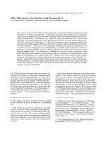
Fig. 2. Model for integration of membrane
proteins into minimal cells. Nascent poly-
peptides that are being synthesized on
ribosomes become associated with signal
recognition particle (SRP). The ribosome–
polypeptide–SRP complex is targeted to the
Sec translocon, which is embedded in the
membrane through interaction with the SRP
receptor (SR). Following release of SRP and
SR, polypeptides are cotranslationally integ-
rated into the lipid bilayer through the force
of peptide elongation. In contrast, some
small membrane proteins are targeted to
YidC, possibly via an SRP ⁄ SR pathway, and
are integrated through YidC alone. Direct
targeting of nascent polypeptides to the Sec
translocon or YidC may occur in the artificial
compartments.
Cell-free translation for protein engineering Y. Shimizu et al.
4136 FEBS Journal 273 (2006) 4133–4140 ª 2006 The Authors Journal compilation ª 2006 FEBS
additional codon–anticodon interactions and expand-
ing the genetic code [42,43]. Thus, reconstituted cell-
free systems have enabled a rewriting of the genetic
code and the incorporation of unnatural amino acids
into proteins [44,45].
Recently, a protein evolution system based on cell-
free translation has been developed (Fig. 3). This
technology is an expanded version of the SELEX (sys-
tematic evolution of ligands by exponential enrich-
ment) system [46], in which functional RNA molecules
can be selected from large libraries through successive
cycles of selection, RNA reverse transcription and
DNA amplification. Because proteins cannot be ampli-
fied by themselves, genotype and phenotype are physi-
cally linked in the system, enabling enrichment of
specific genotypes through successive selection of the
synthesized proteins. Although similar methodology,
such as phage display [47], is widely used for the same
purpose, amplification of the initial library through the
cell-free system enables the use of simple manipulation
techniques and bypasses the need for living cells.
At present, there are a number of ways to link geno-
type and phenotype within the cell-free translation sys-
tem (Fig. 3). The first technique to be demonstrated
was ribosome display [48]; this technique utilizes the
ribosome complex that has peptidyl-tRNA and mRNA
bound noncovalently to the ribosome, to form a link
consisting of protein ⁄ tRNA ⁄ ribosome ⁄ mRNA. In the
in vitro virus or mRNA display, a covalently linked
mRNA ⁄ puromycin ⁄ protein complex that is formed by
the ribosome via a peptide bond is substituted for the
stalled ribosome complex in ribosome display [49,50].
These methodologies select functional peptides or pro-
teins from large libraries and have been used to isolate
antibodies or scaffolding proteins that bind specific
proteins with high affinity [51,52], streptavidin-binding
peptide [53] and ATP-binding protein [54]. In addition,
these methods have also been used for proteomic ana-
lyses of protein–protein interactions [55,56].
Recently, CIS display achieved noncovalent linkage
between DNA and the synthesized protein in a cell-
free, coupled transcription ⁄ translation system [57]. CIS
display uses fusions between DNA encoding random
peptides and the DNA replication initiator protein
(RepA), which binds exclusively to the DNA from
which it has been expressed, resulting in a selectable
library of proteinÆDNA complexes. The formation of
proteinÆDNA complexes can also be achieved by using
cell-free translation system compartmentalized in
water-in-oil emulsions [58,59]. This technology is based
on the adjustment of the concentration of DNA and
the size of the emulsions to express a single molecule
of DNA in each compartment. Because these novel
technologies are performed using a DNAÆprotein com-
plex, they have the potential to overcome the unrelia-
bility of RNAÆprotein complex selection, which
is subject to the instability of RNA. Finally, compart-
mentalization has also been achieved using the
Fig. 3. A system for protein evolution based
on cell-free translation. An initial DNA library
is used as the template for cell-free transla-
tion. Following genotype–phenotype
(RNAÆprotein or DNAÆprotein) complex for-
mation, the complexes are selected accord-
ing to protein function. Subsequently, the
RNA of the selected complex is reverse
transcribed (this stage can be omitted for
DNAÆprotein complexes), amplified by PCR
and used as the template for cell-free trans-
lation. Successive rounds of selection result
in enrichment of the desired genotype–phe-
notype complex. Typical complex formations
include: ribosome display, which utilizes a
protein–tRNA–ribosome–mRNA complex
[48]; mRNA display [49] or in vitro virus [50],
which utilize a protein–puromycin–mRNA
complex; CIS display, which utilizes a pro-
tein–RepA–DNA complex [57]; and streptavi-
din–biotin linkage in emulsions (STABLE)
display, which utilizes a protein–streptavi-
din–biotin–DNA complex [58].
Y. Shimizu et al. Cell-free translation for protein engineering
FEBS Journal 273 (2006) 4133–4140 ª 2006 The Authors Journal compilation ª 2006 FEBS 4137
molecular colony technique, in which reactions are
separated by two-dimensional geometry in an acryla-
mide gel [60].
Conclusion
Proteins are attractive polymers that exhibit an enor-
mous variety of structures and functions. However,
this variation can sometimes cause problems for pro-
duction and handling, for as long as production is con-
strained by in vivo expression, improvements are
limited by the difficulty in introducing expression sub-
systems into host cells. In contrast, a large variety of
systems can be integrated into cell-free translation,
simply by adding the supplements required for the pro-
tein product. Furthermore, prompt and reliable evalua-
tion of both supplement and product can be achieved
in vitro. Thus, we believe that further progress in the
development of subsystems, as well as improvement of
the cell-free translation system itself, will make these
techniques more widely available and will contribute
greatly to the field of protein science.
References
1 Huttenhofer A, Schattner P & Polacek N (2005) Non-
coding RNAs: hope or hype? Trends Genet 21, 289–297.
2 Costa FF (2005) Non-coding RNAs: new players in
eukaryotic biology. Gene 357, 83–94.
3 Spirin AS, Baranov VI, Ryabova LA, Ovodov SY &
Alakhov YB (1988) A continuous cell-free translation
system capable of producing polypeptides in high yield.
Science 242, 1162–1164.
4 Kim DM & Choi CY (1996) A semicontinuous prokar-
yotic coupled transcription ⁄ translation system using a
dialysis membrane. Biotechnol Prog 12, 645–649.
5 Sawasaki T, Hasegawa Y, Tsuchimochi M, Kamura N,
Ogasawara T, Kuroita T & Endo Y (2002) A bilayer
cell-free protein synthesis system for high-throughput
screening of gene products. FEBS Lett 514, 102–105.
6 Kim DM, Kigawa T, Choi CY & Yokoyama S (1996)
A highly efficient cell-free protein synthesis system from
Escherichia coli. Eur J Biochem 239, 881–886.
7 Kigawa T, Yabuki T, Yoshida Y, Tsutsui M, Ito Y,
Shibata T & Yokoyama S (1999) Cell-free production
and stable-isotope labeling of milligram quantities of
proteins. FEBS Lett 442, 15–19.
8 Madin K, Sawasaki T, Ogasawara T & Endo Y (2000)
A highly efficient and robust cell-free protein synthesis
system prepared from wheat embryos: plants apparently
contain a suicide system directed at ribosomes. Proc
Natl Acad Sci USA 97, 559–564.
9 Jewett MC & Swartz JR (2004) Mimicking the Escheri-
chia coli cytoplasmic environment activates long-lived
and efficient cell-free protein synthesis. Biotechnol
Bioeng 86, 19–26.
10 Shimizu Y, Inoue A, Tomari Y, Suzuki T, Yokogawa
T, Nishikawa K & Ueda T (2001) Cell-free translation
reconstituted with purified components. Nat Biotechnol
19, 751–755.
11 Bukau B, Deuerling E, Pfund C & Craig EA (2000)
Getting newly synthesized proteins into shape. Cell 101,
119–122.
12 Hartl FU & Hayer-Hartl M (2002) Molecular chaper-
ones in the cytosol: from nascent chain to folded pro-
tein. Science 295, 1852–1858.
13 Ryabova LA, Desplancq D, Spirin AS & Pluckthun A
(1997) Functional antibody production using cell-free
translation: effects of protein disulfide isomerase and
chaperones. Nat Biotechnol 15, 79–84.
14 Merk H, Stiege W, Tsumoto K, Kumagai I & Erdmann
VA (1999) Cell-free expression of two single-chain
monoclonal antibodies against lysozyme: effect of
domain arrangement on the expression. J Biochem
(Tokyo) 125, 328–333.
15 Kolb VA, Makeyev EV & Spirin AS (2000) Co-transla-
tional folding of an eukaryotic multidomain protein in a
prokaryotic translation system. J Biol Chem 275, 16597–
16601.
16 Mattingly JR Jr, Yanez AJ & Martinez-Carrion M
(2000) The folding of nascent mitochondrial aspartate
aminotransferase synthesized in a cell-free extract can
be assisted by GroEL and GroES. Arch Biochem
Biophys 382, 113–122.
17 Ying BW, Taguchi H, Kondo T & Ueda T (2005) Co-
translational involvement of the chaperonin GroEL in
the folding of newly translated polypeptides. J Biol
Chem 280, 12035–12040.
18 Ying BW, Taguchi H, Ueda H & Ueda T (2004) Cha-
perone-assisted folding of a single-chain antibody in a
reconstituted translation system. Biochem Biophys Res
Com 320, 1359–1364.
19 Lumb RA & Bulleid NJ (2002) Is protein disulfide iso-
merase a redox-dependent molecular chaperone? EMBO
J 21, 6763–6770.
20 Kawasaki T, Gouda MD, Sawasaki T, Takai K & Endo
Y (2003) Efficient synthesis of a disulfide-containing
protein through a batch cell-free system from wheat
germ. Eur J Biochem 270, 4780–4786.
21 Jiang X, Ookubo Y, Fujii I, Nakano H & Yamane T
(2002) Expression of Fab fragment of catalytic antibody
6D9 in an Escherichia coli in vitro coupled
transcription ⁄ translation system. FEBS Lett 514, 290–
294.
22 Shimizu Y, Kanamori T & Ueda T (2005) Protein
synthesis by pure translation systems. Methods 36, 299–
304.
23 Luisi PL (2002) Toward the engineering of minimal liv-
ing cells. Anat Rec 268, 208–214.
Cell-free translation for protein engineering Y. Shimizu et al.
4138 FEBS Journal 273 (2006) 4133–4140 ª 2006 The Authors Journal compilation ª 2006 FEBS
24 Gli R, Silva FJ, Pereto J & Moya A (2004) Determina-
tion of the core of the minimal bacteria gene set. Micro-
biol Mol Biol Rev 68, 518–537.
25 Noireaux V, Bar-Ziv R, Godefroy J, Salman H &
Libchaber A (2005) Toward an artificial cell based on
gene expression in vesicles. Phys Biol 2, 1–8.
26 Yu W, Sato K, Wakabayashi M, Nakaishi T,
Ko-Mitamura EP, Shima Y, Urabe I & Yomo T (2001)
Synthesis of functional protein in liposome. J Biosci
Bioeng 92, 590–593.
27 Ishikawa K, Sato K, Shima Y, Urabe I & Yomo T
(2004) Expression of a cascading genetic network within
liposomes. FEBS Lett 576, 387–390.
28 Oberholzer T, Nierhaus KH & Luisi PL (1999) Protein
expression in liposomes. Biochem Biophys Res Commun
261, 238–241.
29 Noireaux V & Libchaber A (2004) A vesicle bioreactor
as a step toward an artificial cell assembly. Proc Natl
Acad Sci USA 101, 17669–17674.
30 Kuruma Y, Nishiyama K, Shimizu Y, Muller M &
Ueda T (2005) Development of a minimal cell-free
translation system for the synthesis of presecretory and
integral membrane proteins. Biotechnol Prog 21, 1243–
1251.
31 Koch HG, Moser M & Muller M (2003) Signal recogni-
tion particle-dependent protein targeting, universal to
all kingdoms of life. Rev Physiol Biochem Pharmacol
146, 55–94.
32 Economou A & Wickner W (1994) SecA promotes pre-
protein translocation by undergoing ATP-driven cycles
of membrane insertion and deinsertion. Cell 78, 835–
843.
33 Dalbey RE & Chen M (2004) Sec-translocase mediated
membrane protein biogenesis. Biochem Biophys Acta
1694, 37–53.
34 Samuelson JC, Chen M, Jiang F, Moller I, Wiedmann
M & Kuhn A (2000) YidC mediates membrane protein
insertion in bacteria. Nature 406, 637–641.
35 van der Laan M, Bechtluft P, Kol S, Nouwen N &
Driessen AJ (2004) F
1
F
0
ATP synthase subunit c is a
substrate of the novel YidC pathway for membrane pro-
tein biogenesis. J Cell Biol 165, 213–222.
36 Hecht SM, Alford BL, Kuroda Y & Kitano S (1978)
‘Chemical aminoacylation’ of tRNA’s. J Biol Chem 253,
4517–4520.
37 Noren CJ, Anthony-Cahill SJ, Griffith MC & Schultz
PG (1989) A general method for site-specific incorpora-
tion of unnatural amino acids into proteins. Science
244, 182–188.
38 Wang L, Brock A, Herberich B & Schultz PG (2001)
Expanding the genetic code of Escherichia coli. Science
292, 498–500.
39 Kowal AK, Kohrer C & RajBhandary UL (2001)
Twenty-first aminoacyl-tRNA synthetase-suppressor
tRNA pairs for possible use in site-specific
incorporation of amino acid analogues into proteins in
eukaryotes and in eubacteria. Proc Natl Acad Sci USA
98, 2268–2273.
40 Ramaswamy K, Saito H, Murakami H, Shiba K &
Suga H (2004) Designer ribozymes: programming the
tRNA specificity into flexizyme. J Am Chem Soc 126,
11454–11455.
41 Hohsaka T & Sisido M (2002) Incorporation of non-
natural amino acids into proteins. Curr Opin Chem Biol
6, 809–815.
42 Bain JD, Switzer C, Chamberlin AR & Benner SA
(1992) Ribosome-mediated incorporation of a non-stan-
dard amino acid into a peptide through expansion of
the genetic code. Nature 356, 537–539.
43 Hirao I, Ohtsuki T, Fujiwara T, Mitsui T, Yokogawa
T, Okuni T, Nakayama H, Takio K, Yabuki T, Kigawa
T, Kodama K, Yokogawa T, Nishikawa K &
Yokoyama S (2002) An unnatural base pair for incor-
porating amino acid analogs into proteins. Nat Biotech-
nol 20, 177–182.
44 Forster AC, Tan Z, Nalam MN, Lin H, Qu H, Corn-
ish VW & Blacklow SC (2003) Programming peptido-
mimetic syntheses by translating genetic codes
designed de novo. Proc Natl Acad Sci USA 100, 6353–
6357.
45 Josephson K, Hartman MC & Szostak JW (2005) Ribo-
somal synthesis of unnatural peptides. J Am Chem Soc
127, 11727–11735.
46 Tuerk C, MacDougal S & Gold L (1992) RNA pseudo-
knots that inhibit human immunodeficiency virus type 1
reverse transcriptase. Proc Natl Acad Sci USA 89,
6988–6992.
47 Hoogenboom HR (2002) Overview of antibody phage-
display technology and its applications. Methods Mol
Biol 178, 1–37.
48 Mattheakis LC, Bhatt RR & Dower WJ (1994) An
in vitro polysome display system for identifying ligands
from very large peptide libraries. Proc Natl Acad Sci
USA 91, 9022–9026.
49 Roberts RW & Szostak JW (1997) RNA-peptide fusions
for the in vitro selection of peptides and proteins. Proc
Natl Acad Sci USA 94, 12297–12302.
50 Nemoto N, Miyamoto-Sato E, Husimi Y & Yanagawa
H (1997) In vitro virus: bonding of mRNA bearing pur-
omycin at the 3
¢-terminal end to the C-terminal end of
its encoded protein on the ribosome in vitro. FEBS Lett
414, 405–408.
51 Hanes J, Schaffitzel C, Knappik A & Pluckthun A
(2000) Picomolar affinity antibodies from a fully syn-
thetic naive library selected and evolved by ribosome
display. Nat Biotechnol 18, 1287–1292.
52 Binz HK, Amstutz P, Kohl A, Stumpp MT, Briand C,
Forrer P, Grutter MG & Pluckthun A (2004) High-affi-
nity binders selected from designed ankyrin repeat pro-
tein libraries. Nat Biotechnol 22, 575–582.
Y. Shimizu et al. Cell-free translation for protein engineering
FEBS Journal 273 (2006) 4133–4140 ª 2006 The Authors Journal compilation ª 2006 FEBS 4139
53 Wilson DS, Keefe AD & Szostak JW (2001) The use of
mRNA display to select high-affinity protein-binding
peptides. Proc Natl Acad Sci USA 98, 3750–3755.
54 Keefe AD & Szostak JW (2001) Functional proteins
from a random-sequence library. Nature 410, 715–718.
55 Miyamoto-Sato E, Ishizaka M, Horisawa K, Tateyama
S, Takashima H, Fuse S, Sue K, Hirai N, Masuoka K
& Yanagawa H (2005) Cell-free cotranslation and selec-
tion using in vitro virus for high-throughput analysis of
protein–protein interactions and complexes. Genome Res
15, 710–717.
56 Shen X, Valencia CA, Szostak JW, Dong B & Liu R
(2005) Scanning the human proteome for calmodulin-
binding proteins. Proc Natl Acad Sci USA 102, 5969–
5974.
57 Odegrip R, Coomber D, Eldridge B, Hederer R, Kuhl-
man PA, Ullman C, FitzGerald K & McGregor D
(2004) CIS display: In vitro selection of peptides from
libraries of protein-DNA complexes. Proc Natl Acad Sci
USA 101, 2806–2810.
58 Yonezawa M, Doi N, Kawahashi Y, Higashinakagawa
T & Yanagawa H (2003) DNA display for in vitro selec-
tion of diverse peptide libraries. Nucleic Acids Res 31,
e118.
59 Griffiths AD & Tawfik DS (2003) Directed evolution of
an extremely fast phosphotriesterase by in vitro com-
partmentalization. EMBO J 22, 24–35.
60 Samatov TR, Chetverina HV & Chetverin AB (2005)
Expressible molecular colonies. Nucleic Acids Res 33,
e145.
Cell-free translation for protein engineering Y. Shimizu et al.
4140 FEBS Journal 273 (2006) 4133–4140 ª 2006 The Authors Journal compilation ª 2006 FEBS