Tài liệu Báo cáo khoa học: Oxidative stress in the hippocampus after pilocarpineinduced status epilepticus in Wistar rats doc
Bạn đang xem bản rút gọn của tài liệu. Xem và tải ngay bản đầy đủ của tài liệu tại đây (104.49 KB, 6 trang )
Oxidative stress in the hippocampus after pilocarpine-
induced status epilepticus in Wistar rats
Rivelilson M. Freitas, Silva
ˆ
nia M. M. Vasconcelos, Francisca C. F. Souza, Glauce S. B. Viana
and Marta M. F. Fonteles
Department of Physiology and Pharmacology, Laboratory of Neuropharmacology, School of Medicine, Federal University of Ceara
´
, Fortaleza,
Brazil
Status epilepticus (SE) is a neurological emergency
with an associated mortality of 10–12% [1]. Pilocar-
pine-induced seizure models have provided information
on the behavioral and neurochemical characteristics
associated with seizure activity [2,3]. Other studies sug-
gest permanent changes in different biochemical sys-
tems during SE. An increase in lipid peroxidation, a
decrease in GSH content, and excessive free radical
formation may occur during SE induced by pilocarpine
[4,5].
This model can be used to investigate the develop-
ment of neuropathology in SE [6]. Despite numerous
studies clearly indicating the importance of enzyme
activity in the epileptic phenomenon, the mechanisms
by which these enzymes influence SE are not com-
pletely understood [7,8]. Therefore, we decided to
study enzymatic activity related to oxidative stress
mechanisms during SE [9].
Oxidative stress, which is defined as the over-produc-
tion of free radicals, can dramatically alter neuronal
function and has been related to SE [10,11]. It is partic-
ularly facilitated in the brain, as the brain contains
large quantities of oxidizable lipids and metals, and,
moreover, has fewer antioxidant mechanisms than
other tissues [8].
Free radicals are chemical entities characterized by an
orbital containing an unpaired electron [12]. This elec-
tron confers on these molecules a strong propensity to
react with target molecules by giving or withdrawing
one electron from the target molecules to complete their
own orbital [13]. Superoxide, a free radical, can be gen-
erated in the brain by several mechanisms such as
Keywords
hippocampus; oxidative stress; pilocarpine;
seizures; status epilepticus
Correspondence
R. M. Freitas, Rua Frederico Severo 201,
Ap 103, Bl 07, Messejana, Fortaleza,
60830-310, Brazil
Tel ⁄ Fax: +55 85 3274 6091
E-mail:
(Received 23 October 2004, revised 28
November 2004, accepted 20 December
2004)
doi:10.1111/j.1742-4658.2004.04537.x
The role of oxidative stress in pilocarpine-induced status epilepticus was
investigated by measuring lipid peroxidation level, nitrite content, GSH con-
centration, and superoxide dismutase and catalase activities in the hippo-
campus of Wistar rats. The control group was subcutaneously injected with
0.9% saline. The experimental group received pilocarpine (400 mgÆkg
)1
,
subcutaneous). Both groups were killed 24 h after treatment. After the
induction of status epilepticus, there were significant increases (77% and
51%, respectively) in lipid peroxidation and nitrite concentration, but a
55% decrease in GSH content. Catalase activity was augmented 88%, but
superoxide dismutase activity remained unaltered. These results show evi-
dence of neuronal damage in the hippocampus due to a decrease in GSH
concentration and an increase in lipid peroxidation and nitrite content.
GSH and catalase activity are involved in mechanisms responsible for elim-
inating oxygen free radicals during the establishment of status epilepticus in
the hippocampus. In contrast, no correlations between superoxide dismutase
and catalase activities were observed. Our results suggest that GSH and
catalase activity play an antioxidant role in the hippocampus during status
epilepticus.
Abbreviations
ROS, reactive oxygen species; SE, status elipticus.
FEBS Journal 272 (2005) 1307–1312 ª 2005 FEBS 1307
inefficiency of the electron-carrying components of the
mitochondrial transport chain, monoamine degradation,
xanthine oxidase reaction, and metabolism of arachidon-
ic acid. However, the superoxide produced can be meta-
bolized by superoxide dismutase which is present in
both cytosol (copper–zinc-associated isoform) and mito-
chondria (manganese-associated isoform) [14,15].
Reactive oxygen species (ROS), such as superoxide,
hydroxyl radical, nitric oxide, nitrite, nitrate and H
2
O
2
,
are normally produced in the brain. H
2
O
2
is converted
into water by catalase and glutathione peroxidase,
which involves GSH, a cofactor of this enzyme [5,8].
GSH is one of the most important agents of the cellular
antioxidant defense system [16]. The resulting hydroxyl
radical reacts with nonradical molecules, transforming
them into secondary free radicals. This reaction takes
place during lipid peroxidation and produces hydroper-
oxides [7,11]. In the nervous system, the phenomenon
known as excitotoxicity has been related to over-pro-
duction of free radicals [17]. Neuronal hyperactivity
and ⁄ or excitotoxicity may induce an increase in free rad-
ical concentrations during pilocarpine-induced SE [18].
This work was performed to determine lipid peroxida-
tion, nitrite content, GSH concentration, and super-
oxide dismutase and catalase activities in the hippocampus
of adult rats after SE induced by pilocarpine.
Results
Behavioral alterations after treatment with
pilocarpine
According to previous studies [2,19,20], immediately
after pilocarpine administration, animals persistently
show behavioral changes, including initial akinesia,
ataxic lurching, peripheral cholinergic signs (miosis,
piloerection, chromodacriorrhea, diarrhea and mastica-
tory automatisms), stereotyped movements (continuous
sniffing, paw licking, rearing and wet dog shakes that
persist for 10–15 min), clonic movements of forelimbs,
head bobbing and tremors [21,22]. These behavioral
changes progress to motor limbic seizures as previously
described by Tursky et al. [23]. Limbic seizures persist
for 30–50 min, progressing to SE. In the latter experi-
ments, 63% of animals died during the 24 h observa-
tion period.
Lipid peroxidation and nitrite and GSH content
in the hippocampus of adult rats after
pilocarpine-induced SE
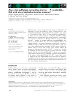
Lipid peroxidation and nitrite and GSH concentrations
are presented in Fig. 1. Lipid peroxidation was
markedly increased in this model compared with cor-
responding values for the control group. After pilocar-
pine-induced SE, there was a significant (77%) increase
in thiobarbituric-acid-reacting substances [T(14) ¼
18.282; P < 0.0001]. SE produced a significant increase
in hippocampal nitrite content of 51% [T(18) ¼ 25.959;
P < 0.0001] compared with the control group. On the
other hand, a 55% decrease in GSH concentration
[T(10) ¼ 27.452; P < 0.0001] compared with the con-
trol group was detected (Fig. 1).
Superoxide dismutase and catalase activities
in the hippocampus of adult rats after
pilocarpine-induced SE
Table 1 shows superoxide dismutase and catalase activ-
ities in the hippocampus after seizures and SE induced
by pilocarpine. Post hoc comparison of means indicated
similar superoxide dismutase activity [T(16) ¼ 0.5892;
P ¼ N.S.]. However, hippocampal catalase activity
showed a marked (88%) increase [T(10) ¼ 10.722;
P < 0.0001] compared with the control group
(Table 1).
Discussion
SE and oxidative stress are thought to be closely inter-
related. Our findings show that GSH was reduced
whereas lipid peroxidation and nitrite content were
increased after SE. Lipid peroxidation in the brain can
Fig. 1. Biochemical alterations in the hippocampus of adult rats
after pilocarpine-induced SE. Male rats (250–280 g, 2 months old)
were treated with a single dose of pilocarpine (400 mgÆkg
)1
, subcu-
taneously). The control group was treated with 0.9% saline.
Animals were observed for 24 h and then killed. Results are
mean ± SEM for the number of animals shown inside the bars.
a
P < 0.05 compared with control animals (Student-Newman-Keuls
test). The differences in the experimental groups were determined
by analysis of variance.
Oxidative stress after status epilepticus in rats R. M. Freitas et al.
1308 FEBS Journal 272 (2005) 1307–1312 ª 2005 FEBS
be induced by many chemical compounds and brain
injury such as epilepsy [24,25]. The brain is more vul-
nerable to injury by lipid peroxidation products than
other tissues [8]. Moreover, lipid peroxidation is an
index of irreversible neuronal damage of cell mem-
brane phospholipid and has been suggested as a poss-
ible mechanism of epileptic activity [11,18,26].
In normal conditions, there is a steady-state balance
between the production of nitric oxide and metabolites
(nitrite and nitrate) and their destruction by antioxid-
ant systems. Our results show an increase in nitrite
formation after SE, suggesting that there is a possible
increase in concentrations of ROS, which are often
involved in neuronal damage [7,15]. Other studies have
shown that nitrite and nitrate concentrations are not
raised in epileptic patients [27]. Other mechanisms may
be associated with the increase in ROS levels in the
epilepsy model as well as in neurodegeneration
observed in epileptic humans [18,28].
During ROS scavenging, glutathione disulfide pro-
duction and GSH reduction occur. When the balance
between ROS formation and ROS elimination is func-
tionally normal, there is GSH recovery [29]. As men-
tioned above, we can conclude that during SE there is
over-formation of free radicals and ⁄ or a deficiency of
antioxidant systems, as evidenced by the augmented
nitrite content, the unaltered superoxide dismutase
activity, and the GSH consumption, all of which char-
acterize oxidative stress.
Our findings show that pilocarpine induces SE,
which can produce alterations in superoxide dismutase
and catalase activities in different areas, thereby pro-
tecting the brain from neuronal damage induced by
lipid peroxidation products [11]. However, we found
no changes in hippocampal superoxide dismutase
activity. It is unlikely that the unaltered superoxide
dismutase activity is related to the mechanisms
involved in the initiation and ⁄ or propagation of
seizures induced by pilocarpine. Our results are in
agreement with another study showing unaltered
superoxide dismutase activity after 24 h, suggesting
that superoxide dismutase activity only changes during
the initiation of seizures [14]. When studying this epi-
lepsy model, we found increased catalase activity in
the hippocampus, indicating that this enzyme, in
association with GSH, provides neuroprotection
against the increase in lipid peroxidation and nitrite
content. These data suggest that the hippocampus does
not use superoxide dismutase as the major free-radical-
scavenging system [9,30]. It probably uses other scav-
enging systems (catalase and GSH).
Pilocarpine-induced SE produces several changes in
variables related to the generation and elimination of
oxygen free radicals in adult rats [18,30]. An increase
in free radical formation is accompanied by an imme-
diate compensatory increase in catalase activity, which
may be a long-term compensatory mechanism inclu-
ding activity modulation of enzymes [31]. In addition,
in the normal physiological state, changes in neuronal
activity are accompanied by alterations in the meta-
bolic rate (oxygen and energy metabolism) [1,8], which
induce modifications in cerebral blood flow [10]. In
pathological states, blood flow may not occur in the
same way. There is clinical and experimental evidence
of alterations in oxygen levels because of reduced
oxygen availability after SE [10]. Considering that
increased metabolic demand was observed, we suggest
that catalase would be one of the enzymes with aug-
mented activity, as this effect was not observed for the
superoxide dismutase.
Evidence for the role of free radicals in SE has been
found by using exogenously enzymatic and nonenzy-
matic antioxidant treatment for protection against
seizures and SE-induced neuronal damage [15,26]. A
steady-state level of superoxide and H
2
O
2
is always
present in cells as a result of normal metabolism.
Superoxide dismutase and catalase are responsible for
degradation of superoxide and H
2
O
2
, respectively, and
the balance between these antioxidant enzymes is rele-
vant for cell and neuronal functions [8,18]. The fact
that an increase in catalase activity may not result in
neurotoxic effects during SE indicates that basal ROS
production is damaging to the neurons and should be
controlled [9,28].
The biochemical alterations observed can produce
neuronal damage in the hippocampus. Our results indi-
cate that SE alters brain antioxidant defenses and that
there may be extensive participation of enzymes in sei-
zures. Further studies need to be carried out to ascer-
tain whether ROS are involved in the pathogenesis of
temporal lobe epilepsy.
Table 1. Superoxide dismutase [UÆ(mg protein)
)1
] and catalase
[mmolÆmin
)1
Æ(lg protein)
)1
] activities in the hippocampus of adult
rats after pilocarpine-induced SE. Male rats (250–280 g, 2 months
old) were treated with a single dose of pilocarpine (400 mgÆkg
)1
,
subcutaneously). The control group was treated with 0.9% saline.
Animals were observed for 24 h and then killed. Results are
mean ± SEM for the number of animals shown in parentheses.
The differences in experimental groups were determined by ana-
lysis of variance.
Group Superoxide dismutase Catalase
Control 2.35 ± 0.14 (10) 14.50 ± 0.65 (9)
Pilocarpine 2.45 ± 0.10 (8) 27.25 ± 1.03 (8)
a
a
P < 0.05 compared with control animals (Student–Newman–Keuls
test).
R. M. Freitas et al. Oxidative stress after status epilepticus in rats
FEBS Journal 272 (2005) 1307–1312 ª 2005 FEBS 1309
Experimental procedures
Treatment of animals and preparation of samples
Male Wistar rats (250–280 g; 2 months old) were used. Ani-
mals were housed in cages with free access to food and
water and with a standard light ⁄ dark cycle (lights on at
07:00 h). The experiments were performed according to the
Guide for the Care and Use of Laboratory Animals of the
US Department of Health and Human Services, Washing-
ton, DC (1985). Control animals received 0.9% saline sub-
cutaneously (control group; n ¼ 48), and the pilocarpine
group were treated with a single dose of pilocarpine hydro-
chloride (400 mgÆkg
)1
; subcutaneous; n ¼ 43). Behavioral
changes were observed over 24 h. The variables assessed
were: number of peripheral cholinergic signs, tremors, ste-
reotyped movements, seizures, SE and mortality. SE was
defined as continuous seizures for a period longer than
30 min. SE was induced by method of Turski et al. [23].
For biochemical assays, both pilocarpine and control
groups were killed by decapitation 24 h after treatment.
Their brains were dissected on ice to remove the hippocam-
pus for determination of lipid peroxidation, nitrite content,
GSH concentration, and superoxide dismutase and catalase
activities. Detailed criteria for determining the periods after
pilocarpine administration have been reported by Cavalhe-
iro et al. [32]. The pilocarpine group consisted of rats that
had seizures, SE for a period longer than 30 min, and that
did not die within 24 h of observation.
Determination of lipid peroxidation and nitrite
content
For all of the experimental procedures, 10% (w ⁄ v) homo-
genates of the area of the brain investigated were prepared
for both groups. Lipid peroxidation in the pilocarpine
group (n ¼ 7) and control animals (n ¼ 9) was analyzed by
measuring thiobarbituric-acid-reacting substances in homo-
genates, as previously described by Draper & Hadley [33].
Briefly, the samples were mixed with 1 mL 10% trichloro-
acetic acid and 1 mL 0.67% thiobarbituric acid. They were
then heated in a boiling water bath for 15 min, and butanol
(2 : 1, v ⁄ v) was added to the solution. After centrifugation
(800 g, 5 min), thiobarbituric-acid-reacting substances were
determined from the absorbance at 535 nm.
To determine nitrite content of the control rats (n ¼ 10)
and pilocarpine group (n ¼ 10), the 10% (w ⁄ v) homogenates
were centrifuged (800 g, 10 min). The supernatants were col-
lected, and nitric oxide production was determined based on
the Griess reaction [25]. Briefly, 100 lL supernatant was
incubated with 100 lL of the Griess reagent [1% sulfanila-
mide in 1% H
3
PO
4
⁄ 0.1% N-(1-naphthyl)ethylenediamine
dihydrochloride ⁄ 1% H
3
PO
4
⁄ distilled water, 1 : 1 : 1 : 1,
v ⁄ v ⁄ v ⁄ v) at room temperature for 10 min. A
550
was meas-
ured using a microplate reader. Nitrite concentration was
determined from a standard nitrite curve generated using
NaNO
2
.
Determination of GSH
GSH in the pilocarpine group (n ¼ 10) and control animals
(n ¼ 10) was analyzed. The hippocampus was homogenized
in 0.02 m EDTA. Immediately thereafter, 10% (w ⁄ v) homo-
genates were assayed for GSH as described by Sedlak &
Lindsay [34], and the results expressed in lgÆ(g tissue wet
weight)
)1
.
Determination of superoxide dismutase and
catalase activities
The hippocampus was ultrasonically homogenized in 1 mL
0.05 m sodium phosphate buffer, pH 7.0. Protein concen-
tration was measured by the method of Lowry et al. [35].
The 10% homogenates were centrifuged (800 g, 20 min),
and the supernatants used to assay superoxide dismutase
and catalase. Superoxide dismutase activity in the pilocar-
pine group (n ¼ 8) and control animals (n ¼ 10) was
assayed by using xanthine and xanthine oxidase to generate
superoxide radicals [24]. They react with 2,4-iodophenyl-
3,4-nitrophenol-5-phenyltetrazolium chloride to form a red
formazan dye. The degree of inhibition of this reaction was
measured to assess superoxide dismutase activity. The
standard assay substrate mixture contained 3 mL xanthine
(500 lm), 7.44 mg cytochrome c, 3.0 mL KCN (200 lm),
and 3.0 mL EDTA (1 mm) in 18.0 mL 0.05 m sodium phos-
phate buffer, pH 7.0. The sample aliquot (20 lL) was
added to 975 lL of the substrate mixture plus 5 lL xan-
thine oxidase. After 1 min, the initial absorbance was recor-
ded and the timer was started. The final absorbance after
6 min was recorded. The reaction was followed at 550 nm.
Purified bovine erythrocyte superoxide dismutase (Randox
Laboratories, Belfast, Northern Ireland, UK) was used
under identical conditions to obtain a calibration curve
showing the correlation of the inhibition percentage of
formazan dye formation and superoxide dismutase activity.
Superoxide dismutase activity in the samples was deter-
mined from this curve, and the results expressed as
UÆ(mg protein)
)1
.
Catalase activity was measured in the control (n ¼ 9) and
pilocarpine (n ¼ 8) groups by the method that uses H
2
O
2
to
generate H
2
O and O
2
[36]. The activity was measured by the
degree of this reaction. The standard assay substrate mix-
ture contained 0.30 mL H
2
O
2
in 50 mL 0.05 m sodium
phosphate buffer, pH 7.0. The sample aliquot (20 lL) was
added to 980 lL substrate mixture. The initial absorbance
was recorded after 1 min, and the final absorbance after
6 min. The reaction was followed at 230 nm. A standard
curve was established using purified catalase (Sigma,
St Louis, MO, USA) under identical conditions. All samples
were diluted with 0.1 mmolÆL
)1
sodium phosphate buffer
Oxidative stress after status epilepticus in rats R. M. Freitas et al.
1310 FEBS Journal 272 (2005) 1307–1312 ª 2005 FEBS
(pH 7.0) to provoke a 50% inhibition of the diluent rate
(i.e. the uninhibited reaction). Results are expressed as
mmolÆmin
)1
Æ(lg protein)
)1
[36,37].
Statistical analysis
Results are expressed as means ± SEM for the number of
experiments, with all measurements performed in duplicate.
The Student–Newman–Keuls test was used for multiple
comparison of means of two groups of data. Differences
were considered significant at P < 0.05. Differences in
experimental groups were determined by two-tailed analysis
of variance.
Acknowledgements
This work was supported by a research grant from the
Brazilian National Research Council (CNPq). R.M.F.
is a fellow of the CNPq. The technical assistance of
Maria Vilani Rodrigues Bastos and Steˆ nio Gardel
Maia are gratefully acknowledged.
References
1 Smith BN & Shibley H (2002) Pilocarpine-induced sta-
tus epilepticus results in mossy fiber sproutng and spon-
taneous seizures in C57BL ⁄ 6 and CD-1 mice. Epilepsy
Res 49, 109–120.
2 Brozek G, Hort J, Koma
´
rek V, Langmeier M & Mares
P (2000) Interstrain differences in cognitive functions in
rats in relation to status epilepticus. Behav Brain Res
112, 77–83.
3 Treiman DM (1955) Electroclinical features of status
epilepticus. J Clin Neurophysiol 12, 343–362.
4 Freitas RM, Souza FCF, Vasconcelos SMM, Viana
GSB & Fonteles MMF (2003) Acute alterations of neu-
rotransmitters levels in striatum of young rat after pilo-
carpine-induced status epilepticus. Arq Neuropsiquiatr
61, 430–433.
5 Simonie
´
A, Laginja J, Varljen J, Zupan G & Erakovie
´
V (2000) Lithium plus pilocarpine induced status epilep-
ticus: biochemical changes. Neurosci Res 36, 157–166.
6 Cavalheiro EA, Fernandes MJ, Turski L & Naffah-
Mazzacoratti MG (1994) Spontaneous recurrent seizures
in rats: amino acid and monoamine determination in
the hippocampus. Epilepsia 35, 1–11.
7 McCord JM (1989) Superoxide radical: controversies,
contradiction and paradoxes. Proc Soc Exp Biol Med
209, 112–117.
8 Naffah-Mazzacoratti MG, Cavalheiro EA, Ferreira EC,
Abdalla DSP, Amado D & Bellissimo MI (2001) Super-
oxide dismutase, glutathione peroxidase activities and
the hydroperoxide concentration are modified in the
hippocampus of epileptic rats. Epilepsy Res 46, 121–128.
9 Michiels C, Raes M, Toussaint O & Remacle J (1994)
Importance of Se-glutathione peroxidase, catalase, and
Cu ⁄ Zn-SOD for cell survival against oxidative stress.
Free Radic Biol Med 17, 235–248.
10 Dymond AM & Crandall PH (1976) Oxygen availabil-
ity and blood flow in the temporal lobes during
spontaneous epileptic seizures in men. Brain Res 102,
191–196.
11 Walz R, Moreira JCF, Benfato MS, Quevedo J, Schorer
N, Vianna MMR, Klamt F & Dal-Pizzol F (2000) Lipid
peroxidation in hippocampus early and late after status
epilepticus induced by pilocarpine of kainic acid in Wis-
tar rats. Neurosci Lett 291, 179–182.
12 Castagne V, Gastschi M, Lefevre K, Posada A &
Clarke PGH (1999) Relationship between neuronal
death and cellular redox status, focus on the developing
nervous system. Prog Neurophysiol 59, 397–423.
13 Halliwell B & Gutteridge JMC (1999) Free Radicals in
Biology and Medicine. Oxford Science Publications,
London.
14 Hussain S, Slikker W Jr & Ali SF (1995) Age-related
changes in antioxidant enzymes, superoxide dismutase,
catalase, glutathione peroxidase and glutathione in dif-
ferent regions of mouse brain. Int J Dev Neurosci 13,
24–34.
15 Kudin AP, Bimpong-Buta NY, Vielhaber S, Elger CE
& Kunz WS (2004) Characterization of superoxide-pro-
ducing sites in isolated brain mitochondria. J Biol Chem
279, 4127–4135.
16 Galleano M & Puntarulo S (1995) Role of antioxidants
in the erythrocyte’s resistance to lipid peroxidation after
acute iron overload in rats. Biochim Biophys Acta 1271,
321–326.
17 Freitas RM, Viana GSB & Fonteles MMF (2003) Stria-
tal monoamine levels during status epilepticus. Rev Psi-
quiatr Clı
´
n 30, 76–79.
18 Pazdernik TL, Emerson MR, Cross R, Nelson SR &
Samson FE (2001) Soman-induced seizures: limbic activ-
ity, oxidative stress and neuroprotective proteins. J Appl
Toxicol 21, S87–S94.
19 Costa-Lotufo LV, Fonteles MMF, Lima ISP, Oliveira
AA, Nascimento VS, Bruin VMS & Viana GSB (2002)
Attenuating effects of melatonin on pilocarpine-induced
seizures in rats. Comp Biochem Physiol C Pharmacol
Toxicol Endocrinol 131, 521–529.
20 Marinho MMF, Sousa FCF, Bruin VMS, Vale MR &
Viana GSB (1998) Effects of lithium, alone or asso-
ciated with pilocarpine, on muscarinic and dopaminer-
gic receptors and on phosphoinositide metabolism in
rat hippocampus and striatum. Neurochem Int 33,
299–306.
21 Viana GSB, Marinho MMF, Sousa FCF, Bruin VMS,
Aguiar LMV & Pinho RSN (1997) Inhibitory action of
a calcium channel blocker (nimodipine) on seizures and
R. M. Freitas et al. Oxidative stress after status epilepticus in rats
FEBS Journal 272 (2005) 1307–1312 ª 2005 FEBS 1311
brain damage induced by pilocarpine and lithium-pilo-
carpine in rats. Neurosci Let 235, 13–16.
22 Viana GSB, Marinho MMF, Sousa FCF & Bruin VMS
(1999) Behavioural and neurochemical alterations after
lithium-pilocarpine administration in young and adult
rats: a comparative study. Pharmacol Biochem Behav 65,
547–551.
23 Turski WA, Cavalheiro EA, Schwarz M, Czuczwar SJ,
Kleinronk Z & Turski L (1983a) Limbic seizures pro-
duced by pilocarpine in rats: behavioural, eletroence-
phalographic and neuropathological study. Behav Brain
Res 9, 315–336.
24 Flohe L & Otting F (1984) Superoxide dismutase assays.
Methods Enzymol 105, 93–104.
25 Green LC, Tannenbaum SR & Goldman P (1981)
Nitrate synthesis in the germfree and conventional rat.
Science 212, 56–58.
26 Lapin IP, Mirzaev SM, Ryzov IV & Oxenkrug GF
(1998) Anticonvulsant activity of melatonin against sei-
zures induced by quinolinate, kainate, glutamte,
NMDA, and pentylenetetrazol in mice. J Pineal Res 24,
215–218.
27 Vanhatalo S & Riikonen R (1999) Cerebrospinal fluid
levels of nitric oxide metabolites (nitrates ⁄ nitrites) in
infantile spasms. Epilepsia 40, 210–212.
28 Mello FAC, Hoffamn ME & Meneghini R (1983) Cell
killing and DNA damage by hydrogen peroxide are
mediated by intracellular iron. Biochem J 218, 273–275.
29 Halliwell B (1993) The role of oxygen radicals in human
disease, with particular reference to the vascular system.
Homeostasis 23, 118–126.
30 Oyanagui Y (1984) Reevaluation of assay methods and
establishment of kit for superoxide dismutase activity.
Anal Biochem 142, 290–296.
31 Frantseva MV, Perez VJL, Hwang PA & Carlen PL
(2000) Free radical production correlates with cell death
in an in vitro model of epilepsy. Eur J Neurosci 12,
1431–1439.
32 Cavalheiro EA, Leite JP, Bortolotto ZA, Turski WA,
Ikonomidou C & Turski L (1991) Long-term effects of
pilocarpine in rats: structural damage of the brain trig-
gers kindling and spontaneous recurrent seizures.
Epilepsia 32, 778–782.
33 Draper HH & Hadley M (1990) Malondialdehyde deter-
mination as an index of lipid peroxidation. Methods
Enzymol 186, 421–431.
34 Sedlak J & Lindsay RH (1968) Estimation of total pro-
tein bound and nonprotein sulfhydryl groups in tissues
with Ellman’s reagent. Anal Biochem 25, 192–205.
35 Lowry H, Rosebrough NJ, Farr AL & Randall RJ
(1951) Protein measurements with the folin phenol
reagent. J Biol Chem 193, 265–275.
36 Maehly AC & Chance B (1954) The assay of catalases
and peroxidases. Methods Biochem Anal 1, 357–359.
37 Chance B & Maehly AC (1955) Assay of catalases and
peroxidases. Methods Enzymol 2, 764–768.
Oxidative stress after status epilepticus in rats R. M. Freitas et al.
1312 FEBS Journal 272 (2005) 1307–1312 ª 2005 FEBS