Tài liệu Báo cáo khoa học: Novel aggregate formation of a frame-shift mutant protein of tissue-nonspecific alkaline phosphatase is ascribed to three cysteine residues in the C-terminal extension pdf
Bạn đang xem bản rút gọn của tài liệu. Xem và tải ngay bản đầy đủ của tài liệu tại đây (329.68 KB, 14 trang )
Novel aggregate formation of a frame-shift mutant protein
of tissue-nonspecific alkaline phosphatase is ascribed
to three cysteine residues in the C-terminal extension
Retarded secretion and proteasomal degradation
Keiichi Komaru
1,2
, Yoko Ishida
1
, Yoshihiro Amaya
1
, Masae Goseki-Sone
3
, Hideo Orimo
4
and Kimimitsu Oda
1,5
1 Division of Biochemistry, Niigata University Graduate School of Medical and Dental Sciences, Gakkocho-dori, Niigata, Japan
2 Kitasato Junior College of Health and Hygienic Sciences, Yamatomachi, Minami-Uonuma-shi, Niigata, Japan
3 Department of Food and Nutrition, Japan Women’s University, Mejirodai, Bunkyo-ku, Tokyo, Japan
4 Department of Biochemistry and Molecular Biology, Nippon Medical School, Tokyo, Japan
5 Center for Transdisciplinary Research, Niigata University, Japan
Keywords
aggregation; alkaline phosphatase;
degradation; hypophosphatasia; proteasome;
ubiquitin
Correspondence
K. Oda, Division of Biochemistry, Course for
Oral Life Science, Niigata University,
Graduate School of Medical and Dental
Sciences, 2–5274, Gakkocho-dori, Niigata,
951–8514, Japan
Fax: +81 25 227 2831
Tel: +81 25 227 2827
E-mail:
(Received 21 December 2004, revised 30
January 2005, accepted 3 February 2005)
doi:10.1111/j.1742-4658.2005.04597.x
In the majority of hypophosphatasia patients, reductions in the serum lev-
els of alkaline phosphatase activity are caused by various missense muta-
tions in the tissue-nonspecific alkaline phosphatase (TNSALP) gene. A
unique frame-shift mutation due to a deletion of T at cDNA number 1559
[TNSALP (1559delT)] has been reported only in Japanese patients with
high allele frequency. In this study, we examined the molecular phenotype
of TNSALP (1559delT) using in vitro translation⁄ translocation system and
COS-1 cells transiently expressing this mutant protein. We showed that the
mutant protein not only has a larger molecular size than the wild type
enzyme by 12 kDa, reflecting an 80 amino acid-long extension at its
C-terminus, but that it also lacks a glycosylphosphatidylinositol anchor. In
support of this, alkaline phosphatase activity of the cells expressing TNS-
ALP (1559delT) was localized at the juxtanucleus position, but not on the
cell surface. However, only a limited amount of the newly synthesized pro-
tein was released into the medium and the rest was polyubiquitinated,
followed by degradation in the proteasome. SDS ⁄ PAGE and analysis by
sucrose-density-gradient analysis indicated that TNSALP (1559delT) forms
a disulfide-bonded high-molecular-mass aggregate. Interestingly, the aggre-
gate form of TNSALP (1559delT) exhibited a significant enzyme activity.
When all three cysteines at positions of 506, 521 and 577 of TNS-
ALP (1559delT) were replaced with serines, the aggregation disappeared
and instead this modified mutant protein formed a noncovalently associ-
ated dimer, strongly indicating that these cysteine residues in the C-ter-
minal region are solely responsible for aggregate formation by cross-linking
the catalytically active dimers. Thus, complete absence of TNSALP on cell
surfaces provides a plausible explanation for a severe lethal phenotype of a
homozygote hypophosphatasia patient carrying TNSALP (1559delT).
Abbreviations
Bz-Asn-Gly-Thr-NH
2
, benzoyl-asparagine-glycine-threonine-amide; DMEM, Dulbecco’s modified Eagle’s medium; ER, endoplasmic reticulum;
ECL, enhanced chemiluminescence; GPI, glycosylphosphatidylinositol; LLnL, N-acetyl-
L-leucinyl-L-leucinyl-L-norleucinal); LLM, N-acetyl-L-
leucinyl-
L-leucinyl-L-methional); MG-132, benzyloxycarbonyl-L-leucinyl-L-leucinyl-L-leucinal; PI-PLC, phosphatidylinositol-specific phospholipase
C; PNGase F, peptide:N-glycosidase F; MEM, minimum essential medium; TNSALP, tissue-nonspecific alkaline phosphatase; sTNSALP,
soluble form of TNSALP.
1704 FEBS Journal 272 (2005) 1704–1717 ª 2005 FEBS
Hypophosphatasia is an inborn error of metabolism
characterized by defective mineralization of hard tis-
sues and reduced levels of tissue nonspecific alkaline
phosphatase (TNSALP, EC 3.1.3.1) [1–3]. Hypophos-
phatasia is classified into at least five categories
depending on age of onset and severity: perinatal,
infantile, childhood, adult and odontohypophosphata-
sia. The disease is caused by various mutations in the
TNSALP gene, which is located on chromosome
1p-36.1–34, and is transmitted in an autosomal reces-
sive or a dominant manner. The severity of the disease
is inversely related to serum levels of alkaline phospha-
tase activity, therefore indicating that reduction of
enzyme activity caused by the defective TNSALP genes
are responsible for poor mineralization of bone and
tooth.
TNSALP-deficient mice develop rickets and osteope-
nia postnatally and many die of seizure [4–6], recapit-
ulating infantile hypophosphatasia. Additionally, as
with hypophosphatasia patients, elevated levels of inor-
ganic pyrophosphate, phosphoethanolamine and pyrid-
oxal-5¢-phosphate have been reported in the serum and
urine of the knockout mice. This raises the possibility
that these phosphocompounds are natural substrates
for TNSALP. Recently Hessle et al. [7] have postulated
that the concerted action of nucleoside triphosphate
pyrophosphohydolase and TNSALP regulates local
concentration of inorganic pyrophosphate at the site of
mineralization, which, as a poison of hydroxyapatite
growth, in turn controls mineralization. According to
this idea, increased levels of inorganic pyrophosphate
resulting from the defect of TNSALP are thought to
be the main cause of hypomineralization.
Since Weiss et al. [8] first identified a missense muta-
tion (Ala162Thr) in the TNSALP gene of a patient
diagnosed with infantile hypophosphatasia, more than
161 mutations have been reported and of all mutations
about 80% of them are missense [3,9,(ep.
uvsq.fr/Database.html)]. To date, biochemical charac-
terization of TNSALP mutant proteins have been lim-
ited to a small number of cases. Nevertheless, some
missense mutations, in particular associated with severe
forms of hypophophatasia, were known to impair
proper folding and correct assembly of TNSALP in the
endoplasmic reticulum (ER), resulting in the loss of
functional TNSALP from the cell surface [10–14]. Dele-
tion of T at position 1559 of the cDNA [TNS-
ALP (1559delT)] of the TNSALP gene was reported
for the first time in hypophosphatasia patients by Orimo
et al. [15] and TNSALP (1559delT) is transmitted as a
recessive trait. TNSALP (1559delT) was originally
referred to as TNSALP (1735delT) [16–18]. According
to the recommended nomenclature, the adenine of the
initiator ATG codon in the cDNA of TNSALP is
denoted as nucleotide +1 instead of the first nucleotide
of the cDNA clone originally isolated by Weiss et al.
[8]. TNSALP (1559delT) is unique in that so far, this
frameshift mutation has been reported only in the Jap-
anese population [18]. The patients are largely com-
pound heterozygotes with different missense mutations,
or in some cases with undetected mutations on the
opposite allele, and exhibit clinical manifestations vary-
ing from infantile to odontohypophosphatasia [18].
Quite recently, a case of a homozygous patient of TNS-
ALP (1559delT) has been reported and classified as the
prenatal lethal form, confirming that this mutation rep-
resents a severe allele [19]. This frameshift mutation
is assumed to eliminate the original translational stop
codon and instead causes the extension consisting of 80
amino acid residues at the C-terminus of TNSALP.
Goseki et al. detected a larger form of TNSALP in vivo
in the serum of the patients carrying this mutation [16];
however, little is known about the molecular phenotype
of TNSALP (1559delT) underlying the clinical symp-
toms.
In this report we have elucidated the biosynthesis
of TNSALP (1559delT) in a heterologous expression
system and in vitro translation ⁄ translocation system.
Our results shows that although TNSALP (1559delT)
is synthesized as a secretory form lacking glycosylphos-
phatidylinositol (GPI), most of newly synthesized mole-
cules form the aggregate and fail to exit from the ER.
Furthermore, the accumulated TNSALP (1559delT)
was found to be polyubiquitinated under the condition
where cellular proteasome activity was blocked, indicat-
ive of ubiquitin ⁄ proteasome pathway as part of an ER
quality control mechanism. We also have demonstrated
that three cysteine residues in the C-terminal extension
of this frameshift mutant protein are responsible for
the formation of the novel aggregate retaining enzyme
activity.
Results
In vitro translation/translocation
The deletion of T at cDNA number 1559 causes a
frameshift downstream from leucine at position 503 of
TNSALP, resulting in the elimination of an original
translational stop codon. Thus, the cDNA of TNS-
ALP (1559delT) was predicted to encode a large sized
TNSALP molecule with an additional 80 amino acid-
long extension at the C-terminus (Fig. 1). To confirm
this prediction, we performed in vitro translation
experiments as shown in Fig. 2A. The molecular mass
of TNSALP (1559delT) was estimated to be 66 kDa
K. Komaru et al. Novel aggregate formation of an alkaline phosphatase frame-shift mutant
FEBS Journal 272 (2005) 1704–1717 ª 2005 FEBS 1705
and larger than the wild type enzyme by 12 kDa.
This value is in close agreement with the calculated
molecular mass (65 796) based on the amino acid
sequence of TNSALP (1559delT). When translation
was carried out in the presence of the canine micro-
some, TNSALP (1559delT) became a 80 kDa form;
however, the appearance of the 80 kDa form was
greatly diminished in the presence of Bz-Asn-Gly-Thr-
NH
2
, an inhibitor of N-glycosylation (Fig. 2A, lane 6).
Furthermore, upon incubation with PNGase F, which
cleaves N-linked oligosaccharides between innermost
N-acetylglucosamine and asparagine residue of glyco-
proteins, the 80 kDa form was completely converted
to the 66 kDa form (Fig. 2B), indicating that TNS-
ALP (1559delT) is cotranslationally N-glycosylated to
become the 80 kDa form in the microsome. Thus, it is
unlikely that the additional 80 amino acid residues
at the C-terminus strongly affect the cotranslational
translocation of TNSALP (1559delT) across the ER
membrane.
Phase separation using Triton X-114
Given our finding that TNSALP (1559delT) is synthes-
ized as a larger protein with a C-terminal extension, we
considered the possibility that this frame-shift mutant
protein fails to be attached by a GPI, because a puta-
tive GPI-anchor signal consisting of a stretch of hydro-
phobic amino acids is abrogated. Previous studies have
demonstrated that the wild type TNSALP expressed in
the COS-1 cell is modified by GPI as shown by its sen-
sitivity to phosphatidylinositol-specific phospholipase C
(PI-PLC), which cleaves between phosphatidylglycerol
and phosphoinositol of GPI, and metabolic labeling
using [
3
H]ethanolamine, a component of GPI [10–13].
To examine if TNSALP (1559delT) is modified by GPI,
we exploited a phase separation method by Bordier
[20]. After metabolic labeling, the cells expressing either
the wild type or TNSALP (1559delT) were lysed in a
buffer containing TX-114 on ice, then warmed at
25 °C. A detergent phase was separated from an aque-
ous phase by centrifugation and both phases were sub-
jected to immunoprecipitation. Newly synthesized wild
type enzyme was largely partitioned into the detergent
phase as shown in Fig. 3 (lanes 1 & 2). The band in the
aqueous phase probably represents GPI-anchor-less
molecules due to overexpression of the enzyme in the
transiently transfected cells. It is noteworthy that a
66 kDa form, but not an 80 kDa form was partitioned
into the aqueous phase. As the 66 kDa form of the wild
type migrates to the Golgi complex and becomes the
80 kDa form as described previously [10,12], this result
suggests that the GPI-less molecules fail to exit the
ER. However, this partition behavior of the wild type
enzyme completely changed upon incubation with
PI-PLC prior to phase separation. All wild type
PNGase F - + - +
80 kDa
80 kDa
66 kDa
66 kDa
54 kDa
54 kDa
1 2 3 4 5 6
TNSALP
1559delT
TNSALP
1559delT
A
B
Fig. 2. In vitro transcription ⁄ translation. (A) Transcription-coupled
translation of TNSALP or TNSALP (1559delT) were carried out in
the absence (lanes 1 and 4) or presence (lanes 2, 3, 5 and 6) of
canine pancreatic microsomes. The N-glycosylation inhibitor
Bz-Asn-Gly-Thr-NH
2
was added at a final concentration of 0.5 mM
(lanes 3 and 6). Aliquots of the translation reactions were analysed
by SDS ⁄ PAGE, followed by fluorography. The leftmost lane shows
14
C-methylated protein markers of 200, 97.4, 66 and 46 kDa, from
the top of the gel. (B) The translation products (A, lanes 2 and 5)
were further incubated in the absence or presence of PNGase F,
followed by SDS ⁄ PAGE ⁄ fluorography. Left lane:
14
C-methylated
protein markers as in Fig. 2A.
TNSALP GPLLLALALYPLSVLF
506 521
1559delT GPLLLALALYP RASCSEGPGPGHPQARDRCQLPTRQPPSQGARWGPP
LQLQERGPRKPKSAAHLAPLWNLPQGPNPLLASSLCSLPAALWPTG
Fig. 1. Predicted amino acid sequence of TNSALP (1559delT). A
single T deletion in the cDNA at nucleotide 1559 of tissue nonspe-
cific alkaline phosphatase (TNSALP) changes the amino acid
sequence at leucine 503 and downstream until the new stop codon
appears. Accordingly, this frame-shift mutation predicts that TNS-
ALP (1559delT) is 80 amino acids longer than the wild type TNS-
ALP. Three cysteine residues at positions of 506, 521 and 577 are
marked.
Novel aggregate formation of an alkaline phosphatase frame-shift mutant K. Komaru et al.
1706 FEBS Journal 272 (2005) 1704–1717 ª 2005 FEBS
TNSALP molecules were now partitioned into the
aqueous phase (Fig. 3, lanes 3 & 4), indicating that the
wild type TNSALP molecule in the detergent phase
represents a GPI-anchored membrane form. In contrast
to the wild type, TNSALP (1559delT) was exclusively
found in the aqueous phase even without PI-PLC diges-
tion (Fig. 3, lanes 7 & 8), strongly arguing that
TNSALP (1559delT) lacks a GPI. For comparison, we
also expressed and analyzed a soluble truncated form
of TNSALP (sTNSALP), which lacks the C-terminal
23 amino acids including a putative GPI-anchor signal
sequence [21]. As expected, sTNSALP was found to be
recovered only in the aqueous phase like TNSALP
(1559delT) (Fig. 3, lanes 5 and 6), further support-
ing that TNSALP (1559delT) is not modified by a GPI.
Biosynthesis of TNSALP (1559delT)
If TNSALP (1559delT) is not attached by a GPI-
anchor, a prediction is that this mutant is no longer
embedded into the lipid bilayer via GPI, which helps
anchor TNSALP to the plasma membrane, but is
secreted out of the cell. To investigate whether this
mutant protein is secreted, we labeled the transfected
cells with [
35
S]methionine ⁄ cysteine and followed the
kinetics of TNSALP secretion. As reported previously
[10,12], the wild type TNSALP was synthesized as the
66 kDa Endo H-sensitive form and underwent process-
ing of N-linked oligosaccharides to become the 80 kDa
Endo H-resistant mature species. Both the 66 kDa and
80 kDa forms of TNSALP were detected in the trans-
fected cells, though the conversion of the precursor to
the 80 kDa form obviously is not efficient in our tran-
sient expression system (Fig. 4A, lanes 1–3). In con-
trast, in the cells expressing TNSALP (1559delT), an
80 kDa form – which corresponds in molecular mass
to the in vitro 80 kDa translational product (Fig. 2) –
was the only molecular species throughout the chase
time (Fig. 4A, lane 6). The intensity of the 80 kDa
form of TNSALP (1559delT) rapidly decreased as the
chase time elapsed. However, this decline is not simply
accounted for by the secretion of the mutant protein
into the medium, as no band was detectable even in
the 6 h chase culture medium (Fig. 4A, lane 8). Only
after a prolonged exposure, however, a 90 kDa form
of TNSALP (1559delT) was found in the medium
(results not shown). To confirm that TNSALP
(1559delT) is indeed secreted into the medium, albeit
in a lesser amount, culture media were collected from
continuously radiolabeled transfected cells and any
secreted TNSALP (1559delT) was immunoprecipitated.
A 90 kDa form of TNSALP (1559delT) became evi-
dent in the medium (Fig. 4B, lane 4), suggesting that
the 80 kDa form was processed to the 90 kDa form
in the Golgi apparatus before being released into the
medium. In support of this, this secretory form was
found to be sensitive to PNGase F but resistant to
Endo H (Fig. 4B, lanes 5 & 6). On the other hand, the
GPI-anchor-less sTNSALP was efficiently secreted out
of the cells even after 0.5 h chase (Fig. 4C), indicating
that sTNSALP behaves like a genuine secretory pro-
tein. Thus, we conclude that TNSALP (1559delT) is
newly synthesized as the 80 kDa soluble form and
mostly undergoes degradation, resulting in only a por-
tion of it being secreted as the 90 kDa Endo H-resist-
ant form.
Degradation and ubiquitination of TNSALP
(1559delT)
We next examined the effect of several protease
inhibitors on the degradation of TNSALP (1559delT).
Inhibitors of proteasome function, such as LLnL and
MG-132, but not a calpain inhibitor (LLM) remark-
ably blocked the degradation of TNSALP (1559delT)
(Fig. 5A,B), indicative of involvement of the protea-
some. Consistent with this observation, leupeptin and
pepstatin A (inhibitors of lysosomal proteases) had no
effect on the degradation (results not shown). Quite
recently we have reported that TNSALP (D289V),
which is associated with perinatal hypophosphatasia,
undergoes polyubiquitination prior to the degradation
in the proteasome in the transfected COS-1 cells. This
det aq det aq det aq det aq
PI-PLC
- - + + - - - -
TNSALP sTNSALP 1559delT
80 kDa
66 kDa
1 2 3 4 5 6 7 8
Fig. 3. Phase separation. COS-1 cells expressing the wild type TNS-
ALP (lanes 1–4), sTNSALP (lanes 5 and 6) or TNSALP (1559delT)
(lanes 7 and 8) were labeled with [
35
S]methionine ⁄ cysteine for 3 h.
The cells were lysed in buffer containing Triton X-114 and parti-
tioned into detergent (det) and aqueous (aq) phases before (lanes 1,
2, 5–8) or after (lanes 3 and 4) PI-PLC treatment. Each phase was
subjected to immunoprecipitation. The immune complexes were
analysed by SDS ⁄ PAGE, followed by fluorography. Left lane:
14
C-
methylated protein markers of 97.4, 66 and 46 kDa.
K. Komaru et al. Novel aggregate formation of an alkaline phosphatase frame-shift mutant
FEBS Journal 272 (2005) 1704–1717 ª 2005 FEBS 1707
1 2 3 4 5 6 7 8
TNSALP 1559delT
TNSALP 1559delT
cell medium
80 kDa
66 kDa
cell medium
1 2 3 4 5 6
80 kDa
90 kDa
54 kDa
cell medium
72 kDa
1 2 3 4 5 6 7
1559delT
sTNSALP
A
B
C
Fig. 4. Pulse-chase experiment. (A) Cells expressing TNSALP (lanes
1–3, 7) or TNSALP (1559delT) (lanes 4–6, 8) were pulse-labeled
with [
35
S]methionine ⁄ cysteine for 30 min (lanes 1 and 4) and
chased for 3 h (lanes, 2 and 5) or for 6 h (lanes 3 and 6). At 6 h
chase period, the media (lanes 7 and 8) were removed and the
cells were lysed for immunoprecipitation. The immune complexes
were analysed by SDS ⁄ PAGE and fluorography. Left lane:
14
C-methylated protein markers of 97.4, 66 and 46 kDa. (B) Cells
expressing TNSALP (1559delT) were labeled for 6 h with
[
35
S]methionine ⁄ cysteine. After 6 h, the medium was removed
(lanes 4–6) and the cells (lanes 1–3) were lysed for immunoprecipi-
tation. The immunoprecipitates were incubated in the absence
(lanes 1 and 4) or presence of PNGase F (lanes 2 and 5) or Endo H
(lanes 3 and 6) prior to SDS ⁄ PAGE ⁄ fluorography. Left lane:
14
C-methylated protein markers of 97.4, 66 and 46 kDa. (C) Cells
expressing sTNSALP were pulse-labeled with [
35
S]methionine ⁄ cys-
teine for 30 min and chased for 0 h (lane 1), for 0.5 h (lanes 2 and
5), for 1 h (lanes 3 and 6) or 2 h (lanes 4 and 7). The media and cell
lysates were subjected to immunoprecipitation and the immune
complexes were analysed by SDS ⁄ PAGE ⁄ fluorography. Left lane:
14
C-methylated protein markers of 200, 97.4, 66, 46 and 30 kDa.
Control LLM LLnL MG132
1 2 3 4 5 6 7 8
-LLnL +LLnL
80 kDa
80 kDa
1 2 3 4 5 6
HA-Ub
- - + + - - + +
LLnL - + - + - + - +
anti-Ub anti-HA
PolyUb
A
B
C
Fig. 5. Degradation and ubiquitination. (A) Cells expressing TNS-
ALP (1559delT) were pulse-labeled with [
35
S]methionine ⁄ cysteine
for 30 min and chased for 6 h in the absence (lanes 1 and 2) or
presence of 50 l
M LLM (lanes 3 and 4), 50 lM LLnL (lanes 5 and
6) or 50 l
M MG-132 (lanes 7 and 8). The cell lysates were subjec-
ted to immunoprecipitation and the immune complexes were ana-
lysed by SDS ⁄ PAGE and fluorography. Left lane:
14
C-methylated
protein markers of 200, 97.4, 66, 46 and 30 kDa. (B) Cells expres-
sing TNSALP (1559delT) were pulse-labeled with [
35
S]methion-
ine ⁄ cysteine for 30 min and chased for 0 h (lanes 1 and 4), 3 h
(lanes 2 and 5) or 6 h (lanes 3 and 6) in the absence (lanes 1–3) or
presence of 50 l
M LLnL (lanes 4–6). The immune complexes were
analysed by SDS ⁄ PAGE and fluorography. Left lane:
14
C-methylated
protein markers (Fig. 4A). (C) Cells expressing TNSALP (1559delT)
alone or TNSALP (1559delT) and HA-ubiquitin were incubated in
the absence (–) or presence (+) of 50 l
M LLnL for 6 h. Then the
cells were lysed and subjected to immunoprecipitated with anti-
TNSALP. After transfer, membranes were reacted with antiubiquitin
(anti-Ub) or anti-influenza hemagglutinin epitope (anti-HA) Igs.
Novel aggregate formation of an alkaline phosphatase frame-shift mutant K. Komaru et al.
1708 FEBS Journal 272 (2005) 1704–1717 ª 2005 FEBS
finding prompted us to determine if TNSALP
(1559delT) also is ubiquitinated prior to degradation
in the proteasome. To this end we transfected the cells
with the plasmid encoding TNSALP (1559delT) with
or without the plasmid encoding ubiquitin bearing the
N-terminal influenza HA epitope. TNSALP (1559delT)
was immnoprecipitated with anti-TNSALP Igs and
subsequently the immunoprecipitates were subject to
immunoblotting using either anti-ubiquitin or anti-HA
Igs (Fig. 5C). Not only did proteasome inhibitors did
not affect ubiquitination, but also overall biosynthesis
of the wild type enzyme (results not shown) [14].
Remarkably, TNSALP (1559delT) was found to be
heavily ubiquitinated in the presence of the inhibitor
of proteasome function. Furthermore, the extent of
ubiquitination of TNSALP (1559delT) was further
augmented in the cells expressing ubiquitin, strongly
demonstrating that this mutant protein is degraded via
ubiquitin ⁄ proteasome pathway.
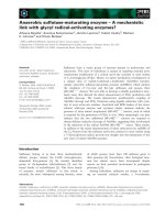
Catalytic activity of TNSALP (1559delT)
Figure 6 shows cytohistochemistry for alkaline phos-
phatase. In contrast to the wild type enzyme, virtually
no alkaline phosphatase activity was detected on the
cell surface of cells expressing TNSALP (1559delT). A
faint staining might be attributed to secreted TNS-
ALP (1559delT) trapped on the cell surface because of
its aggregate nature (see below).
These observations are compatible with the finding
that the wild type, but not the mutant, protein is
attached by GPI as shown in Fig. 2. Interestingly, we
detected strong alkaline phosphatase activity at a
juxtanucleus position in the cells expressing TNSALP
(1559delT) as well as the wild type, suggesting that this
mutant protein possesses catalytic activity and is con-
centrated in the Golgi apparatus on its way to being
discharged. In keeping with this morphological obser-
vation we found a low but significant enzyme activity
in both the homogenate and culture medium of the
cells expressing TNSALP (1559delT) (Fig. 7A). How-
ever, an immunoblotting experiment demonstrated that
the amount of TNSALP (1559delT) in the cell was less
than one tenth of that of the wild type at steady state
(Fig. 7B, lanes 1 and 6), probably reflecting its rapid
degradation as shown in Fig. 5. Taking these values
into consideration, the relative specific enzyme activity
of the mutant protein was calculated to be about one
third of that of the wild type (Fig. 7C). In contrast to
the culture medium of the cells expressing the wild
type enzyme, very high enzyme activity was detected in
that of the cells expressing sTNSALP (Fig. 7A), con-
sistent with a metabolic labeling study showing that
sTNSALP is rapidly secreted out of the cell (Fig. 4C).
Aggregation of TNSALP (1559delT)
Previously, we have reported that several TNSALP
missense mutants tend to form a disulfide-bonded
high-molecular-mass aggregate in transfected cells pre-
sumably due to defective folding and random associ-
ation of mutant proteins [10–14]. To investigate if this
is also the case for TNSALP (1559delT), the newly
synthesized mutant protein was immunoprecipi-
tated and analysed by SDS ⁄ PAGE under reducing or
nonreducing condition. TNSALP (1559delT) formed a
large aggregate bonded by multiple disulfide-bonds at
the top of the resolving gel (Fig. 8A, lanes 2 & 4). In
contrast, only a small amount of the wild type enzyme
formed the aggregate (lanes 1 and 3). The aggregate
saponin - saponin +
TNSALP
1559delT
Fig. 6. Cytohistochemical staining for alka-
line phosphatase. Cells expressing TNSALP
or TNSALP (1559delT) were stained for alka-
line phosphate activity in the absence or
presence of saponin.
K. Komaru et al. Novel aggregate formation of an alkaline phosphatase frame-shift mutant
FEBS Journal 272 (2005) 1704–1717 ª 2005 FEBS 1709
thus found in the cells expressing the wild type could
be GPI-anchor-less molecules, which are retained in
the ER (Fig. 3). Note that the secreted TNS-
ALP (1559delT) also formed large aggregates (Fig. 8A,
lanes 6 & 8). Addition of dithiothreitol in the culture
medium did not enhance the secretion of the mutant,
but rather inhibited it (results not shown). In good
agreement with the SDS ⁄ PAGE, sucrose gradient cen-
trifugation further demonstrated that TNSALP
(1559delT) tends to form large aggregates. Consider-
able amount of cellular activity and most of secreted
activity was recovered in the bottom three fractions
(Fig. 8B). In contrast, sTNSALP peaked at fraction 7
(Fig. 8B). Because the wild type enzyme also appeared
in fractions 6 and 7 in a similar analysis [12,13], this
result indicates that sTNSALP forms a dimer. Import-
antly, K
m
values estimated by Lineweaver–Burk plots
were 4.3 · 10
)4
m (wild type, cell homogenate),
1.9 · 10
)4
m [TNSALP (1559delT), fractions 10–12 of
the medium] and 5.5 · 10
)4
m (sTNSALP, medium).
This finding indicates that the C-terminal extension of
TNSALP (1559delT) does not significantly affect the
substrate affinity of this mutant, thus differentiating
TNSALP (1559delT) from other missense TNSALP
mutants possessing no catalytic activity, such as TNS-
ALP (R54C), TNSALP (N153D), TNSALP (E218G),
TNSALP (D289V) and TNSALP (G317D) [10–14].
Addition of dithiothreitol into the culture media and
cell lysates of the cells expressing TNSALP (1559delT)
did not enhance the enzyme activity (results not
shown).
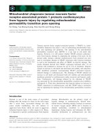
Replacement of three cysteines with serine
residues in the C-terminus region
Despite its aggregation state, TNSALP (1559delT)
shows catalytic activity comparable to that of the wild
type and sTNSALP as described above. We therefore
speculated that TNSALP (1559delT) becomes correctly
folded and assembled by the time that the cysteine resi-
dues in the C-terminal region emerge through the tran-
slocon of the ER, and that it eventually undergoes
1 2.5 5 10 10 10 10
1 2 3 4 5 6 7
80 kDa
66 kDa
TNSALP sTNSALP 1559delT
0
500
1000
1500
2000
2500
3000
TNSALP sTNSALP 1559delT
0
500
1000
1500
2000
2500
3000
TNSALP 1559deT
Alkaline phosphatase (unit/mg protein or ml)
Alkaline phosphatase activity
AC
B
Fig. 7. Enzyme activity of TNSALP
(1559delT). (A) COS-1 cells, which had been
transfected with the plasmids encoding the
wild type TNSALP, sTNSALP or TNSALP
(1559delT), were cultured for 24 h and then
homogenized in the 50 m
M Tris ⁄ HCl
(pH 7.5). The cell homogenates (white bars)
and media (black bars) were assayed for
alkaline phosphatase and expressed in unit
per mg protein (cell) or unit per mL culture
medium, respectively. (B) In addition to the
cell homogenates (lanes 1–6) prepared as
described in (A), cells expressing
TNSALP (1559delT) were incubated in the
presence of of LLnL (10 l
M) for 24 h and
then homogenized (lane 7). The homo-
genates were analysed by immunoblotting
with anti-TNSALP. The numbers above the
fluorogram shows the amounts (lg) of pro-
tein applied on SDS ⁄ PAGE. (C) The relative
specific enzyme activities of the cell homo-
genates prepared from cells expressing
TNSALP or TNSALP (1559delT) described as
in A were calculated based on the relative
amount (10 : 1) of both proteins in the
homogenates as described in B (ordinate,
arbitrary unit).
Novel aggregate formation of an alkaline phosphatase frame-shift mutant K. Komaru et al.
1710 FEBS Journal 272 (2005) 1704–1717 ª 2005 FEBS
multiple cross-linking reactions via the cysteine resi-
dues. To address this possibility, three cysteines were
substituted for serine residues in the C-terminal ex-
tension of TNSALP (1559delT) (Fig. 1). Initially we
attempted to simultaneously replace all three cysteine
residues at positions 506, 521 and 577. However, only
two plasmids were obtained in which two out of three
cysteine residues were replaced [TNSALP (1559delT-
C506C ⁄ C521S ⁄ C577S), TNSALP (1559delT-C506S ⁄
C521C ⁄ C577S)]. When these two proteins were
expressed in COS-1 cells, the amount of the large
aggregate was markedly reduced and instead the cross-
linked dimer became prominent (Fig. 9A, lanes 11–14).
Note the decrease in the aggregate on the stacking
gel. Next, we introduced the third mutation into
TNSALP (1559delT-C506S ⁄ C521C ⁄ C577S). The aggre-
gation state was dramatically changed in the cells
expressing TNSALP (1559delT-C506S ⁄ C521S ⁄ C577S).
1 2 3 4 5 6 7 8
red nonred red nonred
cell medium
90 kDa
66 kDa
80 kDa
0
5
10
15
20
25
123456789101112
0
1
2
3
4
5
123456789101112
0
500
1000
1500
2000
2500
3000
3500
4000
4500
123456789101112
0
10
20
30
40
50
60
70
80
90
123456789101112
b
a
sTNSALP (cell)
sTNSALP (medium)
1559delT (medium)
1559delT (cell)
alkaline phosphatase (unit/mg protein)
alkaline phosphatase (unit/ml)
c
c
alkaline phosphatase (unit/mg protein)
alkaline phosphatase (unit/ml)
A
B
Fig. 8. Sucrose-density-gradient analysis of
TNSALP (1559delT). (A) Cells expressing
TNSALP (lanes 1, 3, 5 and 7) or TNSALP
(1559delT) (lanes 2, 4, 6 and 8) were
continuously labeled with [
35
S]methion-
ine ⁄ cysteine for 4 h. The media and cell
lysates were subjected to immunoprecipita-
tion. The immune complexes were boiled in
the absence (nonreducing condition) or pres-
ence (reducing condition) of 2-mercaptoeth-
anol and analysed by SDS ⁄ PAGE, followed
by fluorography. An arrowhead indicates the
top position of the resolving gel. Left lane:
14
C-methylated protein markers of 200,
97.4, 66 and 46 kDa. (B) After 24 h post-
transfection, the lysates and media prepared
from cell cultures expressing sTNSALP or
TNSALP (1559delT) were directly applied on
the top of sucrose-density-gradient analysis
(5–35%). After centrifugation, each 400 lL
fraction was collected from the top of the
gradient and assayed for alkaline phospha-
tase activity (ordinate, unit per mL fraction).
BSA (b, 68 kDa), alcohol dehydrogenase
(a, 141 kDa) and catalase (250 kDa) were
loaded on to a separate gradient as mole-
cular mass markers.
K. Komaru et al. Novel aggregate formation of an alkaline phosphatase frame-shift mutant
FEBS Journal 272 (2005) 1704–1717 ª 2005 FEBS 1711
Not only the aggregate but also the covalently linked
dimer almost disappeared (Fig. 9A, lanes, 15 and 16).
This modified TNSALP (1559delT) was found to sedi-
ment at a dimer position as judged by sucrose-density
centrifugation (Fig. 9B). We therefore concluded that
TNSALP (1559delT-C506S ⁄ C521S ⁄ C577S) formed a
noncovalently assembled dimer similarly to sTNSALP
(Fig. 8B) and the wild type enzyme [12,13]. As
expected, TNSALP (1559delT-C506S ⁄ C521S ⁄ C577S)
was secreted threefold more than TNSALP (1559delT)
(Fig. 9B; compare ordinates).
Discussion
TNSALP (1559delT) is a large-sized secretory
protein lacking GPI
A growing number of genetic diseases have been rela-
ted to defective post-translational folding and resultant
degradation in the ER as part of the ER quality con-
trol system [22–24]. TNSALP missense mutant pro-
teins, in particular associated with severe form
hypophosphatasia, fall into this category. The missense
C M C M C M C M C M C M C M C M
red nonred
1 2 3 4 5 6 7 8 9 10 11 12 13 14 15 16
90 kDa
a
b
c
dimer
)muidem lm/nim/lomn(
e
satahpsohp eni
l
aklA
1559delT
1559deT (serines)
0
50
100
150
200
250
300
350
123456789101112
0
20
40
60
80
100
120
123456789101112
A
B
Fig. 9. Replacement of the cysteine residues in the C-terminal extension. (A) Cells were transfected with pALTER
Ò
-MAX encoding TNS-
ALP (1559delT) (lanes 1, 2, 9 and 10), pALTER
Ò
-MAX encoding TNSALP (1559delT-C506S ⁄ C521C ⁄ C577S) (lanes 3, 4, 11 and 12), pALTER
Ò
-
MAX encoding TNSALP (1559delT-C506C ⁄ C521S ⁄ C577S) (lanes 5, 6, 13 and14) or pAltermax encoding TNSALP (1559delT-C506S ⁄
C521S ⁄ C577S) (lanes 7, 8, 15 and 16). After 24 h the cells were continuously labeled with [
35
S]methionine ⁄ cysteine. After 3 h, the media
(M) and cell lysates (C) were subjected to immnoprecipitation. Iodoacetoamide was added to both the cell lysates and media (final concen-
tration of 25 m
M). The immune complexes were analysed by SDS ⁄ PAGE in the absence (nonreducing condition) or presence (reducing con-
dition) of 2-mercaptoethanol, followed by fluorography. Double and single arrowheads indicate the top of the stacking and resolving gels,
respectively. Left lane:
14
C-methylated protein markers of 200, 97.4, 66, 46 and 30 kDa. (B) After 24 h post-transfection, the media were
removed from the cell cultures expressing either TNSALP (1559delT) or TNSALP (1559delT-C506S ⁄ C521S ⁄ C577S) [1559delT (serines)] and
directly applied on the top of the sucrose-density-gradient. After centrifugation, each 400 lL fraction was collected from the top of the gradi-
ent and assayed for alkaline phosphatase activity (ordinate, unit per mL fraction). BSA (b, 68 kDa), alcohol dehydrogenase (a, 141 kDa) and
catalase (c, 250 kDa) were loaded on to a separate gradient as molecular mass markers.
Novel aggregate formation of an alkaline phosphatase frame-shift mutant K. Komaru et al.
1712 FEBS Journal 272 (2005) 1704–1717 ª 2005 FEBS
mutations such as TNSALP (R54C), TNSALP
(N153D), TNSALP (E218G), TNSALP (D289V) and
TNSALP (G317D) are causes for severe molecular
phenotypes and exhibit only negligible alkaline phos-
phatase activity when expressed in the cell ectopically.
These mutants were found to form disulfide-bonded
high-molecular-mass aggregates and accumulate in the
ER and ⁄ or cis-Golgi, followed by degradation via the
proteasome. In contrast to these missense mutants,
TNSALP (1559delT) is unique in that it has the long
C-terminal extension due to the frameshift mutation.
In vitro translation ⁄ translocation experiments demon-
strated that the translational product (66 kDa) of the
mutant protein is larger than that of the wild type by
12 kDa, compatible with an additional 80 amino
acid residues at C-terminus (Fig. 1). This 66 kDa prod-
uct becomes the 80 kDa form in the presence of the
microsome. Probably the increase in molecular mass is
solely due to the acquisition of N-linked oligosaccha-
rides, as supported by two lines of evidence. First, the
molecular shift was remarkably diminished when trans-
lation ⁄ translocation experiments were carried out in
the presence of an inhibitor of N-linked oligosaccha-
ride attachment. Second, the 80 kDa form was conver-
ted into the 66 kDa form by digestion with PNGase F.
Consistent with the in vitro translation, we observed
the 80 kDa form immediately following a pulse-period
in the cultured cells expressing TNSALP (1559delT)
(Fig. 4A, lane 4).
Another feature of this mutant is its solubility. In
contrast with the missense mutants mentioned above,
TNSALP (1559delT) is a soluble enzyme lacking a
GPI-anchor. This was examined by phase separation
using Triton X-114. The wild type enzyme is largely
partitioned into the detergent phase and moved into
the aqueous phase only after PI-PLC digestion
(Fig. 3). TNSALP (1559delT) was exclusively parti-
tioned into the aqueous phase without PI-PLC diges-
tion. As a control, a soluble truncated form of
TNSALP (sTNSALP) was also partitioned into the
aqueous phase, further supporting the hypothesis that
TNSALP (1559delT) lacks GPI.
With respect to secretion, it is of interest that several
missense TNSALP mutant proteins are reported to be
secreted out of the cell, such as sTNSALP, when they
are synthesized as soluble forms lacking GPI [25].
TNSALP (1559delT) forms an aggregate
and is degraded
Although TNSALP (1559delT) is a soluble enzyme, its
secretion was far less efficient than that of sTNSALP
(Fig. 4A,C). Nevertheless, a small portion of TNSALP
(1559delT) progressed to the Golgi apparatus, acquired
Endo H-resistance and was then released as the
90 kDa form into the medium (Fig. 4B). Analyses by
SDS ⁄ PAGE and sucrose-density-gradient analysis
demonstrated that TNSALP (1559delT) formed a
disulfide-bonded high-molecular-mass aggregate in the
transfected cells (Fig. 8), implying that this aggregation
state is probably a cause of impaired secretion of TNS-
ALP (1559delT). The aggregation may lower the prob-
ability of TNSALP (1559delT) being segregated into
COP II vesicles at the exit site from ER and therefore
the mutant protein remains longer in the lumen of the
ER and finally is diverted to the degradation pathway.
Because the degradation is blocked by inhibitors of
proteasome function (Fig. 5A,B), it is likely that TNS-
ALP (1559delT) is eventually degraded in the protea-
some in the cytoplasm. We also found that
TNSALP (1559delT) is polyubiquitinated before being
destroyed in the proteasome (Fig. 5C). TNSALP
(1559delT) is not the only TNSALP mutant protein
that is degraded via the ubiquitin ⁄ proteasome path-
way. TNSALP (D289V), which is associated perinatal
hypophosphatasia, is another example [14]. These find-
ings suggest that the biosynthesis of TNSALP is under
scrutiny of the ER quality control system. Improperly
folded and incorrectly assembled molecules are moved
into cytoplasm in the early stage of the secretory path-
way [26–28]. However, much remains to be learned
regarding the molecular mechanism leading to degra-
dation from the ER. How are mutant forms of
TNSALP but not the wild type recognized and
retrotranslocated into the cytoplasm? What type of
ubiquitin ligase(s) is involved in the ubiquitination of
TNSALP mutant proteins prior to destruction in
the proteasome? Furthermore, as both TNSALP
(1559delT) and TNSALP (D289V) are present in
aggregate state, is it an obligatory process to reduce
the disulfide-bonded aggregate prior to translocation
in an opposite direction? Two molecules have recently
emerged as key components of the ER quality control
system, namely a Man
8
GlcNAc
2
-binding lectin
(EDEM) [29,30], and SCF
Fbs2
ubiquitin ligase com-
plex, which specifically targets N-linked high-mannose-
type oligosaccharide chains of glycoproteins [31]. The
involvement of EDEM and ⁄ or SCF
Fbs2
in the degrada-
tion of TNSALP mutant proteins is currently being
investigated.
The aggregate form of TNSALP (1559delT)
possesses enzyme activity
TNSALP (1559delT) retains the catalytic function
comparable to the wild type enzyme, even though it
K. Komaru et al. Novel aggregate formation of an alkaline phosphatase frame-shift mutant
FEBS Journal 272 (2005) 1704–1717 ª 2005 FEBS 1713
forms large aggregates in both the cell and the med-
ium. This is supported by several lines of evidence as
follows: (a) cytohistochemistry for alkaline phospha-
tase activity (Fig. 6); (b) enzyme assay of the cell
homogenate and culture medium of the cells expressing
TNSALP (1559delT) (Fig. 7); (c) the K
m
value of
TNSALP (1559delT); and (d) SDS ⁄ PAGE in conjunc-
tion with sucrose-density-gradient analysis (Fig. 8). At
first glance this finding was quite puzzling as several
missense TNSALP mutant proteins (R54C, N153D,
E218G, D289V and G317D), which form similar high-
molecular-mass aggregates in the transfected cells,
exhibit no enzyme activity [10–14]. However, the sub-
stitution of cysteines for serines at position of 506, 521
and 577 of TNSALP (1559delT) provided a clue. The
disulfide-bonded aggregation almost disappeared in
the cell lysate and the culture medium of the
cells expressing TNSALP (1559delT-C506S ⁄ C521S ⁄
C577S) (Fig. 9). Importantly, thus modified TNS-
ALP (1559delT) formed a noncovalently assembled
homodimer like sTNSALP (Figs 8 and 9) and the wild
type [12,13], as judged by sucrose-density-gradient ana-
lysis. Collectively, our findings strongly indicate that as
it emerges through the translocon into the ER lumen,
TNSALP (1559delT) adopts its proper conformation
and assembles into the dimer structure; however, this
catalytically active dimer further undergoes multiple
cross-linkings among the dimers via the three cysteine
residues in the C-terminal region.
Molecular phenotype and disease
So far TNSALP (1559delT) has been reported only in
the Japanese population. In addition, this mutation
has been found in about 71% of the Japanese hypo-
phosphatasia patients with an allele frequency of 36%
[18]. However, it is unlikely that this specific mutation
derives from a single founder, based on haplotype ana-
lysis [18]. For Caucasians, TNSALP (E174K) has been
repeatedly reported [32]. Our results raise the possibil-
ity that TNSALP (1559delT) may be secreted into the
circulation of patients carrying this mutation, albeit in
a limited amount, as an aggregate form still possessing
enzyme activity. With regard to this, it is of interest
that a large-sized TNSALP was detected in the sera of
patients carrying this frame-shift mutation [16]. How-
ever, this soluble form of TNSALP (1559delT) may
quickly lose its catalytic activity during circulation, as
the serum level of alkaline phosphatase activity in a
homozygous patient was reported to be quite low [19].
TNSALP on cell surfaces, but not circulating alkaline
phosphatase is physiologically important. Intravenous
infusions of plasma from Paget disease or purified
alkaline phosphatase to hypophosphatasia patients
failed to improve clinical conditions [33].
Experimental procedures
Materials
Express
35
S
35
S protein labeling mix (> 1000 CiÆmmol
)1
)
was obtained from Dupont-New England Nuclear (Boston,
MA, USA).
14
C-methylated proteins and enhanced chemilu-
minescence western blotting detection reagent, peroxidase-
conjugated donkey anti-(rabbit IgG) Ig and Protein
A-Sepharose CL-4B from Amersham Pharmacia Biotech
(Arlington Heights, IL, USA). pALTERÒ-MAX, Altered
sitesÒ II mammalian mutagenesis system, T
N
TÒT7 coupled
reticulocyte lysate system, T7 polymerase, Flexi rabbit reti-
culocyte lysate and canine pancreas microsome were from
Promega (Madison, WI, USA); benzoyl-asparagine-glycine-
threonine-amide (Bz-Asn-Gly-Thr-NH
2
), from BACHEM
AG (Bubendorf, Switzerland); Lipofectamine Plus Reagent
from Invitrogen (Carlsbad, CA, USA); N-acetyl-l-leucinyl-
l-leucinyl-l-norleucinal (LLnL), N-acetyl-l-leucinyl-l-leuci-
nyl-l-methional (LLM), aprotinin and saponin (Quillaja
Bark) from Sigma Chemical Co. (St. Louis, MO, USA);
peptide:N-glycosidase F (PNGase F) from New England
Biolabs, Inc. (Beverly, MA, USA); anti-HA Igs from BAb-
CO (Richmond, CA, USA); anti-multiubiquitin Igs from
MBL (Nagoya, Japan); peroxidase-conjugated goat anti-
(mouse IgG) from Molecular Probes, Inc. (Eugene, OR,
USA); antipain, chymostatin, elastatinal, leupeptin and
MG-132 (benzyloxycarbonyl-l-leucinyl-l-leucinyl-l-leucinal)
and pepstatin A from Protein Research Foundation
(Osaka, Japan); phosphatidylinositol-specific phospholipase
C (PI-PLC) from Funakoshi Co. (Tokyo, Japan); Triton
X-114 from Nacalai Tesque, Inc. (Kyoto, Japan). Anti-
serum against recombinant human TNSALP was raised in
rabbits as described previously [21]. COS-1 cells were cul-
tured in Dulbecco’s modified Eagle’s minimum essential
medium (DMEM) supplemented with 10% (v ⁄ v) fetal
bovine serum [10]. MG-132, LLnL and LLM were dis-
solved in dimethylsulfoxide (50 mm stock solution) and
stored at )20 °C.
Plasmids and transfection
The plasmids encoding the wild type TNSALP, TNS-
ALP (1559delT) or secretory form of TNSALP (sTNSALP)
were constructed as described previously [10–12,21]. For
mutations, the cDNAs of wild type TNSALP and TNS-
ALP (1559delT) were subcloned into pALTERÒMAX.
Mutations were introduced at specific sites to replace three
cysteine residues with serines using Altered sitesÒ II mam-
malian mutagenesis system as described previously [13,14].
Oligonucleotides used were: C506S, 5¢-CCCTCAGAACTG
Novel aggregate formation of an alkaline phosphatase frame-shift mutant K. Komaru et al.
1714 FEBS Journal 272 (2005) 1704–1717 ª 2005 FEBS
GACGCTC-3¢; C521S, 5¢-GTGTGGGAAGTTGAGAT
CTGTCACGGG-3¢; C577S, 5¢-GGGAGGGAGCTAAGG
CTGG-3¢. The mutations were verified by DNA sequen-
cing. A plasmid encoding influenza hemaggulutinin (HA)-
tagged ubiquitin was provided by D. Bohmann (EMBL,
Heidelberg, Germany). Cells (1.0–1.3 · 10
5
cells per 35 mm
dish) were transfected with 0.8–1 lg of each plasmid using
Lipofectamine Plus according to the manufacturer’s proto-
col as described previously [13,14] and the transfected cells
were incubated for 24 h in 5% CO
2
⁄ 95% air (v ⁄ v) incuba-
tor before use.
In vitro transcription/translation
Transcription-coupled translation was performed using the
T
N
TÒT7 coupled reticulocyte lysate system essentially
according to the manufacturer’s protocol. Transcrip-
tion ⁄ translation was carried out with [
35
S]methionine ⁄ cys-
teine at 30 °C for 90 min in the absence or presence of
canine pancreatic microsomal membrane as described previ-
ously [14].
Metabolic labelling and immunoprecipitation
For pulse-chase experiments, cells were preincubated for
0.5–1 h in the methionine ⁄ cysteine-free DMEM and labeled
with 50–100 lCi of [
35
S]methionine ⁄ cysteine for 0.5 h in the
fresh methionine ⁄ cysteine-free MEM. After a pulse period,
cells were washed and chased in DMEM as described previ-
ously [10,14]. When protease inhibitors were included,
inhibitors were added at the start of starvation and present
throughout entire pulse ⁄ chase experiments. After metabolic
labeling, the medium was removed, and the cells were lysed
in 0.5 mL of lysis buffer [1% (w ⁄ v) Triton X-100 ⁄ 0.5%
(w ⁄ v) sodium deoxycholate ⁄ 0.05% (w ⁄ v) SDS in NaCl ⁄ P
i
].
A protease inhibitors cocktail (antipain, aprotinin, chy-
mostatin, elastatinal, leupeptin, pepstatin A) was added to
cell lysates and media (10 lgÆmL
)1
for each). Unless stated
otherwise, iodoacetoamide was not added to the lysates and
media. The lysates were incubated for 20 min at 37 °Cto
extract TNSALP. The lysates and media were subjected to
immunoisolation as described previously [10,14]. The
immune complexes ⁄ Protein A beads were boiled in the
absence or presence of 1% (v ⁄ v) 2-mercaptoethanol, and
then analyzed by SDS ⁄ PAGE [9% (w ⁄ v) gels], followed by
fluorography [10].
Phase separation using Triton X-114
Following metabolic labeling, cells were collected, sonicated
in the 20 mm Tris ⁄ HCl buffer (pH 7.5) containing 150 mm
NaCl and 0.1% (w ⁄ v) Triton X-114 and incubated in the
absence or presence of PI-PLC (0.05 unit) for 3 h at 37 °C.
Samples were then adjusted to a final concentration of 1%
(w ⁄ v) Triton X-114 and subjected to phase separation
essentially according to Bordier [20]. TNSALP molecules
recovered in aqueous and detergent phases were immuno-
precipitated.
Miscellaneous procedures
Cytohistochemical staining for alkaline phosphatase was
performed as described previously [12]. Sucrose-density-
gradient analysis was performed as described previously
[12,13]. Electric transfer of proteins and subsequent proce-
dures were described as before [13,14]. Proteins on mem-
branes were detected with enhanced chemiluminescence
western blotting detection reagents. Digestion of [
35
S]TNS-
ALP with PNGase F and Endo H was carried out as des-
cribed previously [10], as were the protein and alkaline
phosphatase assays [12]. One unit of alkaline phosphatase
activity is defined as nmol of p-nitrophenylphosphate
hydrolyzed per min at 37 °C.
Acknowledgements
We would like to thank Dr Dirk Bohmann for sending
plasmids. This work was supported in part by a
Grant-in-Aid for Scientific Research from the Ministry
of Education, Culture, Sports and Technology of
Japan (to K.O.) and by a grant for the Promotion of
Niigata University Research Project (to K.O.).
References
1 Harris H (1989) The human alkaline phosphatases: what
we know and what we don’t know. Clin Chim Acta 186,
133–150.
2 Whyte MP (2001) Hypophosphatasia. In The Metabolic
and Molecular Basis of Inherited Disease (Scriver CR,
Beaudet AL, Sly WS, Valle D, Childs B, Kinzler KW &
Vogelstein B, eds), 8th edn, Vol. 4, pp. 5313–5329.
McGraw-Hill, New York, NY.
3 Mornet E, Stura E, Lia-Baldin A-S, Stigbrand T, Menez
A & Lu Du, M-H. (2001) Structural evidence for a
functional role of human tissue nonspecific alkaline
phosphatase in bone mineralization. J Biol Chem 276,
31171–31178.
4 Waymire KG, Mahuren JD, Jaje M, Guilarte TR,
Coburn SP & Macgregor GR (1995) Mice lacking tissue
non-specific alkaline phosphatase die from seizures due
to defective metabolism of vitamin B-6. Nat Genet 11,
45–51.
5 Narisawa S, Frohlander N & Milla
´
n JL (1997) Inactiva-
tion of two mouse alkaline phosphatase genes and
establishment of a model of infantile hypophosphatasia.
Dev Dyn 208, 432–446.
K. Komaru et al. Novel aggregate formation of an alkaline phosphatase frame-shift mutant
FEBS Journal 272 (2005) 1704–1717 ª 2005 FEBS 1715
6 Fedde KN, Blair L, Silverstein J, Coburn SP, Ryan
LM, Weinstein RS, Waymire K, Narisawa S, Milla
´
n JL,
Macgregor GR & Whyte MP (1999) Alkaline phospha-
tase knock-out mice recapitulate the metabolic and ske-
letal defects of infantile hypophosphatasia. J Bone
Mineral Res 14, 2015–2026.
7 Hessle L, Johnson KA, Anderson HC, Narisawa S, Sali
A, Goding JW, Terkeltaub R & Milla
´
n JS (2002) Tis-
sue-nonspecific alkaline phosphatase and plasma cell
membrane glycoprotein-1 are central antagonistic regu-
lators of bone mineralization. Proc Natl Acad Sci USA
99, 9445–9449.
8 Weiss JM, Henthorn PS, Lafferty MA, Slaughter C,
Raducha M & Harris H (1986) Isolation and characteri-
zation of a cDNA encoding a human liver ⁄ bone ⁄ kid-
ney-type alkaline phosphatase. Proc Natl Acad Sci USA
83, 7182–7186.
9 Mumm S, Jones J, Finnega P & Whyte MP (2001)
Hypophosphatasia: Molecular diagnosis of rathbun’s
original case. J Bone Minerl Res 16, 1724–1727.
10 Shibata H, Fukushi M, Igarashi A, Misumi Y, Ikehara
Y, Ohashi Y & Oda K (1998) Defective intracellular
transport of tissue-nonspecific alkaline phosphatase
with an Ala162Thr mutation associated with lethal
hypophosphatasia. J Biochem (Tokyo) 123, 968–977.
11 Fukushi M, Amizuka N, Hoshi K, Ozawa H, Kumagai
H, Omura S, Misumi Y, Ikehara Y & Oda K (1998)
Intracellular retention and degradation of tissue-nonspe-
cific alkaline phosphatase with a Gly317Asp substitution
associated with lethal hypophosphatasia. Biochem Bio-
phys Res Commun 246, 613–618.
12 Fukushi-Irie M, Ito M, Amaya Y, Amizuka N, Ozawa
H, Omura S, Ikehara Y & Oda K (2000) Possible inter-
ference between tissue-non-specific alkaline phosphatase
with an Arg54Cys substitution and a counterpart with
an Asp277Ala substitution found in a compound het-
erozygote associated with severe hypophosphatasia.
Biochem J 15, 633–642.
13 Ito M, Amizuka N, Ozawa H & Oda K (2002) Reten-
tion at the cis-Golgi and delayed degradation of tissue-
non-specific alkaline phosphatase with an Asn153-Asp
substitution, a cause of perinatal hypophosphatasia.
Biochem J 361, 473–480.
14 Ishida Y, Komaru K, Ito M, Amaya Y, Kohno S &
Oda K (2003) Tissue-nonspecific alkaline phosphatase
with an Asp289-Val mutation fails to reach the cell sur-
face and undergoes proteasome-mediated degradation.
J Biochem (Tokyo) 134, 63–70.
15 Orimo H, Hayashi Z, Watanabe A & Hirayama T
(1994) Shimada T. Novel missense and frameshift muta-
tions in the tissue-nonspecific alkaline phosphatase gene
in a Japanese patient with hypophosphatasia. Hum Mol
Genet 3, 1683–1684.
16 Goseki-Sone M, Orimo H, Iimura T, Miyazaki H, Oda
K, Shibata H, Yanagishita M, Takagi Y, Watanabe H,
Shimada T & Oida S (1998) Expression of the mutant
(1735T-DEL) tissue-nonspecific alkaline phosphatase
gene from hypophosphatasia patients. J Bone Miner Res
13, 1827–1834.
17 Cai G, Michigami T, Yamamoto T, Yasui N, Satomura
K, Yamagata M, Shima M, Nakajima S, Mushiake S,
Okada S & Ozono K (1998) Analysis of localization of
mutated tissue-nonspecific alkaline phosphatase proteins
associated with neonatal hypophosphatasia using green
fluorescent protein chimeras. J Clin Endocrinol Metab
83, 3936–3942.
18 Orimo H, Goseki-Sone M, Inoue M, Tsubakio Y,
Sakiyama T & Shimada T (2002) Importance of deletion
of T at nucleotide 1559 in the tissue-nonspecific alkaline
phosphatase gene in Japanese patients with hypopho-
sphatasia. J Bone Miner Metab 20, 28–33.
19 Sawai H, Kanazawa N, Tsukahara Y, Koike K, Uda-
gawa H, Koyama K & Mornet E (2003) Severe perina-
tal hypophosphatasia due to homozygous deletion of T
nucleotide 1559 in the tissue nonspecific alkaline phos-
pahtase gene. Prenat Diagn 23, 743–746.
20 Bordier C (1981) Phase separation of integral mambrane
proteins in Triton X-114. J Biol Chem 256, 1604–1607.
21 Oda K, Amaya Y, Fukushi-Irie M, Kinameri Y, Ohsuye
K, Kubota I, Fujimura S & Kobayashi J (1999) A gen-
eral method for rapid purification of soluble versions
of glycosylphosphatidylinositol-anchored proteins
expressed in insect cells: An application for human tis-
sue-nonspecific alkaline phosphatase. J Biochem
(Tokyo) 126, 694–699.
22 Amara JF, Cheng SH & Smith AE (1992) Intracellular
protein trafficking defects in human diseases. Trend Cell
Biol 2, 145–149.
23 Thomas PJ, Qu B-H & Pederson PL (1995) Defective
protein folding as a basis of human disease. Trends Bio-
chem Sci 20, 456–459.
24 Dobson CM (2001) The structural basis of protein fold-
ing and its links with human disease. Phil Trans R Soc
Lond B 356, 135–145.
25 Mauro SD, Manes T, Hessel L, Kozlenkov A, Pizauro
JR, Hoylaerts MF & Milla
´
n JL (2002) Kinetic charac-
terization of hypophosphatasia mutations with
physiological substrates. J Bone Mineral Res 17,
1383–1391.
26 Ellgaard L & Helenius A (2003) Quality control in the
endoplasmic reticulum protein. Nat Rev Mol Cell Biol 4,
181–191.
27 Brodsky JL & McCracken AA (1999) ER protein qual-
ity control and proteasome-mediated protein degrada-
tion. Semin Cell Dev Biol 10, 507–513.
28 Tsai B, Ye Y & Rapoport TA (2002) Retro-transloca-
tion of proteins from the endoplasmic reticulum into
the cytosol. Nat Rev 3, 246–255.
29 Hosokawa N, Wada I, Hasegawa K, Yorihuzi T,
Tremblay LO, Herscovics A & Nagata K (2001) A
Novel aggregate formation of an alkaline phosphatase frame-shift mutant K. Komaru et al.
1716 FEBS Journal 272 (2005) 1704–1717 ª 2005 FEBS
novel ER a-mannosidase-like protein accelerates ER-
associated degradation. EMBO Report 2, 415–422.
30 Jakob AC, Bodmer D, Spirig U, Battig P, Marcil A,
Dignard D, Bergeron JJM, Thomas DY & Aebi M (2001)
Htmlp, a-mannosidas-like protein, is involved in glyco-
protein degradation in yeast. EMBO Report 2 , 423–430.
31 Yoshida Y, Chiba T, Tokunaga F, Kawasaki H, Iwai
K, Suzuki T, Ito Y, Matsuoka K, Yoshida M, Tanaka
K & Tai T (2002) E3 ubiquitin ligase that recognizes
sugar chains. Nature 418, 438–442.
32 Herrase M, Spentchian M, Taillandier A & Mornet E
(2002) Evidence of a founder effect for the tissue-non-
specific alkaline phosphatase (TNSALP) gene E174K
mutation in hypophosphatasia patients. Eur J Hum
Genet 10, 666–668.
33 Whyte MP (1994) Hypophosphatasia and the role
of alkaline phosphatase in skeletal mineralization.
Endocrine Rev 15, 439–461.
K. Komaru et al. Novel aggregate formation of an alkaline phosphatase frame-shift mutant
FEBS Journal 272 (2005) 1704–1717 ª 2005 FEBS 1717