Tài liệu Báo cáo khoa học: Evidence for interactions between domains of TatA and TatB from mutagenesis of the TatABC subunits of the twin-arginine translocase docx
Bạn đang xem bản rút gọn của tài liệu. Xem và tải ngay bản đầy đủ của tài liệu tại đây (546.51 KB, 15 trang )
Evidence for interactions between domains of TatA
and TatB from mutagenesis of the TatABC subunits
of the twin-arginine translocase
Claire M. L. Barrett and Colin Robinson
Department of Biological Sciences, University of Warwick, Coventry, UK
The twin-arginine translocation (Tat) system operates
in the plasma membranes of a wide range of bacteria
as well as the thylakoid membrane in plant chloro-
plasts (reviewed in [1,2]). Working in parallel with the
Sec system, it is responsible for the export of a subset
of proteins into the periplasm, outer membrane or
extracellular medium, and the primary defining attrib-
ute of the system is its ability to transport proteins in
a fully folded state [3,4]. Particular attention has
centred on a series of periplasmic proteins that are
exported only after binding redox cofactors such as
FeS or molybdopterin centres [5–8] although it should
also be emphasized that the system also transports
proteins that do not bind cofactors [1,2].
Substrates for the Tat pathway are exported post-
translationally [8] after synthesis with cleavable, N-ter-
minal signal peptides that almost invariably contain an
essential twin-arginine motif in the N-terminal domain
[9,10]. They then interact with a translocon in the
inner membrane that consists, minimally, of three sub-
units (TatABC) in Escherichia coli and several other
Gram-negative bacteria studied to date. Genetic stud-
ies indicate that the tatABC genes are all important
for Tat activity although a fourth gene, tatE, encodes
Keywords
green fluorescent protein (GFP); Tat system;
twin-arginine; protein transport; signal
peptide
Correspondence
C. Robinson, Department of Biological
Sciences, University of Warwick, Coventry,
CV4 7AL, UK
Fax: +44 2476523701
Tel: +44 2476523557
E-mail:
(Received 13 December 2004, revised 25
February 2005, accepted 8 March 2005)
doi:10.1111/j.1742-4658.2005.04654.x
The twin-arginine translocation (Tat) system transports folded proteins
across the bacterial plasma membrane. Three subunits, TatA, B and C, are
known to be involved but their modes of action are poorly understood, as
are the inter-subunit interactions occurring within Tat complexes. We have
generated mutations in the single transmembrane (TM) spans of TatA and
TatB, with the aim of generating structural distortions. We show that sub-
stitution in TatB of three residues by glycine, or a single residue by proline,
has no detectable effect on translocation, whereas the presence of three gly-
cines in the TatA TM span completely blocks Tat translocation activity.
The results show that the integrity of the TatA TM span is vital for Tat
activity, whereas that of TatB can accommodate large-scale distortions.
Near-complete restoration of activity in TatA mutants is achieved by the
simultaneous presence of a V12P mutation in the TatB TM span, strongly
implying a direct functional interaction between the TatA ⁄ B TM spans.
We also analyzed the predicted amphipathic regions in TatA and TatB and
again find evidence of direct interaction; benign mutations in either subunit
completely blocked translocation of two Tat substrates when present in
combination. Finally, we have re-examined the effects of previously ana-
lyzed TatABC mutations under conditions of high translocation activity.
Among numerous TatA or TatB mutations tested, TatA F39A alone
blocked translocation, and only substitutions of P48 and F94 in TatC
blocked translocation activity.
Abbreviations
GFP, green fluorescent protein; Tat, twin-arginine translocation; TM, transmembrane; TMAO, trimethylamine N-oxide; TorA, TMAO
reductase.
FEBS Journal 272 (2005) 2261–2275 ª 2005 FEBS 2261
a TatA paralog of minor importance in some species
[7,8,11–13]. Only two tat genes (designated tatC and
tatA) are thought to be important in some Gram-
positive species, with a single gene product apparently
fulfilling both TatA and TatB functions [14–16].
Studies on the Tat mechanism are at an early stage.
The Tat subunits are not related to any proteins in the
database and most studies point to a mechanism that
is unique among known protein transport systems.
However, recent studies have begun to unravel some
salient features of this system. Protein expression ⁄ puri-
fication approaches have resulted in the characteriza-
tion of two distinct complexes in E. coli: a TatABC
complex and homo-oligomeric TatA complex. The
TatABC complex has a mass of 600 kDa in deter-
gent and contains multiple copies of TatABC; within
this complex, TatB and TatC are in stoichiometric
amounts and the two subunits appear to function as a
unit [17]. Approximately equal numbers of TatA sub-
units are present [17,18] but the vast majority of TatA
is found as separate, apparently homo-oligomeric com-
plexes [17,19,20]. In vitro cross-linking studies on the
plant thylakoid [21] or E. coli Tat system [22] have
shown that substrates initially bind to the TatB and
TatC subunits, and it thus appears that these subunits
cooperate to form the substrate binding site. In plants,
the TatA homolog was only found to cross-link to
the Hcf106 ⁄ cpTatC complex (corresponding to bacterial
TatBC) in the presence of substrate and a proton
motive force [23]. On the basis of these studies, it has
been proposed that binding of substrate to the TatBC
subunits triggers the recruitment of the separate TatA
complex to form an active translocation system.
In an effort to pinpoint important regions of the Tat
subunits, the three proteins have been subjected to
site-specific mutagenesis and a number of key regions
or residues have been identified [24–27]. TatA and
TatB are single-span proteins with C-terminal, cyto-
plasmic domains and each has also been truncated
from the C-terminus in order to delineate the regions
important for activity [28]. Site-specific mutagenesis
has also been used to assess the importance of residues
in the predicted amphipathic domains and cytoplasmic
regions of TatA and TatB, and the highly conserved
residues of TatC have also been probed [24–27]. In this
report we have analyzed the transmembrane regions of
TatA and TatB, in an effort to analyze their import-
ance for Tat function. We show that mutations
designed to destabilize the TatB TM span through sub-
stitution by proline or multiple glycine residues have
no detectable effect, whereas some of the TatA
mutants are severely affected or blocked in transloca-
tion activity. We also present evidence for interactions
between the TM spans of TatA and TatB, and
between the amphipathic regions. Finally, we have
re-examined the numerous mutations made previously
in TatA, B and C and we present new information on
potentially important TatA and TatC mutants.
Results
Analysis of TatA and TatB mutants
The overall structures of the TatA and TatB subunits
are similar: both contain a single TM span, with very
short periplasmic N-terminal regions and cytoplasmic
domains that are relatively small in the case of TatA
( 40 residues) and larger in TatB ( 90 residues).
The TM spans and cytoplasmic domains are separated
by regions that are strongly predicted to form amphi-
pathic a-helices [19,20]. In the present study we have
generated mutations in the TM and amphipathic
regions of TatA and TatB (see below) in order to
probe the importance of this region, especially with
respect to a possible role for TatA as the translocation
channel. In order to present a comprehensive analysis
we have analyzed in parallel the translocation activity
of TatABC mutants described in several previous stud-
ies [24–27]. This was considered important because one
of the mutants exhibited unexpected properties when
compared with previous findings.
The effects of the mutations were analyzed using
two types of export assay. The first involves expression
of the mutated tatABC operon in the arabinose-indu-
cible pBAD24 vector in a tat null background (Dtat-
ABCDE strain). The cells were fractionated and the
distribution of a known Tat substrate, trimethylamine
N-oxide (TMAO) reductase (TorA) was analysed using
a native gel activity assay. This assay is not quantita-
tive but defects in translocation are usually apparent
through an increased accumulation of TorA in the
cytoplasm. It should be noted that this vector expres-
ses the tatABC operon by a factor of 10–20 fold
higher compared with wild-type TatABC levels. This
means that minor, and even moderate defects in trans-
location activity may not be revealed because the
higher levels of Tat apparatus might be able to com-
pensate for defects. Moreover, this assay is qualitative
rather than quantitative because the appearance of the
TorA signal in the native gels is not linear with time.
In summary, this assay is best suited for identification
of major defects in translocation activity.
The second assay involves synthesis of a construct
comprising the presequence of TorA linked to green
fluorescent protein (GFP), which is efficiently exported
by the Tat pathway under these conditions [24,26,28].
Mutagenesis of TatABC C. M. L. Barrett and C. Robinson
2262 FEBS Journal 272 (2005) 2261–2275 ª 2005 FEBS
In these experiments, synthesis of TorA-GFP was
induced for 2 h using the pBAD vector, after which
the arabinose was removed and IPTG was added to
induce expression of tatABC from the compatible
pEXT22 plasmid (although this plasmid is relatively
leaky, thus TatABC are synthesized at appreciable
rates throughout the growth of the cells). Membranes
were isolated after a 3 h induction with IPTG. This
assay is effectively semiquantitative (as the cells are
again analyzed over a relatively long period) but is
more reproducible than the TorA export assay and the
pEXT22 plasmid produces TatABC at lower levels (we
estimate approximately three- to fivefold more than
wild-type [28]). Most of the data presented below
involve the use of this assay but it should be empha-
sized that all mutants were tested several times using
the both types of export assay. TorA export data are
shown only where there were minor discrepancies with
the TorA-GFP data.
The experimental system varied from those of previ-
ous studies [24,26] in important respects. We recently
found [28] that the Tat system is inhibited by the pres-
ence of arabinose (for unknown reasons) and TorA
export assays were conducted in a slightly different
manner compared to previous studies: only 50 lm ara-
binose was used for induction (instead of 200 lm).
With the TorA-GFP export assays, TorA-GFP was
induced with arabinose for 2 h, after which the arabi-
nose was removed and the cells incubated with IPTG
for 3 h to induce expression of the mutated tatABC
operons. We have found that these conditions give
more reproducible results and the Tat pathway of
wild-type cells is shown below to be highly active at
the time of analysis.
First we analyzed TatABC levels in cells expressing
the various mutated subunits, and the data for the
TatA and TatB mutants (expressed using the pBAD
vector) are shown in Fig. 1. The expression of the
wild-type tatABC from the pBAD-ABC is illustrated
in the indicated lane, with wild-type cells in the adja-
cent MC4100 lane; it is evident that the TatA and
TatB proteins are produced from pBAD-ABC at eleva-
ted levels as described above. No TatC signal is evi-
dent in wild-type cells because this protein is detected
using antibodies to the Strep-tag II on the TatC sub-
unit in the pBAD-ABC vector. In the other control
lane, no Tat components are detected using mem-
branes from DtatABCDE cells (denoted DABCDE in
Fig. 1 and other figures) as expected. The remaining
lanes contain membrane samples from DtatABCDE
cells expressing pBAD-ABC in which mutations are
present in the TatA or TatB subunit as indicated.
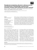
Fig. 1. TatABC expression levels in cells expressing wild-type or mutated TatA ⁄ B subunits. Membranes were isolated from wild-type
MC4100 cells, DtatABCDE cells (DABCDE)andDtatABCDE cells expressing pBAD-ABC containing mutations in the TatA or TatB subunit as
indicated. Samples were immunoblotted using antibodies to TatA, TatB or the Strep-tag II on TatC. Asterisks denote strains in which a
Strep-tag II is not detected by immunoblotting.
C. M. L. Barrett and C. Robinson Mutagenesis of TatABC
FEBS Journal 272 (2005) 2261–2275 ª 2005 FEBS 2263
Although the expression levels vary to some extent, all
of the mutant proteins are present at similar levels,
with the possible exception of TatA ⁄ G2A which exhib-
its a relatively low TatB signal for unknown reasons.
One anomaly is, however, evident with two newly gen-
erated TatB mutants, E8Q and L63A: TatA and TatB
are formed at typical levels but the TatC signal is com-
pletely absent (lanes denoted by asterisks). This is
reproducible and, because these mutants display high
levels of Tat activity (see below), we assume that the
C-terminal Strep-tag II has been removed from the
proteins after expression. Some of the other mutants
also have this property (see below).
Although the primary aim was to analyze new
mutants affected in the amphipathic or TM regions,
previously analysed TatA mutants containing single
amino acid changes were also analyzed in terms of
their ability to export TorA and TorA-GFP, and the
data for the TatA mutants are shown in Fig. 2. With
the TorA export assays in Fig. 2A, it is observed that
the bulk of the activity is found in the periplasm in
wild-type cells and cells expressing pBAD-ABC, as
expected (lanes P) with very little cytoplasmic signal
evident. TorA is found exclusively in the cytoplasm in
DtatABCDE cells, where it migrates more slowly in the
gel system (denoted by an asterisk). The TatA mutants
all export TorA with high efficiency with the exception
of F39A, where all of the TorA is present as the cyto-
plasmic form. Some cytoplasmic TorA is also evident
with L25A.
The TorA-GFP export assays are in good agreement
with the TorA data (Fig. 2B). In pEXT-ABC-expres-
sing cells, the bulk of GFP is found as mature-size
protein in the periplasm (P), whereas GFP is found
only in the cytoplasm and membrane fractions (C, F)
in cells expressing the pEXT22 vector. Some of this
protein is present as precursor form (TorA-GFP) and
some mature-size GFP is also present, presumably
due to proteolytic clipping. No signal is observed in
DtatABCDE cells that do not synthesize TorA-GFP (a
control for the specificity of the GFP antibodies). All
of the TatA mutants export TorA-GFP with high effi-
ciency except F39A, which is again completely defect-
ive in translocation. Whereas some cytoplasmic TorA
A
B
C
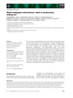
Fig. 2. The TatA F39A mutant is inactive whereas other TatA ⁄ B
mutants show no detectable loss of translocation activity. (A) Dtat-
ABCDE cells expressing pBAD-ABC or the same vector containing
mutations in tatA were induced using 50 l
M arabinose for 3.5 h,
and cytoplasmic (C) and periplasmic fractions (P) were prepared as
detailed in Experimental procedures. These fractions were electro-
phoresed on native polyacrylamide gels that were subsequently
stained for TMAO reductase (TorA) activity. Asterisk denotes
slower-migrating cytoplasmic form of TorA. (B) A TorA-GFP con-
struct was expressed in DtatABCDE cells using an arabinose-
inducible vector for 2 h. The arabinose was then washed out and
wild-type or tatABC operons containing the same tatA mutations as
in (A) were expressed for 3 h using the isopropyl thio-b-
D-galacto-
side-inducible pEXT22 plasmid as detailed in Experimental proce-
dures (pEXT-ABC plasmid contains wild-type tatABC operon).
Cytoplasmic, membrane and periplasmic fractions (C, M, P), were
isolated and immunoblotted using antibodies to GFP. The mobility
of mature-size GFP is indicated. (C) TatB mutants described in
Fig. 1 were analyzed for export of TorA-GFP (B) exactly as des-
cribed in (B) for TatA mutants.
Mutagenesis of TatABC C. M. L. Barrett and C. Robinson
2264 FEBS Journal 272 (2005) 2261–2275 ª 2005 FEBS
was evident with the L25A mutation, as described
above, no defect is apparent using TorA-GFP as a
substrate. Because signal strengths are not linearly
related to protein activity in the native gel TorA assay,
we are more inclined to regard the TorA-GFP data as
evidence of high translocation activity, although other
possibilities can not be excluded.
Similar tests on the TatB mutants are shown in
Fig. 2C. The results show that all of the strains effi-
ciently export TorA-GFP, including the E8Q and
L63A mutants that exhibited no signal with the Strep-
tag II immunoblots for TatC in Fig. 1.
Inhibitory effects of mutations in the predicted
amphipathic regions of TatA and TatB
Considerable attention has centred on possible roles of
conserved predicted amphipathic regions that are
highly conserved in both TatA and TatB. These
regions effectively bridge the transmembrane helices
and soluble cytoplasmic domains, and truncation ana-
lysis [29] has shown that they are essential for translo-
cation activity. Their sequences are shown in Fig. 3A.
In a previous report [24], we analyzed the effects of
changing three lysine residues in TatA (residues 37, 40
and 41) to glutamine, and in a second mutant we addi-
tionally changed K24 to alanine. These mutants,
denoted TatA ⁄ )3K and TatA ⁄ )4K previously [24]
were shown to be active albeit with reduced efficien-
cies. These TatA mutants have been re-made (and re-
named TatA ⁄ 3K > Q and TatA ⁄ K24A,3K > Q) after
finding several revertants in recent studies of previ-
ously analyzed mutants. In the case of TatB, it was
previously found that changing two arginines (residues
37 and 40) to asparagine (TatB ⁄ 2R > N), or three
lysines (residues 65, 67 and 68) to glutamine
(TatB ⁄ 3K > Q) had little effect on the efficiency of
Tat-dependent export [24]. In the present report we
have re-assessed these mutants and another mutant
combining the two sets of mutations in TatB
(TatB ⁄ 2R > N,3K > Q). This new mutant is indica-
ted by ‘Ù’ in Fig. 3. The expression profiles are shown
in Fig. 3B, which shows TatA and TatB to be synthes-
ized in all cases, although again at slightly varying lev-
els. Strep-tagged TatC is formed in every case except
TatA ⁄ K24A,3K > Q, but because this mutant is act-
ive (see below) we again believe that the TatC protein
is present but lacking the Strep-tag II.
Export assays using these mutants are shown in
Fig. 4A. The data show that all three TatB mutants
TatA
TatA
TatB
TatC
TatA
TatB
TatC
pBAD-ABCs
pBAD-ABCs
2R>N
K24A
K24A
K24A
K24A
K24A
3K>Q
3K>Q
3K>Q
3K>Q
3K>Q
3K>Q/
2R>N
2R>N
3K>Q
3K>Q
3K>Q
3K>Q
2R>N 3K>Q
2R>N 3K>Q
∆ABCDE
∆ABCDE
TatB
16
25
30
*
A
BC
Fig. 3. Mutations in the predicted amphi-
pathic regions of TatA and TatB. (A) Primary
sequences of the amphipathic regions with
the targeted residues indicated by arrows
and numbered. The changes introduced in
the various mutants are indicated and under-
lined. (B) pBAD-ABC cells, DtatABCDE cells
and DtatABCDE cells expressing pBAD-ABC
containing the ‘amphipathic’ mutations
shown in (A) were analyzed by immunoblot-
ting with antibodies to TatA, TatB and the
Strep-tag II on TatC. Asterisks denote
strains that exhibit no signal with Strep-tag
II antibodies. (C) as in (B), except with
strains expressing combinations of muta-
tions in TatA + TatB. ‘^’ denotes new muta-
tions analyzed in this study. Mutations in
TatA are shown underlined and in grey font.
Mobilities of molecular mass markers are
given on the left.
C. M. L. Barrett and C. Robinson Mutagenesis of TatABC
FEBS Journal 272 (2005) 2261–2275 ª 2005 FEBS 2265
exhibit efficient export of TorA-GFP, in that the vast
majority of protein is found in the periplasmic fraction.
This includes the new TatB mutant (TatB ⁄ 2R > N,
3K > Q) in which five basic residues are substituted;
surprisingly, there is little evident effect. Among the
TatA mutants, TatA ⁄ 3K > Q was previously described
as active [24] but Fig. 4A shows this not to be the case
with the newly generated mutant, which fails to export
TorA-GFP to any detectable extent. No export of TorA
was observed either (data not shown). Surprisingly, the
presence of an additional mutation (K24A) enables
export to take place, with the TatA ⁄ K24A,3K > Q
quadruple mutant exhibiting moderate export activity.
Some precursor form of TorA-GFP is present in the
cytoplasm and ⁄ or membrane fractions but considerable
amounts of periplasmic TorA and GFP are present. It is
likely that the original TatA ⁄ 3K > Q mutant [24] simi-
larly acquired an additional mutation prior to analysis
that enabled translocation to occur, although this has
yet to be confirmed. Other TatABC mutations described
previously [24,26] have been remade and shown to have
unchanged properties.
We also expressed tatABC operons in which these
multiple mutations in the amphipathic regions were
combined; in the relevant Figures the TatA mutations
are shown underlined for simplicity. We previously
reported data [24] on two such combinations:
(TatA ⁄ K24A, 3K > Q + TatB ⁄ 3K > Q) and (TatA ⁄
3K > Q + TatB ⁄ 2R > N), and both mutants were
described as active. In this report we have reassessed
the effects of these mutations (as the former TatA
mutant TatA ⁄ 3K > Q is now known to be inactive on
its own, as shown above) and have constructed several
new permutations as detailed in Fig. 3. These new
mutations are again denoted by ‘Ù’. Figure 3C shows
that strains synthesizing all of the multiple TatAB
mutations contain TatABC at similar levels and activ-
ity assays are shown in Fig. 4(B,C).
These ‘mixed amphipathic’ mutations exhibit very
interesting properties. Figures 2 and 4A showed that
the TatA ⁄ K24A and TatB ⁄ 2R > N mutations support
wild-type levels of export activity but Fig. 4B shows
that the combined (TatA ⁄ K24A + TatB ⁄ 2R > N)
mutations severely disrupt activity, with no periplasmic
A
B
C
Fig. 4. Combinations of mutations in the
TatA ⁄ TatB amphipathic regions have partic-
ularly severe effects on translocation activ-
ity. (A) Mutants containing alterations in the
predicted amphipathic regions of either TatA
or TatB (as detailed in Fig. 3) were analyzed
for export of TorA-GFP using the assay pro-
tocols detailed in Fig. 2. For clarity, muta-
tions in TatA are shown underlined and in
grey. (B) Combinations of mutations in the
amphipathic regions of TatA and TatB,
whose structures and expression profiles
are illustrated in Fig. 3, were tested for the
export of TorA-GFP. Mutations in TatA are
shown underlined and in grey font; addition-
ally, labels on the right indicate whether
mutations are in TatA or TatB. (C) Dtat-
ABCDE cells, or DtatABCDE expressing
pBAD-ABC or the same vector containing
mutations in both TatA and TatB as indica-
ted, were assayed for export of TorA using
the protocol described in Fig. 2.
Mutagenesis of TatABC C. M. L. Barrett and C. Robinson
2266 FEBS Journal 272 (2005) 2261–2275 ª 2005 FEBS
mature-size GFP detected at all after synthesis of
TorA-GFP. Note that mature-size GFP accumulates in
the cytoplasm, and not the full precursor protein; this
is due to proteolytic clipping of the signal peptide
when export is blocked [28,30]. Figure 4C shows TorA
export assays in which the vast majority of TorA is
found in the cytoplasm. A minor fraction of TorA
is found in the periplasm, indicating that the mutant
is not completed blocked in Tat-dependent transloca-
tion, but this mutant is clearly badly compromised. An
almost identical result is obtained with (TatA ⁄ K24A
+ TatB ⁄ 3K > Q), and it should again be noted that
the individual TatA and TatB mutants show no appar-
ent defects. These data show that the mutations have
synergistic effects and the particularly severe effects on
TorA-GFP export raise the intriguing possibility that
these mutations somehow affect the export of GFP
differently when compared to TorA.
The next two mutants in Fig. 4B (from left to right)
contain TatA ⁄ 3K > Q in combination with either
TatB ⁄ 2R > N or TatB ⁄ 3K > Q. Both combinations
exhibit no detectable Tat activity and this is unsurpris-
ing as we showed above that the ‘new’ TatA ⁄ 3K > Q
mutant is inactive on its own. Of the remaining
mutants (TatA ⁄ K24A, 3K > Q + TatB ⁄ 3K > Q) is
completely inactive, although the individual TatA and
TatB mutants did exhibit activity, and the final mutant
containing five changes in TatA plus four in TatB is
likewise totally inactive. In all, these mutants empha-
size the importance of the amphipathic region but they
also show for the first time that combinations of
TatA ⁄ TatB mutations can have far more dramatic
effects than the individual mutations.
Mutations in the transmembrane spans
of TatA and TatB
Deletion of the TM spans of TatA or TatB leads to a
loss of activity [31] but the important characteristics of
these regions have not been probed. We constructed
a series of new TatA ⁄ B mutants containing changes
within the TM spans, for two reasons. First, it has
been suggested that TatA may form the translocation
channel, in which case drastic structural alterations
may be expected to selectively block the translocation
event. Secondly, mutations affecting the structure and
orientation of the TM span may be expected to disrupt
the interactions with other Tat components, and this
would provide information on the inter-subunit associ-
ations occurring within and between Tat complexes.
Both of these areas are poorly understood at present.
It has been shown with other proteins that the intro-
duction of proline residues has a marked effect on the
structures of TM spans [32,33], usually introducing dis-
tortions of major proportions, and such substitutions
were made in the TM spans of TatA and TatB in the
present study. We also substituted three residues in the
TatA and TatB TM spans by glycine. The presence
of glycine can also lead to increased flexibility in TM
helices [34]. However, glycine residues can also play
important roles in modulating inter-helix interactions
[35] and the effects of inserting or removing these resi-
dues may therefore be less predictable than with pro-
line mutations. Nevertheless, the presence of three
consecutive glycines should lead to a significant struc-
tural effect in either case. The proline and glycine sub-
stitutions were made near the centre of the TM span
in order to maximize possible structural effects.
Figure 5A shows the sequences of the TatA and
TatB TM spans, together those of the mutated forms.
With TatA, residues 11–13 were changed to glycine in
one case (TatA ⁄ 3Gly) and a single residue was chan-
ged to proline in the centre of the TM span in another
(TatA ⁄ I12P). We also changed three additional
residues to proline in the TatA ⁄ 3Gly mutant (TatA ⁄
3Pro3Gly) and then a further three residues to glycine
in the same subunit (TatA ⁄ 3Pro6Gly). With TatB, resi-
dues 11–13 were changed to glycine (TatB ⁄ 3Gly) or a
single residue to proline (TatB ⁄ V12P).
The expression characteristics of these mutants are
shown in Fig. 5B. With the simplest TatA mutants,
TatA ⁄ 3Gly and TatA ⁄ I12P, TatABC are all present
at expected levels. With TatA ⁄ 3Pro3Gly, TatA and
TatB are present at the usual levels but TatC is not
detected at all. However, as this mutant is highly act-
ive (see below) it appears that this is another example
of the C-terminal Strep-tag II being clipped or modi-
fied. With the most drastic of the TatA mutants,
TatA ⁄ 3Pro6Gly, TatB and TatC are synthesized but
no TatA is detected. Fractionation of cells synthes-
izing TatA ⁄ 3Pro3Gly shows the presence of full-
length protein in the cytosol, consistent with a slight
defect in membrane-insertion; in contrast, smaller
fragments of the TatA ⁄ 3Gly6Pro protein are found in
the cytosol (data not shown). This suggests that the
TatA ⁄ 3Gly6Pro protein is degraded either within the
membrane or, perhaps more likely, after failure to
insert into the membrane.
Cells expressing the simpler of the tatB mutants,
TatB ⁄ V12P, appear to contain TatABC at expected
levels but the TatB ⁄ 3Gly mutations result in a much-
reduced TatB signal, presumably reflecting problems in
insertion and ⁄ or stability. No TatC signal is evident,
but this again appears to reflect problems in detection
of the Strep-tag II as this mutant is active in Tat-
dependent transport (see below).
C. M. L. Barrett and C. Robinson Mutagenesis of TatABC
FEBS Journal 272 (2005) 2261–2275 ª 2005 FEBS 2267
Figure 6 shows TorA and TorA-GFP export assays
for these TM span mutants. Of the TatA mutants,
TatA ⁄ I12P shows no detectable defect because GFP
and TorA are found predominantly in the periplasm.
The TatA ⁄ 3Gly mutant, on the other hand is blocked
in export and no translocation activity can be detected.
The same applies to the TatA ⁄ 3Pro6Gly mutant,
although this is expected because the immunoblots in
Fig. 5 show no signal for TatA. The real surprise is
the TatA ⁄ 3Pro3Gly mutant, which is shown to export
both TorA and TorA-GFP with high efficiency. Given
that the parent TatA ⁄ 3Gly mutant is completely inac-
tive, this result indicates that the three proline residues
somehow compensate for the inhibitory effects of the
glycine residues and enable translocation to occur.
Finally, Fig. 6 shows that the two TatB mutants,
TatB ⁄ 3Gly and TatB ⁄ V12P, are highly active in
export; perhaps surprisingly given the drastic effects of
the 3Gly mutations in TatA.
We also tested the effects of expressing tatABC
operons carrying combinations of these mutations in
the TM spans of both TatA and TatB, namely (TatA ⁄
3Gly + TatB ⁄ 3Gly) (TatA ⁄ 3Gly + TatB ⁄ V12P) (TatA ⁄
I12P + TatB ⁄ 3Gly) and (TatA ⁄ I12P + TatB ⁄ V12P).
Immunoblots confirmed that the TatABC were syn-
thesized at approximately the same levels as the wild-
type subunits generated from pBAD-ABC (data not
shown), and activity assays are shown in Fig. 7. With
the TorA assays, the control tests show efficient export
with wild-type TatABC and a complete block in export
in the tat null mutant (denoted DABCDE), as expected.
The combination of (TatA ⁄ 3Gly + TatB ⁄ 3Gly) is
blocked in Tat function, and this is not unexpected
given that the TatA ⁄ 3Gly mutant itself shows no
export activity. However, a combination of (TatA ⁄
3Gly + TatB ⁄ V12P) displays very efficient export of
TorA and this shows that the TatB ⁄ V12P mutation
compensates for the drastic effects of the 3Gly muta-
tions in TatA. This is confirmed by the TorA-GFP
export assays, which reveal a complete block in export
with the (TatA ⁄ 3Gly + TatB ⁄ 3Gly) mutant but near
wild-type export efficiency with (TatA ⁄ 3Gly + TatB ⁄
V12P). The remaining (TatA ⁄ I12P + TatB ⁄ 3Gly) and
(TatA ⁄ I12P + TatB ⁄ V12P) mutants export both TorA
and TorA-GFP efficiently, in keeping with the finding
that none of the mutations affect export to a detect-
able extent when present in TatA or TatB alone (see
Fig. 6). In conjunction with the data shown in Fig. 6,
these data show that the severe effects of the
TatA ⁄ 3Gly mutations can be rescued by the presence
of additional mutations either elsewhere in the TatA
TM span or in the TatB TM span (TatB ⁄ V12P
mutation).
Mutagenesis of TatC
TatC has also been studied in previous reports and a
number of mutations have been characterized [26,27],
but some apparent differences were reported in studies
on the same mutants by different groups (see below).
Few TatC residues are highly conserved but of these, a
high proportion is clustered in the N-terminal cyto-
plasmic domain and the first cytoplasmic loop. In our
previous analysis [26] only two residues were found to
A
B
Fig. 5. Expression of TatA and TatB mutants containing alterations
in the transmembrane spans. (A) Sequences of the TM regions of
TatA and TatB, with residues numbered and the substitutions indi-
cated. (B) Expression of the various mutations constructed within
the pBAD-ABC vector. Samples were analyzed by immunoblotting
with antibodies to TatA, TatB and the Strep-tag II on TatC. Mobili-
ties of molecular mass markers are given on the left.
Mutagenesis of TatABC C. M. L. Barrett and C. Robinson
2268 FEBS Journal 272 (2005) 2261–2275 ª 2005 FEBS
be absolutely essential for TatC function: R17A
(N-terminal cytoplasmic region) and P48A (first peri-
plasmic loop). The deletion of three residues (20–22)
also disrupted function, prompting the suggestion that
this cytoplasmic domain played an important role. In
a separate study [27], R17 was again found to be
important but two acidic residues, E103 and D211
were found to be particularly critical; E103A ⁄ Q ⁄ R
mutants and D211A were completely inactive,
although D211E ⁄ N mutants were partially active.
These residues are located on the first cytoplasmic loop
and third periplasmic loop, respectively. Substitution
of F94 (at the interface between cytoplasmic loop I
and the membrane bilayer) was also reported to block
transport activity [27]. D211 was not analyzed in our
previous study [26] but we did find that E103A showed
no translocation defect at all, and so we have made
new mutants in all three residues in order to analyse
the effects using our expression and assay systems.
As with other mutants, we carried out expression
studies using all of the TatC mutants; in each case the
TatABC subunits were present at similar levels and in
A
B
Fig. 6. Effects of mutations in the TM
regions of TatA and TatB. The TatA and
TatB mutants containing alterations in TM
spans (detailed in Fig. 5) were tested for
effects on translocation activity. (A) Muta-
tions within the pBAD-ABC vector were
assayed for export of TorA. (B) mutations
within pEXT-ABC were assayed for export
of TorA-GFP, as detailed in Fig. 2. Cells
expressing pBAD-ABC or pEXT-ABC without
mutations were analyzed as controls, and
DtatABCDE cells were analyzed for export
of TorA, again as a control. Other symbols
are as in Fig. 2.
A
B
Fig. 7. A mutation in the TatB TM span can compensate for the
severe effects of the TatA ⁄ 3Gly mutation. This figure illustrates the
translocation activities of four DtatABCDE strains expressing pBAD-
ABC or pEXT-ABC in which mutations are present in the TM spans
of both TatA and TatB (mutations in TatA are shown underlined and
labels on the right indicate whether the mutations are in TatA or
TatB). Cells were analyzed for the export of TorA (A) or TorA-GFP
(B) using protocols described in Fig. 2.
C. M. L. Barrett and C. Robinson Mutagenesis of TatABC
FEBS Journal 272 (2005) 2261–2275 ª 2005 FEBS 2269
these instances the TatC Strep-tag II were all found to
be intact (data not shown).
Mutations in the first cytoplasmic loop are charac-
terized in Fig. 8(A). None of these mutants display
any detectable defects in translocation efficiency, inclu-
ding the E103A and E103Q mutants which do not
even contain elevated levels of the precursor protein
TorA-GFP. These data do not agree with reports that
the two residues are critical [27], and with this appar-
ent contradiction in mind we made each mutant again
and again found them to be highly active (data not
shown). Figure 8(B) shows the effects of mutations in
TM spans two to six. Most of these mutations again
have no detectable effect with the notable exception of
F94A which is completely inactive. These results agree
with those reported in [27].
The final study on TatC is shown in Fig. 9, where
mutations in the periplasmic loops are analyzed. We
have previously shown that the P48A mutation des-
troys activity, and the data confirm this result with no
export detected using either assay system. The K73A
and Y154S mutants are active, as expected from previ-
ous studies [26] and so too are the D211A and D211N
mutants that were described as completely or partially
inactive, respectively, in a previous study [27]. In
Fig. 9, no translocation defects are apparent with
either D211A or D211N and these mutants were again
A
B
Fig. 8. Mutations in several residues of the
cytoplasmic loops in TatC cause no detect-
able loss of translocation activity. (A) TatC
mutants carrying substitutions in the 1st
cytoplasmic loops were tested for export of
TorA-GFP as described in Fig. 2; mutations
were made within the pEXT-ABC vector and
the Figure shows assays using pEXT-ABC
as a control. (B) Similar tests carried out
using TatC mutants carrying substitutions in
TM spans (numbered above the lanes).
A
B
Fig. 9. Effects of mutations in the periplasmic loops of TatC. Muta-
tions in the three periplasmic loops of TatC (numbers indicated
above the lanes) were assessed using export assays for TorA
(upper panel) or TorA-GFP (lower panel) as described earlier for
other mutants (Fig. 2).
Mutagenesis of TatABC C. M. L. Barrett and C. Robinson
2270 FEBS Journal 272 (2005) 2261–2275 ª 2005 FEBS
generated and re-tested, with the same result (data not
shown).
Discussion
Structure–function studies on protein translocases have
benefited from mutagenesis studies that pinpoint
important residues or regions of the proteins. Previous
studies on the bacterial Tat system have been carried
out with this aim in mind but the roles of (and poss-
ible interactions between) the TM spans of TatA and
TatB have not been probed in detail. In addition, some
potential discrepancies have arisen regarding poten-
tially crucial residues in TatC. Here, we have
attempted to be comprehensive and have analyzed pre-
existing mutants in concert with new ones so that the
various sets are tested under the same assay condi-
tions. The findings can be summarized as follows.
Mutations in TatA and TatB ‘hinge’ regions and
predicted amphipathic domains
TatA and TatB are generally regarded as containing:
(a) a single TM span; (b) adjacent hinge region; (c) a
predicted amphipathic region; and finally (d) a cyto-
solic domain that is small in TatA and slightly larger
in TatB. Very few residues are conserved in these pro-
teins, even within Gram-negative bacteria, but the very
highly conserved residues are found in the apparent
hinge region. In TatA, the sequence F20-G21 is essen-
tially invariant, and F39 is very highly conserved. In
bacterial TatB proteins the most highly conserved resi-
dues are G21-P22, and other highly conserved residues
include E8 and P26. In our own study [24] it was
found that the TatA F39A was almost completely
blocked in Tat-dependent export. Several other
mutants exhibited lower levels of translocation activity
when compared to wild-type cells, but none showed
the severe reduction present in F39A. A separate study
[25] found TatA F39 to be essential. Here, analysis of
this mutant under conditions that promote highly act-
ive translocation efficiencies shows the F39A mutant
to be completely blocked in Tat function and our data
thus agree with this study [25].
In this report we have expressed several other
TatA ⁄ B mutant forms and have confirmed that none
of these residues are essential (indeed, none of these
mutants even exhibit markedly lower export efficien-
cies). In this respect, our data differ at first sight from
those described in [25], where several TatA⁄ TatB
mutants were found to exhibit export efficiencies that
were much-reduced. For example, the TatA F20A
and G21A mutants were compromised, as were the
G21A ⁄ P ⁄ L and P22L TatB mutants. However, it is
important to stress that the mutations were tested in
different ways. In the above study [25], individual
mutated subunits were expressed in a background con-
taining chromosomal copies of the remaining subunits.
This should mean that Tat complexes are synthesized
at wild-type levels, but a drawback is that the mutated
subunit is synthesized at higher levels than the others
and this may have adverse consequences for assembly.
In this report we have used a multicopy vector to
express the tatABC operon; this has the advantage that
the subunits are coordinately synthesized in the correct
stoichiometry, but the disadvantage is that minor ⁄
moderate effects on translocation activity are masked.
As a result, our approach is best suited for the identifi-
cation of moderate-severe effects on translocation
activity. Numerous TatABC mutants have been
analyzed in this study and the first observation is that
very few are affected at this level, confirming previous
findings that few of the highly conserved residues are
actually essential for Tat function. Taking these previ-
ous reports into consideration, the logical conclusion is
that several residues (F20, G21 in TatA, G21, P22,
P26 in TatB) are probably important for full transloca-
tion activity but certainly not essential. The only
mutant devoid of translocation activity in the present
study and [25] is TatA ⁄ F39A.
Mutations in the predicated amphipathic region can
have more drastic effects, and we previously analyzed
the effects of substituting groups of basic residues in
these regions of TatA and TatB [24]. The present study
has increased our understanding of this region in two
ways. First, the removal of three lysines completely
blocks translocation activity and this reinforce s the
importance of this region. Truncation analysis has
similarly pointed to essential roles for this region in
both TatA and TatB [29]. While the precise effects of
the individual sets of mutations await further analysis,
the data obtained with new combinations of TatA and
TatB mutants have unexpected and interesting proper-
ties. For example, the TatA K24A mutant exhibits
wild-type levels of TorA and TorA-GFP export, as
does the TatB 2R > N mutant. However, a combina-
tion of the two mutations leads to a near-complete loss
of TorA export and an absolute loss of TorA-GFP
export. The same phenomenon is observed with the
combination of K24A in TatA and 3K > Q in TatB,
each of which exhibits no translocation defects in
isolation. We propose that these regions interact with
each other, and that these mutations can be safely
accommodated in either subunit separately, whereas
the simultaneous presence of both mutations leads to
an undue disturbance of the inter-subunit interaction.
C. M. L. Barrett and C. Robinson Mutagenesis of TatABC
FEBS Journal 272 (2005) 2261–2275 ª 2005 FEBS 2271
Mutations in the TM spans of TatA and TatB
Most of the previous studies on TatA ⁄ B have focused
on the more highly conserved residues and we consid-
ered it important to test the effects of introducing struc-
tural rearrangements of the TM spans of these subunits.
It has not been established whether these TM spans
simply anchor the amphipathic regions and cytosolic
domains to the membrane, or whether they serve more
important roles such as the formation of the transloca-
tion channel. Proline residues usually introduce distor-
tions of major proportions into TM helices [32,33] and
glycine residues are also thought to alter their structures
although the precise effects are less well-characterized.
We set out to introduce such disruptions without affect-
ing the membrane insertion properties of either protein.
Table 1. Primers used for site-specific mutagenesis of TatABC. Only the forward primers are shown.
Name of primer Sequence 5’ to 3’ Substitution
TatA mutants
G2A G2A F gaggaattcaccatggctggtatcagtatttgg Gly2 to Ala
3Pro3Gly W7P F ggtggtatcagtattccgccgccattgggtggtggcg Tyr7, Glu8 and Leu9 to Pro
I12P I12P F ggcagttattgattccggccgtcatcgttgtactg Ile12 to Pro
3Pro6Gly V16G F ggcgtcatcggtggagggctttttggcaccaaaaag Val16, Val17 and Leu18 to Gly
F20A F20A F catcgttgtactgcttgctggcaccaaaaagctc Phe20 to Ala
G21A G21A F gttgtactgctttttgccaccaaaaagctcgg Gly21 to Ala
K24A K24A F gctttttggcaccaaagccctcggctccatcgg Lys24 to Ala
L25A L25A F gctttttggcaccaaaaaggccggctccatcg Leu25 to Ala
G33A G33A F ggttccgatcttgctgcgtcgatcaaagg Gly33 to Ala
F39A F39A F gcgtcgatcaaaggcgctaaaaaagcaatgagcg Phe39 to Ala
3K > Q K37Q F ggtgcgtcgatccaaggctttcaacaagcaatgag Lys37, Lys40 and Lys41 to Gln
TatB mutants
E8Q E8Q F cggttttagccaactgctattggtgttcatcatc Glu8 to Gln
E8A E8A F cggttttagcgcactgctattggtgttcatc Glu8 to Ala
3Gly L11G F cggttttagcgaactgctaggggggggcatcatcggc Leu11, Val12 and Phe13 to Gly
V12P V12P F gcgaactgctattgcccttcatcatcggcctcg Val12 to Pro
G21A G21A F ctcgtcgttctggcaccgcaacgactgcc Gly21 to Ala
P22G P22G F ctcgtcgttctggggggacaacgactgcc Pro22 to Gly
P22L P22L F gtcgttctggggctacaacgactgcctgtg Pro22 to Leu
L25A L25A F ggggccgcaacgagcgcctgtggcgg Leu25 to Ala
P26A P26A F cgcaacgactggctgtggcggtaaaaac Pro26 to Ala
L63A L63A F ggagtttcaggacagtgcgaaaaaggttgaaaagg Leu63 to Ala
2R > N R37N F gtagcgggctggattaacgcgttgaattcactggcg Arg37 and Arg40 to Asn
3K > Q K64Q F caggacagtctgcagcaggttgaacaagcgagcctcac Lys64, Lys65 and Lys68 to Gln
TatC mutants
P48A P48A F ggtatccgcggcattgatcaagcagttg Pro48 to Ala
I81M I81M F ggtgtcgctgatgctgtcagcgccg Ile81 to Met
P85A P85A F gattctgtcagcggcggtgattctctatcag Pro85 to Ala
F94A F94A F caggtgtgggcagctatcgccccagcgc Phe94 to Ala
P97A P97A F ggcatttatcgccgctgcgctgtataagcatg Pro97 to Ala
L99A L99A F cgccccagcggcgtataagcatgaacgtcg Leu99 to Ala
E103Q E103Q F gctgtataagcatcagcgtcgcctggtggtg Glu103 to Gln
E103A E103A F gctgtataagcatgcacgtcgcctggtg Glu103 to Ala
R104A R104A F ctgtataagcatgaagctcgcctggtggtgcc Arg104 to Ala
R105A R105A F gtataagcatgaacgtgccctggtggtgccg Arg105 to Ala
F118A F118A F ccagctctctgctggcttatatcggcatggc Phe118 to Ala
G121A G121A F ctgctgttttatatcgccatggcattcgcctac Gly121 to Ala
Y154S Y154S F ccgacatcgccagcagcttaagcttcgttatggc Tyr154 to Ser
F169A F169A F gtttggtgtctccgctgaagtgccgg Phe169 to Ala
P172A P172A F cctttgaagtggcggtagcaattgtgctgc Phe172 to Ala
T208A T208A F gtcgggatgttgctggcaccgccggatg Thr208 to Ala
P209A P209A F gatgttgctgacggcaccggatgtcttctcg Pro209 to Ala
D211N D211N F gacgccgccgaacgtcttctcgcaaacgc Asp211 to Asn
D211A D211A F ctgacgccgccggctgtcttctcgc Asp211 to Ala
L225A L225A F cccgatgtactgtgcgtttgaaatcggtgtcttc Leu225 to Ala
Mutagenesis of TatABC C. M. L. Barrett and C. Robinson
2272 FEBS Journal 272 (2005) 2261–2275 ª 2005 FEBS
The results show that the presence of three glycine resi-
dues in the TatA TM span leads to a total block in
translocation activity. The TatA protein is present in
the membrane fraction and these data suggest that the
TM region is not merely a membrane anchor, but rather
plays a much more important role in either the assembly
of Tat complexes or the actual translocation process (or
both). Curiously, the further introduction of three pro-
line residues actually restores translocation activity sug-
gesting that the distorting effect of these residues
somehow counterbalances the effects of the glycine resi-
dues. In contrast to the TatA data, substitution by
either a single proline or three glycine residues in the
TatB TM span has little detectable effect, and this may
represent a very preliminary argument against a role
in participating in the translocation channel, although
further studies are certainly required to obtain a clear
picture of TatB function.
Again, an analysis of mixed TatA ⁄ TatB mutations
provides strong indications for critical interactions
between these subunits. The TatA 3Gly mutant is
unable to export either TorA or TorA-GFP, but the
simultaneous presence of the V12P mutation in TatB
restores export to levels that are very close to those of
cells expressing the wild-type tatABC operon. Appar-
ently, the latter mutation compensates almost com-
pletely for the effects of introducing three glycines in
the TatA TM span, and the logical interpretation is that
these TM spans must undergo direct interactions with
each other. One caveat should, however, be noted: it
has very recently been shown that surprisingly minor
mutations in the extreme N-terminus of TatA can ren-
der TatB dispensable for translocation [36]. Given that
TatA and TatB are not as distinct as previously ima-
gined, it is possible that the TatB V12P mutation has
rendered TatA dispensable. This is, in our view unlikely
but further studies on the specific roles of the mutant
TatA ⁄ TatB proteins could be useful in this context.
In conjunction with the results described above, these
data provide indirect evidence of interactions between
at least two regions of TatA and TatB: the predicted
amphipathic regions and the TM spans. It will be
important to probe these issues in detail because little is
currently known about the molecular details of the pro-
posed TatA–TatB interaction under conditions where
TatABC are expressed at physiological stoichiometries.
Previous coimmunoprecipitation studies [17,37] have
shown that TatA and TatB are present within a single
complex but these studies did not prove that TatA and
TatB actually contact each other. A more direct cross-
linking approach found evidence TatA dimers and
trimers, as well as TatB dimers, but no TatA-TatB
cross-links were observed [31].
TatC mutants
Most TatC mutations do not exhibit major defects in
translocation but a few have been reported to block
translocation, including R17A and P48A in one study
[26], and F94A, E103A and D211A in another [27]. In
the present study we have re-affirmed the effects of the
P48A and F94A mutations, which completely block
Tat-dependent export. However, we have also studied
the effects of mutating E103 and D211 and have
observed no effects on translocation activity. Again,
these differences probably reflect differences in expres-
sion ⁄ analysis methods, because in [27] the mutated
subunits were generated in a background of wild-type
levels of the remaining Tat subunits. These residues
are of real interest because acidic side-chains could
participate in proton translocation or, in the case of
E103, in the binding of the twin-arginine motif in the
signal peptide. The absence of any translocation
defects leads us to conclude that these residues are not
in fact essential for translocation activity.
Experimental procedures
Bacterial strains, plasmids and growth conditions
E. coli strain MC4100 [38] was the parental strain; Dtat-
ABCDE has been described previously [13], and arabinose
resistant derivatives were used as described [17,37]. All of
the mutated Tat subunits were expressed using the arabi-
nose-inducible pBAD24 vector or the compatible IPTG-
inducible pEXT22 vector [39] (tac promoter, R100 origin),
with E. coli tatABC expressed coordinately as described
[24,28]. The TatC protein is encoded with a C-terminal
Strep-tag II in each case. Assays for TorA-GFP export
involved use of the pJDT1 plasmid [30] which expresses
TorA-GFP from the arabinose-inducible pBAD24 vector.
E. coli was grown at 37 °C in Luria broth (LB) as described
in [17], and this medium was supplemented with glycerol
(0.5%, v ⁄ v), TMAO (0.4%, v ⁄ v), and sodium molybdate
(1 lm) for TorA export assays. Media supplements were
used at the following final concentrations: ampicillin,
100 lgÆmL
)1
; kanamycin, 50 lgÆmL
)1
; arabinose 50 lm
unless stated otherwise.
Mutagenesis of tatABC
Site-directed mutaganesis was used to generate a vector that
encodes the tat operon within pBAD-ABC with point
mutations in the tatA, tatB or tatC genes using the Quik-
Change
TM
mutagenesis system (Stratagene, La Jolla, CA,
USA) according to the manufacturer’s instructions. All
mutated operons were sequenced in full. For studies on the
effects of these mutations on the export of TorA-GFP, the
C. M. L. Barrett and C. Robinson Mutagenesis of TatABC
FEBS Journal 272 (2005) 2261–2275 ª 2005 FEBS 2273
tatABC sequences were cloned into the pEXT22 vector [39],
generating variants of pEXT-ABC compatible with pJDT1
encoding TorA-GFP [28]. All pBAD-ABC and pEXT-ABC
mutations were expressed in the DtatABCDE strain. Prim-
ers used in this study were as shown in Table 1.
Export assays
Tat-dependent translocation activity was assayed by deter-
mining the localization of TMAO reductase (TorA), a
known periplasmic Tat substrate, or by determining the
location of a TorA-GFP construct that is exported to the
periplasm in wild-type cells. For the TorA assays, cells
expressing pBAD-ABC or mutated derivatives were grown
in LB ⁄ glycerol ⁄ TMAO medium (described above) plus
50 lm arabinose [17]. After 3.5 h growth the cells were ana-
lyzed by fractionation as detailed below. For studies on the
export of TorA-GFP, cells expressing pJDT1 and pEXT-
ABC were grown for 2 h in the presence of 50 lm arabi-
nose to induce expression of TorA-GFP, and the cells were
then pelleted and resuspended in fresh medium containing
1mm isopropyl thio-b-d-galactoside. This leads to induc-
tion of TatABC synthesis from the pEXT-ABC plasmid,
and after growth for 3 h the cells were fractionated and
analyzed by immunoblotting to determine the location of
TorA-GFP or processed, mature-size GFP. Periplasm and
spheroplasts were prepared by the EDTA ⁄ lysozyme ⁄ cold
osmoshock procedure [17]. Spheroplasts were lysed by soni-
cation, and intact cells and cellular debris were removed by
centrifugation (5 min at 10 000 g). From the supernatant
generated, membranes were separated from the cytoplasmic
fraction by centrifugation (30 min at 250 000 g). Protein
concentration was determined using a BCA-linked assay
(Pierce, Rockford, IL, USA). Protein fractions (periplasm,
cytoplasm and membrane) were separated on a 10% non-
denaturing polyacrylamide gel and stained for TorA as
described [40] or analyzed by SDS ⁄ PAGE, transferred to
poly(vinylidene difluoride) membranes and immunoblotted
using antibodies to TatA, TatB, the Strep-tag II on TatC
(monoclonal antibody from IBA, Stuttgart, Germany) or
green fluorescent protein (GFP) using a monoclonal anti-
body from BD Clontech (Palo Alto, CA, USA).
References
1 Robinson C & Bolhuis A (2001) Protein targeting by
the twin-arginine translocation pathway. Nat Rev Mol
Cell Biol 2, 350–355.
2 Dalbey RE & Kuhn A (2000) Evolutionarily related
insertion pathways of bacterial, mitochondrial, and
thylakoid membrane proteins. Annu Rev Cell Dev Biol
16, 51–87.
3 Clark SA & Theg SM (1997) A folded protein can be
transported across the chloroplast envelope and thyla-
koid membranes. Mol Biol Cell 8, 923–934.
4 Hynds PJ, Robinson D & Robinson C (1998) The Sec-
independent twin-arginine translocation system can
transport both tightly folded and malfolded proteins
across the thylakoid membrane. J Biol Chem 273,
34868–34874.
5 Berks BC (1996) A common export pathway for pro-
teins binding complex redox cofactors? Mol Microbiol
22, 393–404.
6 Santini C-L, Ize B, Chanal A, Mu
¨
ller M, Giordano G
& Wu L-F (1998) A novel Sec-independent periplasmic
protein translocation pathway in Escherichia coli.
EMBO J 17, 101–112.
7 Weiner JH, Bilous PT, Shaw GM, Lubitz SP, Frost L,
Thomas GH, Cole JA & Turner RJ (1998) A novel and
ubiquitous system for membrane targeting and secretion
of cofactor-containing proteins. Cell 93, 93–101.
8 Sargent F, Bogsch EG, Stanley NR, Wexler M, Robin-
son C, Berks BC & Palmer T (1998) Overlapping func-
tions of components of a bacterial Sec-independent
export pathway. EMBO J 17, 3640–3650.
9 Chaddock AM, Mant A, Karnauchov I, Brink S, Herr-
mann RG, Klo
¨
sgen RB & Robinson C (1995) A new
type of signal peptide: central role of a twin-arginine
motif in transfer signals for the DpH-dependent thyla-
koidal protein translocase. EMBO J 14, 2715–2722.
10 Stanley NR, Palmer T & Berks BC (2000) The twin
arginine consensus motif of Tat signal peptides is
involved in Sec-independent protein targeting in Escheri-
chia coli. J Biol Chem 275, 11591–11596.
11 Bogsch EG, Sargent F, Stanley NR, Berks BC, Robin-
son C & Palmer T (1998) An essential component of a
novel bacterial protein export system with homologues
in plastids and mitochondria. J Biol Chem 273, 18003–
18006.
12 Sargent F, Stanley NR, Berks BC & Palmer T (1999)
Sec-independent protein translocation in Escherichia
coli: a distinct and pivotal role for the TatB protein.
J Biol Chem 274, 36073–36082.
13 Wexler M, Sargent F, Jack RL, Stanley NR, Bogsch
EG, Robinson C, Berks BC & Palmer T (2000) TatD is
a cytoplasmic protein with DNase activity: no require-
ment for TatD family proteins in Sec-independent pro-
tein export. J Biol Chem 275, 16717–16722.
14 Yen MR, Tseng YH, Nguyen EH, Wu LF & Saier MH
Jr (2002) Sequence and phylogenetic analyses of the
twin-arginine targeting (Tat) protein export system.
Arch Microbiol 177, 441–450.
15 Jongbloed JD, Martin U, Antelmann H, Hecker M,
Tjalsma H, Venema G, Bron S, van Dijl JM & Muller J
(2000) TatC is a specificity determinant for protein
secretion via the twin-arginine translocation pathway.
J Biol Chem 275, 41350–41357.
16 Pop O, Martin U, Abel C & Muller JP (2002) The twin-
arginine signal peptide of PhoD and the TatAd ⁄ Cd
proteins of Bacillus subtilis form an autonomous
Mutagenesis of TatABC C. M. L. Barrett and C. Robinson
2274 FEBS Journal 272 (2005) 2261–2275 ª 2005 FEBS
Tat translocation system. J Biol Chem 277, 3268–
3273.
17 Bolhuis A, Mathers JE, Thomas JD, Barrett C &
Robinson C (2001) TatB and TatC form a structural
and functional unit of the twin-arginine translocase of
Escherichia coli. J Biol Chem 276, 20213–20219.
18 Oates J, Mathers J, Mangels D, Ku
¨
hlbrandt W, Robin-
son C & Model K (2003) Consensus structural features
of purified bacterial TatABC complexes. J Mol Biol
330, 277–286.
19 De Leeuw E, Granjon T, Porcelli I, Alami M, Carr S,
Muller M, Sargent F, Palmer T & Berks BC (2002)
Membrane interactions and self association of the TatA
and TatB components of the twin-arginine translocation
pathway. J Mol Biol 322, 1135–1146.
20 Porcelli I, de Leeuw E, Wallis R, van den Brink-van der
Laan E, de Kruijff B, Wallace BA, Palmer T & Berks
BC (2002) Characterization and membrane assembly of
the TatA component of the Escherichia coli twin-argi-
nine protein transport system. Biochemistry 41, 13690–
13697.
21 Cline K & Mori H (2001) Thylakoid DpH-dependent
precursor proteins bind to a cpTatC-Hcf106 complex
before Tha4-dependent transport. J Cell Biol 154, 719–
729.
22 Alami M, Luke I, Deitermann S, Eisner G, Koch HG,
Brunner J & Muller M (2003) Differential interactions
between a twin-arginine signal peptide and its trans-
locase in Escherichia coli. Mol Cell 12, 937–946.
23 Mori H & Cline K (2002) A twin arginine signal peptide
and the pH gradient trigger reversible assembly of the
thylakoid Delta pH ⁄ Tat translocase. J Cell Biol 157,
205–210.
24 Barrett CML, Mathers JE & Robinson C (2003) Identi-
fication of key residues within the Escherichia coli
TatAB subunits. FEBS Letts 537, 42–46.
25 Hicks MG, de Leeuw E, Porcelli I, Buchanan G, Berks
BC & Palmer T (2003) The Escherichia coli twin-argi-
nine translocase: conserved residues of TatA and TatB
family components involved in protein transport. FEBS
Lett 539, 61–67.
26 Allen SCH, Barrett CML, Ray N & Robinson C (2002)
Essential cytoplasmic domains in the Escherichia coli
TatC protein. J Biol Chem 277, 10362–10366.
27 Buchanan G, De Leeuw E, Stanley NR, Wexler M,
Berks BC, Sargent F & Palmer T (2002) Functional
complexity of the twin-arginine translocase TatC
component revealed by site-directed mutagenesis. Mol
Microbiol 43, 1457–1470.
28 Barrett CML, Ray N, Thomas JD, Robinson C &
Bolhuis A (2003) Quantitative export of a heterologous
protein, GFP, by the twin-arginine translocation path-
way in Escherichia coli. Biochem Biophys Res Comm
304, 279–284.
29 Lee PA, Buchanan G, Stanley NR, Berks BC & Palmer
T (2002) Truncation analysis of TatA and TatB defines
the minimal functional units required for protein trans-
location. J Bacteriol 184, 5871–5879.
30 Thomas JD, Daniel RA, Errington J & Robinson C
(2001) Export of active green fluorescent protein to the
periplasm by the twin-arginine translocase (Tat) path-
way in Escherichia coli. Mol Microbiol 39, 47–52.
31 De Leeuw E, Porcelli I, Sargent F, Palmer T & Berks B
(2001) Membrane interactions and self association of
the TatA and TatB components of the twin-arginine
translocation pathway. FEBS Letts 506, 143–148.
32 Yohannan S, Yang D, Faham S, Boulting D, Whitel-
egge J & Bowie JU (2004) Proline substitutions are not
easily accommodated in a membrane protein. J Mol
Biol 341, 1–6.
33 Reiersen H & Rees AR (2001) The hunchback and its
neighbours: proline as an environmental modulator.
Trends Biochem Sci 26, 679–684.
34 MacKinnon R (2003) Potassium channels. FEBS Letts
555, 62–65.
35 Curran AR & Engelman DM (2003) Sequence motifs,
polar interactions and conformational changes in helical
membrane proteins. Curr Opin Struct Biol 13, 412–417.
36 Blaudeck N, Kreutzenbeck P, Muller M, Sprenger GA
& Freudl R (Year?) Isolation and characterization of
bifunctional Escherichia coli TatA mutant proteins that
allow efficient Tat-dependent protein translocation in
the absence of TatB. J Biol Chem, in press.
37 Bolhuis A, Bogsch EG & Robinson C (2000) Subunit
interactions in the twin-arginine translocase complex of
Escherichia coli. FEBS Lett 472, 88–92.
38 Casadaban MJ & Cohen SN (1979) Lactose genes fused
to exogenous promoters in one step using a Mu-lac
bacteriophage: in vivo probe for transcriptional control
sequences. Proc Natl Acad Sci USA 76, 4530–4533.
39 Dykxhoorn DM, St Pierre R & Linn T (1996) A set of
compatible tac promoter expression vectors. Gene 177,
133–136.
40 Silvestro A, Pommier J, Pascal MC & Giordano G
(1989) The inducible trimethylamine N-oxide reductase
of Escherichia coli K12: its localization and inducers.
Biochim Biophys Acta 999, 208–216.
C. M. L. Barrett and C. Robinson Mutagenesis of TatABC
FEBS Journal 272 (2005) 2261–2275 ª 2005 FEBS 2275