Tài liệu Báo cáo khoa học: Enlarging the gas access channel to the active site renders the regulatory hydrogenase HupUV of Rhodobacter capsulatus O2 sensitive without affecting its transductory activity docx
Bạn đang xem bản rút gọn của tài liệu. Xem và tải ngay bản đầy đủ của tài liệu tại đây (190.02 KB, 10 trang )
Enlarging the gas access channel to the active site renders
the regulatory hydrogenase HupUV of Rhodobacter
capsulatus O
2
sensitive without affecting its transductory
activity
Ophe
´
lie Duche
´
1
, Sylvie Elsen
1
, Laurent Cournac
2
and Annette Colbeau
1
1 Laboratoire de Biochimie et Biophysique des Syste
`
mes Inte
´
gre
´
s (UMR 5092 CNRS-CEA-UJF), De
´
partement Re
´
ponse et Dynamique
Cellulaires, Grenoble, France
2 CEA Cadarache, De
´
partement des Sciences du Vivant, De
´
partement d’Ecophysiologie Ve
´
ge
´
tale et de Microbiologie, Laboratoire
d’Ecophysiologie de la Photosynthe
`
se, UMR 6191 CNRS-CEA-Aix Marseille II, Saint Paul-lez-Durance, France
Hydrogenases are enzymes involved in H
2
metabolism.
They occur widely in bacteria and in some eukaryotes
[1]. The various hydrogenases differ in their metal con-
tent (FeFe, NiFe), their localization in the cell, their
relationship with metabolism, and the way their synthe-
sis is regulated [2]. They catalyze the reversible reaction
H
2
« 2H
+
+2e
)
and are known to be O
2
sensitive. In
general, iron hydrogenases, which actively evolve H
2
,
are quickly and irreversibly inactivated in the presence
of O
2
[3]. In contrast, most [NiFe] hydrogenases are
only reversibly inhibited by O
2
.
The structure of the bimetallic active site and the
mechanisms of hydrogen oxidation in [NiFe] hydro-
genases have been thoroughly studied by various bio-
physical methods (reviewed in [4,5]). The information
obtained has given clues to the inactivation of the
enzyme by O
2
.InDesulfovibrio hydrogenases, it has
been shown that the Fe atom is linked to three non-
protein ligands: 1 CO and 2 CN
–
[6]. The Ni and Fe
ions are asymmetrically bridged by two cysteine sulfur
atoms and one oxygenic species (O
2
–
or OH
–
), which
appears in the oxidized enzyme [7–9]. The catalytic
Keywords
gas access channel; hydrogenases; oxygen
sensitivity; Rhodobacter capsulatus
Correspondence
A. Colbeau, Laboratoire de Biochimie et
Biophysique des Syste
`
mes Inte
´
gre
´
s, DRDC,
CEA ⁄ Grenoble, 17 rue des martyrs,
38054 Grenoble Cedex 9, France
Fax: +33 4 38 78 51 85
Tel: +33 4 38 78 30 74
E-mail:
Website: />(Received 13 May 2005, revised 26 May
2005, accepted 6 June 2005)
doi:10.1111/j.1742-4658.2005.04806.x
In the photosynthetic bacterium Rhodobacter capsulatus, the synthesis of
the energy-producing hydrogenase, HupSL, is regulated by the substrate
H
2
, which is detected by a regulatory hydrogenase, HupUV. The HupUV
protein exhibits typical features of [NiFe] hydrogenases but, interestingly,
is resistant to inactivation by O
2
. Understanding the O
2
resistance of
HupUV will help in the design of hydrogenases with high potential for bio-
technological applications. To test whether this property results from O
2
inaccessibility to the active site, we introduced two mutations in order to
enlarge the gas access channel in the HupUV protein. We showed that such
mutations (Ile65 fi Val and Phe113 fi Leu in HupV) rendered HupUV
sensitive to O
2
inactivation. Also, in contrast with the wild-type protein,
the mutated protein exhibited an increase in hydrogenase activity after
reductive activation in the presence of reduced methyl viologen (up to 30%
of the activity of the wild-type). The H
2
-sensing HupUV protein is the first
component of the H
2
-transduction cascade, which, together with the two-
component system HupT ⁄ HupR, regulates HupSL synthesis in response to
H
2
availability. In vitro, the purified mutant HupUV protein was able to
interact with the histidine kinase HupT. In vivo, the mutant protein exhib-
ited the same hydrogenase activity as the wild-type enzyme and was equally
able to repress HupSL synthesis in the absence of H
2
.
Abbreviations
MG medium, malate ⁄ glutamate medium; MN medium, malate ⁄ ammonia medium; RH, regulatory hydrogenase; SH, soluble NAD-linked
hydrogenase.
FEBS Journal 272 (2005) 3899–3908 ª 2005 FEBS 3899
activity of [NiFe] hydrogenases, i.e. binding and
oxidation of H
2
, is preceded by an activation step in
the presence of H
2
or a reductant. During this reduc-
tive activation, the oxygenic species is lost and reap-
pears during reoxidation, as shown by X-ray analyses.
This ligand is thus a signature of the inactive, unready
state [10,11].
The regulatory hydrogenases (RHs) form a subclass
of [NiFe] hydrogenases, identified in Rhodobacter cap-
sulatus and Bradyrhizobium japonicum (HupUV) [12–
15] and in Ralstonia eutropha (HoxBC) [16,17]. They
are able to catalyze the three typical reactions of
hydrogenases (H
2
uptake, H
2
evolution and H–D
exchange) [14,18] but are unable to sustain growth
[16,19]. These hydrogenases are the first element of a
multicomponent system that regulates the synthesis of
the energy-linked hydrogenase in response to H
2
; their
role is to detect the availability of H
2
. In addition to
the H
2
sensor protein, this system comprises a histidine
kinase and a response regulator (HupT and HupR
respectively in R. capsulatus), which form a two-
component regulatory system functioning by phosphate
transfer [20]. We have demonstrated that the H
2
sen-
sor, HupUV, interacts directly with the histidine kinase
HupT [13], thus promoting its autophosphorylation in
the absence of H
2
. The phosphate is then transfered to
the response regulator HupR, which, in contrast with
most response regulators, is active in the unphosphory-
lated state [20]. Consequently, this phosphorylation
leads to the inactivation of the transcriptional factor
HupR and to the decrease in the synthesis of HupSL
hydrogenase in the absence of H
2
. A homologous
system has been found in R. eutropha, namely the
HoxBC ⁄ HoxJ ⁄ HoxA system [21].
Compared with standard hydrogenases, RHs from
R. capsulatus and R. eutropha exhibit unusual bio-
chemical features. The most interesting feature is that
they are O
2
insensitive [14,16,18,22], and thus could
offer an attractive option for applications in a future
hydrogen economy. However, the hydrogenase activity
of RHs is low, and the reason for the O
2
insensitivity
is not well understood. It has been suggested that this
insensitivity results from limited O
2
access to the active
site [16]. Indeed, hydrophobic channels have been iden-
tified that may serve as pathways for gas access to the
deeply buried active site [23–25]. As both molecular H
2
and O
2
are hydrophobic gases, they probably use the
same access pathway to the hydrogenase active site.
The amino-acid sequences of the O
2
-resistant RHs
have been compared with those of the O
2
-sensitive
hydrogenases from Desulfovibrio species [25]; five of
the six amino acids lining the putative channel were
found to be different in the H
2
sensors. In a mutated
model of Desulfovibrio fructosovorans hydrogenase
with two of these amino acids, Val74 and Leu122,
replaced by Ile and Phe, respectively, the accessibility
of the active site was predicted to be significantly
decreased, suggesting that a partial blocking of the gas
channel by the presence of bulky residues may indeed
explain the O
2
insensitivity of the sensor enzymes [25].
In this study, we replaced Ile65 and Phe113 (corres-
ponding to amino acids 74 and 122 in the large sub-
unit of D. fructosovorans hydrogenase) of the large
subunit (HupV) of HupUV with Val and Leu, respect-
ively, and showed that these amino acids are indeed
involved in the O
2
insensitivity of the isolated protein.
We have also shown that the mutated HupUV protein
is as active in vivo as wild-type HupUV and is func-
tional in the H
2
-transduction system.
Results
Overproduction of mutated HupUV proteins
in R. capsulatus
We used site-directed mutagenesis to modify two bulky
residues lining the putative gas access channel in the
large subunit HupV (Ile65 and Phe113 replaced by
Val and Leu, respectively) After mutagenesis, the
hupUV genes were cloned into the expression vector
pSE102. In pSE103 and pOD7, the wild-type and
mutated hupUV genes, respectively, are expressed from
the strong nif promoter.
To assess H
2
-uptake activity catalyzed by these pro-
teins in whole cells, the plasmids pSE103 and pOD7
were introduced into R. capsulatus JP91 cells devoid
of HupSL enzyme. When grown under conditions that
promote nitrogenase synthesis (under light and in the
absence of oxygen and ammonia), the two strains
exhibited similar hydrogenase activity, assayed by
reduction of methylene blue in the presence of H
2
[spe-
cific activity in whole cells ranging from 0.08 to
0.15 lmol reduced methylene blueÆmin
)1
Æ(mg pro-
tein)
)1
, compared with 0.01–0.02 in the JP91 strain
without any plasmid]. The production level of the two
proteins was also similar, as shown by western immuno-
blotting of entire cells revealed by antibodies against
His
6
tag (not shown).
For the purification of the HupUV proteins, the two
plasmids were introduced into a HupUV
–
strain of
R. capsulatus, BSE16, which was grown under light
and in the absence of oxygen and ammonia. As the
HupU subunit was produced as a fusion protein with
an N-terminal His
6
tag, we were able to purify the
complex His
6
HupUHupV by affinity chromatography
on a Ni
2+
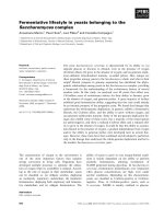
-charged column. Figure 1A shows the last
O
2
sensitivity of the regulatory hydrogenase HupUV O. Duche
´
et al.
3900 FEBS Journal 272 (2005) 3899–3908 ª 2005 FEBS
step of the purification of wild-type and mutated pro-
teins, with the two subunits in a stoichiometric ratio.
Under native conditions (Fig. 1B), the two proteins
displayed the same pattern. The molecular masses of
the two bands ( 80 and 170 kDa) were estimated by
runs in native gels with different acrylamide concentra-
tions [26], as previously described [13]. They corres-
pond, respectively, to the dimeric form, HupUV, and
the tetrameric form, Hup(UV)
2
, both of which exhibit
hydrogenase activity (see below). These results suggest
that the quaternary structure of the mutated protein
was well conserved.
Hydrogenase activity and stability of purified
mutated HupUV protein
We observed that, after breakage in a French Press of
BSE16 cells producing mutated protein, hydrogenase
activity decreased noticeably in the soluble extracts
(which contained only HupUV, HupSL being retained
in the membrane fraction), suggesting inactivation by
air. The hydrogenase activity of the purified proteins,
assayed by H
2
uptake in the presence of benzyl violo-
gen, was about fivefold lower in the mutant protein
OD7 than that of wild-type HupUV [9.2 vs. 2.0 lmol
reduced benzyl viologenÆmin
)1
Æ(mg protein)
)1
). To
check the stability of the proteins in the presence of
O
2
, soluble extracts and purified proteins were stored
in air at 4 °C for several days, and, each day,
H
2
-uptake activity was determined by measuring ben-
zyl viologen reduction in an aliquot. As shown in
Fig. 2, the mutant OD7 had lost 80% of its activity
after 3 days under air in soluble extracts, and in
1.5 days when purified. The mutant protein was par-
tially protected when stored under N
2
; it exhibited
50% activity during the same time (as compared
with 20% under air) (not shown). Thus in the mutant
protein, there was specific inactivation of the catalytic
activity by O
2
, but the mutation could also modify the
conformation of the protein, rendering it unstable.
H–D exchange activity catalysed by wild-type and
mutated HupUV proteins
The effect of O
2
on the activity of aerobically purified
HupUV proteins was then assessed directly by a MS
method monitoring continuously the H–D exchange in
either the absence or presence of O
2
. The results are
given in Table 1.
In the wild-type HupUV protein, the activity and
the rate of HD and H
2
formation were similar under
aerobic and anaerobic conditions. These results are in
agreement with a previous study reporting that the H–D
exchange reaction catalyzed by the HupUV protein was
high in the presence of O
2
[18]. In this study, we
observed that the activity of the mutant OD7 was repro-
ducibly twofold higher in the absence of O
2
than under
aerobiosis [1.3 ± 0.3 vs. 0.7 ± 0.1 lmolÆmin
)1
Æ(mg
protein)
)1
, respectively]. In all cases, the rate of HD
formation was twice that of H
2
formation.
To check whether the low activity of the mutant
protein OD7 was due to the fact that O
2
could now
reach the active site and partially inactivate it, we
repeated the assays in the presence of reduced methyl
viologen. It is well known that standard hydrogenases
need to be activated by reduction to become catalyti-
cally competent [27]. The activities of the aerobically
purified proteins were assayed under anaerobiosis by
H–D exchange, as described above, and then 0.16 mm
MV
+
was added. Table 1 shows that addition of
MV
+
did not further activate the HupUV protein,
whereas, interestingly, the activity of the OD7 protein
AB
Fig. 1. SDS ⁄ polyacrylamide gel (A) and native gel (B) of wild-type
and mutated HupUV proteins. (A) Cell extracts from 5 L were puri-
fied on two successive Ni
2+
-charged columns. Then 10 lL of the
pools purified on the second HiTrap column and eluted with
250 m
M imidazole were loaded on to an SDS ⁄ 12% polyacrylamide
gel. Lane 1, wild-type; lane 2, mutant. (B) An 8-lg sample of each
protein was run on a native polyacrylamide gel and stained with
Coomassie Brilliant Blue. Lane 1, wild-type; lane 2, mutant.
Fig. 2. Inactivation of wild-type and mutated HupUV proteins in air.
Soluble extracts obtained after centrifugation of sonicated cells at
50 000 r.p.m. for 1 h (A) and purified proteins (B) were kept at 4 °C
under air, and H
2
-uptake hydrogenase activity was assayed every
day during 1 week. Wild-type, diamonds; mutant, circles. Data rep-
resent the mean results from two or three independent assays.
O. Duche
´
et al. O
2
sensitivity of the regulatory hydrogenase HupUV
FEBS Journal 272 (2005) 3899–3908 ª 2005 FEBS 3901
was fourfold higher after reduction by MV
+
. This
suggests that the mutated HupUV protein was inacti-
vated during aerobic purification, but partial activity
could be recovered under reducing conditions, this
activity remaining threefold lower than that of the
wild-type. Figure 3 illustrates the effect of reduced
MV
+
on H–D exchange activity catalysed by the puri-
fied HupUV proteins (note the difference in the scale).
In vitro interaction of the mutated HupUV protein
with HupT
In a previous study, we showed that HupUV (probably
the HupU subunit) interacts with the N-terminal
domain of the histidine kinase HupT to transduce the
signal of H
2
availability [13]. We therefore addressed
the question of whether the mutation of some amino
acids of HupUV modifies the conformation of the pro-
tein and consequently its interaction with the histidine
kinase. Mutated and wild-type HupUV proteins were
incubated with HupT, and their interactions visualized
directly on native acrylamide gels (Fig. 4). When
HupT was incubated with any one of the HupUV
proteins, a new active band appeared with a higher
molecular mass, representing a Hup(UV)
2
–HupT
2
complex, as previously determined for wild-type
HupUV [13], and the amount of free HupUV decreas-
ed. There was no difference in migration between the
two HupUV–HupT complexes, suggesting that the
mutations did not substantially modify the interaction.
Table 1. H–D exchange activity and rate of H
2
and HD formation by wild-type and mutated HupUV proteins of R. capsulatus. The values are
initial rates corrected for gas consumption by the mass spectrometer. Activity and H
2
or HD rate of formation are expressed as lmol formedÆ
min
)1
Æ(mg protein)
)1
as described [44]. Assays under aerobiosis and anaerobiosis were performed separately. When noted, reduced methyl
viologen (MV
+
) was present at 0.16 mm. Data are means from two or three independent experiments, with variation of less than 15%.
Proteins
Aerobiosis Anaerobiosis Anaerobiosis + MV
+
Activity
HD
formation
H
2
formation Activity
HD
formation
H
2
formation Activity
HD
formation
H
2
formation
HupUV (wild-type) 18.5 ± 0.6 8.0 ± 0.8 4.7 ± 0.1 15.6 ± 1.2 7.0 ± 0.5 4.0 ± 0.2 15.5 ± 1.7 7.0 ± 0.7 3.7 ± 0.5
OD7 (mutant) 0.7 ± 0.1 0.3 ± 0.1 0.2 ± 0.1 1.3 ± 0.3 0.7 ± 0.1 0.3 ± 0.1 5.2 ± 1.3 2.5 ± 0.6 1.2 ± 0.5
Fig. 3. Reductive activation by reduced MV
+
in HupUV proteins assayed by MS. The vessel containing 1.5 mL Mes buffer was saturated
with D
2
and made anaerobic as explained in Experimental procedures. Then 3 lg wild-type or mutated HupUV protein was added. Exchange
activity was assayed under anaerobiosis for 1–2 min, then Zn-reduced MV
+
was added and the activity followed for 2 or more minutes. (A)
Wild-type HupUV protein; (B) mutant OD7 protein.
Fig. 4. Interaction between HupT and HupUV proteins. Lanes 1 and
2, wild-type HupUV; lanes 3 and 4, mutant OD7. HupT was present
in lanes 2 and 4.
O
2
sensitivity of the regulatory hydrogenase HupUV O. Duche
´
et al.
3902 FEBS Journal 272 (2005) 3899–3908 ª 2005 FEBS
The mutated HupUV protein can regulate the
synthesis of HupSL hydrogenase
The next question we addressed was to check whether
the O
2
-sensitive mutated protein was able to function
in vivo, i.e. to transduce the H
2
signal, and in the
absence of H
2
, to repress hydrogenase synthesis, even
in presence of O
2
. The plasmids pSE103 and pOD7,
which expressed hupUV genes from the nif promoter,
were not suitable for in vivo experiments, because this
promoter is not active under aerobiosis [28]. For this
reason, the plasmids pSE60 and pOD15, in which the
hupUV genes were cloned under control of the fruc-
tose-induced fru promoter, were constructed and used
to complement the hupUV mutant strain BSE16. The
complemented cells were grown under aerobiosis or
anaerobiosis in the presence of 3 mm fructose and in
either the presence (derepressing conditions) or absence
(repressing conditions) of H
2
.H
2
was produced endo-
genously as a by-product of nitrogenase activity during
anaerobic growth in malate ⁄ glutamate (MG) medium.
The presence of O
2
and ammonia inhibits activity and
synthesis of nitrogenase; when indicated, H
2
was added
externally during aerobic growth in malate ⁄ ammonia
(MN) medium. Table 2 summarizes the results. In all
conditions tested, the BSE16 mutant strain exhibited a
high level of hydrogenase activity compared with the
wild-type B10, because, in the absence of the HupUV
protein, which is part of the repressing system, hupSL
gene expression remains fully activated [12]. As shown
in Table 2, mutated HupUV protein, produced from
plasmid pOD15, was able to repress hydrogenase syn-
thesis in the absence of H
2
to the same extent as the
wild-type protein. Thus, the availability of H
2
was still
detected even when growth was carried out in the pres-
ence of O
2
.
Discussion
In regulatory hydrogenases, it has been hypothesized
that bulky residues lining the gas channel participate
in O
2
resistance by blocking O
2
access to the active site
[25]. To check this hypothesis, we replaced, by site-
directed mutagenesis, two amino acids that line the gas
access channel, Ile65 and Phe113, with Val and Leu,
respectively. Interestingly, these replacements rendered
the protein O
2
sensitive, demonstrating that these resi-
dues are involved in O
2
sensitivity of the RH. This was
corroborated by experiments showing that the H–D
exchange activity of the mutant protein increased
greatly in the presence of reduced MV, at variance
with that of the wild-type protein. However, even after
reductive activation, the hydrogenase activity of puri-
fied mutated HupUV protein remained twice as low as
that of the wild-type, suggesting that O
2
may also irre-
versibly inactivate the active site. Another explanation
is that the mutations could also modify the structure
around the active site and⁄ or the binding of ligands,
thus decreasing the catalytic efficiency of the enzyme.
Our results suggest that, in vivo, the mutated HupUV
protein is protected from O
2
inactivation, as it exhibited
about the same hydrogenase activity as the wild-type
one. This was further corroborated by complementation
experiments, which showed that the OD7 mutated pro-
tein produced in a hupUV mutant was able to restore
the regulation of HupSL synthesis. This implies that it
was able to transmit the information about the availab-
ility of H
2
to the histidine kinase, HupT. Indeed, we
showed that the mutated protein was able to interact
with HupT in vitro at the same HupUV ⁄ HupT ratios
and under the same conditions as the wild-type one [13].
‘Standard’ [NiFe] hydrogenases are known to be
reversibly inactivated by O
2
.O
2
could affect either the
enzyme during the activation step and ⁄ or the active
enzyme in the catalytic cycle. For instance, in the
hydrogenase from Allochromatium vinosum, it has been
observed that O
2
added during the activation step of the
ready enzyme increases the lag phase without preventing
the activation [29]. On the other hand, when added to
the active enzyme, O
2
would react directly with the act-
ive NiFe site, thus inactivating the reaction with H
2
[30].
It should be noted that the occurrence of direct binding
of O
2
to the active NiFe site is under debate and was not
observed for hydrogenase from Desulfovibrio gigas [8].
Some hydrogenases, however, are able to consume
H
2
in the presence of O
2
, and exhibit noticeable resist-
ance to this gas. The best-known enzyme is the soluble
Table 2. Hydrogenase activities of the wild-type B10 and hupUV
BSE16 strains from R. capsulatus, complemented with wild-type
and mutated hupUV genes. Cells were grown overnight at 30 °C
anaerobically in the light (MN or MG medium) or aerobically in the
dark (MN or MN medium + 10% H
2
)toanA
660
of 1.5. In MG
medium, H
2
was evolved from nitrogenase activity. Fructose
(3 m
M) was added at the beginning of growth at an A
660
of 0.6.
Hydrogenase activity was assayed with methylene blue and was
expressed as lmol reduced MBÆh
)1
Æ(mg protein)
)1
. The values are
the means from at least three independent experiments.
Strains
Hydrogenase activity
Anaerobiosis Aerobiosis
MN MG (H
2
)MN MN+H
2
B10 19 ± 10 44 ± 3 24 ± 1 80 ± 17
BSE16 92 ± 21 36 ± 10 82 ± 8 116 ± 14
BSE16 (pSE60) 16 ± 13 52 ± 12 18 ± 3 65 ± 16
BSE16 (pOD15) 17 ± 2 68 ± 9 16 ± 1 64 ± 17
O. Duche
´
et al. O
2
sensitivity of the regulatory hydrogenase HupUV
FEBS Journal 272 (2005) 3899–3908 ª 2005 FEBS 3903
NAD-linked hydrogenase (SH) from the strictly aero-
bic bacterium R. eutropha [31]. This hydrogenase har-
bours an active site different from that of ‘standard’
hydrogenases with two additional CN
–
groups, tenta-
tively assigned to the Fe atom and the Ni atom [6,32].
It has been hypothesized that these two CN
–
groups
may shield the active site from O
2
attack by steric hin-
drance [32,33]. The Ni-bound CN
–
seems to be respon-
sible for the O
2
insensitivity of the enzyme, and is
linked to the presence of the hypX gene [34,35], found
also in other aerobic bacteria such as Rhizobium [36].
Indeed, in SH purified from an HypX
–
strain, the cata-
lytic turnover (the hydrogenase activity) was shown to
be independent of the presence of O
2
, but the enzyme
was irreversibly inactivated if O
2
was present during
the autocatalytic activation [35], probably because of
formation of some peroxide or superoxide. In a recent
study, a mutant of HoxH, the active-site-containing
subunit of the SH, was constructed by replacement of
Leu118 with Phe; this mutation led to an O
2
-sensitive
phenotype, and it was postulated that this bulky resi-
due impaired the incorporation of the Ni-linked CN
–
,
thus conferring O
2
sensitivity [37]. Interestingly, this
mutated HoxH subunit contains the two bulky
residues corresponding to Phe113 and Ile65 of the
wild-type HupUV proteins, and conserved in the other
H
2
-sensing RHs, such as R. eutropha HoxBC [21] and
B. japonicum HupUV [15]. Therefore, and paradoxic-
ally, the presence of these residues, which seem in our
study to confer O
2
resistance on HupUV, did not pro-
tect the protein against O
2
in the HypX
–
mutant.
Although the active site of HupUV from R. capsula-
tus has not yet been studied, that of the homologous
protein, HoxBC, of R. eutropha was shown to be very
similar to that of standard hydrogenases, with a Fe
atom liganded by 1 CO and 2 CN
–
[16], and the binding
of an hydride to Ni and Fe after H
2
reduction has
recently been demonstrated [38]. However, in contrast
with standard hydrogenases, the RH exists only as two
redox forms, i.e. ready oxidized and reduced. The O
2
and MV
+
responses observed in the mutant HupUV
protein suggest that it has reached unready states, and
further studies will be needed to determine which ones.
In a recent study using X-ray absorption spectroscopy,
Haumann et al. [39] suggested that the specific Ni
co-ordination may also be crucial to the O
2
insensitivity
of the R. eutropha RH. In particular, the number of S
ligands was decreased by one upon formation of the
active state, but binding of O
2
to the active site was pre-
vented because an O ⁄ N ligand from an amino acid was
already bound at the free position at the Ni site.
In any case, it appears that in the O
2
-resistant
hydrogenases, O
2
is prevented from contacting the
active site, even if various mechanisms are certainly
involved. In the case of the regulatory HupUV protein,
our results favour the hypothesis of Volbeda et al.
[25], which explains the O
2
resistance of RHs by
limited accessibility of the active site to O
2
. In the
mutated protein, O
2
has access to the active bimetallic
site, which would remain in the inactive form, and,
consequently, this protein exhibits some features of the
standard hydrogenases that must be activated in the
presence of H
2
or a reductant [27]. Our conclusions
are strengthened by a recent paper from Friedrich’s
group [40], which shows the O
2
sensitivity of HoxBC
proteins mutated in residues lining the gas access chan-
nel in R. eutropha. In this respect, the comparative
analysis of wild-type, O
2
-resistant and mutated,
O
2
-sensitive HupUV proteins by biophysical methods
may lead to the improved understanding of the mecha-
nisms of O
2
resistance ⁄ sensitivity in [NiFe] hydrogen-
ases in general. RHs that are insensitive to O
2
and, as
isolated, ready to function are potentially of great bio-
technological interest, but their activity is low. When
the basis of their O
2
resistance is understood, it will be
possible to design a hydrogenase that exhibits high
activity together with O
2
insensitivity.
Experimental procedures
Bacterial strains and plasmids
The strains and plasmids used in this study are listed in
Table 3. R. capsulatus strains were grown heterotrophically
at 30 °C under anaerobiosis in the light or under aerobiosis
in the dark with shaking, in MG medium (7 mm glutamate,
30 mmdl-malate) or MN medium (7 mm ammonium sul-
fate, 30 mmdl-malate) [19]. Escherichia coli strains were
grown at 37 °C in Luria–Bertani medium. Antibiotics were
used at the following concentrations: 100 (ampicillin) and
10 (tetracycline) mgÆL
)1
for E. coli and 1 (tetracycline)
mg ⁄ L
)1
for R. capsulatus.
DNA manipulation and bacterial mating
Standard recombinant DNA techniques were performed as
described by Sambrook et al. [41]. Restriction enzymes were
used as indicated by the manufacturers. Triparental matings
were performed with the plasmid helper pRK2013 as des-
cribed previously [42].
Construction of plasmids with mutations
in the hupV gene
A 3.2-kb fragment bearing hupUV genes cloned into pUC18
was used to modify two amino acids with the QuikChange
O
2
sensitivity of the regulatory hydrogenase HupUV O. Duche
´
et al.
3904 FEBS Journal 272 (2005) 3899–3908 ª 2005 FEBS
Site-Directed Mutagenesis Kit (Stratagene, La Jolla, CA,
USA). The mutagenesis leading to plasmid pOD585 was
carried out in two successive steps with the following sets of
oligonucleotides as primers: UV5 (5¢-CGCGGATCTGCG
TCTGCTCGATCTCGC-3¢) and UV6 (5¢-GCGAGATCG
AGCAGACGCCGCAGATCCGCG-3¢) to replace Ile65
from HupV with Val; UV7 (5¢-GCATTTCAACCTCCT
GTTCATGCCCGATTTC-3¢) and UV8 (5¢-GAAATCG
GGCATGAACAGGAGGTTGAAATGC-3¢) to replace
Phe113 from HupV with Leu. The 3.2-kb fragment corres-
ponding to the mutated hupUV genes was excised from plas-
mid pOD585 with NdeI–BamHI and cloned into pSE50
digested with the same enzymes in place of the wild-type
hupUV genes, leading to plasmid pSE504. Plasmid pSE102
was cleaved with NcoI–BamHI, to clone 3.2-kb fragments
from pSE50 and pSE504 digested with the same enzymes,
leading to plasmids pSE103 and pOD7, respectively, which
were introduced into R. capsulatus hupUV mutant BSE16 or
hupSL mutant JP91 by conjugation. From these plasmids,
the HupU subunit will carry an N-terminal His
6
tag for easy
purification of the HupUV complex.
Purification of the His
6
-HupUV proteins
In the plasmids pSE103 and pOD7, wild-type and
mutated hupUV genes were expressed from the nifHDK
promoter. For this reason, cells (from 5 L culture) were
grown under conditions allowing strong expression of the
nif promoter (MG medium, anaerobiosis, under light).
Proteins were purified on a HiTrap chelating column
(Amersham Pharmacia Biotech, Piscataway, NJ, USA) as
described previously [13]. Elution of the 5-mL column
with buffer containing 100 mm imidazole gave an active
pool, which was concentrated on a 1-mL column by elu-
tion with 250 mm imidazole in the buffer. The pools were
dialyzed three times in 25 mm Tris ⁄ HCl (pH 8) contain-
ing 10% (v ⁄ v) glycerol and 150 mm NaCl, at 4 °C. The
purified proteins were divided into aliquots and stored at
)80 °C.
Enzyme assays
Hydrogenase activity was assayed by the rate of H
2
uptake or H–D exchange. H
2
uptake was determined spec-
trophotometrically in 20 mm Tris ⁄ HCl buffer (pH 8),
either in whole cells with 0.15 mm methylene blue (MB)
as artificial electron acceptor, at A
565
, or in cell extracts
and purified proteins with 2 mm benzyl viologen (BV), at
A
555
[43]. In native gels, hydrogenase activity was revealed
by incubating the gels under H
2
for 10–40 min in 20 mm
Tris ⁄ HCl buffer (pH 8), containing 2 mm BV. The reac-
tion was stabilized by adding 1 mm triphenyltetrazolium
chloride. The H–D exchange reaction was measured at
30 °C and determined by a MS method as previously des-
cribed in detail [18,4]. Briefly, the reaction vessel was filled
with 1.5 mL Mes buffer (50 mm, pH 6) and then sparged
with D
2
until saturation, and the vessel was closed. Then
3 lg purified wild-type or mutated HupUV proteins were
introduced into the vessel, and the changes in D
2
,HD
and H
2
were monitored by scanning masses 4, 3 and 2,
Table 3. Bacterial strains and plasmids used in this study.
Strain or plasmid Relevant characteristics Source or reference
Strains
R. capsulatus
B10 Wild-type [45]
BSE16 hupUV Hup
c
[12]
JP91 hupSL Hup
–
[46]
Plasmids
pUC18 Ap
r
[47]
pFRK-I Ap
r
Ble
r
Gm
r
Km
r
; fruP fusion vector [48]
pRK2013 Km
r
; plasmid helper [49]
pSE50 Ap
r
; pET-15b with 3.2 kb NdeI-SalI insert
containing hupUV
[12]
pSE102 Tc
r
; pnif expression vector [13]
pPHU231 Tc
r
; pRK290 with a 388-bp HaeII insert
containing pUC18 polylinker
P. Hu
¨
bner,
unpublished
observations
pOD585 Ap
r
; pUC18 with mutated hupUV genes This work
pSE504 Ap
r
; pSE50 with 3.2-kb NdeI-BamHI from pOD585 This work
pSE103 Tc
r
; pSE102 with 3.2-kb NcoI-BamHI from pSE50 This work
pOD7 Tc
r
; pSE102 with 3.2-kb NcoI-BamHI from pSE504 This work
pOD12 Ap
r
Gm
r
; pFRK-I with a 3.2-kb NdeI-BamHI from pOD585 This work
pSE60 Tc
r
; pPHU234 with 6.2-kb HindIII containing hupUV [12]
pOD15 Tc
r
; pPHU231 with 6-kb HindIII-BamHI from pOD12 This work
O. Duche
´
et al. O
2
sensitivity of the regulatory hydrogenase HupUV
FEBS Journal 272 (2005) 3899–3908 ª 2005 FEBS 3905
respectively. When required, the medium was made aero-
bic by the addition of H
2
O
2
(5 lL 0.3% H
2
O
2
) decom-
posed by the addition of catalase (500 U) thus liberating
O
2
, or was made anaerobic by the addition of catalase
(500 U), glucose (5 mm) and glucose oxidase (40 U). Zn-
reduced methyl viologen (MV
+
0.16 mm) was added to
the anaerobic medium in some experiments. The rates of
D
2
consumption and H
2
and HD production were correc-
ted for simultaneous consumption by the spectrometer.
This consumption, which showed first-order kinetics, was
assayed in the absence of protein.
In vitro interaction of HupUV and HupT
The proteins (50 pmol HupUV and 250 pmol HupT) were
incubated for 10 min at 30 °C in buffer containing 10 mm
Tris ⁄ HCl (pH 8), 20 mm NaCl, 10% (v ⁄ v) glycerol, 1 mm
EDTA and 1 mm dithiothreitol as previously described [13].
Proteins were then run on a native acrylamide gel in
0.5 · Laemmli buffer, and the gel was revealed by hydro-
genase activity staining in the presence of BV.
Complementation of hupUV mutant with
mutated hupUV genes
The mutated hupUV genes excised from plasmid pOD585
by NdeI–BamHI digestion were used to replace a 1.7 kb
NdeI–BamHI fragment (deletion of the Ble
r
Km
r
cartridge)
of the plasmid pFRK-I, leading to plasmid pOD12.
pFRK-I contains a fructose-activated promoter, pfru, from
R. capsulatus. From pOD12, the HindIII–BamHI fragment
containing mutated hupUV genes downstream of pfru was
cloned into the broad host range plasmid pPHU231 diges-
ted with the same enzymes. The resulting plasmid, pOD15,
was introduced into the R. capsulatus hupUV mutant
BSE16 by triparental conjugation [13].
Acknowledgements
We thank P. M. Vignais and M. Satre for critical read-
ing of the manuscript. We also thank P. Carrier for
excellent technical assistance. O.D. was supported by a
two-year postdoctoral grant from the Commissariat a
`
l’Energie Atomique (CEA). The work was supported
by research grants from the CEA, the Centre National
de la Recherche Scientifique (CNRS: ACI ‘Energie Con-
ception Durable’) and the Universite
´
Joseph Fourier
(UJF) de Grenoble.
References
1 Vignais PM, Billoud B & Meyer J (2001) Classification
and phylogeny of hydrogenases. FEMS Microbiol Rev
25, 455–501.
2 Vignais PM & Colbeau A (2004) Molecular biology
of microbial hydrogenases. Curr Issues Mol Biol 6,
159–188.
3 Adams MW (1990) The structure and mechanism of
iron-hydrogenases. Biochim Biophys Acta 1020,
115–145.
4 Albracht SPJ (2001) Spectroscopy-the functional puzzle.
In Hydrogen as a Fuel. Learning from Nature (Cammack
R, Frey M & Robson R, eds), pp. 110–158. Taylor &
Francis, London.
5 Cammack R (2001) The catalytic machinery. In Hydro-
gen as a Fuel. Learning from Nature (Cammack, R, Frey
M & Robson R, eds), pp. 159–180. Taylor & Francis,
London.
6 Happe RP, Roseboom W, Pierik AJ, Albracht SPJ &
Bagley KA (1997) Biological activation of hydrogen.
Nature 385, 126.
7 Bleijlevens B, Faber BW & Albracht SPJ (2001) The
[Ni-Fe] hydrogenase from Allochromatium vinosum was
studied in EPR-detectable states: H ⁄ D exchange experi-
ments that yield new information about the structure of
the active site. J Biol Inorg Chem 6, 763–769.
8 Carepo M, Tierney DL, Brondino CD, Yang TC,
Pamplona A, Teiser D, Moura I, Moura JJG & Hoff-
man BM (2002)
17
O ENDOR detection of a solvent-
derived Ni-(OH
x
) -Fe bridge that is lost upon activation
of the hydrogenase from Desulfovibrio gigas. JAm
Chem Soc 124, 281–286.
9 Volbeda A, Garcin E, Piras C, De Lacey AL, Fernan-
dez VM, Hatchikian EC, Frey M & Fontecilla-Camps
JC (1996) Structure of the [NiFe]hydrogenase active site:
evidence for uncommon Fe-ligands. J Am Chem Soc
118, 12989–12996.
10 Garcin E, Vernede X, Hatchikian EC, Volbeda A, Frey
M & Fontecilla-Camps JC (1999) The crystal structure
of a reduced [NiFeSe]hydrogenase provides an image of
the activated catalytic center. Structure Fold Des 7,
557–566.
11 Higuchi Y, Ogata H, Miki K, Yasuoka N & Yagi T
(1999) Removal of the bridging ligand atom at the
Ni-Fe active site of [NiFe]hydrogenase upon reduction
with H
2
, as revealed by X-ray structure analysis at 1.4
A
˚
resolution. Structure Fold Des 7, 549–556.
12 Elsen S, Colbeau A, Chabert J & Vignais PM (1996)
The hupTUV operon is involved in negative control of
hydrogenase synthesis in Rhodobacter capsulatus.
J Bacteriol 178, 5174–5181.
13 Elsen S, Duche
´
O & Colbeau A (2003) Interaction
between the H
2
sensor HupUV and the histidine kinase
HupT controls HupSL hydrogenase synthesis in Rhodo-
bacter capsulatus. J Bacteriol 185, 7111–7119.
14 Vignais PM, Dimon B, Zorin NA, Colbeau A & Elsen
S (1997) HupUV proteins of Rhodobacter capsulatus can
bind H
2
: evidence from the H-D exchange reaction.
J Bacteriol 179, 290–292.
O
2
sensitivity of the regulatory hydrogenase HupUV O. Duche
´
et al.
3906 FEBS Journal 272 (2005) 3899–3908 ª 2005 FEBS
15 Black LK, Fu C & Maier RJ (1994) Sequences and
characterization of hupU and hupV genes of Bradyrhizo-
bium japonicum encoding a possible nickel-sensing com-
plex involved in hydrogenase expression. J Bacteriol
176, 7102–7106.
16 Bernhard M, Buhrke T, Bleijlevens B, De Lacey AJ,
Fernandez VM, Albracht SPJ & Friedrich B (2001) The
H
2
sensor of Ralstonia eutropha. Biochemical character-
istics, spectroscopic properties, and its interaction with
a histidine protein kinase. J Biol Chem 276, 15592–
15597.
17 Kleihues L, Lenz O, Bernhard M, Buhrke T & Friedrich
B (2000) The H
2
sensor of Ralstonia eutropha is a mem-
ber of the subclass of regulatory hydrogenases. J Bacter-
iol 182, 2716–2724.
18 Vignais PM, Cournac L, Hatchikian EC, Elsen S,
Serebryakova N, Zorin NA & Dimon B (2002) Con-
tinuous monitoring of the activation and activity of
[NiFe]-hydrogenases by membrane-inlet mass spectro-
metry. Int J Hydrogen Energy 27, 1441–1448.
19 Colbeau A & Vignais PM (1992) Use of hupS, lacZ
gene fusion to study regulation of hydrogenase expres-
sion in Rhodobacter capsulatus : stimulation by H
2
.
J Bacteriol 174, 4258–4264.
20 Dischert W, Vignais PM & Colbeau A (1999) The
synthesis of Rhodobacter capsulatus HupSL hydrogenase
is regulated by the two-component HupT ⁄ HupR sys-
tem. Mol Microbiol 34 , 995–1006.
21 Buhrke T, Lenz O, Porthun A & Friedrich B (2004)
The H
2
-sensing complex of Ralstonia eutropha: interac-
tion between a regulatory [NiFe]hydrogenase and a his-
tidine protein kinase. Mol Microbiol 51, 1677–1689.
22 Vignais PM, Dimon B, Zorin NA, Tomiyama M &
Colbeau A (2000) Characterization of the hydrogen-
deuterium exchange activities of the energy-transducing
HupSL hydrogenase and H
2
-signaling HupUV hydro-
genase in Rhodobacter capsulatus. J Bacteriol 182, 5997–
6004.
23 Fontecilla-Camps JC, Frey M, Garcin E, Hatchikian C,
Montet Y, Piras C, Vernede X & Volbeda A (1997)
Hydrogenase: a hydrogen metabolizing enzyme. What
do the crystal structures tell us about its mode of action.
Biochimie 79, 661–666.
24 Montet Y, Amara P, Volbeda A, Vernede X, Hatchik-
ian EC, Field MJ, Frey M & Fontecilla-Camps JC
(1997) Gas access to the active site of NiFe hydro-
genases probed by X-ray crystallography and molecular
dynamics. Nat Struct Biol 4, 523–526.
25 Volbeda A, Montet Y, Vernede X, Hatchikian EC &
Fontecilla-Camps JC (2002) High-resolution crystallo-
graphic analysis of Desulfovibrio fructosovorans [NiFe]
hydrogenase. Int J Hydrogen Energy 27, 1449–1461.
26 Bryan JK (1977) Molecular weights of protein multi-
mers from polyacrylamide gel electrophoresis. Anal
Biochem 78, 513–519.
27 Cammack R (2001) Hydrogenases and their activities.
In Hydrogen as a Fuel. Learning from Nature (Cammack
R, Frey M & Robson R, eds), pp. 73–92. Taylor &
Francis, London.
28 Hu
¨
bner P, Willison JC, Vignais PM & Bickle TA (1991)
Expression of regulatory nif genes in Rhodobacter capsu-
latus. J Bacteriol 173, 2993–2999.
29 Kurkin S, George SJ, Thorneley RNF & Albracht
SPJ (2004) Hydrogen-induced activation of the [NiFe]-
hydrogenase from Allochromatium vinosum as studied
by stopped-flow infrared spectroscopy. Biochemistry
43, 6820–6831.
30 George SJ, Kurkin S, Thorneley RNF & Albracht SPJ
(2004) Reaction of H
2
, CO, and O
2
with active [NiFe]-
hydrogenase from Allochromatium vinosum. A stopped-
flow infrared study. Biochemistry 43, 6808–6819.
31 Schneider K, Schlegel HJ, Cammack R & Hall DO
(1979) The iron-sulphur centres of soluble hydrogenase
from Alcaligenes eutrophus. Biochim Biophys Acta 578,
445–461.
32 Van der Linden E, Burgdorf T, Bernhard B, Bleijlevens
B, Friedrich B & Albracht SPJ (2004) The soluble
[NiFe]-hydrogenase from Ralstonia eutropha contains
four cyanides in its active site, one of which is respon-
sible for the insensitivity towards oxygen. J Biol Inorg
Chem 9, 616–626.
33 Happe RP, Roseboom W, Egert G, Friedrich CG, Mas-
sanz C, Friedrich B & Albracht SPJ (2000) Unusual
FTIR and EPR properties of the H
2
-activating site of
the cytoplasmic NAD-reducing hydrogenase from
Ralstonia eutropha. FEBS Lett 466, 259–263.
34 Buhrke T & Friedrich B (1998) hoxX (hypX) is a func-
tional member of the Alcaligenes eutrophus hyp gene
cluster. Arch Microbiol 170, 460–463.
35 Bleijlevens B, Buhrke T, Van der Linden E, Friedrich B
& Albracht SPJ (2004) The auxiliary protein HypX pro-
vides oxygen tolerance to the soluble [NiFe]-hydroge-
nase of Ralstonia eutropha H16 by way of a cyanide
ligand to nickel. J Biol Chem 279, 46686–46689.
36 Rey L, Fernandez D, Brito B, Hernando Y, Palacios
JM, Imperial J & Ruiz-Argueso T (1996) The hydroge-
nase gene cluster of Rhizobium leguminosarum bv viciae
contains an additional gene (hypX) which encodes a
protein with sequence similarity to the N10-formyltetra-
hydrofolate-dependent enzyme family and is required
for nickel-dependent hydrogenase processing and activ-
ity. Mol General Genet 252, 237–248.
37 Burgdorf T, De Lacey AL & Friedrich B (2002) Func-
tional analysis by site-directed mutagenesis of the
NAD
+
-reducing hydrogenase from Ralstonia eutropha.
J Bacteriol 184, 6280–6288.
38 Brecht M, Van Gastel M, Burhke T, Friedrich B &
Lubitz W (2003) Direct detection of a hydrogen ligand
in the [NiFe] center of the regulatory H
2
-sensing hydro-
genase from Ralstonia eutropha in its reduced state by
O. Duche
´
et al. O
2
sensitivity of the regulatory hydrogenase HupUV
FEBS Journal 272 (2005) 3899–3908 ª 2005 FEBS 3907
HYSCORE and ENDOR spectroscopy. J Am Chem
Soc 125, 13075–13083.
39 Haumann M, Porthun A, Buhrke T, Liebisch P, Meyer-
Klaucke W, Friedrich B & Dau H (2003) Hydrogen-
induced structural changes at the nickel site of the
regulatory [NiFe]hydrogenase from Ralstonia eutropha
detected by X-ray absorption spectroscopy. Biochemistry
42, 11004–11015.
40 Buhrke T, Lenz O, Krauss N & Friedrich B (2005) Oxy-
gen tolerance of the H
2
-sensing [NiFe] hydrogenase
from Ralstoniae utropho H16 is based on limited access
of oxygen to the active site. J Biol Chem in press.
41 Sambrook J, Fritsch EF & Maniatis T (1989) Molecular
Cloning: a Laboratory Manual, 2nd edn. Cold Spring
Harbor Laboratory Press, Cold Spring, N.Y.
42 Colbeau A, Godfroy A & Vignais PM (1986) Cloning
of DNA fragments carrying hydrogenase genes of Rho-
dopseudomonas capsulata. Biochimie 68, 147–155.
43 Colbeau A, Kelley BC & Vignais PM (1980) Hydro-
genase activity in Rhodopseudomonas capsulata: relation-
ship with nitrogenase activity. J Bacteriol 144, 141–148.
44 Cournac L, Guedeney G, Peltier G & Vignais PM
(2004) Sustained photoevolution of molecular hydrogen
in a mutant of Synechocystis sp. strain PCC 6803 defi-
cient in the type I NADPH-dehydrogenase complex.
J Bacteriol 186, 1737–1746.
45 Marrs B (1974) Genetic recombination in Rhodopseudo-
monas capsulata. Proc Natl Acad Sci USA 71, 971–973.
46 Colbeau A, Magnin JP, Cauvin B, Champion T &
Vignais PM (1990) Genetic and physical mapping of an
hydrogenase gene cluster from Rhodobacter capsulatus.
Mol General Genet 220, 393–399.
47 Yanisch-Perron C, Vieira J & Messing J (1985)
Improved M13 phage cloning vectors and host strains:
nucleotide sequences of the M13mp18 and pUC19 vec-
tors. Gene 33, 103–119.
48 Duport C, Meyer C, Naud I & Jouanneau Y (1994) A
new gene expression system based on a fructose-depen-
dent promoter from Rhodobacter capsulatus. Gene 145,
103–108.
49 Ditta G, Stanfield S, Corbin D & Helinski DP (1980)
Broad-host range DNA cloning system for gram-
negative bacteria: construction of a gene bank of
Rhizobium meliloti . Proc Natl Acad Sci USA 77,
7347–7351.
O
2
sensitivity of the regulatory hydrogenase HupUV O. Duche
´
et al.
3908 FEBS Journal 272 (2005) 3899–3908 ª 2005 FEBS