Tài liệu Báo cáo khoa học: Functional interaction between RNA helicase II⁄Gua and ribosomal protein L4 pptx
Bạn đang xem bản rút gọn của tài liệu. Xem và tải ngay bản đầy đủ của tài liệu tại đây (478.08 KB, 15 trang )
Functional interaction between RNA helicase II⁄Gua and
ribosomal protein L4
Hushan Yang, Dale Henning and Benigno C. Valdez
Department of Pharmacology, Baylor College of Medicine, Houston, Texas, USA
Ribosome biogenesis is a complicated cellular process
which occurs in the nucleolus [1]. The entire scenario
begins at the end of mitosis and includes ribosomal
DNA transcription, pre-ribosomal RNA (pre-rRNA)
modifications and processing as well as assembly of
rRNAs and ribosomal proteins into preribosome sub-
units which are then exported to the cytoplasm to
form the mature ribosomes [2]. Errors in this process
are reported to be associated with several diseases
[3–8]. The availability of genetic manipulations makes
ribosome biogenesis much better studied in yeast than
in higher eukaryotes such as mammalian and frog sys-
tems, resulting in the identification of more than 80
yeast ribosomal proteins and numerous trans-acting
elements including small nucleolar RNAs (snoRNAs)
as well as nonribosomal proteins. However, in mam-
malian systems, ribosome biogenesis is far from being
thoroughly understood due to the increased complex-
ity. To date, only a few nucleolus-localized nonribo-
somal proteins have been implicated in pre-rRNA
processing in mammalian cells and include B23 ⁄
NO38 ⁄ NPM [9], C23 ⁄ nucleolin [3], fibrillarin [10,11],
p120 [12], EBP1 [13], Bop1 [14] and p19
Arf
[15]. No
bona fide RNA helicase has been implicated in this
process in higher eukaryotes except RNA helicase
II ⁄ Gua [16,17].
RNA helicase II ⁄ Gua is a multifunctional nucleolar
protein with in vitro RNA-dependent ATPase activity,
ATP-dependent RNA helicase activity and GTP-stimu-
lated RNA foldase activity [17–19]. The presence of
both RNA unwinding and RNA folding activities in
two distinct domains of the same protein highly sug-
gests a role of Gua in rRNA biogenesis [19]. Using
antisense oligodeoxynucleotide and siRNA to down-
regulate Gua expression in Xenopus oocytes [16] and
mammalian cells [17], respectively, we demonstrated
that Gua is important for 18S and 28S rRNA produc-
tion in both systems. In addition, Gua was also showed
to participate in other major cellular activities such as
cell growth and differentiation [19,20], regulation of
Keywords
ribosomal protein; ribosomal RNA
biogenesis; RNA helicase; nucleolus
Correspondence
B. C. Valdez, Department of Pharmacology,
Baylor College of Medicine, Houston,
TX 77030, USA
Fax: +1 713 798 3145
Tel: +1 713 798 7908
E-mail:
(Received 11 April 05, revised 19 May 05,
accepted 9 June 05)
doi:10.1111/j.1742-4658.2005.04811.x
RNA helicase II ⁄ Gua is a multifunctional nucleolar protein involved in
ribosomal RNA processing in Xenopus laevis oocytes and mammalian cells.
Downregulation of Gua using small interfering RNA (siRNA) in HeLa
cells resulted in 80% inhibition of both 18S and 28S rRNA production.
The mechanisms underlying this effect remain unclear. Here we show that
in mammalian cells, Gua physically interacts with ribosomal protein L4
(RPL4), a component of 60S ribosome large subunit. The ATPase activity
of Gua is important for this interaction and is also necessary for the func-
tion of Gua in the production of both 18S and 28S rRNAs. Knocking
down RPL4 expression using siRNA in mouse LAP3 cells inhibits the pro-
duction of 47 ⁄ 45S, 32S, 28S, and 18S rRNAs. This inhibition is reversed
by exogenous expression of wild-type human RPL4 protein but not the
mutant form lacking Gua-interacting motif. These observations have sug-
gested that the function of Gua in rRNA processing is at least partially
dependent on its ability to interact with RPL4.
Abbreviations
aa, amino acid; GST, glutathione S-transferase; Gua, RNA helicase II ⁄ Gua; HA, hemagglutinin; IPTG, isopropylthio-b-
D-galactoside; NLS,
nuclear localization signal; NoLS, nucleolar localization signal; RPL4, Ribosomal Protein L4; rDNA, ribosomal DNA; rRNA, ribosomal RNA;
RNP, ribonucleoprotein; siRNA, small interfering RNA; snRNA, small nuclear RNA; snoRNA, small nucleolar RNA.
3788 FEBS Journal 272 (2005) 3788–3802 ª 2005 FEBS
c-Jun-mediated gene expressions [21] and in vitro clea-
vage by PIAS1 [22]. It is unclear whether the effect of
Gua on cell proliferation is due to its involvement
in ribosomal RNA production, c-Jun-mediated gene
expression or, as yet, other undiscovered mechanisms.
RNA helicases are believed to function through
regulation of RNA structure rearrangement and RNA-
RNA, RNA-protein or protein–protein interactions
[23,24]. To date, at least 18 putative ATP-dependent
RNA helicases have been suggested to contribute to
ribosome production in yeast S. cerevisiae [25]. It is
hypothesized that RNA helicases interact with other
trans-acting protein factors in preribosomal particles
during pre-rRNA processing and ribosome assembly
to modulate specific intracellular RNA structures
[26–28]. To test the hypothesis that Gua may function
by interacting with other nucleolar proteins in rRNA
production, we did immunoprecipitation and identified
ribosomal protein L4 physically interacting with Gua
in an RNA-independent manner. It was also demon-
strated that the interaction is necessary for the function
of Gua in 28S rRNA production in mammalian cells.
Results
Ribosomal protein L4 physically interacts with
RNA helicase II ⁄ Gua
In our search for Gua-interacting proteins, we used
isopropylthio-b-d-galactoside (IPTG) to induce a stable
LAP3 clone expressing FLAG epitope-tagged mouse
Gua protein. Two days after induction, the cells were
lysed, treated with RNase A to rule out any protein that
may be in the complex due to binding to the same RNA,
and subjected to immunoprecipitation using anti-FLAG
Ig resin (Sigma, St Louis, MO). The immunoprecipitates
were resolved on a sodium dodecyl sulfate–polyacryl-
amide (10%) gel and analyzed by silver staining. Several
stained bands were detected in the FLAG-Gua lane but
not in the control lane (Fig. 1A). One of the major
bands with molecular weight of approximately 50 kDa
was recovered and sent for mass spectrometry sequen-
cing. It turned out to be ribosomal protein L4. There
were other faster-migrating bands in the FLAG-Gua
lane which were of greater abundance than those in
the control lane (Fig. 1A). We did not sequence these
bands, but we suspect they represent other ribosomal
proteins and ⁄ or trans-acting factors of a large nucleolar
complex essential for ribosome biogenesis.
The yeast two-hybrid system was used to prove the
direct interaction between Gua and RPL4. We sub-
cloned human Gua and RPL4 into pGBKT7 and
pGADT7 yeast expression vectors, respectively. The
growth of yeast cells containing both Gua and RPL4
in a triple drop-out medium that lacks tryptophan,
leucine, and histidine indicates interaction of the two
proteins (Fig. 1B, right). The specificity of Gua–RPL4
interaction is shown by the inability of the yeast clones
that harbor RPL4 and p68, a DEAD-box helicase
implicated in RNA splicing and export [29], or RPL4
and p53, to grow in a triple drop-out medium
(Fig. 1B, right). The growth of yeast cells containing
the above expression constructs in a double drop-out
medium that lacks tryptophan and leucine indicates
that these constructs were expressed (Fig. 1B, left).
Because protein–protein interactions shown by yeast
two-hybrid system are not always direct, we performed
an in vitro pull down assay using bacterially expressed
GST-RPL4 and untagged Gua mixed together and
pulled down with GSH-resin. Gua was pulled down
with GST-RPL4, which further supported the direct
interaction between Gua and RPL4 (Fig. 1C).
The in vivo association of Gua with RPL4 was
shown in both human HeLa cells and mouse LAP3
cells. We cotransfected either FLAG-tagged Gua and
protein A-tagged RPL4, or FLAG-tagged RPL4 and
protein A-tagged Gua into HeLa cells and did immu-
noprecipitation using anti-FLAG resin. Figure 1D
shows that both overexpressed (proA-RPL4) and
endogenous RPL4 interact with Gua. Figure 1E shows
that both overexpressed (proA-Gua) and endogenous
Gua interact with RPL4. We tried using either anti-
Gua or anti-RPL4 Ig to do similar experiments to
show an association of endogenous Gua and endo-
genous RPL4. However, neither antibody worked for
immunoprecipitation although they performed well in
western blot analyses.
To determine the expression and cellular localization
of RPL4 protein, anti-FLAG Ig was used for indirect-
immunofluorescence of HeLa cells transfected with
FLAG-tagged RPL4. GFP-tagged Gua was cotrans-
fected as a control to show the positions of nucleoli.
The localization of RPL4 to the nucleolus further indi-
cates the role it may play in rRNA processing and
ribosome assembly (Fig. 1F). RPL4 is a component of
60S ribosome large subunit with proposed cytoplasmic
localization. However, we did not observe strong cyto-
plasmic fluorescent signal for FLAG-RPL4. This
observation has been shown with other reported ribo-
somal large subunit proteins such as L23 [30].
A DEVD (Asp-Glu-Val-Asp) mutant of RNA
helicase II ⁄ Gua does not interact with RPL4
The DEVD motif of RNA helicases is critical for their
ATPase activity which is necessary for RNA helicase-
H. Yang et al. Gua–RPL4 interaction in mammalian rRNA production
FEBS Journal 272 (2005) 3788–3802 ª 2005 FEBS 3789
mediated reorganization of RNA ⁄ protein structure
[23]. We previously showed that the DEVD motif of
Gua is important to 18S and 28S rRNA production
[17]. Since we surmised that Gua–RPL4 interaction is
necessary for Gua to function in pre-rRNA processing,
we sought to determine if the DEVD motif is also
important to the Gua–RPL4 interaction. DEVD was
mutated to ASVD with reported abolishment of both
ATPase and helicase activities [16]. An SAT mutant, in
which the SAT motif was mutated to LET, was used
as a control. This mutant still retains ATPase activity
but not RNA helicase activity [19]. We cotransfected
FLAG-tagged human RPL4 with hemagglutinin (HA)-
tagged wild-type, DEVD mutant or SAT mutant form
of human Gua into HeLa cells and did immunopreci-
pitation with anti-FLAG resin. Figure 2A shows that
FLAG-RPL4 was pulled-down efficiently in all three
precipitates. However, only wild-type Gua was also
present in the precipitate, suggesting the relevance of
the DEVD and SAT motifs in its association with
F
D
C
A
B
E
FLAG-vector
FLAG-Guα
Supernatant
IP: anti-FLAG IP: anti-FLAG
WB: anti-FLAG
FLAG-Guα
Guα
FLAG-RPL4
ProA-Guα
ProA-RPL4
RPL4
WB: anti-Guα
WB: anti-FLAG
Input
Sup’t
Wash
IP
Input
Sup’t
Wash
IP
WB: anti-RPL4
Pellet
Mock
GST-RPL4
GST
Mock
GST-RPL4
GST
Guα
Guα
RPL4
Other
proteins
Double drop-out
(No Trp, No Leu)
Triple drop-out
(No Trp, No Leu, No His)
RPL4 Guα
IgG-H
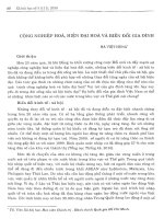
Fig. 1. Gua interacts with RPL4. (A) Stable LAP3 clones were induced with 2 mM IPTG for 48 h to express FLAG-tagged mouse Gua. RNase
A-treated lysates were used in immunoprecipitation using anti-FLAG resin. Silver staining shows the precipitation of 50-kDa protein (RPL4)
in cells expressing mouse Gua but not in cells expressing vector alone. (B) Yeast two-hybrid analysis showing the interaction of human
RPL4 with human Gua and Gub. Yeast clones were grown on selection media. Growth in the absence of tryptophan and leucine would indi-
cate presence of the appropriate vectors used to clone RPL4 and its candidate partner. Presence of colonies in the triple drop-out medium
(no tryptophan, no leucine, no histidine) would indicate interaction between RPL4 and the other protein. (C) In vitro interaction of RPL4 with
Gua. Purified GST, GST-RPL4 or blank control was mixed with purified untagged Gua in a binding buffer prior to addition of GSH-resin. Cen-
trifugation separated the supernatant from the resin. Both the supernatant and resin were analyzed by western blot analysis using anti-Gua
Ig. (D) Overexpressed Gua interacts with both endogenous and overexpressed RPL4. Extracts from HeLa cells cotransfected with FLAG-
tagged Gua and protein A-tagged RPL4 were immunoprecipitated using anti-FLAG resin and probed with the indicated antibodies. (E) Over-
expressed RPL4 interacts with both endogenous and overexpressed Gua in HeLa cells. Extracts from HeLa cells cotransfected with
FLAG-tagged RPL4 and protein A-tagged Gua were immunoprecipitated with anti-FLAG resin and probed with indicated antibodies. (F) HeLa
cells transfected with FLAG-tagged human RPL4 were stained by indirect immunofluorescence using anti-FLAG Ig. Anti-mouse IgG coupled
to rhodamine was used as secondary antibody. GFP-tagged human Gua was cotransfected and visualized directly under microscope, as a
control showing the position of nucleoli.
Gua–RPL4 interaction in mammalian rRNA production H. Yang et al.
3790 FEBS Journal 272 (2005) 3788–3802 ª 2005 FEBS
RPL4. This experiment further proved the specificity
of Gua–RPL4 interaction since B23, an abundant
nucleolar phosphoprotein which is also implicated in
ribosomal RNA processing [31], was not pulled-down
by RPL4 (Fig. 2A, bottom).
In a similar experiment using mouse cell line,
Xpress-tagged mouse RPL4 was transfected into a sta-
ble LAP3 clone which expressed either IPTG-induced
FLAG-tagged wild-type, DEVD mutant or SAT
mutant form of mouse Gua. Immunoprecipitation was
again carried out using anti-FLAG resin and the pre-
cipitate was analyzed by western blot analysis.
Figure 2B shows that all three forms of Gua pulled-
down Xpress-tagged mouse RPL4 but with different
efficiencies. Wild-type Gua interacted with RPL4 with
the highest efficiency, while the DEVD mutant showed
the least. This is demonstrated by comparing the signal
intensities of the precipitates with those of the original
inputs (Fig. 2B). The ratio (input : IP) is approxi-
mately 1 : 5 for wild-type, 1 : 1 for SAT mutant and
5 : 1 for DEVD mutant. The observed difference in
the sensitivity of the two experiments (Fig. 2A,B)
might be attributed to difference in the levels of
expression of FLAG-Gua and FLAG-RPL4. The sig-
nal intensity of the FLAG-Gua (Fig. 2B) is greater
than FLAG-RPL4 (Fig. 2A), which is possibly due to
higher expression level of FLAG-Gua in the LAP3
stable cell line that was induced with IPTG compared
to FLAG-RPL4 that was expressed by transient trans-
fection.
The DEVD motif is important for the function of
Gua in both 18S and 28S rRNA production
We have demonstrated that in Xenopus oocytes, wild-
type Gua can reverse the aberrant rRNA processing
pattern while the DEVD mutant cannot [16], highlight-
ing the importance of the DEVD motif to the function
of Gua in both 18S and 28S rRNA production in
Xenopus. In the mammalian system, we were able to
demonstrate that an SAT mutant, which lacks helicase
activity, can restore 28S but not 18S rRNA production
in mouse LAP3 cells [17], which suggests that the SAT
motif is important in 18S but not 28S rRNA produc-
tion. Because the helicase activity is dependent on the
presence of the ATPase activity of Gua [18,19], it is
reasonable to expect that mutation of the DEVD motif
would consequently result in defects of 18S matur-
ation. However, whether or not the DEVD motif is
necessary for 28S production in mammalian cells is
unknown. Here, a rescue experiment was performed
exactly as described [17] to address this issue. Briefly, a
stable LAP3 clone was induced with IPTG to over-
express a DEVD mutant form of the human Gua,
after which the cells were treated with si935, an effect-
ive siRNA that specifically targets mouse Gua mRNA
but not human Gua mRNA. Figure 3 shows that treat-
ment of the cells with si935 effectively inhibited the
production of both 18S and 28S rRNAs (lane 3, com-
pared with lanes 1 and 2), which conforms to our pre-
vious results [17]. However, in this experiment the
expression of a DEVD mutant form of human Gua
protein did not restore 18S nor 28S rRNA (Fig. 3. lane
4) as the wild-type did [17]. Thus, we conclude that the
DEVD motif is indispensable for the function of
human Gua in both 18S and 28S rRNA production,
consistent with our results in the Xenopus oocyte [16].
Amino acids 264–333 of human RPL4 is important
to its interaction with Gua
Human RPL4 has not been extensively studied after
its cloning [32]. Human and mouse RPL4 are 90%
A
B
Input
Sup’t
IP
Input
IP
Input
IP
Input
IP
Input
Sup’t
IP
Input
Sup’t
IP
WB: anti-FLAG
FLAG-RPL4
HA-Gu (WT)
FLAG-RPL4
HA-Gu (SAT-M)
FLAG-RPL4
HA-Gu (DEVD-M)
IP: anti-FLAG
IP: anti-FLAG
WB: anti-HA
WB: anti-B23
WB: anti-FLAG
WB: anti-Xpress
WB: anti-B23
FLAG-Guα
Xpress-RPL4
Xpress-RPL4
FLAG-Guα (WT)
Xpress-RPL4
FLAG-Guα (SAT-M)
Xpress-RPL4
FLAG-Gu (DEVD-M)
B23
FLAG-RPL4
HA-Guα
B23
Fig. 2. The DEVD motif of Gua is important to Gua–RPL4 interac-
tion. (A) HeLa cells were cotransfected with FLAG-tagged human-
RPL4 and plasmids encoding HA-tagged wild-type (WT), SAT
mutant (SAT-M) or DEVD mutant (DEVD-M) form of human Gua.
Whole cell extracts were immunoprecipitated using anti-FLAG resin
and probed with anti-FLAG, anti-HA or anti-B23 Ig. (B) LAP3 cells
were transfected with Xpress-tagged mouse RPL4 and induced
with 2 m
M IPTG for 48 h to express FLAG-tagged wild-type, SAT
mutant or DEVD mutant of mouse Gua. Whole cell extracts were
immunoprecipitated using anti-FLAG resin and probed with anti-
FLAG, anti-Xpress or anti-B23 Ig.
H. Yang et al. Gua–RPL4 interaction in mammalian rRNA production
FEBS Journal 272 (2005) 3788–3802 ª 2005 FEBS 3791
homologous in their cDNA-derived amino acid
sequences. The 98 amino acid C-terminal of human
RPL4 protein has little homology with its mouse
homologue. However, their N-termini (amino acids 1–
333) are 99% identical with differences in only three
amino acid residues. In higher eukaryotes, other than
the proposed involvement in ribosome assembly, RPL4
has been implicated in cell proliferation and differenti-
ation during rat neurogenesis [33] with unknown mech-
anism. In yeast, ribosomal protein RPL2 is most
homologous to human RPL4. The yeast RPL2 has
two copies, RPL2A and RPL2B [34]. A decrease in the
expression of RPL2A leads to reduced production of
60S large subunits and mature ribosomes, which conse-
quently results in slower growth rates [34]. These data
suggest the relevance of RPL4 in lower and higher
eukaryotes. We hypothesize that the function of RPL4
is partly regulated by protein–protein interactions.
To determine the RPL4 domains involved in Gua
interaction, we generated FLAG-tagged human RPL4
deletion mutants (Fig. 4A,D), expressed them in HeLa
cells and tested their ability to bind with Gua via
immunoprecipitation. Analysis of the overexpressed
proteins by indirect immunofluorescence showed that
wild-type RPL4 (amino acids 1–428), N1 mutant
(amino acids 1–264) and C1 mutant (amino acids 131–
428) predominantly localize to the nucleolus but the
C2 mutant (amino acids 264–428) is dispersed within
the nucleus but not in the nucleolus (Fig. 4B), indica-
ting the region of amino acids 131–264 probably con-
tains both the nuclear (NLS) and nucleolar (NoLS)
localization signals while amino acids 264–428 may
harbor another NLS but no NoLS.
Figure 4C reveals that RPL4 C1 and C2 mutants,
but not its N1 mutant form, coimmunoprecipitate with
Gua, suggesting the Gua-interacting domain resides in
amino acids 264–428 of RPL4. We speculated that if
Gua–RPL4 interaction is important to cellular func-
tions, then the chance should be high that the Gua-
interacting domain in RPL4 would be in a conserved
region. As the region of amino acids 333–428 is not
highly conserved among different species, we focused
on amino acids 264–333 as a possible Gua-interacting
motif in RPL4. This hypothesis was proved to be cor-
rect by coimmunoprecipitation of three mutants har-
boring amino acids 264–333 (Fig. 4F, M3, M5, M6).
The other three RPL4 mutants that lack amino acids
264–333 did not coimmunoprecipitate with Gua
(Fig. 4F, M1, M2, M4). We observed that two bands
are recognized by the anti-FLAG Ig in mutant M1.
The lower band should be the correct deletion mutant
expression product according to its expected molecular
size. The identity of the upper band remains to be
determined.
Localizations of M2 (amino acids 131–264) and M3
(amino acids 131–333) mutants to the nucleolus are in
accordance with the finding that both NLS and NoLS
are within amino acids 131–264. Mutant M1 (amino
acids 131–196) is dispersed within the whole cell but
with stronger signal intensity in the cytoplasm than in
the nucleus, suggesting that both the NLS and NoLS
should be in the region of amino acids 196–264.
Because both M4 (amino acids 204–264) and M5
(amino acids 204–333) mutants localize to the nucleo-
plasm but not to the nucleolus, it would follow that
the major NoLS for human RPL4 is within amino
acids 196–204. For mutant M4, the fluorescent signal
is mainly in the nucleoplasm, however, a significant
proportion was also found in the cytoplasm. The locali-
zations of M5 and M6 mutants, consistent with that of
C2 mutant, are predominantly in the nucleus excluding
the nucleolar region (Fig. 4E). Thus, we suspect that
a strong NLS is within amino acids 264–333 while a
IPTG
si935
47S/45S
32S
28S
28S
18S
12 34
18S
–
–
+
–
–
+
+
+
Fig. 3. The DEVD motif of Gua is important to both 18S and 28S
rRNA production. LAP3 cells were induced with 2 m
M IPTG to
express DEVD mutant of human Gua. Cells were then treated with
si935 for 48 h followed by pulse-labeling with [
32
P]orthophosphate
for 1.5 h and a chase for 3 h with normal growth medium. Total
RNAs were extracted, resolved on a 1.2% agarose-formaldehyde
gel and blotted onto a membrane for phosphorimager analysis.
Gua–RPL4 interaction in mammalian rRNA production H. Yang et al.
3792 FEBS Journal 272 (2005) 3788–3802 ª 2005 FEBS
weak NLS may be within amino acids 196–264. Com-
bined with the NoLS, this weak NLS is capable to
cause most RPL4 molecules to enter the nucleolus. It
is not uncommon to have more than one nuclear local-
ization signal within a protein [35,36].
Downregulation of RPL4 inhibits rRNA production
in mouse LAP3 cell line
RPL4 is a component of the 60S ribosome large sub-
unit. To date, there is no report showing a direct
involvement of RPL4 in pre-rRNA processing. Ribo-
somal proteins are always produced in the cytoplasm,
and then imported into the nucleoli to participate in
preribosome assembly. The ribosomes are then expor-
ted back into the cytoplasm where they direct protein
production [37]. As we hypothesize the interaction
between RPL4 and Gua is important to the function of
Gua in rRNA production, it will aid to our hypothesis
if we could determine whether downregulation of
RPL4 has any effect on this process. A sequence near
the 3¢ end of mouse RPL4 was used to design a small
interfering RNA (si-L4-M1), which targets mouse but
not human RPL4 mRNA (Fig. 5A). Downregulation
effects were examined at both mRNA and protein lev-
els, using RT-PCR and western blot analysis, respect-
ively. Treatment of LAP3 cells with 100 nm si-L4-M1
for 48 h resulted in a decrease of the mouse RPL4
mRNA level by 70% (Fig. 5B, lanes 7 and 8). This
decrease was dose-dependent. When 5 nm or 10 nm
si-L4-M1 was used, the mRNA level decreased by
about 42% or 55%, respectively (Fig. 5B, lanes 3–6).
C
F
E
D
A
B
WT
1
1
131
Constructs
Guα-binding Localization
264
WT
anti-FLAG
IP: anti-FLAG
WB: anti-FLAG
FLAG-C1
FLAG-N1
FLAG-C2
HA-Guα
FLAG-M3
FLAG-M5
FLAG-M2
FLAG-M6
FLAG-M4
FLAG-M1
HA-Guα
WB: anti-HA
WB: anti-FLAG
WB: anti-HA
IP: anti-FLAG
Hoechst
Phase
anti-FLAG
Hoechst
Phase
N1 C1 C2
N1 C1 C2
M1 M2 M3 M4 M5 M6
M1 M2 M3 M4 M5 M6
264
333
428
131
131
131
196
264
333
264
204
204
264
–
–
+
–
+
+
Nucleoplasm and cytoplasm
Nucleoli
Nucleoli
Nucleoplasm and cytoplasm
Nucleoplasm
Nucleoplasm
333
333
428
428
+
–
+
+
Nucleoli
Nucleoli
Nucleoli
Nucleoplasm
N1
C1
C2
M1
M2
M3
M6
M5
M4
Constructs
Guα-binding Localization
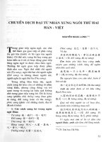
Fig. 4. Mapping of Gua-binding domain in human RPL4. (A) Schematic representation of wild-type and mutant forms of human RPL4. The
open bar represents regions conserved between human and mouse RPL4. The shaded bar represents nonconserved regions. (B) Cellular
localization of human RPL4 mutants. HeLa cells transfected with FLAG-tagged human RPL4 and various mutants were stained by indirect
immunofluorescence using anti-FLAG Ig. Anti-mouse IgG coupled to FITC was used as secondary antibody. Nuclei were visualized by Hoe-
chst stain. The phase images show dark phase nucleoli. (C) Whole cell extracts from HeLa cells cotransfected with HA-tagged human Gua
and FLAG-tagged human RPL4 deletion mutants were immunoprecipitated using anti-FLAG resin and blotted as indicated. (D) Schematic rep-
resentations of human RPL4 mutants M1 to M6 and their (E) cellular localization. (F) Whole cell extracts from HeLa cells cotransfected with
HA-tagged human Gua and FLAG-tagged human RPL4 deletion mutant shown in (D) were immunoprecipitated using anti-FLAG resin and
blotted as indicated.
H. Yang et al. Gua–RPL4 interaction in mammalian rRNA production
FEBS Journal 272 (2005) 3788–3802 ª 2005 FEBS 3793
Lacking an antibody against mouse RPL4 protein,
we instead used an indirect way to determine the effect
of si-L4-M1 on the protein level of mouse RPL4. We
cotransfected HeLa cells with si-L4-M1 and an
Xpress-tagged mouse RPL4 construct. In this experi-
ment, the remaining exogenously expressed mouse
RPL4 protein level after si-L4-M1 treatment could be
measured with anti-Xpress Ig. Figure 5C shows that
mouse RPL4 mRNA level was decreased by 83% after
treatment of the cell with 100 nm si-L4-M1 for 48 h
while the exogenously expressed mouse RPL4 protein
level was downregulated by 72%. The siRNA did not
have significant influence on mRNA levels of human
RPL4 and U1C, and the protein levels of human
RPL4 and B23.
We hypothesized that downregulation of RPL4
would lead to an aberrant rRNA processing pattern if
Gua–RPL4 interaction is necessary for the function of
Gua in rRNA production. After 48 h of si-L4-M1
treatment, LAP3 cells were pulse-labeled with [
32
P]-
orthophosphate and chased with growth medium for
3 h. As a negative control, a nonrelated siRNA
(si934Scr) was included [17]. Total RNA was extracted,
resolved on a 1.2% agarose-formaldehyde gel and
transferred to a hybond-N nitrocellulose filter for phos-
phorimager analysis. We found that all four main
visible species of rRNA (47 ⁄ 45S, 32S, 28S and 18S)
were dramatically decreased in samples treated with
si-L4-M1 (Fig. 5D). However, the decreases in 47 ⁄ 45S
rRNAs were not as great as those in mature 28S rRNA
(Fig. 5D), indicating that only part of the decrease in
28S was due to less precursors while the remaining
changes resulted from the influence by downregulation
of RPL4 on other pathways involved in rRNA produc-
tion. Ethidium bromide-stained gel (Fig. 5D, bottom)
is shown to indicate equal loading of the RNA.
Wild-type RPL4 but not its mutant form which
lacks the Gua-interacting domain reverses
inhibition of rRNA production
We constructed a deletion mutant of human RPL4,
D264-333, which lacks the Gua-interacting domain
amino acids 264–333 and another mutant D204-264 as
a control (Fig. 6A). We had already localized the
NoLS of RPL4 to amino acids 196–204, which was
supported by the nucleolar localization of both
mutants (Fig. 6B). The immunoprecipitation experi-
ment shows their Gua-binding activity (Fig. 6C), and
supports our earlier findings (Fig. 4F). Because the
region including amino acids 264–333 seems to be the
RPL4-Gua-interacting domain, the D204-264 mutant,
A
B
C
D
Human 1120
Mouse 1129
si934Scr
mRPL4
mU1C
Relative mRPL4
Relative mL4
si-L4-M1
47S/45S
si934Scr
si-L4-M1
32S
28S
18S
18S
28S
RT-PCR Western
hU1C
hRPL4
mRPL4
Xpress-mRPL4
hRPL4
hB23
100
94 65 52 40 49 35 100 17 100 2226
1
2345678
si934Scr
si934Scr
20 nM si-L4-M1
5 nM si-L4-M1
100 nM si-L4-M1
100 nM si-L4-M1
100 nM si-L4-M1
_ _ _ __ _________ ___
Fig. 5. Downregulation of mouse RPL4 using si-L4-M1 resulted in aberrant rRNA processing. (A) Comparison of human and mouse RPL4
partial cDNA sequences containing the si-L4-M1 region. Underscored nucleotides differ from human to mouse. (B) siRNA-mediated down-
regulation of mouse RPL4 mRNA. LAP3 cells were transfected with increasing concentrations of si-L4-M1. Total RNA was isolated after
48 h and analyzed by RT-PCR to determine the mRNA levels of mouse RPL4 and mouse U1C. (C) siRNA-mediated downregulation of mouse
RPL4 protein. HeLa cells were cotransfected with Xpress-tagged mouse RPL4 and si-L4-M1. Total RNA was isolated after 72 h using TRIzol
Reagent (Invitrogen) and analyzed by RT-PCR to determine the mRNA levels of mouse RPL4, human RPL4 and human U1C (left panel). A
parallel experiment was done to analyze changes in the protein levels of mouse RPL4, human RPL4 and human B23 after si-L4-M1 treat-
ment. (D) LAP3 cells were treated with 100 n
M si-L4-M1 for 48 h. Total
32
P-labeled RNA was analyzed as described in the legend to Fig. 3.
Ethidium bromide staining of both 18S and 28S rRNA is shown at the bottom.
Gua–RPL4 interaction in mammalian rRNA production H. Yang et al.
3794 FEBS Journal 272 (2005) 3788–3802 ª 2005 FEBS
but not the D264-333 mutant form, coimmunoprecipi-
tated with Gua (Fig. 6C).
To determine the significance of Gua–RPL4 inter-
action in rRNA biogenesis, we first used si-L4-M1 to
downregulate endogenous mouse RPL4 expression and
then exogenously expressed the human orthologue and
looked for reversal of inhibition of rRNA production.
Figure 6(D) (lanes 1 and 2) shows that si-L4-M1
effectively inhibited production of all four species of
rRNAs, which is consistent with the results in
Fig. 5(D). This effect was reversed by the exogenous
expression of human wild-type RPL4, suggesting that
the human orthologue can functionally replace mouse
RPL4 (Fig. 6D, lane 3). However, the expression of
mutant human RPL4 lacking the Gua-interacting
domain could not reverse the aberrant rRNA process-
ing pattern as effectively as the wild-type while the
mutant lacking amino acids 204–264 had a similar
effect to that of the wild-type, indicating that the
Gua–RPL4 interaction is important to the function of
Gua in rRNA processing (Fig. 6D, lanes 4 and 5).
Human RPL4 associates with 28S but not 18S
rRNA
To determine if RPL4 associates with 18S or 28S
rRNA, we performed RNA immunoprecipitation using
HeLa cells transiently transfected with either FLAG
vector or FLAG-tagged human RPL4. RNA–RPL4
complexes in the nucleolar extracts were immunopre-
cipitated with anti-FLAG resin, and the RNA compo-
nents were resolved on a 1.2% agarose-formaldehyde
gel, blotted onto a nitrocellulose membrane and sub-
jected to northern blot analysis. Figure 7 shows that
A
BC
D
∆204-264
∆204-264
anti-FLAG
Hoechst
Phase
IP: anti-FLAG
WB: anti-FLAG
WB: anti-FLAG
1
st
transfection
si934Scr
FLAG
vector
FLAG
vector
FLAG-RPL4
(WT)
FLAG-RPL4
(∆204-264)
FLAG-RPL4
(∆264-333)
si-L4-M1 si-L4-M1 si-L4-M1 si-L4-M1
2
nd
transfection
18S
28S
18S
28S
32S
47S/45S
28S± SE 49±4
54±5
84±10
84±10
73±6
83±5
42±7
47±6
1 234 5
100
100
Total± SE
WB: anti-HA
204 264
428
428
333
264 333
∆264-333
∆264-333
∆204-264
∆264-333
FLAG-∆204-264
FLAG-RPL4 (WT)
FLAG-RPL4 (∆204-264)
FLAG-RPL4 (∆264-333)
FLAG-∆264-333
HA-Guα
1
1
Fig. 6. Reversal of inhibition of rRNA production. (A) Schematic representation of human RPL4 deletion mutants D204-264 and D264-333.
The open bar represents regions conserved between human and mouse RPL4. The shaded bar represents nonconserved regions. (B) Indi-
rect immunofluorescence showing both D204-264 and D264-333 are localized to nucleoli. (C) Whole cell extracts from HeLa cells cotransfect-
ed with HA-tagged human Gua and FLAG-tagged human RPL4 deletion mutant were immunoprecipitated using anti-FLAG resin and blotted
as indicated. (D) LAP3 cells were transfected with either 100 n
M si934Scr or 100 nM si-L4-M1 as indicated. After 48 h, cells were next trans-
fected with FLAG-vector, FLAG-tagged wild-type human RPL4 or FLAG-tagged human RPL4 mutants as indicated. After an additional 48 h,
cells were pulse-labeled with [
32
P]orthophosphate for 1.5 h and chased with cold medium for 3 h. Total RNA was extracted and analyzed as
described in Fig. 3. Ethidium bromide staining for 18S and 28S rRNAs is shown in the middle panel. The lowest panel shows expression of
the FLAG-tagged human wild-type RPL4 (WT) and its mutant forms (D204-264 and D264-333) by western blot analysis using anti-FLAG Ig.
The numbers below the upper panel correspond to the amount of 28S rRNA or total rRNA ± standard error relative to samples in lane 1 (set
at 100) calculated with
IMAGE-QUANT software. Results were average of three independent experiments ± SE.
RNA-Total
28S probe
28S rRNA
18S rRNA
18S probe
12 34 65
RNA-IP-
FLAG-vector
RNA-IP-
FLAG-RPL4
Fig. 7. Human RPL4 associates with 28S but not 18S rRNA. HeLa
cells were transfected with either FLAG-vector only or FLAG-
tagged human RPL4. After 48 h, cells were collected and RNA-
RPL4 complexes were immunoprecipitated from nucleolar extracts
using anti-FLAG resin as described under Experimental procedures.
RNA components were isolated and resolved in a 1.2% agarose-
formaldehyde gel and blotted onto a nitrocellulose membrane,
which was subjected to northern blot analysis as described under
Experimental procedures.
H. Yang et al. Gua–RPL4 interaction in mammalian rRNA production
FEBS Journal 272 (2005) 3788–3802 ª 2005 FEBS 3795
overexpressed RPL4 pulled-down 28S but not 18S
rRNA in a dose-dependent manner (lanes 5 and 6).
We did not observe any signal from 47 ⁄ 45S, 36S and
32S pre-rRNAs, the precursors of 28S rRNA. Based
on our previous experience [16], a single oligodeoxy-
nucleotide probe we used to detect 28S could not
detect higher molecular weight pre-rRNAs under our
hybridization conditions for unknown reasons. As we
used nucleolar extract as the starting material for
the immunoprecipitation experiment, the 28S rRNA
pulled down by RPL4 should be newly produced.
Discussion
RNA helicase II ⁄ Gua is the first nucleolar RNA heli-
case shown to be directly involved in rRNA processing
in both the metazoan and mammalian systems [16,17].
There are several other nonhelicase nucleolar proteins
which have been demonstrated to also function in
rRNA processing in higher eukaryotes including
B23 ⁄ nucleophosmin, C23 ⁄ nucleolin, Bop1, p120 and
p19
Arf
. Each of these proteins was found to function
at least partially through RNA–protein or protein–
protein interactions [12,14,38–42]. Among the 18 RNA
helicases which have been directly implicated in yeast
ribosome biogenesis, at least nine were demonstrated
to functionally interact with other protein factors
[25,37,43]. Based on the bona fide helicase activity of
Gua and its demonstrated role in rRNA processing, it
is conceivable that Gua will be shown to have partners
that facilitate its function in the ribosome biogenesis
pathway.
In this paper, we report the identification of ribo-
somal protein L4 as a Gua-interacting partner through
immunoprecipitation in mouse LAP3 cells (Fig. 1A).
We noticed that several other fast migrating bands
were also pulled down by anti-FLAG resin (Fig. 1A,
other proteins), which we suspect to be proteins associ-
ated with either Gua or RPL4. The high concentration
of RNase A (200 lgÆmL
)1
), which was used in previ-
ous reports to isolate specific target-associated proteins
[31,44], suggests that these additional interactions
might not be RNA-mediated. The Gua–RPL4 inter-
action was further confirmed by immunoprecipitation
from HeLa cells (Fig. 1D,E), yeast two-hybrid analysis
(Fig. 1B) and in vitro binding assay (Fig. 1C). It is
noteworthy that Gub also interacts with RPL4 as
shown by the two-hybrid analysis (Fig. 1B, lower
right). As a paralogue of Gua,Gub also possesses
in vitro ATPase and helicase activities, but no RNA
foldase activity [45]. The current data suggest that
both paralogues arose through gene duplication but
the resulting genes are differentially regulated and
might possess different functions [46]. Overexpression
of Gub in mouse LAP3 cells leads to inhibition of
total rRNA production, suggesting contrasting roles
for Gub and Gua [17]. It would be valuable to deter-
mine whether the inhibitory effect of Gub on rRNA
biogenesis is through its competitive interaction with
RPL4. Indirect immunofluorescence showed a predom-
inant localization of newly produced FLAG-tagged
RPL4 protein to the nucleolus (Fig. 1F) which is con-
sistent with the published report that most newly
formed ribosomal proteins are highly concentrated in
the nucleolus [47]. Burial of FLAG epitope within the
highly structured mature ribosome subunit might
account for the absence of strong fluorescent signal in
the cytoplasm. The distribution of RPL4 in the nucleo-
lus seems more localized compared with the more dis-
persed localization of Gua throughout the entire
nucleolus (Fig. 1F). This subtle discrepancy between
the localizations of the two proteins may indicate that
Gua interacts with other partners in different sub-
nucleolar regions, which is consistent with the presence
of other additional bands shown in Fig. 1A. Moreover,
our previous immunoelectron microscopy experiments
showed that rat Gua is localized to the dense fibrillar
component (DFC) and granular component (GC)
within the nucleolus [16].
An interesting finding was the importance of the
DEVD motif in Gua–RPL4 interaction (Fig. 2). We
previously showed that in Xenopus, the DEVD motif
of Gua was important for both 18S and 28S rRNA
production [16]. However, in mammalian cells, we
were only able to prove that SAT motif is necessary
for 18S maturation [17]. As the unwinding activity is
dependent on the ATPase activity, we speculated that
the DEVD motif of Gua is also necessary for 18S pro-
duction in mammalian cells. In this report, we showed
the conserved importance of the DEVD motif of Gua
to 28S maturation in mouse LAP3 cells (Fig. 3).
Because DEVD is important for 28S production as
well as Gua–RPL4 interaction, and because RPL4 is a
component of the ribosome large subunit which con-
tains 28S but not 18S rRNA, it is reasonable to sus-
pect that RPL4 might be involved in the function of
Gua in 28S rRNA production.
Through a series of deletion mutants of RPL4 used
in the immunoprecipitation and indirect immunofluo-
rescence experiments, we identified the NLS and NoLS
as well as the Gua-interacting domains in RPL4
(Figs 4 and 5). However, it is worth mentioning
that the use of deletion mutants may not accurately
reflect the exact functional states of protein inter-
actions since the possibility exists that the shortened
proteins may be unfolded and thus nonfunctional.
Gua–RPL4 interaction in mammalian rRNA production H. Yang et al.
3796 FEBS Journal 272 (2005) 3788–3802 ª 2005 FEBS
Subtle point mutations in the identified Gua-interacting
domain might lend more support to our conclusions.
Downregulation of RPL4 via si-L4-M1 resulted in the
inhibition of production of all four rRNA species
(Figs 5D and 6D lane 2), strongly suggesting a general
mechanism whereby RPL4 modulates rRNA biogen-
esis through rDNA transcription, rRNA turn over,
ribosome production rate, ribosome stability or rRNA
degradation. It is possible that the amount of RPL4 in
the cell correlates with the assembly or stabilization of
pre-rRNA processing machineries or preribosomal par-
ticles. Perhaps RPL4 is actually a component of the
pre-rRNA processing machinery. If so, a dramatic
change in the amount of RPL4 protein level might
lead to disassembly of the specific machineries or parti-
cles, which would send feedback signals to RNA
polymerase I to advance more slowly or even to rRNA-
degrading complexes to degrade the unincorporated
mature rRNA [17]. This hypothesis might help to
interpret the involvement of other nucleolar proteins in
pre-rRNA production such as p19
Arf
[15]. It might also
explain why there is a decrease of 18S rRNA. More-
over, inhibition of the 28S pathway might concomit-
antly result in the reduction of 18S rRNA through an
unidentified mechanism. For example, many yeast
mutants with 25S rRNA production defects also show
an inhibition in 35S pre-rRNA cleavages which lead to
decrease in 18S biogenesis [48].
Other than the proposed general function of RPL4
in overall rRNA production, we also hypothesize that
RPL4 plays a direct role in 28S production through its
interaction with Gua. Several arguments and lines of
evidence support this: (a) our rescue experiment
showed that wild-type human RPL4 reversed the aber-
rant rRNA processing pattern (Fig. 6D, compare lanes
2 and 3) but the mutant lacking the Gua-interacting
domain had no effect (Fig. 6, compare lanes 2 and 5);
(b) inhibition of 28S rRNA production is more signifi-
cant than that of 47 ⁄ 45S when RPL4 is downregulated
(Fig. 5D, lanes 1 and 2); (c) RPL4 is an important
component of the 60S ribosome subunit which con-
tains the 28S but not the 18S rRNA; (d) RNA immu-
noprecipitation revealed coprecipitation of RPL4 with
28S but not with 18S rRNA (Fig. 7); (e) ATPase activ-
ity of Gua is important for both 28S production and
Gua–RPL4 interaction. These five lines of evidence
support a more direct role for the Gua–RPL4 inter-
action in 28S production than a possible general mechan-
ism, although it is likely that both mechanisms coexist.
It is not uncommon for a nucleolar protein to function
in different pathways. For example, the function of
C23 in ribosome biogenesis is reflected in almost all
steps of the process including rDNA transcription,
pre-rRNA processing, preribosome assembly and
nucleocytoplasmic transport [39].
What then could be a mechanism whereby the Gua–
RPL4 interaction facilitates 28S rRNA biogenesis? The
fact that Gua and RPL4 have been identified in ribo-
nucleoprotein (RNP) particles [31,49] indicates that
their interaction might cause them to be localized into
pre-rRNA processing machineries essential for pre60S
ribosome particles. It is known that interruption of
early assembly steps results in disassembly of the parti-
cles and destabilization of pre-rRNAs [43]. Moreover,
we did observe several other bands in the immunopre-
cipitation assay (Fig. 1A) coimmunoprecipitating with
Gua, which may represent other proteins in the same
processing machinery as Gua. Once Gua has been
incorporated into the RNP particle, it might function
in early rRNA processing steps such as regulating
interactions between guide snoRNAs and pre-rRNAs,
helping the endo- and exo-nucleases in removing inter-
nal or external transcribed spacer sequences as well as
modulating the numerous trans-acting factors and
ribosomal proteins in the pre60S particles through
regulation of RNA-RNA, RNA–protein and protein–
protein interactions [43]. In yeast S. cerevisiae, involve-
ment of ATP-dependent RNA helicase has been
implicated in each of these possible roles [50–54]. The
functional diversity of an RNA molecule is based on
its extreme flexibility. With the help of proteins, RNA
retains or loses its active configuration in response to
various different cellular signals [55]. RNA helicase is
a candidate to function in an energy-dependent
manner in this process. In addition, Gua has another
activity, GTP-stimulated RNA folding activity which
resides within a domain separate from the ATPase ⁄
helicase activity. Proteins utilizing GTP as an energy
source have recently been found to participate in ribo-
some biogenesis [56,57]. In addition, it was recently
reported that GTP-binding state might influence the
nucleolar targeting of nucleostamin, a nucleolar pro-
tein which shuttles between nucleoplasm and nucleolus
with suspected roles in cell cycle and cell proliferation
regulation [58–60]. Interestingly, the C-terminal
FRGQR-containing region of Gua has also been
reported to be critical for both GTP-stimulated RNA
foldase activity and nucleolar localization [20,61]. It
remains to be identified if the FRGQR region of Gua
is relevant to GTP–binding or RPL4 interaction. It
would not be surprising if the Gua–RPL4 interaction
is found to be important in all three roles mentioned
considering the complexity of ribosome assembly,
which highly demands versatile energy producers and
consumers. The multifunctional property of Gua
makes it a good candidate.
H. Yang et al. Gua–RPL4 interaction in mammalian rRNA production
FEBS Journal 272 (2005) 3788–3802 ª 2005 FEBS 3797
In summary, the data in this report suggest that
RPL4 functions in a general manner in rRNA biogen-
esis whereas the Gua–RPL4 interaction is more direct
in the production of 28S rRNA and maturation of
pre60S particles. More questions arise such as whether
RPL4 recruits Gua or vise versa, what is the exact
mechanism of Gua ⁄ RPL4 involvement in rRNA pro-
cessing, which specific step requires Gua–RPL4 inter-
action and what other cis-ortrans-acting factors are
in the RNP particle containing Gua and RPL4. Ribo-
some biogenesis in yeast has been shown to be a highly
intricate cellular process with the involvement of about
80 ribosomal proteins and numerous trans-acting fac-
tors [43]. It is believed that the process is more com-
plex in higher eukaryotes [43]. Yeast S. cerevisiae has
been shown to be an effective model to defining this
field due to the availability of genetic screens. How-
ever, the yeast orthologue for Gua remains to be
identified. Further studies aimed at isolation and
investigation of Gua orthologue in yeast, and the
large-scale proteomics study combined with molecular
analysis of specific factors involved should provide a
clearer understanding of the function of the Gua–
RPL4 interaction and its significance in a more inclu-
sive picture of the ribosome biogenesis in higher
eukaryotes.
Experimental procedures
Cell culture and transfection
HeLa cells were grown in Dulbecco’s modified Eagle’s med-
ium containing 10% fetal bovine serum with 5% CO
2
.
LAP3 cells [14] were grown in Dulbecco’s modified Eagle’s
medium containing 10% newborn calf serum, 100 IUÆmL
)1
penicillin G, and 100 lgÆmL
)1
streptomycin sulfate with
5% CO
2
. Cell transfections were carried out using Lipofec-
tamine 2000 (Invitrogen, Carlsbad, CA) according to the
manufacturer’s instructions.
Immunoprecipitation and identification of
FLAG-tagged Gua-associated proteins
Stable clones of LAP3 cells [17] were induced with 2 mm
IPTG to express either FLAG-tagged mouse Gua or
FLAG vector only. Two days after induction, cells were
homogenized in a gentle lysis buffer (10 mm Tris ⁄ HCl
pH 7.5, 10 mm NaCl, 10 mm EDTA, 0.5% NP-40,
0.35 mgÆmL
)1
phenylmethanesulfonyl fluoride, 20 lgÆmL
)1
aprotinin, 10 lgÆmL
)1
leupeptin). After centrifugation at
10 000 g at 4 °C for 2 min, the supernatant was transferred
to a clean tube and RNase A was added to a final concen-
tration of 200 lgÆmL
)1
. After incubation on ice for 10 min
followed by another centrifugation at 10 000 g at 4 ° C for
10 min, the supernatant was separated, mixed with anti-
FLAG-M2 resin (Sigma), and tumbled at 4 °C for 4 h. The
mixture was then centrifuged at 1500 g at 4 °C for 2 min.
The pellet was washed five times with wash buffer (50 mm
Tris ⁄ HCl, pH 7.5, 150 mm NaCl, 0.5% NP-40) and subjec-
ted to further analysis. For identification of Gua-associated
proteins, the immunoprecipitate was resolved on a sodium
dodecyl sulfate–polyacrylamide (10%) gel followed by silver
staining. The protein bands showing only in the Gua-
expressed samples were excised and analyzed by mass
spectrometry.
Yeast two-hybrid analysis
The cDNAs for human Gua and human RPL4 were
subcloned into pGBKT7 and pGADT7 yeast expression
vectors, respectively. Protein–protein interaction was deter-
mined in yeast exactly as previously described [62].
In vitro binding assay
Recombinant proteins were expressed in Escherichia coli.
Purified GST or GST-RPL4 protein was mixed with puri-
fied untagged Gua [18] in NETN buffer [63] and tumbled
for 2 h at 4 °C. GSH-resin was then added and tumbled
for additional 1 h. Followed by centrifugation, the resin
was washed in NETN buffer and boiled in Laemmli buffer.
The supernatant was also boiled in Laemmli buffer. Sam-
ples were analyzed on immunoblots using anti-Gua Ig.
Immunoprecipitation to confirm the interaction
between Gua and RPL4 in mammalian cells
Actively growing HeLa cells or stable clones of LAP3 cells,
either transiently transfected or induced with IPTG, were
suspended in NET2 buffer (50 mm Tris ⁄ HCl pH 7.4,
150 mm NaCl and 0.05% NP-40) and sonicated for 30 s, 6
times with 30-s intervals. Lysates were centrifuged at
10 000 g at 4 ° C for 10 min. The concentration of the
supernatant was determined using Bradford reagent (Bio-
Rad, Hercules, CA). Total proteins, 500 lg, were mixed
with 50 lL anti-FLAG-M2 resin (Sigma) and 10 lL prote-
inase inhibitor cocktail (Sigma), tumbled overnight, washed
three times with NET2 buffer and centrifuged at 10 000 g
at room temperature for 20 s. The immunoprecipitates were
boiled in Laemmli buffer, separated on a sodium dodecyl
sulfate–polyacrylamide (10%) gel and subjected to immuno-
blots using appropriate antibodies.
Immunofluorescence
HeLa cells grown on slides were analyzed by indirect
immunofluorescence staining as described [62]. Cells were
Gua–RPL4 interaction in mammalian rRNA production H. Yang et al.
3798 FEBS Journal 272 (2005) 3788–3802 ª 2005 FEBS
examined with a Nikon Eclipse TE2000-U inverted micro-
scope equipped with a Coolsnap digital color camera.
Metabolic labeling of cells and subsequent RNA
analysis
The procedure is similar to that described elsewhere [17,62].
Briefly, LAP3 cells were transfected with 100 nm siRNA.
After 2 days, cells were incubated in phosphate-free med-
ium (Sigma) for 3.5 h. The medium was replaced with fresh
phosphate-free medium containing 40 lCiÆmL
)1
[
32
P]ortho-
phosphate (Amersham Pharmacia Biosciences, Piscataway,
NJ), incubated for 1.5 h and chased with the regular
growth medium for 3 h.
32
P-labeled RNA was isolated and
analyzed as described [17].
RNA immunoprecipitation and northern blot
analysis
HeLa cells were transfected with either FLAG vector alone
or FLAG-tagged human RPL4. After 48 h, cells were har-
vested by centrifugation at 3000 g for 5 min and washed
three times with 1· NaCl ⁄ P
i
. Cell pellets were resuspended
in resuspension buffer (RSB) (10 mm Tris ⁄ HCl pH 7.4,
10 mm NaCl, 1.5 m m magnesium acetate). After incubation
on ice for 30 min followed by a centrifugation at 1000 g
for 8 min, cell pellets were resuspended in RSB buffer
with 0.5% NP-40. Cells were homogenized with a Dounce
homogenizer until the nuclei were released (which were
visualized under microscope). After another centrifugation
at 1000 g for 8 min, cell pellets were resuspended in 0.88 m
sucrose, 5 mm magnesium acetate and centrifuged at 1500 g
for 20 min. The supernatant was discarded and the nuclear
pellet was suspended by gentle Dounce homogenization in
0.34 m sucrose, 0.5 m m magnesium acetate and sonicated
for 10 s, six times with 10-second intervals. The sonicated
fraction was layered with three times the volume of 0.88 m
sucrose and centrifuged at 2600 g for 20 min. The super-
natant was discarded and the nucleolar pellets were
resuspended in NET2 buffer and subjected to immunopre-
cipitation as described above. RNAs were isolated from the
immunoprecipitate with TRIzol Reagent (Invitrogen). Nor-
thern blot analysis was carried out as previously described
[16]. 18S and 28S rRNAs were detected using
32
P-end-
labeled oligodeoxynucleotide probes.
siRNAs, probes and RT-PCR primers si935 (targets
mouse Gua) and si934Scr were described previously [17].
The sequence of si-L4-M1 (targets mouse RPL4) is shown
in Fig. 5A. RT-PCR primers and northern blot analysis
probes are shown in Table 1.
Acknowledgements
This work was supported by Public Health Service
grant DK52341 from the National Institute of Diabe-
tes and Digestive and Kidney Diseases to B.C.V. We
thank Bianca Gonzales for the suggestions in writing
the manuscript.
References
1 Gerbi SA, Borovjagin AV, Ezrokhi M & Lange TS
(2001) Ribosome biogenesis: role of small nucleolar
RNA in maturation of eukaryotic rRNA. Cold Spring
Harb Symp Quant Biol 66, 575–590.
2 Scheer U, Thiry M & Goessens G (1993) Structure,
function and assembly of the nucleolus. Trends Cell Biol
3, 236–241.
3 Tuteja R & Tuteja N (1998) Nucleolin: a multifunc-
tional major nucleolar phosphoprotein. Crit Rev Bio-
chem Mol Biol 33, 407–436.
4 Derenzini M, Ceccarelli C, Santini D, Taffurelli M
& Trere D (2004) The prognostic value of the
AgNOR parameter in human breast cancer depends
on the pRb and p53 status. J Clin Pathol 57, 755–
761.
5 Xue D, Shi H, Smith JD, Chen X, Noe DA, Cedervall
T, Yang DD, Eynon E, Brash DE, Kashgarian M et al.
(2003) A lupus–like syndrome develops in mice lacking
the Ro 60-kDa protein, a major lupus autoantigen. Proc
Natl Acad Sci USA 100, 7503–7508.
Table 1. Primers used for RT-PCR and probes used for northern blot analysis.
Name 5¢ to 3¢ sequence Gene
U1C5¢ GCAACATGCCCAAGTTTTATTGTG RT-PCR primer, human U1C, sense
U1C3¢ TATCCTTATCTGTCTGGTCGAGTC RT-PCR primer, human U1C, antisense
BV974 AGCATCATGCCCAAGTTTTATTGTGA RT-PCR primer, mouse U1C, sense
BV976 TTTCTCCCTCCAAAAATATTCAGTTA RT-PCR primer, mouse U1C, antisense
YH34 TCTCCTCTCCTCGAGATGGCGTGTGCTCGCCCACTG RT-PCR primer, human and mouse L4, sense
YH47 ACCGCCGCCTTCTCATCTGA RT-PCR primer, human L4, antisense
YH48 TTCTCTGGAACAACCTTCTCG RT-PCR primer, mouse L4, antisense
YH9 ATGGCCTCAGTTCCGAAAACCAACAAAATAGA Northern blot analysis probe, for 18S rRNA
YH11 TTCTGACTTAGAGGCGTTCAGTCATAATCCCA Northern blot analysis probe, for 28S rRNA
H. Yang et al. Gua–RPL4 interaction in mammalian rRNA production
FEBS Journal 272 (2005) 3788–3802 ª 2005 FEBS 3799
6 Stefanovsky VY, Pelletier G, Hannan R, Gagnon-
Kugler T, Rothblum LI & Moss T (2001) An immediate
response of ribosomal transcription to growth factor
stimulation in mammals is mediated by ERK phosphor-
ylation of UBF. Mol Cell 8, 1063–1073.
7 Gallagher ED & Cobb MH (2001) ERKs weigh in on
ribosome mass. Mol Cell 8, 932–933.
8 Giordano E, Peluso I, Senger S & Furia M (1999) mini-
fly, a Drosophila gene required for ribosome biogenesis.
J Cell Biol 144, 1123–1133.
9 Dumbar TS, Gentry GA & Olson MO (1989) Interac-
tion of nucleolar phosphoprotein B23 with nucleic acids.
Biochemistry 28, 9495–9501.
10 Baserga SJ, Yang XD & Steitz JA (1991) An intact Box
C sequence in the U3 snRNA is required for binding of
fibrillarin, the protein common to the major family of
nucleolar snRNPs. EMBO J 10, 2645–2651.
11 Omer AD, Ziesche S, Ebhardt H & Dennis PP (2002)
In vitro reconstitution and activity of a C ⁄ D box methyl-
ation guide ribonucleoprotein complex. Proc Natl Acad
Sci USA 99, 5289–5294.
12 Gustafson WC, Taylor CW, Valdez BC, Henning D,
Phippard A, Ren Y, Busch H & Durban E (1998)
Nucleolar protein p120 contains an arginine-rich
domain that binds to ribosomal RNA. Biochem J 331,
387–393.
13 Squatrito M, Mancino M, Donzelli M, Areces LB &
Draetta GF (2004) EBP1 is a nucleolar growth-regulat-
ing protein that is part of pre-ribosomal ribonucleopro-
tein complexes. Oncogene 23, 4454–4465.
14 Strezoska Z, Pestov DG & Lau LF (2002) Functional
inactivation of the mouse nucleolar protein Bop1 inhi-
bits multiple steps in pre-rRNA processing and blocks
cell cycle progression. J Biol Chem 277, 29617–29625.
15 Sugimoto M, Kuo ML, Roussel MF & Sherr CJ (2003)
Nucleolar Arf tumor suppressor inhibits ribosomal
RNA processing. Mol Cell 11, 415–424.
16 Yang H, Zhou J, Ochs RL, Henning D, Jin R & Valdez
BC (2003) Downregulation of RNA helicase II ⁄ Gu
results in the depletion of 18 and 28 S rRNAs in Xeno-
pus oocyte. J Biol Chem 278, 38847–38859.
17 Henning D, So RB, Jin R, Lau LF & Valdez BC (2003)
Silencing of RNA helicase II ⁄ Gualpha inhibits mamma-
lian ribosomal RNA production. J Biol Chem 278,
52307–52314.
18 Valdez BC, Henning D, Busch RK, Woods K, Flores-
Rozas H, Hurwitz J, Perlaky L & Busch H (1996) A
nucleolar RNA helicase recognized by autoimmune anti-
bodies from a patient with watermelon stomach disease.
Nucleic Acids Res 24, 1220–1224.
19 Valdez BC, Henning D, Perumal K & Busch H (1997)
RNA-unwinding and RNA-folding activities of RNA
helicase II ⁄ Gu: Two activities in separate domains of
the same protein. Eur J Biochem 250, 800–807.
20 Ou Y, Fritzler MJ, Valdez BC & Rattner JB (1999)
Mapping and characterization of the functional domains
of the nucleolar protein RNA helicase II ⁄ Gu. Exp Cell
Res 247, 389–398.
21 Westermarck J, Weiss C, Saffrich R, Kast J, Musti AM,
Wessely M, Ansorge W, Seraphin B, Wilm M, Valdez
BC et al. (2002) The DEXD ⁄ H-box RNA helicase
RHII ⁄ Gu is a co-factor for c-Jun-activated transcrip-
tion. EMBO J 21, 451–460.
22 Valdez BC, Henning D, Perlaky L, Busch RK & Busch
H (1997) Cloning and characterization of Gu ⁄ RH-II
binding protein. Biochem Biophys Res Commun 234,
335–340.
23 Jankowsky E, Gross CH, Shuman S & Pyle AM (2001)
Active disruption of an RNA–protein interaction by a
DExH ⁄ D RNA helicase. Science 291, 121–125.
24 de la Cruz J, Kressler D & Linder P (1999) Unwinding
RNA in Saccharomyces cerevisiae: DEAD-box proteins
and related families. Trends Biochem Sci 24, 192–198.
25 de la Cruz J, Lacombe T, Deloche O, Linder P &
Kressler D (2004) The putative RNA helicase Dbp6p
functionally interacts with Rpl3p, Nop8p and the novel
trans-acting factor Rsa3p during biogenesis of 60S ribo-
somal subunits in Saccharomyces cerevisiae. Genetics
166, 1687–1699.
26 Watkins NJ, Gottschalk A, Neubauer G, Kastner B,
Fabrizio P, Mann M & Luhrmann R (1998) Cbf5p, a
potential pseudouridine synthase, and Nhp2p, a putative
RNA-binding protein, are present together with Gar1p
in all H BOX ⁄ ACA-motif snoRNPs and constitute a
common bipartite structure. RNA 4, 1549–1568.
27 Venema J & Tollervey D (1995) Processing of pre-ribo-
somal RNA in Saccharomyces cerevisiae. Yeast 11,
1629–1650.
28 Venema J, Bousquet-Antonelli C, Gelugne JP, Caizer-
gues-Ferrer M & Tollervey D (1997) Rok1p is a puta-
tive RNA helicase required for rRNA processing. Mol
Cell Biol 17, 3398–3407.
29 Bates GJ, Nicol SM, Wilson BJ, Jacobs AM, Bourdon
JC, Wardrop J, Gregory DJ, Lane DP, Perkins ND &
Fuller-Pace FV (2005) The DEAD box protein p68: a
novel transcriptional coactivator of the p53 tumour
suppressor. EMBO J 24, 543–553.
30 Jin A, Itahana K, O’Keefe K & Zhang Y (2004) Inhibi-
tion of HDM2 and activation of p53 by ribosomal pro-
tein L23. Mol Cell Biol 24, 7669–7680.
31 Bertwistle D, Sugimoto M & Sherr CJ (2004) Physical
and functional interactions of the Arf tumor suppressor
protein with nucleophosmin ⁄ B23. Mol Cell Biol 24,
985–996.
32 Bagni C, Mariottini P, Annesi F & Amaldi F (1993)
Human ribosomal protein L4: cloning and sequencing
of the cDNA and primary structure of the protein.
Biochim Biophys Acta 1216, 475–478.
Gua–RPL4 interaction in mammalian rRNA production H. Yang et al.
3800 FEBS Journal 272 (2005) 3788–3802 ª 2005 FEBS
33 Ueno M, Nakayama H, Kajikawa S, Katayama K,
Suzuki K & Doi K (2002) Expression of ribosomal pro-
tein L4 (rpL4) during neurogenesis and 5-azacytidine
(5AzC)-induced apoptotic process in the rat. Histol
Histopathol 17, 789–798.
34 Lucioli A, Presutti C, Ciafre S, Caffarelli E, Fragapane
P & Bozzoni I (1988) Gene dosage alteration of L2
ribosomal protein genes in Saccharomyces cerevisiae:
effects on ribosome synthesis. Mol Cell Biol 8, 4792–
4798.
35 Heiss NS, Girod A, Salowsky R, Wiemann S, Pepper-
kok R & Poustka A (1999) Dyskerin localizes to the
nucleolus and its mislocalization is unlikely to play a
role in the pathogenesis of dyskeratosis congenita. Hum
Mol Genet 8, 2515–2524.
36 Choi J, Ko MK & Kay EP (2000) Subcellular localiza-
tion of the expressed 18 kDa FGF-2 isoform in corneal
endothelial cells. Mol Vis 6, 222–231.
37 Fromont-Racine M, Senger B, Saveanu C & Fasiolo F
(2003) Ribosome assembly in eukaryotes. Gene 313,
17–42.
38 Scheer U & Hock R (1999) Structure and function of
the nucleolus. Curr Opin Cell Biol 11, 385–390.
39 Ginisty H, Sicard H, Roger B & Bouvet P (1999) Struc-
ture and functions of nucleolin. J Cell Sci 112, 761–772.
40 Ginisty H, Serin G, Ghisolfi-Nieto L, Roger B, Libante
V, Amalric F & Bouvet P (2000) Interaction of nucleo-
lin with an evolutionarily conserved pre-ribosomal
RNA sequence is required for the assembly of the pri-
mary processing complex. J Biol Chem 275, 18845–
18850.
41 Li YP, Busch RK, Valdez BC & Busch H (1996) C23
interacts with B23, a putative nucleolar-localization-
signal-binding protein. Eur J Biochem 237, 153–158.
42 Zhang Y (2004) The ARF-B23 connection: implications
for growth control and cancer treatment. Cell Cycle 3,
259–262.
43 Kressler D, Doere M, Rojo M & Linder P (1999)
Synthetic lethality with conditional dbp6 alleles identi-
fies rsa1p, a nucleoplasmic protein involved in the
assembly of 60S ribosomal subunits. Mol Cell Biol 19,
8633–8645.
44 Itahana K, Bhat KP, Jin A, Itahana Y, Hawke D,
Kobayashi R & Zhang Y (2003) Tumor suppressor
ARF degrades B23, a nucleolar protein involved in
ribosome biogenesis and cell proliferation. Mol Cell 12,
1151–1164.
45 Valdez BC, Perlaky L & Henning D (2002) Expression,
cellular localization, and enzymatic activities of RNA
helicase II ⁄ Gub. Exp Cell Res 276, 249–263.
46 Valdez BC, Yang H, Hong E & Sequitin AM (2002)
Genomic structure of newly identified paralogue of
RNA helicase II ⁄ Gu: detection of pseudogenes and
multiple alternatively spliced mRNAs. Gene 284, 53–
61.
47 Warner JR (1979) Distribution of newly formed ribo-
somal proteins in HeLa cell fractions. J Cell Biol 80,
767–772.
48 Venema J & Tollervey D (1999) Ribosome synthesis
in Saccharomyces cerevisiae. Annu Rev Genet 33, 261–
311.
49 Fujiyama S, Yanagida M, Hayano T, Miura Y, Isobe
T, Fujimori F, Uchida T & Takahashi N (2002) Isola-
tion and proteomic characterization of human parvulin-
associating preribosomal ribonucleoprotein complexes.
J Biol Chem 277, 23773–23780.
50 O’Day CL, Chavanikamannil F & Abelson J (1996) 18S
rRNA processing requires the RNA helicase-like protein
Rrp3. Nucleic Acids Res 24, 3201–3207.
51 Daugeron MC & Linder P (1998) Dbp7p, a putative
ATP-dependent RNA helicase from Saccharomyces cer-
evisiae, is required for 60S ribosomal subunit assembly.
RNA 4, 566–581.
52 Liang WQ, Clark JA & Fournier MJ (1997) The
rRNA-processing function of the yeast U14
small nucleolar RNA can be rescued by a conserved
RNA helicase-like protein. Mol Cell Biol 17, 4124–
4132.
53 Weaver PL, Sun C & Chang TH (1997) Dbp3p, a puta-
tive RNA helicase in Saccharomyces cerevisiae,is
required for efficient pre-rRNA processing predomi-
nantly at site A3. Mol Cell Biol 17, 1354–1365.
54 Mitchell P, Petfalski E, Shevchenko A, Mann M &
Tollervey D (1997) The exosome: a conserved eukaryo-
tic RNA processing complex containing multiple 3¢?5¢
exoribonucleases. Cell 91, 457–466.
55 Schroeder R, Barta A & Semrad K (2004) Strategies for
RNA folding and assembly. Nat Rev Mol Cell Biol 5,
908–919.
56 Gelperin D, Horton L, Beckman J, Hensold J &
Lemmon SK (2001) Bms1p, a novel GTP-binding pro-
tein, and the related Tsr1p are required for distinct
steps of 40S ribosome biogenesis in yeast. RNA 7,
1268–1283.
57 Wegierski T, Billy E, Nasr F & Filipowicz W (2001)
Bms1p, a G-domain-containing protein, associates with
Rcl1p and is required for 18S rRNA biogenesis in yeast.
RNA 7, 1254–1267.
58 Ritland Politz JC, Polena I, Trask I, Bazett-Jones DP &
Pederson T (2005) A nonribosomal landscape in the
nucleolus revealed by the stem cell protein nucleostemin.
Mol Biol Cell 16, 3401–3410.
59 Tsai RY & McKay RD (2005) A multistep, GTP-driven
mechanism controlling the dynamic cycling of nucleo-
stemin. J Cell Biol 168, 179–184.
60 Misteli T (2005) Going in GTP cycles in the nucleolus.
J Cell Biol 168, 177–178.
61 Valdez BC (2000) Structural domains involved in the
RNA folding activity of RNA helicase II ⁄ Gu protein.
Eur J Biochem 267, 6395–6402.
H. Yang et al. Gua–RPL4 interaction in mammalian rRNA production
FEBS Journal 272 (2005) 3788–3802 ª 2005 FEBS 3801
62 Valdez BC, Henning D, So RB, Dixon J & Dixon MJ
(2004) The Treacher Collins syndrome (TCOF1 ) gene
product is involved in ribosomal DNA gene transcrip-
tion by interacting with upstream binding factor. Proc
Natl Acad Sci USA 101, 10709–10714.
63 Valdez BC, Perlaky L, Henning D, Saijo Y, Chan PK
& Busch H (1994) Identification of the nuclear and
nucleolar localization signals of the protein p120. Inter-
action with translocation protein B23. J Biol Chem 269,
23776–23783.
3802 FEBS Journal 272 (2005) 3788–3802 ª 2005 FEBS
Gua–RPL4 interaction in mammalian rRNA production H. Yang et al.