(Focus on structural biology 2) t j griffin, l m smith (auth ), j nicholas housby (eds ) mass spectrometry and genomic analysis springer netherlands (2002) (1)
Bạn đang xem bản rút gọn của tài liệu. Xem và tải ngay bản đầy đủ của tài liệu tại đây (7.15 MB, 158 trang )
Mass Spectrometry and Genomic Analysis
Edited by
J. NICHOLAS HOUSBY
Oxagen Limited, Abingdon, United Kingdom
KLUWER ACADEMIC PUBLISHERS
NEW YORK, BOSTON, DORDRECHT, LONDON, MOSCOW
www.pdfgrip.com
eBook ISBN:
Print ISBN:
0-306-47595-2
0-7923-7173-9
©2002 Kluwer Academic Publishers
New York, Boston, Dordrecht, London, Moscow
Print ©2001 Kluwer Academic Publishers
Dordrecht
All rights reserved
No part of this eBook may be reproduced or transmitted in any form or by any means, electronic,
mechanical, recording, or otherwise, without written consent from the Publisher
Created in the United States of America
Visit Kluwer Online at:
and Kluwer's eBookstore at:
www.pdfgrip.com
TABLE OF CONTENTS
INTRODUCTION
PREFACE
xiii
xv
CHAPTER 1
TJ. Griffin, LM. Smith
Single-Nucleotide Polymorphism Analysis by MALDI-TOF Mass Spectrometry
1. Introduction
1.1. MALDI-TOF MS
2. Analysis of Peptide Nucleic Acid Hybridisation Probes
2.1. Design of PNA Hybridisation Probes
2.2. Analysis of Polymorphisms in Tyrosinase Exon 4
3. Direct Analysis of Invasive Cleavage Products
3.1. The Invader Assay
3.2. Direct Analysis of SNPs From Human Genomic DNA
4. Conclusions
5. Experimental Methods
5.1. PNA Probe Synthesis and Preparation
5.2. PCR Amplification of Exon 4 of the Tyrosinase Gene
5.3. Hybridisation of PNA Probes to Immobilised Gene Targets
5.4. MALDI-TOF MS Analysis of PNA Probes
5.5. Invader Squared Reaction
5.6. MALDI-TOF MS Sample Preparation of Cleavage Products
5.7. MALDI-TOF MS Analysis of Cleavage Products
6. Affiliations
7. References
1
2
2
3
4
5
6
8
11
11
11
11
12
12
13
13
14
14
14
CHAPTER 2
LA. Haff, AC. Belden, LR. Hall, PL. Ross, IP. Smirnov
SNP Genotyping by MALDI-TOF Mass Spectrometry
1. Introduction
2. SNP Analysis by Single Base Extension of Primers
3. Materials and Methods
4. Design Considerations for the SNP Genotyping Assay
4.1. Design of PCR Product
4.2. PCR Product Polishing
4.3. Primer Design Rules for Monoplex SNP Typing
vii
www.pdfgrip.com
16
17
19
19
19
20
20
viii
4.4. Mass Calculations
4.5. Primer Design Rules for Multiplexed Reactions
4.5.1. Multiplexing with Primer Pools of Six or Fewer Primers
4.5.2.Recommended Primer Pool Design: More Than Six Primers
4.6 Primer Quality
5. The Single Base Extension Reaction
5.1. Desalting of Primer Extension Reactions
5.2. MALDI-TOF Conditions
5.3. Determination of Bases Added to the Primer
6. Modification of the SNP Typing Assay to Support Allele Frequency
Determination
7. Conclusions
8. References
21
22
22
23
25
26
27
27
27
28
31
32
CHAPTER 3
Hubert Köster
MASSARRAY™: Highly Accurate and Versatile High Throughput Analysis of
Genetic Variations
1. Introduction
2. MassARRAY™ Technology
3. Methodology of MassARRAY™ Technology
4. Diagnostic Applications of MassARRAY™ Technology for Analysis of DNA
Sequence Variations
5. Application of MassARRAY™ for Confirmation and Validation of Single
Nucleotide Polymorphisms
6. Conclusions
7. Materials and Methods
8. Acknowledgements
9. References
33
34
36
38
43
45
47
48
48
CHAPTER 4
S. Sauer, D. Lechner, IG. Gut
The GOOD Assay
1. Introduction
2. SNP Genotyping by MALDI
3. How to Improve the Analysis of DNA by MALDI
4. Principles of the GOOD Assay
www.pdfgrip.com
50
50
51
54
ix
57
60
62
62
62
62
63
64
65
5. Variations of the GOOD Assay
6. Materials and Method of the GOOD Assay
7. Applications of the GOOD Assay
8. The Issue of DNA Quality
9. Physical Haplotyping by the GOOD Assay
10. Quantitation
11. Automation of the GOOD Assay
12. Outlook
13. References
CHAPTER 5
PH. Tsatsos, V. Vasiliskov, A. Mirzabekov
Microchip Analysis of DNA Sequence by
Oligonucleotides and Mass Spectrometry
Contiguous
Stacking of
1. Introduction
2. Magichip properties
2.1 Production of MAGIChip
2.2 Activation of Probes
2.3 Chemical Immobilisation of Probes
2.4 Preparation of the Target
3. Hybridisation
3.1 Theoretical Considerations of Hybridisation
3.2 Hybridisation on Microchips
4. Generic Microchip
5. Principle of Contiguous Stacking Hybridisation
6. Monitoring
6.1 Fluorescence
6.2 Laser Scanner
6.3 Mass Spectrometry
6.4 Example of Mutation Detection by CSH and MALDI-TOF Mass
Spectrometry
7. Conclusions
8. Acknowledgements
9. References
CHAPTER 6
PE. Jackson, MD. Friesen, JD. Groopman
Short Oligonucleotide Mass Analysis (SOMA): an ESI-MS Application for
Genotyping and Mutation Analysis
www.pdfgrip.com
66
67
67
67
68
68
68
68
69
69
70
71
71
71
71
72
73
74
74
x
1. Introduction
2. Short Oligonucleotide Mass Analysis
2.1. Method Outline
2.2. Design of PCR Primers and Fragments for Analysis
2.3. Typical PCR Reaction Conditions
3. Electrospray Ionisation Mass Spectrometry
3.1. Formation of Ions
3.2. Tandem Mass Spectrometry
3.3. Typical ESI-MS Settings for SOMA
4. Purification Procedures
4.1. Phenol/Chloroform Extraction and Ethanol Precipitation
4.2. In-line HPLC Purification
5. Genotyping Using SOMA
5.1. APC Genotyping in Human Subjects
5.2. APC Genotyping in Min Mice
5. Mutation Detection Using SOMA
6.1. Analysis of p53 Mutations in Liver Cancer Patients
6.1.1. p53 Mutations in Liver Tumours
6.1.2. p53 Mutations in Plasma Samples
7. Advantages and Disadvantages of SOMA
8. Future Perspectives
9. Acknowledgements
10. References
76
76
76
78
79
79
79
79
80
80
80
81
81
81
85
86
86
87
88
89
90
91
91
CHAPTER 7
WV. Bienvenut, M. Müller, PM. Palagi, E. Gasteiger, M. Heller, E. Jung, M.
Giron, R. Gras, S. Gay, PA. Binz, G J. Hughes, JC. Sanchez, RD. Appel, DF.
Hochstrasser
Proteomics and Mass Spectrometry: Some Aspects and Recent Developments
1. Introduction to Proteomics
93
2. Protein Biochemical and Chemical Processing Followed by Mass Spectrometric
Analysis
94
2.1. 2-DE Gel Protein Separation
95
96
2.2. Protein Identification Using Peptide Mass Fingerprinting and Robots
98
2.2.1. MALDI-MS Analysis
2.2.2. MS/MS Analysis
102
2.2.3. Improvement of the Identification by Chemical Modification of Peptides 106
2.3. The Molecular Scanner Approach
113
115
2.3.1. Double Parallel Digestion Process
2.3.2.
Quantitation of the Transferred Product and Diffusion
116
3. Protein Identification Using Bioinformatics Tools
119
120
3.1. Protein Identification by PMF Tools Using MS Data
www.pdfgrip.com
xi
3.1.1 Peak Detection
3.1.2 Identification Tools
3.2 MS/MS Ions Search
3.3 De Novo Sequencing
3.4 Other Tools Related to Protein Identification
3.5. Data Storage and Treatment with LIMS
3.6. Concluding Remarks
4. Bioinformatics Tools for the Molecular Scanner
4.1 Peak Detection and Spectrum Intensity Images
4.2 Protein Identification
4.2.1 Validation of Identifications
4.3 Concluding Remarks
5. Conclusions
6. Acknowledgements
7. References
121
122
126
127
128
129
131
132
132
134
134
140
140
141
141
INDEX
147
www.pdfgrip.com
INTRODUCTION
The human genome project has created intense interest from academics, commercial
business and, not least, the general public. This is not surprising, as understanding
the genetic make up of each individual gives us clues as to the genetic factors that
predispose one to a particular genetic disease. In this way the human genome
sequence is set to revolutionise the way we treat people for genetic diseases and/or
predict patients future health regimes. Single Nucleotide Polymorphisms (SNPs),
single base changes in the nucleotide DNA sequence of individuals, are thought to
be the main cause of genetic variation. It is this variation that is so exciting as it
underpins the way(s) in which the human body can respond to drug treatments,
natural defence against disease susceptibility or the stratification of the disease in
terms of age of onset or severity. These SNPs can be either coding (cSNP),
appearing within coding regions of genes or in areas of the genome that do not
encode for proteins. The coding cSNPs may alter the amino acid protein sequence
which in turn may alter the function of that particular protein. Much effort is
directed towards identifying the functions of SNPs, whether that be within genes
(cSNPs) or within regulatory regions (eg. promoter region) that affect the level of
transcription of the gene into mRNA.
If an SNP is proved to be truly polymorphic, i.e. it appears in many samples of
the population, then individuals can be genotyped for the homozygous form of the
allele, the same variation on both chromosomes, or a heterozygous form with a
different variation of the SNP on each chromosome. An international SNP working
group has been set up to map all of the known human SNPs, it is envisaged that
every single gene in the human genome will have a variation within or close to it. By
comparing patterns of SNP allele frequencies between disease affected and control
populations, disease associated SNPs can be identified and potential disease gene(s)
located. These types of study require genotyping of thousands of SNPs which
requires the use of powerful, high throughput, systems of analysis. There are many
competing new technology platforms which attempt this but the one that ‘stands out
from the crowd’ is mass spectrometry. This book contains a collection of
descriptions of some of the most outstanding advances in this field of mass
spectrometry (chapters 1-6), from which, I hope, the reader will be able to learn both
the principles and the most up to date methods for its use.
Analysis of the proteins produced from mRNA will lead to another level of
information analysis. Not all of the proteins produced from mRNA correlates to its
expression. Many proteins have alterations at the post-translational stage, mostly by
glycosylation or phosphorylation events. It is this that may cause alteration in
function of the protein product. It is therefore necessary to investigate at both the
gene level and at the protein level. The study of proteomics, the comprehensive
study of proteins in a given cell, is discussed in chapter 7. This gives the reader a
broader perspective in the uses of mass spectrometry in this fast changing analytical
environment of genome research.
J. NICHOLAS HOUSBY
xiii
www.pdfgrip.com
PREFACE
My interest in mass spectrometry stemmed from working in the laboratory of
Professor Edwin Southern at the department of Biochemistry, Oxford University,
UK. It was there that I was given an ambitious project which involved the analysis
of arrays of nucleic acids using mass spectrometry. I must certainly thank him for
his tremendous insights into this field and for stimulating my interest in this area of
research. Having now moved on from Professor Southern’s lab I have become
extremely interested in the use of novel technologies for genetic analysis. I am
convinced, that over the next decade, mass spectrometry will lead the way in
polymorphism screening, genotyping and in other genetic testing environments. It is
for this reason that I have put together this book. I have attempted to bring together
descriptions, from some of the world leaders in this field of research, of the most
recent advances in genomic analysis using mass spectrometry. I make no attempt to
make this an exhaustive collection but a text that will ‘whet’ the appetite of those
interested in this fast moving and provocative arena. The final chapter describes the
use of mass spectrometry in proteomics, the comprehensive (high throughput) study
of proteins in cells. I think that this is a necessary addition for the reader to have a
broader insight into the current uses of mass spectrometry in research and
development. I hope that this book will be a useful companion to investigators
already at the ‘cutting edge’ but also a guide to those who are interested in learning
more about this powerful analytical tool.
J. NICHOLAS HOUSBY
xv
www.pdfgrip.com
CHAPTER 1
SINGLE-NUCLEOTIDE POLYMORPHISM ANALYSIS
BY MALDI-TOF MASS SPECTROMETRY
1. Analysis of Peptide Nucleic Acid Hybridisation Probes
2. Direct Analysis of Invasive Cleavage Products
T.J. Griffin and L.M. Smith
Department of Chemistry, University of Wisconsin-Madison, 1101 University
Avenue, Madison, WI 53706-1396. Tel:608-263-2594; Fax:608-265-6780; E-mail
1. INTRODUCTION
As the sequencing of the human genome draws near to completion, it has become
evident that there is substantial variation in DNA sequence between any two
individuals at many points throughout the genome. Sequence variation most
commonly occurs at discrete, single-nucleotide positions referred to as singlenucleotide polymorphisms (SNPs), which are estimated to occur at a frequency of
approximately one per 1000 nucleotides [1-4]. SNPs are biallelic polymorphisms,
meaning that the nucleotide identity at these polymorphic positions is always
constrained to one of two possibilities in humans, rather than the four nucleotide
possibilities that could occur in principle [4].
SNPs are important to genetic studies for several reasons: First, a subset of
SNPs occur within protein coding sequences [3, 4]. The presence of a specific SNP
allele may be implicated as a causative factor in human genetic disorders, so that
screening for such an allele in an individual may allow the detection of a genetic
predisposition to disease. Second, SNPs can be used as genetic markers for use in
genetic mapping studies [2-5], which locate and identify genes of functional
importance. It has been proposed that a set of 3,000 biallelic SNP markers would be
sufficient for whole-genome mapping studies in humans; a map of 100,000 or more
SNPs has been proposed as an ultimate goal to enable effective genetic mapping
studies in large populations [6]. Therefore, technologies capable of genotyping
thousands of SNP markers from large numbers of individual DNA samples in an
accurate, rapid and cost-effective manner are needed to make these studies feasible.
1
J.N. Housby (ed.), Mass Spectrometry and Genomic Analysis, 1-15.
© 2001 Kluwer Academic Publishers. Printed in the Netherlands
www.pdfgrip.com
2
T.J.
GRIFFIN AND L.M. SMITH
1.1. MALDI-TOF MS
Among the more promising technologies for SNP genotyping is matrix-assisted laser
desorption/ionisation (MALDI) time-of-flight (TOF) mass spectrometry (MS) [7].
Introduced in 1988 by Karas and Hillenkamp [8], MALDI revolutionised the mass
analysis of large biomolecules. MALDI-TOF MS has several advantages for
analysing nucleic acids, including speed, in that ionisation, separation by size, and
detection of nucleic acids takes milliseconds to complete. As signals from multiple
laser pulses (~20-100 pulses) are usually averaged to obtain a final mass spectrum,
the total analysis time can take as little as 10 seconds. By contrast, conventional
electrophoretic methods for separating and detecting nucleic acids can take hours to
complete. Additionally, the results are absolute, being based on the intrinsic
property of mass-to-charge ratio (m/z). This is inherently more accurate than
electrophoresis-based or hybridisation-array-based methods, which are both
susceptible to complications from secondary structure formation in nucleic acids.
Furthermore, the absolute nature of detection, combined with the detection of
predominantly single-charged molecular ions, makes the analysis of complex
mixtures possible by MALDI-TOF MS. Finally, the complete automation of all
steps, from sample preparation through to the acquisition and processing of the data,
is feasible [9], giving MALDI-TOF MS great potential for high-throughput nucleic
acid analysis applications.
We describe two approaches to SNP analysis by MALDI-TOF MS, one
involving the analysis of peptide nucleic acid hybridisation probes [10], and the
other the analysis of products of a novel, enzymatic invasive cleavage assay [11].
2. ANALYSIS OF PEPTIDE NUCLEIC ACID HYBRIDISATION PROBES
Peptide nucleic acid (PNA) [12, 13] is a DNA analogue containing the four
nucleobases of DNA attached to a neutrally charged amide backbone (Figure 1a)
that retains the ability to base-pair specifically with complementary DNA. The
neutral backbone confers unique characteristics on the hybridisation of PNA with
DNA, including increased thermal stability of the resulting duplex, the ability to
hybridise under very low ionic strength conditions and an increased hybridisation
specificity for complementary DNA sequences [12-14], making PNA oligomers
useful as allele-specific hybridisation probes. PNA is easily analysed by MALDITOF MS [15], because the peptide backbone does not fragment, unlike DNA
molecules, which may undergo substantial fragmentation during the MALDI process
[16]; also, PNA oligomers do not tend to form adducts with metal cations, which is
detrimental to MALDI-TOF mass spectrometric analysis [17], because annealing of
these oligomers can be done in buffers containing low salt concentrations and also
the neutral amide backbone does not have the tendency to bind to cations that may
be present to the same extent as the negatively-charged backbone of DNA.
The approach using PNA hybridisation probes for MALDI-TOF mass
spectrometric analysis is comprised of the following steps (Figure 1b):
immobilisation of biotinylated target DNA (e.g. a PCR amplicon) by binding to
streptavidin coated magnetic beads; dissociation and removal of the non-biotinylated
www.pdfgrip.com
SNP ANALYSIS BY MALDI-TOF MASS SPECTROMETRY
3
strand; hybridisation of the PNA probes; washing to achieve proper discrimination;
and finally direct analysis by MALDI-TOF MS. During the MALDI process, the
PNA probes hybridised to the immobilised DNA targets are dissociated and
desorbed from the immobilised target strand, enabling their detection by MALDITOF MS, whereas the target DNA remains immobilised on the MALDI probe tip
and thus is not detected in the resulting mass spectrum.
2.1. Design of PNA Hybridisation Probes
The model system employed in this study was the 182 base pair exon 4 of the human
tyrosinase gene. Tyrosinase is a copper-containing enzyme in the melanin
biosynthetic pathway. Mutations in the tyrosinase gene have been implicated in type
I oculocutaneous albinism. For each of these four polymorphic positions, two allelespecific PNA probes were designed, one complementary to the wild-type allele, the
other complementary to the single-base substituted variant allele. Each pair of PNA
probes were designated as either wt (wild-type sequence) or var (variant sequence)
along with the corresponding number of the codon in tyrosinase exon 4 where the
polymorphic base occurs within each probe sequence. Table 1 shows the sequences
www.pdfgrip.com
4
T.J.
GRIFFIN AND
L.M.
SMITH
and design of the PNA probes employed in this study. Each probe was uniquely
mass labelled to give a distinct, easily resolved, single-charged molecular ion peak
when analysed by MALDI-TOF MS. The mass labels attached to the amino
terminus of the probes were 8-amino-3,6-dioxaoctanoic acid molecules, each with a
molecular weight of 146 Daltons (Figure 1a).
2.2. Analysis of Polymorphisms in Tyrosinase Exon 4
For all samples analysed, hybridisation and wash steps with an added pair of PNA
probes (wild-type and variant) were performed separately for each of the four
polymorphic positions, as was the subsequent MALDI-TOF MS analysis. The
separate spectra obtained for each of the four polymorphic positions were then
added together to give a final, composite mass spectrum for each sample. In order to
initially optimise the hybridisation and wash conditions, control experiments were
done using synthetic oligonucleotide targets containing sequences corresponding to
the possible alleles at each of the four point mutation positions.
Figure 2 shows representative results obtained from PCR amplicons obtained
from two different human genomic DNA samples. Individual 1 was heterozygous at
codon 446, and homozygous wild-type for the other three polymorphic positions
examined; individual 2 was heterozygous at codon 448 and wild-type at all other
positions. These results demonstrate the ability of this approach not only to analyse
multiple polymorphic positions on human DNA samples, but also to unambiguously
identify heterozygotes, which is critical to effective genetic analysis.
The benefits offered by the use of PNA hybridisation probes in this approach are
quite substantial. Not only do they offer a high degree of sequence specificity as
described above, but also the ability to hybridise in a buffer containing no salt,
which decreases the potential for secondary structure to form in the immobilised
DNA target. Additionally, the elimination of salt from the PNA containing solution
as well as the decreased tendency of the neutral charged PNA backbone to form salt
adducts eliminates the need for extra washing steps which are required to remove
salts in DNA based analyses17. The results show that the PNA probes give robust,
www.pdfgrip.com
SNP ANALYSIS BY MALDI-TOF MASS SPECTROMETRY
5
well-resolved, molecular ion signals in the MALDI-TOF MS analysis, with no base
loss, backbone fragmentation or loss of mass labels.
A limitation of this approach lies in the fact that each set of PNA probes requires
different wash conditions in order to obtain good discrimination between the wild-
type and variant probes. This is due to highly variable, sequence dependent, thermal
stabilities of the duplexes formed between the PNA probes and DNA targets [10].
Optimally, the hybridisation and washing steps for all the polymorphic positions
being analysed in an individual sample would be done in one reaction tube, and
multiplex MALDI-TOF MS analysis could then be done on one spot on the probe
tip. This simultaneous detection of the probes from all of the polymorphic positions
would eliminate the need for separate spectra to be taken and then summed together
to give a composite spectrum. To this end, approaches to predicting the thermal
stabilities of PNA:DNA duplexes have been developed [18, 19] that may allow for
the design of PNA probes having similar duplex stabilities, allowing for true
multiplex analyses.
3. DIRECT ANALYSIS OF INVASIVE CLEAVAGE PRODUCTS
Common to almost all existing methods of SNP analysis, including the approach
described above, is an initial target amplification step using the polymerase chain
reaction (PCR), followed by further hybridisation or enzymatic manipulation of the
resulting PCR amplicon [2-4, 7]. Despite its widespread utility in basic research,
PCR does have significant limitations when used in a high-throughput setting. The
fundamental reason for this is the extraordinary sensitivity conferred by the
www.pdfgrip.com
6
T.J. GRIFFIN AND L.M. SMITH
exponential nature of the PCR process. Although this extreme sensitivity is
advantageous for certain applications, it also means that a sample containing no true
molecules of a specific sequence that is contaminated by only a few copies of that
sequence from another source will amplify the sequence and give a false positive
result. As contamination can result from aerosols produced from simply opening a
tube or pipetting, laboratories performing high-throughput PCR-based analyses have
had to go to extreme lengths to avoid these cross-over contamination problems [20,
21]. Additional issues with the use of PCR for high-throughput analyses include the
need for optimisation of each primer set and the corresponding reaction conditions,
variability of these reaction conditions between different amplification targets,
variability in yields of amplicons produced in different PCR reactions, as well as
differential amplification yields of alleles in regions containing sequence
polymorphisms [21-23]. Given these inherent limitations to PCR-based highthroughput SNP analysis methods, it is clear that the development of simpler and
more direct analysis approaches would be desirable. We describe an alternative
MALDI-TOF MS-based approach to analysing SNPs in human DNA that employs
the Invader assay [24], an isothermal, highly sequence-specific, linear signal
amplification method for the analysis of DNA which does not require an initial PCR
amplification of the target sequence.
3.1. The Invader Assay
The Invader assay [24] involves the hybridisation of two sequence-specific
oligonucleotides, one termed the Invader oligonucleotide and the other termed the
probe oligonucleotide, to a nucleic acid target of interest (Figure 3a). These two
oligonucleotides are designed so that the nucleotide on the 3’ end of the Invader
oligonucleotide (nucleotide “N” in Figure 3a) invades at least one nucleotide into the
downstream duplex formed by the probe oligonucleotide and the target strand,
forming a sequence overlap at that position. The Invader assay is based on the
ability of the 5’ nuclease domains of eubacterial Pol A DNA polymerases and
structurally homologous DNA repair proteins called Flap endonucleases (FENs) to
specifically recognise and efficiently cleave the unpaired region on the 5’ end of the
probe oligonucleotide, resulting in a 3’ hydroxyl terminating DNA cleavage product.
Relative to a flap formed by simple non-complementarity of the 5’ end of the probe
oligonucleotide to the target, a flap that contains sequence overlap between the
Invader and probe oligonucleotide is cleaved at a dramatically enhanced rate 3’ of
the nucleotide located at the position of overlap [25]. Additionally, while the
nucleotide at the position of overlap contained in the probe oligonucleotide has a
strict requirement of complementarity to the target, the overlapped nucleotide on the
3’ end of the Invader oligonucleotide does not have to be complementary to the
target for efficient enzymatic cleavage of the 5’ flap [24, 25]. The use of
thermostable variants of these FENs permits the reaction to be run near the melting
temperature
of the duplex formed between the probe oligonucleotide and target,
such that cleaved and uncleaved probe oligonucleotides will cycle off and on the
target strand. Thus, with excess probe oligonucleotide present in solution, when a
www.pdfgrip.com
SNP ANALYSIS BY MALDI-TOF MASS SPECTROMETRY
7
probe oligonucleotide is cleaved it is replaced by an uncleaved probe
oligonucleotide, which is in turn cleaved and replaced, resulting in a linear
accumulation of cleavage product with respect to both time and target strand
concentration.
A modification of the Invader assay, called the Invader squared assay, has also
been developed [26] (Figure 3b). The Invader squared assay is a two-step reaction,
in which a primary invasive cleavage reaction is directed against a DNA target of
interest, producing an oligonucleotide cleavage product as shown in Figure 3a. This
cleavage product in turn serves as an Invader oligonucleotide in a secondary
invasive cleavage reaction directed against a target oligonucleotide and probe
oligonucleotide that are externally introduced into the reaction mix, producing
secondary cleavage products (signal molecules) which are then detected. This use of
two sequential stages of cleavage reactions approximately squares the amount of
amplification of cleavage product compared to a single-step reaction. The Invader
squared assay was used in this work to obtain signal at a level necessary for robust
detection by MALDI-TOF MS.
www.pdfgrip.com
8
T.J. GRIFFIN AND L.M.
SMITH
Along with the increased amplification of signal molecules when the Invader
squared assay is used, there is also an increased potential for the presence of nonspecific background signal [26]. One step taken to suppress this background
potential was to add an excess of a 2’-O-methyl RNA oligonucleotide to the
secondary reaction mix, called the arrestor oligonucleotide [11], that is
complementary to the target hybridisation sequence of the primary probe
oligonucleotide. This arrestor oligonucleotide anneals to the uncleaved primary
probe oligonucleotide molecules present after the primary reaction, rendering the 5’
cleavage product sequence, still present on these probe molecules, unavailable to
undergo hybridisation with the secondary target. This can lead to background signal
accumulation if allowed to occur. 2’-O-methyl RNA nucleotides are not recognised
by the FEN, thus ensuring no additional enzymatic cleavage of the structure formed
between the arrestor and the probe oligonucleotides. Another step taken to suppress
background was to designate the last five nucleotides on the 3’ end of the secondary
target as 2’-O-methyl RNA (detailed as Xs in Figure 3B), and also to have a 3’
amino group, rendering this end of the target inert to the enzyme. This was
necessary because the 3’ end of the relatively short target has the potential to wrap
around and act as the Invader oligonucleotide, displacing the secondary probe
oligonucleotide and causing non-specific cleavage and background accumulation of
signal molecules.
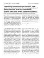
3.2. Direct Analysis of SNPs From Human Genomic DNA
Figure 4 shows the design of the Invader squared assay employed for the analysis of
SNPs in human genomic DNA by MALDI-TOF MS. Figure 4a details the general
design of the primary reaction.
For any SNP, two allele-specific probe
oligonucleotides were designed, each having identical hybridisation sequences
complementary to the target DNA. The probe oligonucleotides had different
nucleotides at the polymorphic nucleotide position (indicated by the asterisk in the
target DNA) which are designated in Figure 4 as “X” and “Y”, each being
complementary to one of the two possible nucleotides at the SNP position. The
nucleotide sequences 5’ of X and Y in the probe oligonucleotides were not
complementary to the target DNA, and were designed specifically for use in the
secondary Invader reaction. The Invader oligonucleotide was designed to be
complementary to the target upstream of the probe oligonucleotide region, with a
one nucleotide invasion into the probe base-pairing region at the SNP position, so
that enzymatic cleavage occurs immediately 3’ of nucleotide X or Y in the probe
oligonucleotide. This design confers three-fold specificity for SNP detection. First,
the Invader oligonucleotide must be complementary to the target and anneal to form
the correct overlap structure with the correctly annealed probe oligonuclaotide;
second, the endonuclease used in the Invader assay has a strict requirement of
absolute complementarity between the target and the nucleotide that occurs at the
overlap position in the probe oligonucleotide. Thus, nucleotides X or Y in the probe
oligonucleotide must be perfectly complementary to the target at the SNP position in
order for the enzyme to recognise the overlap structure and for cleavage to occur;
www.pdfgrip.com
SNP ANALYSIS BY MALDI-TOF MASS SPECTROMETRY
9
third, a mismatch at the polymorphic nucleotide between the probe oligonucleotide
and the target is thermodynamically destabilising when the reaction is run near the
of the duplex. This highly stringent three-fold specificity resulted in the allelespecific accumulation of cleavage products. If the nucleotide complementary to the
allele 1 probe was present, then cleavage product 1 accumulated; if the allele 2
nucleotide was present, cleavage product 2 accumulated; in the case of a
heterozygote, both cleavage products accumulated over time at similar rates.
After allowing the primary reaction to incubate for two hours, the reaction was
decreased and a secondary reaction mix was added that included two allele-specific
secondary target oligonucleotides, two secondary probe oligonucleotides, and one
arrestor oligonucleotide which annealed to the hybridisation sequence common to
each of the primary allele-specific probe oligonucleotides. The sequences of the
secondary target and probe oligonucleotides were designed so that the cleavage
products from the primary Invader reaction would anneal specifically to one of the
secondary targets and act as the Invader oligonucleotide in the secondary reaction.
The two allele-specific secondary systems were designed to produce biotinylated
signal molecules of unique molecular weights, so that in the subsequent MALDITOF MS analysis, the deprotonated, negative, singly-charged molecular ion values
detected (
values) would be distinct from each other
for allele
www.pdfgrip.com
10
T.J. GRIFFIN AND L.M.SMITH
1 product and 1538 for allele 2 product). Figure 4b shows the three possible
MALDI-TOF MS outputs from this Invader system, corresponding to two possible
homozygous genotypes (a single peak at an m/z value of either 1234 or 1538) or a
heterozygous genotype (peaks at both m/z values). The same two primary cleavage
product sequences shown in Figure 4a were used in every pair of SNP-specific
primary probe oligonucleotides, which enabled the use of the same secondary
oligonucleotides and signal outputs for each unique SNP analysed. The nucleotides
X and Y do not have to be complementary to the secondary target, so primary
cleavage products containing any of the four possible nucleotides at the X and Y
positions were effective as Invader oligonucleotides in the secondary reaction. A
biotin-modified deoxythymidine nucleotide was incorporated in the signal molecules
to facilitate solid-phase purification of these molecules using streptavidin coated
magnetic beads prior to analysis by MALDI-TOF MS. The signal molecules were
designed to contain only deoxythymidine nucleotides because these oligonucleotides
are more resistant to fragmentation in the MALDI process than oligonucleotides of
other sequences [16].
This approach has proven effective in the analysis of a variety of SNPs in
multiple individuals [27]. Figure 5 shows representative MALDI-TOF MS results
from the direct analysis of a human genomic DNA sample. All seven of the SNPs
analysed by this approach gave unambiguous mass spectral results, showing a single
peak in the mass spectrum in the case of homozygous genotypes, and two peaks of
approximately equal intensities in the case of heterozygotes. Additionally all
different types of SNPs (G to A transitions, G to C transversions, etc.) have been
effectively analysed [11, 27].
The design of the sequences of the oligonucleotides used in the Invader assay
was straightforward, with the only design criteria being that the sequences had
thermal duplex stabilities that enabled them to be used at the desired reaction
temperature [24, 25]. The primary probe oligonucleotides had hybridisation
sequences that were from 16 to 23 nucleotides in length depending on the target
sequence, and gave predicted
four to seven degrees above the reaction
temperature of 63° C. The primary Invader oligonucleotides were designed to have
www.pdfgrip.com
SNP ANALYSIS BY MALDI-TOF MASS SPECTROMETRY
11
a
about 15° to 20° C above the corresponding probe oligonucleotides, and were
about 30-40 nucleotides in length depending on the target sequence. The secondary
reaction oligonucleotides, were designed similarly to work at a reaction temperature
of 50° C. As the design of oligonucleotide sequences for use in the Invader assay is
simple and robust, the Invader assay should be effective in analysing the vast
majority of SNPs found throughout the human genome. As with any method
involving oligonucleotide hybridisation, sequences that form significant secondary
structures may be problematic, however, because the reaction is run at an elevated
temperature some of these problematic sequences may still be effectively analysed.
Integrating the inherent benefits of the Invader assay (highly specific, direct
signal amplification without the need for target amplification by PCR) with those
conferred by MALDI-TOF MS (extremely rapid and accurate signal detection)
represents a significant advance in the development of approaches for the highthroughput genotyping of SNPs. The relatively simple, isothermal Invader assay
and the solid-phase sample preparation procedure lend themselves nicely to
automated sample handling, giving this approach much potential to the highthroughput genotyping of SNPs for genetic analysis.
4. CONCLUSIONS
We have described two general approaches to SNP analysis by MALDI-TOF MS.
Both are designed to incorporate informative signal molecules (PNA hybridisation
probes and DNA invasive cleavage products) that are robustly analysed by MALDITOF MS and take advantage of the speed and accuracy of this analytical technology.
The approach using PNA hybridisation probes is useful for the routine analysis and
screening of all types of SNPs from PCR amplicons; the approach involving the
Invader assay is ideally suited for the high-throughput analysis of SNPs on a
genome-wide scale, useful in a wide variety of genetic studies.
5. EXPERIMENTAL METHODS
5.1. PNA Probe Synthesis and Preparation
PNA probes were synthesised by Perceptive Biosystems, Framingham, MA. These
were purified by RP-HPLC and quantified by UV absorbance at 260 nm. The purity
and m/z values of the probes were verified by MALDI-TOF MS.
5.2. PCR Amplification of Exon 4 of the Tyrosinase Gene
The
primers
5'-GGAATTCTAAAGTTTTGTGTTATCTCA-3'
and
5’TTAATATATGCCTTATTTTA-3’, employed for the amplification of human
genomic samples, yields a 347 nt fragment from exon 4 and adjacent intronic
sequences. Due to the small amount of genomic DNA sample available, these
products were re-amplified by nested-PCR using the primer set 5’-biotin-
www.pdfgrip.com
12
T.J.
GRIFFIN AND
L.M.
SMITH
CTGAATCTTGTAGATAGCTA-3’
and 5’-TATTTTTGAGCAGTGGCTCC-3’,
and the resulting 182 nt products were analysed.
5.3. Hybridisation of PNA Probes to Immobilised Gene Targets
Purified, double-stranded, biotinylated amplicons from a single PCR amplification
reaction were combined with
of streptavidin Dynabeads M-280 (Dynal,
Hamburg, Germany), and allowed to bind for 15 minutes at room temperature in
of binding buffer (10 mM Tris pH 7.0, 1 M NaCl). These were washed once with
of binding buffer.
of 0.1 M NaOH was then added to the beads and
dissociation of the double-stranded DNA was allowed to occur for 10 minutes. The
beads were washed once with
of 0.1 M NaOH and then three times with
of hybridisation buffer (10 mM Tris pH 7.0, no NaCl added) to remove the
dissociated, non-immobilised DNA strand.
Each immobilised, single-stranded PCR amplicon sample, containing all four of
the tyrosinase exon 4 point mutation targets within its sequence, was divided into
equal portions in four separate tubes and brought up in
of hybridisation buffer.
One pair of PNA probes was then added to one of the four tubes. The PNA probe
pairs were added in the following amounts (pmol WT:pmol VAR): 419-7.5:30; 42215:7.5; 446-7.5:30; 448-7.5:15. Hybridisation took place for 15 minutes at room
temperature. Each reaction tube was then heated for five minutes at the following
temperatures, depending on which pair of PNA probes had been added: 419 probes37 °C; 422 probes-58 °C; 446 probes-58 °C; 448 probes-37 °C. The optimal
amounts of each PNA probe added and also the optimal wash temperatures were
obtained empirically, in experiments using the immobilised oligonucleotides as
targets for the PNA probes. These conditions were considered to be satisfactory if
sufficient discrimination between a one-base mismatched target was obtained, as
well as approximately equal signal intensity for the two PNA probes when both
oligonucleotide targets for a probe pair were present. After this first wash, the
supernatant was then removed from each reaction tube, and
of washing buffer
(10 mM Tris, pH 7.0, 0.1% SDS) was then added to the beads and the tubes were
heated at their respective temperatures for five minutes, the supernatant removed
and the wash repeated for an additional five minutes. The beads were then rinsed
once with wash buffer, and once more with ice-cold hybridisation buffer to remove
the SDS from the beads. The beads were then brought up in
of hybridisation
buffer. This
of beads from each reaction tube was then separately spotted on
the MALDI probe tip and allowed to dry for approximately 10 minutes. To this,
of matrix (2,5-dihydroxybenzoic acid at
in 9:1
was
added and allowed to crystallise. If satisfactory crystals did not form the first time,
an additional
of matrix was then added to the beads.
5.4. MALDI-TOF MS Analysis of PNA Probes
Mass spectra were obtained on a Bruker Reflex II time-of-flight mass spectrometer
(Billerica, MA), equipped with a 337 nm
laser and operated in the linear,
www.pdfgrip.com
SNP ANALYSIS BY MALDI-TOF MASS SPECTROMETRY
13
positive-ion detection mode using delayed extraction with an initial accelerating
voltage of 25 kV. For each sample analysed, separate spectra were acquired for
each of the four polymorphic positions, and these were then summed together using
the mass spectrometer acquisition software to give a composite mass spectrum for
each sample. Calibration of the instrument was achieved by use of bovine insulin as
an external standard.
5.5. Invader Squared Reaction
All oligonucleotides used were synthesised by the University of Wisconsin
Biotechnology Centre (Madison, WI) or Integrated DNA Technologies (Coralville,
IA). All probe oligonucleotides used in the primary Invader reaction were PAGE
purified. All other oligonucleotides were synthesised with the trityl group on and
purified using Sep-Pak C18 reverse-phase purification cartridges (Waters Corp.,
Milford, MA). Each primary Invader reaction consisted of
of nuclease-free
water,
of 10X Reaction Buffer (Third Wave Technologies, Madison, WI),
of
primary Invader oligonucleotide, and
of
human genomic
DNA in water. This reaction mix was incubated at 95° C for 5 minutes to denature
the genomic DNA. The reaction mix was brought to 63° C and immediately
of
a solution containing 75 nanomoles
5 picomoles of each of the two primary
probe oligonucleotides, and 100 ng of the Afu FEN 1 enzyme (Third Wave
Technologies, Madison, WI) was added to give a final reaction volume of
This primary reaction was incubated at 63° C for 2 hours. The reaction was then
brought to 50°C and the secondary reaction mix
was added which contained
40 picomoles of 2’-O-methyl RNA arrestor oligonucleotide, 10 picomoles of each
secondary probe oligonucleotide and 0.5 picomoles of each secondary target
oligonucleotide. The secondary reaction was incubated at 50° C for 2 hours.
5.6. MALDI-TOF MS Sample Preparation of Cleavage Products
To each completed Invader reaction
of Dynabeads M-280 streptavidincoated magnetic beads (Dynal, Oslo, Norway) contained in
of
Immobilisation Buffer (10 mM Tris-HCl, 2 M NaCl, pH 7.0) was added. This
solution was mixed well and incubated at room temperature for 10 minutes with
gentle shaking. The bead solution was transferred to a 1.5 mL microcentrifuge tube
and placed in a Dynal magnetic concentrator (MC). The beads were then washed
once with
of Wash Buffer 1 (10 mM diammonium citrate, 0.1% SDS, pH
7.0) and then twice with
of Wash Buffer 2 (200 mM diammonium citrate).
The beads were then resuspended in
ultra pure deionised water, transferred
to a clean 1.5 mL microcentrifuge tube and washed 3 times with
of ultra
pure water. The washed beads were then resuspended in
of freshly prepared
Elution Buffer (1:1
and incubated at 60° C for 10 minutes.
After this incubation, the microcentrifuge tube was immediately placed in the MC
and the supernatant was removed and transferred to a clean tube, being careful to
remove the magnetic beads as completely as possible. The volatile Elution Buffer
www.pdfgrip.com
14
T.J.
GRIFFIN AND
L.M.
SMITH
was then completely removed by centrifugation under vacuum for about 15 minutes.
The clean, dry sample was then resuspended in
of 1:1
water.
5.7. MALDI-TOF MS Analysis of Cleavage Products
of MALDI matrix (1%
acid in 1:1
water) was spotted on the MALDI sample plate and allowed to airdry. To the dried matrix crystals, the resuspended sample in
of 1:1
water was added and allowed to air dry. MALDI-TOF MS
analysis was done on a Perceptive Biosystems (Framingham, MA) Voyagir DESTR mass spectrometer using a nitrogen laser at 337 nm with an initial accelerating
voltage of 20 kV and a delay time of 100 nanoseconds. The instrument was! run in
reflector mode using negative ion detection with external instrument calibration. All
spectra acquired consisted of averaged signal from 50-100 laser shots and the data
was processed using accompanying Perceptive Biosystems mass spectrometry
software.
6. AFFILIATIONS
All work was conducted at the Department of Chemistry, University of Wisconsin,
Madison, 1101 University Avenue, Madison, WI 53706.
7. REFERENCES
1.
2.
3.
4.
5.
6.
7.
8.
9.
10.
11.
12.
13.
14.
15.
16.
17.
18.
19.
20.
21.
Wang DG, Fan JB, Siao CJ, Berno A, Young P, Sapolsky R, Ghandour G, Perkins N, Winchester E,
Spencer J, Kruglyak L, Stein L, Hsie L, Topaloglou T, Hubbell E, Robinson E, Mittmann M, Morris
MS, Shen N, Kilburn D, Rioux J, Nusbaum C, Rozen S, Hudson TJ, Lander ES, et al. Science 280:
1077, 1998
Schafer AJ, Hawkins JR. Nature Biotechnol 16: 33, 1998
Landegren U, Nilsson M, Kwok PY. Genome Res 8: 769, 1998.
Brookes AJ. Gene 234: 177, 1999.
Kruglyak L. Nature Genet 17: 21, 1998.
Collins FS, Patrinos A, Jordan E, Chakravarti A, Gesteland R, Walters L. Science 282: 682, 1998
Griffin TJ, Smith LM. Trends Biotechnol 18: 77, 2000
Karas M, Hillenkamp F. Anal Chem 60: 2299, 1988
Van Ausdall DA, Marshall WS. Anal Biochem 256: 220, 1998
Griffin TJ, Tang W, Smith LM. Nat Biotechnol 15: 1368, 1997
Griffin TJ, Hall JG, Prudent JR, Smith LM. Proc Natl Acad Sci USA 96: 6301, 1999
Egholm M, Buchardt O, Christensen L, Behrens C, Freier SM, Driver DA, Berg RH, Kim SK,
Norden B, Nielsen PE. Nature 365: 566, 1993
Corey DR. Trends Biotechnol 15: 224, 1997
Tomac S, Sarkar M, Ratilainen T, Wittung P, Nielsen P, Norden B, Graslund A. J Am Chem
Soc118: 5544, 1996
Butler J, Jiang-Baucom P, Huang M, Belgrader P, Girard J. Anal Chem 68: 3283, 1996
Zhu L, Parr G, Fitzgerald M, Nelson C, Smith LM. J Am Chem Soc 117: 6048, 1995
Shaler TA, Wickham JN, Sannes KA, Wu KJ, Becker CH. Anal Chem 68: 576, 1996
Griffin TJ, Smith LM. Anal Biochem 260: 56, 1998
Giesen U, Kleider W, Berding C, Geiger A, Orum H, Nielsen PE. Nucleic Acids Res 26: 5004, 1998
Erlich GD. in PCR-based Diagnostics in Infectious Disease, Blackwell Scientific Publications, pp.
3-18, 1994
Kwok S, Higuchi R. Nature 339: 237, 1989
www.pdfgrip.com
SNP ANALYSIS BY MALDI-TOF MASS SPECTROMETRY
15
22. Farrell RE. Immunol Invest 26: 3, 1997
23. Vaneechoutte M, Van Eldere J. J Med Microbiol 46: 188, 1997
24. Lyamichev V, Mast AL, Hall JG, Prudent JR, Kaiser MW, Takova T, Kwiatkowski RW, Sander TJ,
de Arruda M, Arco DA, Neri BP, Brow MA. Nature Biotechnol 17: 292, 1999
25. Lyamichev V, Brow MA, Varvel VE, Dahlberg JE. Proc Natl Acad Sci U S A 96: 6143, 1999
26. Hall JG, Eis PS, Law SM, Reynaldo LP, Prudent JR, Marshall DJ, Allawi HT, Mast AL, Dahlberg
JE, Kwiatkowski RW, de Arruda M, Neri BP, Lyamichev VI. Proc Natl Acad Sci USA 97: 8272,
2000
27. Griffin TJ, Smith LM. Anal Chem 72: 3298, 2000
www.pdfgrip.com