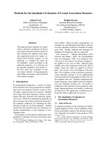
Tài liệu Báo cáo Y học: Insights into the reaction mechanism of Escherichia coli agmatinase by site-directed mutagenesis and molecular modelling ppt
Bạn đang xem bản rút gọn của tài liệu. Xem và tải ngay bản đầy đủ của tài liệu tại đây (406.04 KB, 5 trang )
Insights into the reaction mechanism of
Escherichia coli
agmatinase
by site-directed mutagenesis and molecular modelling
A critical role for aspartate 153
Mo
´
nica Salas
1
, Rolando Rodrı
´
guez
2
, Nelia Lo
´
pez
2
, Elena Uribe
1
, Vasthi Lo
´
pez
1
and Nelson Carvajal
1
1
Departamento de Biologı
´
a Molecular, Facultad de Ciencias Biolo
´
gicas, Universidad de Concepcio
´
n, Casilla 160-C, Concepcio
´
n,
Chile;
2
Center for Genetic Engineering and Biotechnology, Habana, Cuba
Upon mutation of Asp153 by asparagine, the catalytic
activity of agmatinase (agmatine ureohydrolase, EC
3.5.3.11) from Escherichia coli was reduced to about 5% of
wild-type activity. Tryptophan emission fluorescence (k
max
¼ 340 nm), and CD spectra were nearly identical for wild-
type and D153N agmatinases. The K
m
value for agmatine
(1.6 ± 0.1 m
M
),aswellastheK
i
for putrescine inhibition
(12 ± 2 m
M
) and the interaction of the enzyme with the
required metal ion, were also not altered by mutation. Three-
dimensional models, generated by homology modelling
techniques, indicated that the side chains of Asp153 and
Asn153 can perfectly fit in essentially the same position in the
active site of E. coli agmatinase. Asp153 is suggested to be
involved, by hydrogen bond formation, in the stabilization
and orientation of a metal-bound hydroxide for optimal
attack on the guanidinium carbon of agmatine. Thus, the
disruption of this hydrogen bond is the likely cause of the
greately decreased catalytic efficiency of the D153N variant.
Keywords: agmatinase; Asp153; site-directed mutagenesis;
homology-modelling; E. coli.
Agmatinase (agmatine ureohydrolase, EC 3.5.3.11) cata-
lyses the hydrolysis of agmatine to putrescine and urea [1].
Agmatine, which results from decarboxylation of arginine
by arginine decarboxylase [2], is a metabolic intermediate in
the biosynthesis of putrescine and higher polyamines [1] and
may have important regulatory roles in mammals [3–5].
Agmatinases from Escherichia coli and human tissues,
and putative agmatinases from Synechocystic sp. Schizo-
saccharomyces pombe and Bacillus subtilis, have been cloned
and the deduced amino acid sequences indicate their
homology to all sequenced arginases [4–7]; all these enzymes
catalyse an hydrolytic reaction with production of urea. The
question arises therefore as to whether a similar or identical
mechanism is involved in catalysis by these enzymes, which
apparently evolved from a single primordial protein [6,7]. In
this context, both enzymes exhibit an absolute requirement
for Mn
2+
for catalytic activity [8,9]; the well established
requirement of a binuclear metal cluster for full catalytic
activity of arginase [8] is probably also valid for agmatinase
[9]. This is reinforced by the fact that residues known to be
metal ligands in arginase are strictly conserved in the
sequence of agmatinase [7]. Moreover, a critical role for one
conserved histidine residue (His163 in the sequence of
E. coli agmatinase) has been shown by chemical modifica-
tion and site-directed mutagenesis of human and rat liver
arginases [10,11] and E. coli agmatinase [12]; similar infor-
mation was deduced from X-ray crystallographic data for
arginase from Bacillus caldovelox [13].
Based on the crystal structure of rat liver arginase, it was
suggested that arginine hydrolysis involves the participation
of a metal-bound hydroxide, which is stabilized for optimal
nucleophilic attack at the substrate, by donating an
hydrogen bond to Asp128 [8,14,15]. In this connection,
the D128G variant of human liver arginase was described as
inactive [16,17], although the possible influence of structural
changes accompanying the mutation were not examined.
Since this aspartate is conserved among all sequenced
arginases and agmatinases [4–7], a critical role for the
equivalent residue in agmatinase (Asp153), may be reason-
ably expected. This expectation is supported by our present
findings of a markedly decreased activity of a D153N
variant of E. coli agmatinase. From the enzymic properties
of D153N agmatinase and a modelled structure, we
conclude that the lower activity of the mutant may be
ascribed to the loss of an acceptor hydrogen bond to a
metal-bound hydroxide, as a consequence of replacement of
a carboxylate oxygen with an amide group.
MATERIALS AND METHODS
Materials
All reagents were of the highest quality commercially
available (most from Sigma Chemical Co.) and were used
without further purification. Restriction enzymes, as well as
enzymes and reagents for PCR were obtained from
Promega. The plasmid pKA5, bearing the speB gene of
E. coli agmatinase, was kindly supplied by S. Boyle (Vir-
ginina Polytechnic Institute and State University). The
pQE60 E. coli expression vector and the Ni-nitrilotriacetic
acid resin were obtained from Qiagen, and synthetic
Correspondence to N. Carvajal, Departamento de Biologı
´
a Molecular,
Facultad de Ciencias Biolo
´
gicas, Universidad de Concepcio
´
n,
Casilla 160-C, Concepcio
´
n, Chile. Fax: + 56 41 239687;
E-mail:
(Received 5 June 2002, revised 9 September 2002,
accepted 12 September 2002)
Eur. J. Biochem. 269, 5522–5526 (2002) Ó FEBS 2002 doi:10.1046/j.1432-1033.2002.03255.x
nucleotide primers from the Centro de Estudios Avanzados
(Universidad de Chile).
Enzyme preparations and enzyme assays
Bacteria were grown with shaking at 37 °C in Luria broth in
the presence of ampicillin (100 lgÆmL
)1
). The wild-type and
D153N agmatinase cDNAs were directionally cloned into
the histidine tagged pQE60 E. coli expression vector and the
histidine-tagged enzyme was expressed in E. coli strain
JM109, following induction with 1 m
M
isopropyl thio-b-
D
-galactoside. The histidine-tagged enzymes were purified
to homogeneity by metal chelate chromatography over
Ni-nitrilotriacetic acid resin, according to the instructions of
the manufacturer. A single protein band was detected by
SDS/PAGE of purified enzymes.
Enzyme activities were determined by measuring the
formation of urea from agmatine in 50 m
M
glycine/NaOH
(pH 9.0). Urea was determined by a colorimetric method
with a-isonitrosopropiophenone [18] and protein concen-
trations were estimated by the method of Bradford [19],
with bovine serum albumin as standard. Kinetic data were
analyzed by double reciprocal plots, and the K
i
value for
putrescine inhibition was determined from a replot of slopes
vs. inhibitor concentration. All lines were computer-fitted to
the appropriate equations.
Site-directed mutagenesis
The D153N mutant form of E. coli agmatinase was
obtained by a two-step PCR [20], using the plasmid
pKA5 containing the speB gene of E. coli agmatinase as a
template. A first PCR product was obtained using the 5¢
sense primer 5¢-AGTCCATCCATGGGCACCTTAG-3¢
and a 3¢ complementary primer corresponding to nucleo-
tides 448–468 of agmatinase with a CfiT substitution at
nucleotide 457 (sequence: 5¢-CGCATAGGTAT
TG
GTGTGGGC-3¢). Similarly, the second PCR product was
obtained using the 5¢ sense primer corresponding to
nucleotides 448–468 of agmatinase with a GfiA substitu-
tion at nucleotide 457 (sequence: 5¢-GCCCACACC
AAT
ACCTATGCG-3¢) and the 3¢ complementary primer 5¢-AT
TAATGGCATGCTTTACCCGT-3¢.UsingthePCR
products of agmatinase with the GfiAandCfiT substi-
tutions in the coding and noncoding strands, respectively,
and using the 5¢ and 3¢ primers mentioned above, the full
length agmatinase cDNA coding for the D153N mutant
was generated by a second round of PCR. The expected
mutation was confirmed by DNA sequence analysis. That
no unwanted mutations had been introduced during the
mutagenesis process was verified by automated sequencing.
The H163F E. coli agmatinase was obtained as described
previously [10].
Fluorescence spectra
Fluorescence measurements were made at 25 °Cona
Shimadzu RF-5301 spectrofluorimeter. The protein concen-
tration was 30 lgÆmL
)1
and emission spectra were measured
with the excitation wavelength at 295 nm. The slit width for
both excitation and emission was 1.5 nm, and spectra were
corrected by subtracting the spectrum of the buffer solution
(5 m
M
Tris/HCl, pH 7.5) in the absence of protein.
Circular dichroism
CD experiments were performed on a Jasco J-810 spectro-
polarimeter thermostated at 22 °C. CD spectra of wild-type
and D153N mutant enzymes (5.5 l
M
) were measured in the
range 200–250 nm, with a bandwidth of 1 nm and a scan
speed of 50 nmÆmin
)1
. The buffer solution contained 5 m
M
Tris/HCl (pH 7.5) and 2 m
M
Mn
2+
. The reported spectra
represents the averages of five repeat scans. Spectra were
smoothed and analysed for protein secondary structures by
using the software package provided with the instrument.
Molecular modelling
The agmatinase structural model was obtained by homology
methods, using the structure of B. caldovelox arginase (PDB
id: 1CEV) as a template and the modelling package
WHAT IF
[21]. An amino-acid sequence identity of 29% was calculated
for E. coli agmatinase and B. caldovelox arginase. The
sequence alignment used in the modelling experiment was
derived from the structural superposition of two arginase
structures (rat liver and B. caldovelox arginases, PDB id
1RLA and 1CEV, respectively). The agmatinase
SPEB_ECOLI sequence, obtained from the Swissprot
database (accession number P16936), was separately aligned
with the sequences of 1CEV and 1RLA and pasted into the
structural alignment. The alignment was then corrected by
hand. Since the sequences of the two arginases and
agmatinase greately differ in the N terminal region, the
agmatinase and rat liver arginases were stripped of the first
32 and five amino acids, respectively. The location of the
gaps was optimized by repetitive modelling, shuffling those
aminoacids that were not conserved in the structural
alignment, to obtain the shortest possible Ca-Ca distance.
The resulting sequence alignment, along with the secondary
structure elements, is shown in Fig. 1. All loops that were
different due to insertions or deletions in the agmatinase
sequence were modelled using the DGLOOP set of options
in
WHATIF
; the whole loops and the two connecting amino
acids at the beginning and the end, were mutated to glycines
and, after remodelling back to their side chains in agma-
tinase, the whole hydrogen bond network was optimized.
The position of the active site manganese ions was calculated
using the averaged distance template of 1CEV and 1RLA,
and then a full hydrogen bond network optimization was
performed again. Agmatine was added to agmatinase using
the same superposition matrix calculated for manganese
ions, in order to conserve the ligand-manganese distances.
RESULTS AND DISCUSSION
By site-directed mutagenesis, a D153N mutant form of
E. coli agmatinase was obtained. The mutant enzyme
retained about 5% of wild-type activity and it was equally
active when assayed in the presence or absence of Mn
2+
.
However, as shown in Fig. 2, it was half inactivated by
dialysis against 10 m
M
EDTA in 5 m
M
Tris/HCl (pH 7.5)
for 4 h at 4 °C and full recovery of enzyme activity was
produced by incubation of the EDTA-treated species with
2m
M
Mn
2+
for 20 min at 37 °C; as expected, the effect of
the metal ion was again reversed by EDTA. These results
indicate the presence of tightly and weakly bound mangan-
ese ions in fully active species of the mutant enzyme.
Ó FEBS 2002 A critical role for Asp153 in E. coli agmatinase (Eur. J. Biochem. 269) 5523
Identical behaviour was previously described, and con-
firmed here (Fig. 2), for wild-type agmatinase and this was
interpreted as supporting the presence of a binuclear metal
center in the active site of fully activated agmatinase [9]. The
K
m
for agmatine (1.6 ± 0.1 m
M
) and the K
i
for competitive
inhibition by putrescine (12 ± 2 m
M
), were also essentially
equal for wild-type and D153N agmatinases. It is clear
therefore that altered interactions with the substrate or a
significantly altered affinity for the activating metal ion, are
not the explanations for the greatly decreased catalytic
activity of the D153N variant.
To evaluate possible structural changes that may result
from mutation, wild-type and D153N enzymes were com-
pared by using fluorescence and CD spectrometry. The
tryptophan emission fluorescence spectra were not altered
by mutation (k
max
¼ 342 nm), indicating that the environ-
ment of tryptophan residues is essentially conserved in the
mutant enzyme. On the other hand, the absence of major
differences in the CD spectra of the wild-type and D153N
agmatinases indicates that mutation had no effect on their
respective secondary structures (Fig. 3). As an example, the
percentage values calculated for the a-helix were 22.3 and
21.5% for wild-type and D153N enzymes, respectively.
Therefore, based on the criteria used in this study, we may
discard a gross structural change as the explanation for the
lower catalytic activity of D153N agmatinase.
Since an experimentally derived structure is not yet
available for any agmatinase, to further evaluate the
consequences of the Asp153fiAsn substitution, we used a
modelled structure of the E. coli enzyme, constructed by
using homology-modelling techniques and the 3D structure
of the binuclear form of B. caldovelox arginase as a
template. Principal attention was given to the modeling of
the active site. The modelled structure was very similar to
the template, with respect to the number and arrangement
of structural elements (a-helix and b-sheets), and one of the
major differences concerned the surface loops. However, as
showninFig.4,B. caldovelox and rat liver arginases also
Fig. 2. Effect of added Mn
2+
and EDTA on the catalytic activity of
wild-type and D153N agmatinase. The enzymes were assayed before
(Control) and after dialysis against 10 m
M
EDTA in 5 m
M
Tris/HCl
(pH 7.5) for 4 h at 4 °C. Enzyme activities were measured with and
without a previous incubation with 2 m
M
Mn
2+
for 20 min at 37 °C.
Activities are expressed as percentage of the corresponding control not
preincubated with Mn
2+
andassayedintheabsenceofaddedmetal
ion.
Fig. 1. Structural sequence alignment of
B. caldovelox arginase (1cev), E. coli agma-
tinase (AUH) and rat liver arginase (1rla). H, S,
T and 3 stands for a-helix, strand, turn and
3
10
-helix. Conserved residues are marked by
an asterisk.
5524 M. Salas et al. (Eur. J. Biochem. 269) Ó FEBS 2002
differed in these areas. A more specific difference concerned
a loop located at the entrance of the active site cleft, and
defined by residues 124–141 in the sequence of the bacterial
arginase. Whereas the backbone and some of the side chains
are very precisely conserved in both arginases, the agma-
tinase loop was shorter when compared with the same
region of the arginases (Fig. 4). Since the arginase loop
contains residues that interacts with the a-carboxylate
group of the substrate arginine [13], and this is the part of
the molecule that makes arginine different from agmatine, it
seems reasonable to assume that differences in this loop area
are key factors in determining the difference in substrate
specificity between arginase and agmatinase. This aspect is
presently under investigation in our laboratory. In any case,
despite the structural differences between agmatinase and
arginase, agmatine and arginine were fixed in essentially the
same position in the corresponding active site. The same
position for the scissile guanidine carbon of the substrates,
with respect to the metal ions and conserved, catalytically
important residues, agree with a similar, if not identical,
mechanism for both enzymes.
Upon replacement of Asp153 with asparagine, the whole
topology of the active site was found to be conserved in
agmatinase. In the modelled active site structure, the side
chains of Asp153 and Asn153 can be accommodated at
essentially the same position, with the whole distance
network remaining almost intact (Fig. 5). Moreover, one of
the carboxylate oxygens of Asp153 and the carboxamide
oxygen of Asn153 are positioned in such a way as to allow
metal coordination interaction with one of the manganese
ions. This, together with the fact that the positions of other
Fig. 4. A superimposition of the structures of B. caldovelox arginase
(red), rat liver arginase (yellow) and the modelled structure of E. coli
agmatinase (blue). Note the shorter extension, in the case of E. coli
agmatinase, of the loop indicated by the letter a.
Fig. 5. Scheme of the binuclear manganese cluster and the localization
of the side-chains of Asp153 and Asn153 in the modelled structures of
wild-type and D153N mutant forms of E. coli agmatinase. Average
distances (in A
˚
) are indicated by the numbers. For simplicity, other
active site residues, including other metal ligands, are not indicated.
Fig. 3. CD spectra of wild-type (solid line) and D153N mutant (dotted
line) E. coli agmatinases. The far-UV CD spectra were recorded at
22 °C.
Ó FEBS 2002 A critical role for Asp153 in E. coli agmatinase (Eur. J. Biochem. 269) 5525
potential metal ligands are not substantially affected by the
mutation, as revealed by the conservation of the whole
topology of the active site, would explain the essentially
unaltered interaction of the D153N variant with the metallic
cofactor.
In the modelled structure, the noncoordinated carboxy-
late oxygen atom of Asp153 is within hydrogen-bonding
distance of the metal-bound water molecule. This is
interesting if one considers a catalytic mechanism involving
a nucleophilic attack of a metal-bound hydroxide on the
guanidinium carbon of agmatine [8,9]. By donating an
hydrogen bond to Asp153, the nucleophile would be
stabilized and oriented for optimal catalysis. On this basis,
the disruption of this stabilizing hydrogen bond, due to the
replacement of the carboxylic oxygen by the amide nitrogen,
would be expected to result in a less efficient catalysis by the
metal-bound hydroxide in the D153N mutant enzyme. The
low, but significant catalytic activity of the mutant indicates
that, even in the absence of the hydrogen bond to the
noncoordinating carboxyl oxygen, the metal-bound
hydroxide could still serve as a catalytic nucleophile,
although considerably less efficiently. Further studies are,
evidently, required to clarify this aspect. The proposed role
for the critical Asp153 in E. coli agmatinase reinforces the
relationships between this enzyme and the evolutionary
related arginase. It would be, thus, of interest to examine
the effects of the corresponding aspartate to asparagine
mutation in this enzyme.
ACKNOWLEDGEMENTS
This research was supported by Grant 2990049 from FONDECYT and
P.I. 98.031.076-1.0 from the Direccio
´
ndeInvestigacio
´
n, Universidad de
Concepcio
´
n. We are greateful to Dr Enrique Pe
´
rez Paya (Universidad
de Valencia, Espan
˜
a) for assistance with the CD spectra.
REFERENCES
1. Satishchandran, C. & Boyle, S.M. (1986) Purification and prop-
erties of agmatine ureohydrolyase, a putrescine biosynthetic
enzyme in Escherichia coli. J. Bacteriol. 165, 843–848.
2. Buch, J.K. & Boyle, S.M. (1985) Biosynthetic arginine
decarboxylase in Escherichia coli is synthesized as a precursor and
locatedinthecellenvelope.J. Bacteriol. 163, 522–527.
3. Gilad, G.M., Wollam, Y., Iaina, A., Rabey, J.M., Chernihovsky,
T. & Gilad, V.H. (1996) Metabolism of agmatine into urea but not
into nitric oxide in rat brain. Neuroreport 7, 1730–1732.
4. Iyer,R.K.,Kim,H.K.,Tsoa,R.W.,Grody,W.W.&Cederbaum,
S.D., (2002) Molecular cloning and characterization of human
agmatinase. Genet. Metabol. 75, 209–218.
5. Mistry, S.K., Burwell, T.J., Chambers, R.M., Rudolph-Owen, L.,
Spaltmann, F., Cook, W.J. & Morris, S.M. (2002) Cloning of
human agmatinase. An alternate path for polyamine synthesis
induced in liver by hepatitis B virus. Am. J. Physiol. Gastrointest
Liver Physiol. 282, G375–G381.
6. Ouzounis, C.A. & Kyrpides, N.C. (1994) On the evolution of
arginases and related enzymes. J. Mol. Evol. 39, 101–104.
7. Perozich, J., Hempel, J. & Morris, S.M. (1997) Roles of conserved
residues in the arginase family. Biochim. Biophys. Acta 1328,
23–37.
8. Kanyo, Z.F., Scolnick, L.R., Ash, D.E. & Christianson, D.W.
(1996) Structure of a unique binuclear manganese cluster in argi-
nase. Nature 382, 554–557.
9. Carvajal, N., Lo
´
pez,V.,Salas,M.,Uribe,E.,Herrera,P.&Cerpa,
J. (1999) Manganese is essential for catalytic activity of Escherichia
coli agmatinase. Biochem. Biophys. Res. Commun. 258, 808–811.
10. Carvajal, N., Olate, J., Salas, M., Uribe, E., Lo
´
pez, V., Herrera, P.
& Cerpa, J. (1999) Chemical modification and site-directed
mutagenesis of human liver arginase: evidence that the imidazole
group of histidine-141 is not involved in substrate binding. Arch.
Biochem. Biophys. 371, 202–206.
11. Cavalli, R.C., Burke, C., J., Kawamoto, S., Soprano, D.R. & Ash,
D.E. (1994) Mutagenesis of rat liver arginase expressed in
Escherichia coli: role of conserved histidines. Biochemistry 33,
10652–10657.
12. Carvajal, N., Olate, J., Salas, M., Lo
´
pez, V., Cerpa, J., Herrera, P.
& Uribe, E. (1999) Evidence that histidine-163 is critical for cata-
lytic activity, but not for substrate binding to Escherichia coli
agmatinase. Biochem. Biophys. Res. Commun. 264, 196–200.
13. Bewley, M.C., Jeffrey, P.D., Patchett, M.L., Kanyo, Z.F. & Baker,
E.N. (1999) Crystal structures of Bacillus caldovelox arginase in
complex with substrate and inhibitors reveal new insights into
activation, inhibition and catalysis in the arginase superfamily.
Structure 7, 435–448.
14.Ash,D.E.,Cox,J.D.&Christianson,D.W.(2000)Arginase:
a binuclear manganese metalloenzyme. Met Ions Biol. Syst. 37,
407–428.
15. Christianson, D.E. & Cox, J.D. (1999) Catalysis by metal-acti-
vated hydroxide in zinc and manganese metalloenzymes. Annu.
Rev. Biochem. 68, 33–57.
16. Vockley, J.G., Tabor, D.E., Kern, R.M., Goodman, B.K., Wiss-
man,P.B.,Kang,D.S.,Grody,W.W.&Cederbaum,S.D.(1994)
Identification of mutations (D128G, H141L) in the liver arginase
gene of patients with hyperargininemia. Hum. Mutat. 4, 150–154.
17. Ash, D.E., Scolnick, L.R., Kanyo, Z.F., Vockley, J.G., Ceder-
baum, S.D. & Christianson, D.W. (1998) Molecular basis of
hyperargininemia: structure–function consequences of mutations
in human liver arginase. Mol. Genet. Metabol. 64, 243–249.
18. Archibald, R.M. (1945) Colorimetric determination of urea.
J. Biol. Chem. 157, 507–518.
19. Bradford, M.M. (1976) A rapid and sensitive method for the
quantitation of microgram quantities of protein utilizing the
principle of protein-dye binding. Anal. Biochem. 72, 248–254.
20. Ho, S.N., Hunt, H.D., Horton, R.M., Pullen, J.K. & Pease, L.R.
(1989) Site-directed mutagenesis by overlap extension using the
polymerase chain reaction. Gene 77, 51–59.
21. Vriend, G. (1990) WHAT IF: a molecular modelling and drug
design program. J. Mol. Graph. 8, 52–56.
5526 M. Salas et al. (Eur. J. Biochem. 269) Ó FEBS 2002