Tài liệu Báo cáo khoa học: Solution NMR structure of five representative glycosylated polyene macrolide antibiotics with a sterol-dependent antifungal activity doc
Bạn đang xem bản rút gọn của tài liệu. Xem và tải ngay bản đầy đủ của tài liệu tại đây (334.29 KB, 9 trang )
Solution NMR structure of five representative glycosylated polyene
macrolide antibiotics with a sterol-dependent antifungal activity
Laurent Volpon and Jean-Marc Lancelin
Laboratoire de RMN Biomole
´
culaire associe
´
au CNRS, Universite
´
Claude Bernard – Lyon 1 and Ecole Supe
´
rieure de Chimie
Physique & Electronique de Lyon, Villeurbanne, France
Glycosylated polyene macrolide antibiotics, as nystatins and
amphotericins, are amphiphilic structures known to exert
antifungal activity by disrupting the fungal cell membrane,
leading to leakage of cellular materials, and cell death. This
membrane disruption is strongly influenced by the presence
and the exact nature of the membrane sterols. The solution
structures of five representative glycosylated members, three
tetraenes (pimaricin, nystatin A1 and rimocidin) and two
heptaenes (candidin and vacidin A) have been calculated
using geometric restraints derived from
1
H-NMR data and
random searches of their conformational space. Despite a
different apparent structural order, the NMR solutions
structure indicate that the hydroxyl groups all clustered on
one side of the rod-shaped structures, and the glycosyl
moieties are structurally conserved both in their conforma-
tion and their apparent order. The molecular structures
afford an understanding of their selective interaction with the
membrane sterols and the design of new polyene macrolides
with improved activities.
Keywords: antifungal antibiotics; polyene macrolides; sterol-
dependant antibiotics; NMR solution structure; 1, 3-polyols.
The vast family of polyenes antibiotics [1,2] includes
amphiphilic compounds mostly produced by Streptomyces
species with potent antifungal properties. Polyene macro-
lides are of an authentic clinical value for efficient therapies
against animals, and human infectious diseases caused by
pathogenic fungi. In particular, nystatin A1, amphotericin
B, and pimaricin (natamycin) are the most common
polyene macrolides used for the treatment of fungal
infections. Due to its particular low toxicity, pimaricin
also has been used for a decade as a food preservative [3,4]
allowed in the European Union (additive E235) and
USA for preserving foods from mold contamination and
possible inherent risks of mycotoxin poisoning. The
polyene macrolides target the cytoplasmic membranes of
fungi where they interact selectively with ergosterol,
causing a major disorganization of the membrane structure
[5] leading to the leakage of cellular materials and in turn
the cellular death.
Depending on their molecular structures, polyene
macrolides have a more or less toxicity, in part due to a
residual interaction with cholesterol in mammalian cyto-
plasmic membranes. This gives to polyene macrolides
therapies undesired hemolytic and nephrolytic side-effects.
Other relevant effects assigned to some polyene macro-
lides, such as antiviral properties against several groups of
enveloped viruses [6,7] or stimulation of the immune
response at lower concentrations [8,9], have been also
reported. These activities make of polyene macrolides a
source of lead structures for the engineering of future
molecules with improved medicinal purposes. In parti-
cular, the gene clusters involved in the biosynthesis of
pimaricin in S. natalensis [10,11] and nystastin in S. nour-
sei [12] have been recently cloned and new models for
their biosynthetic pathways been proposed. These new
insights make bioengineering possible for new polyene
macrolides in addition to chemical synthesis.
Despite their discovery over 50 years ago, the under-
standing of the selective affinity of polyenes macrolides
for sterols in biomembranes has yet no experimental
molecular explanation at atomic resolution. The con-
formational analysis of different members of the polyene
family is one important step essential in understanding
their structure-to-activity relationships. Three-dimen-
sional structures of only three polyene macrolides of
disparate nature and activity, have been described to
date. Amphotericin B [13,14] and roxaticin [15] were
solved by crystallography, while filipin III was solved
using solution NMR [16].
Full stereochemical information (with the exception of
one chiral center at position 42 of the vacidin A side chain,
Fig. 1) are available for at least five polyene macrolides that
belong to a group of polyenes specifically glycosylated by
mycosamine, a hexose of the
D
-series. The glycosylation by
an amino sugar occurs near a carboxylic acidic function of
the macrolide, so that these polyene macrolides are zwiter-
ionic in addition to being amphiphilic. Nystatin A1 was the
first polyene macrolide discovered [17]. Its covalent struc-
ture (without stereochemistry) was confirmed in 1970 [18]
and 1971 [19]. Pimaricin, or natamycin [20], was isolated in
1957 [21] and its covalent structure was established by
Golding et al. [22]. Rimocidin from S. rimosus was reported
in 1951 [23] and its covalent structure finally described in
1977 [24]. Vacidin A, one of the main components of the
aureofacin complex from S. aureofaciens, belongs to the
Correspondence to J M. Lancelin, Laboratoire de RMN
Biomole
´
culaire, Universite
´
Claude-Bernard – Lyon 1, Domaine
Scientifique de La Doua, CPE – Lyon, 43, boulevard du 11 Novembre
1918, F-69622 Villeurbanne cedex, France.
Fax/Tel.:+33472431395,
E-mail:
Abbreviation: ROE, rotating frame Overhauser effect.
(Received 11 March 2002, revised 17 July 2002, accepted 23 July 2002)
Eur. J. Biochem. 269, 4533–4541 (2002) Ó FEBS 2002 doi:10.1046/j.1432-1033.2002.03147.x
aromatic macrolide group [25]. Finally, candidin is a
main component of the antibiotic complex produced by
S. viridoflavus [26]. These five polyene macrolides have a 26-
to 38-membered macrolactone ring, containing a polyol
and a polyene part, which is the origin of their amphiphilic
nature, and a
D
-mycosamine sugar (Fig. 1). Asymmetric
centers of these five glycosylated polyenes were character-
ized by of NMR spectroscopy and stereo-controlled organic
synthesis for nystatin A1 [27,28], pimaricin [29,30], rimo-
cidin [31], vacidin A [32] and candidin [33].
We took the advantage of the complete knowledge of the
stereochemical information of these five polyene macrolides
to study their conformation in solution, using NMR and
molecular modeling protocols used to explore the confor-
mation space of biopolymers [34]. We report herein, the first
comparative solution NMR structures of these five repre-
sentative 26–38-membered polyene macrolides glycosylated
by
D
-mycosamine.
MATERIALS AND METHODS
NMR experiments
Nystatin A1 sample was obtained from Dr C. Cimarusti,
The Squibb Institute for Medical Research (Princeton, New
Jersey, USA) [35] and rimocidin (Pfizer Lot #4157-47-2)
from Prof Kenneth L. Rinehart, University of Illinois
(Urbana-Champaign, Illinois, USA). The antibiotic solu-
tions were prepared under dry argon in methanol-d
4
at
3–5 m
M
concentration. All NMR spectra were recorded at
25 °C on a Bruker Avance DRX 500 spectrometer
(
1
H ¼ 500 MHz) using a 5-mm (
1
H,
13
C,
15
N) triple-
resonance probe head, equipped with a supplementary self
shielded z-gradient coil. Spectra were processed using
Bruker XWINNMR and GIFA V.4 [36] software. Homo-
nuclear two-dimensional spectra DQF-COSY [37], TOCSY
(HOHAHA) [38,39] and ROESY [40,41], were recorded
with a 1.5-s recovery delay in the phase-sensitive mode using
the States-TPPI method [42] as data matrices of 512
(t
1
) · 1024 (t
2
) complex data points. Mixing times of
80 ms for TOCSY and 250 ms for ROESY spectra were
used. The spectral width in both dimensions was 3500 Hz.
The data were apodized with shifted sine-bell and Gaussian
window functions in both F
1
and F
2
dimensions after zero-
filling in the t
1
dimension to obtain a final matrix of 1024
(F
1
) · 1024 (F
2
) real data points. Chemical shifts were
referenced to the solvent chemical shift (CHD
2
OD,d
(
1
H) ¼ 3.31 p.p.m.).
For heteronuclear spectroscopy, phase-sensitive
13
C-
heteronuclear single quantum coherence [43] were recorded
with a 1.5-s recovery delay using the echo-antiecho method
[44]. The coherence pathway selection was achieved by
applying pulsed-field gradients as coherence-filters [45,46].
The FID was collected as a data matrix of 512 (t
1
,
13
C) · 1024 (t
2
,
1
H) complex data points and 150 scans
per t
1
increment. Spectral widths were 3500 Hz in F
2
and
17450 Hz in F
1
with carrier frequencies at 3.7 and 70 p.p.m.,
respectively.
For the other three polyenes, the NMR data at
1
H ¼ 300 MHz were taken from the literature where
complete
1
H-NMR assignment and experimental restraints
are available. Pimaricin was studied in methanol-d
4
[29,30],
candidin in a methanol-d
4
/pyridine-d
5
/DMSO-d
6
(2:2:1,
v/v) mixture [33], vacidin A in a pyridine-d
5
/methanol-d
4
(9 : 1, v/v) mixture [32].
Experimental NMR restraints
For nystatin A1, pimaricin, rimocidin and vacidin A, all the
interproton-distance restraints between non J-coupled pro-
tons, are derived from the two-dimensional homonuclear
ROESY experiments. Interproton restraints were classified
into three categories. Upper bounds were fixed at 2.8, 3.3
and 4.0 A
˚
for strong, medium and weak correlations,
respectively. For candidin, each of the ROE correlations
were considered as weak correlations as no information
concerning the ROEs relative intensity were given [32]. A
lower bound was fixed at 1.8 A
˚
, which corresponds to the
sum of the hydrogen van der Waals’ radii. The intensity of a
H
i
) H
i+2
ROEs within the polyene part was considered as
reference intensity for strong correlations [29,30]. Pseudo
atom corrections [47] of the upper bounds were applied for
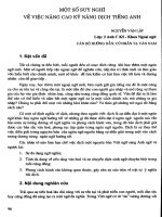
Fig. 1. Molecular structures. (A) Pimaricin (B) nystatin A1 (C) rimo-
cidin (with R
1
:CH
2
–CH
3
;R
2
:CH
2
–CH
2
–CH
3
) (D) candidin and (E)
vacidin A. R or S absolute configurations are indicated for asymmetric
centers. Carbons are numbered according to their position in the
macrolide sequence. Primed indices are assigned to the
D
-mycosamine
glycoside on the right.
4534 L. Volpon and J M. Lancelin (Eur. J. Biochem. 269) Ó FEBS 2002
distance restraints involving the unresolved methylene and
methyl protons (+1 A
˚
). For nonstereospecifically assigned
but spectroscopically resolved diastereotopic methylene
protons, the interproton distances were treated as single
(Ær
)6
æ)
)1/6
average distances. When possible, H–C–C–H
dihedral angle were restrained to dihedral domains accord-
ing to the different
3
J
HC,CH
coupling constants measured
using optimized Karplus dihedral relations [16]. When
different or very large domains were deduced, some of them
could be further restricted from intermediate structure
calculations without dihedral restraints. If the resulting
models with acceptable energy (see Results) gave a parti-
cular dihedral value compatible with the
3
J
HC,CH
,the
corresponding restraint was applied in a narrower domain.
The smallest final dihedral domains were not more restricted
than an arbitrary value of ± 20° (Table 3) in case of a
correct match between the dihedral angles and the measured
couplings. If the dihedral value in the intermediate models
was too dispersed, no further restriction was applied.
Structure calculations
Models were calculated using the
X
-
PLOR
software version
3.851 [48] as previously described [16]. Initial atomic
coordinates and structure files for each polyene macrolides
were generated step by step (given as supporting informa-
tion) from the
X
-
PLOR
libraries and topology files of
different parts of other molecules taken from the Protein
Data Bank [49]. For each molecule, the atoms involved in
of the lactone function (C–CO–O–C) were maintained in a
plane. The hemi-ketal 6-membered rings of the macrolides
were maintained in a chair conformation by definition of
suitable improper angle restraints. The results were visu-
alized using the program
MOLMOL
version 2.4 [50]. Starting
from 30 randomized coordinates, the sampling of the
conformational space was performed following a simulated
annealing protocol (random SA) proposed by Nilges et al.
[51]. The simplified allhdg.pro force field of
X-PLOR
was
used. The nonbonded van der Waals’ interactions were
represented by a simple repulsive quadratic term [34,51].
The experimental distance restraints were represented as
a soft asymptotic potential, and electrostatic interactions
were ignored. The force constant associated with the
distance restraints was kept to 50 kcalÆmol
)1
ÆA
˚
)2
through-
out the protocol. One cycle of random SA consisted of
1500 steps of 3 fs at 1000 K followed by 3000 cooling steps
of 1 fs from 1000 K to 100 K. At the end, each structure
was subjected to 1500 steps of conjugate gradient energy
minimization.
RESULTS
NMR assignments of nystatin A1 and rimocidin in
methanol-
d
4
The structure-specific assignment of the
1
Hand
13
C
resonances of nystatin A1 and rimocidin in neat meth-
anol-d
4
(Table 1) were carried out based on the identifica-
tion of various unambiguous resonances. These signals were
used as starting points for the complete assignments. In
particular, for nystatin A1, starting points were: (a) the well-
defined scalar correlations between the two methylene
protons at C28 and C29 with their neighboring CH (Fig. 2);
(b) the narrow resonance of the anomeric proton H1¢ of the
D
-mycosamine which is located near d 4.6 p.p.m., and
weakly dependent upon the solvent or polyene macrolide
nature (Fig. 3); and (c) methyl, ethyl or propyl resonances in
the aliphatic region (d 0.95–1.25 p.p.m.) located near the
lactone function.
NMR-derived geometric restraint
In addition to the regular H–C–C–H dihedral restraints
derived from the
3
J coupling constants, a particular
geometric restraint could be applied for the structure
calculation of pimaricin. Indeed, a long-range coupling
constant
4
J
HO9,H10a
¼ 0.5–1 Hz was already non ambigu-
ously assigned [30] to a specific coplanar disposition of the
H10a–C10–C9–O9–H9 atoms in the so-called W-arrange-
ment [52]. The dihedral angle restraints derived from NMR
data in neat methanol for nystatin A1 and rimocidin are
summarized in Table 2. Due to some spectrally degenerate
proton resonances in the polyene and the polyol regions,
complete extraction of the J coupling constants and the
ROE information was not possible. The total number of
interproton distance restraints derived from the NMR data
was 21, 22, 25, 20 and 50 distance restraints for pimaricin
(data from [30]), nystatin A1 (this study, Table 1), rimocidin
(this study, Table 1), candidin (data from [33]) and vacidin
A (data from [32]), respectively.
Structure calculations for the five glycosylated
macrolide polyenes
From the 30 structures calculated, 21, 25, 29, 25 and 29
were retained, respectively, for pimaricin, nystatin A1,
rimocidin, candidin and vacidin A on the basis of their low
experimental and nonexperimental potential energies
(F
total
<12kcalÆmol
)1
). The models had no ROE viola-
tions greater than 0.1 A
˚
nor dihedral violations greater than
5°. Structural statistics are given in Table 3.
Structural analysis of pimaricin
The superposition for a minimum rmsd of the pimaricin
heavy atoms led to a single type of main-chain conforma-
tion (Fig. 4A, left). This is likely due in part to the particular
macrolactone ring of pimaricin, which is the smallest of the
series studied here. This can afford an intrinsic constraint
leading to a single conformer under the NMR restraint. The
lactone function with the conjugated C2–C3 double bond,
the C4–C5 epoxide function, the chair conformation of the
C9–C13 heterocycle and the conjugated tetraene C16–C23,
altogether form a tightly constrained molecular topology.
This topology, in addition to the experimental restraints
deduced from ROEs (Table 1) and
3
J information (Table 2)
give a very high apparent structural order. The analysis of
the model ensemble indicates, a possible hydrogen bond
between the hydrogen of OH9 (H
O
9) and the oxygen of
OH7 (O
H
7) which can contribute to the stabilization of the
C7–C9 region of the macrolide.
Structural analysis of nystatin A1
The spectral degeneracy of
1
H resonances in the polyol as
well as in the polyene parts, precluded the derivation of
Ó FEBS 2002 NMR structure of glycosylated polyene antibiotics (Eur. J. Biochem. 269) 4535
structural restraints within and between these two structural
regions. Therefore, the 25 final models of nystatin A1
appeared well ordered in only two regions: the C11–C27
which is used for the superposition for a minimal atomic
rmsd in Fig. 4B (left) and in Table 3, and the C30–C1
segment near the lactone function. An apparent strong
structural and local disorder is present in the C2–C10 polyol
segment as well as for the two methylenes C28 and C29 at
the junction between the tetraene C20–C27 and the diene
C30–C33. The absence of experimental restraints in these
two regions and the conformational space allowed to the
C2–C10 and C28–C29 segments give a strong apparent
disorder as shown in Fig. 4. Correlated to this feature, from
the six hydroxyl and the carbonyl of the C1–C13 segment of
nystatin A1, only the two hydroxyl groups at C11 and C13
are hydrogen bonded in the models (Fig. 4B, left). Other
Table 1.
1
Hand
13
C NMR assignments and ROEs of nystatin A1 and rimocidin (in MeOH-d
4
,25°C).
Nystatin A1
Position
d 1H
a
(p.p.m.)
d 13C
a
(p.p.m.) ROEs
b
Rimocidin
Position
d 1H
a
(p.p.m.)
d 13C
a
(p.p.m.) ROEs
b
2 2.36 43.1 – 2 2.15 57.8 H2¢¢
d
3 4.17 67.9 – 2¢ (CH
2
) 1.57; 1.89 23.2 H3
e
4 1.56 44.1 – 2¢¢ (CH
3
) 0.93 12.1 H3
e
5 3.95 70.4 – 3 4.09 69.4 H24
e
6 1.51 44.6 – 4 2.34; 2.48 45.4
7 3.74 71.5 – 6 2.32; 2.45 49.6
8 1.51 29.9 – 7 2.30; 2.48
9 1.51 29.9 – 8 1.40; 1.65
10 3.29 70.0 – 9 4.11 69.4 H12
d
,H18
c
11 4.03 71.4 H20
c
10 1.28; 1.65 44.9
12ax
f
1.58 43.7 H10
c
, H14ax
c
12ax
f
1.28 44.8 H13
e
12eq
f
1.86 43.7 H10
d
, H14eq
c
12eq
f
2.01 44.8 H13
c
14ax
f
1.28 44.6 – 13 4.26 67.8 H15
c
14eq
f
2.02 44.6 – 14 2.15 59.0
15 4.25 67.5 – 15 4.39 67.1 H16ax
e
, H16eq
c
H18
d
,H1¢
e
,H2¢
e
16 2.05 60.9 H18ax
c
16ax
f
1.72 39.0
17 4.26 67.5 H20
d
,H2¢
e
16eq
f
2.18 39.0 H1¢
c
18ax
f
1.69 39.1 H1¢
d
17 4.49 78.4 H18
d
,H19
c
18eq
f
2.10 39.1 – 18 5.92 136.9
19 4.39 78.6 H21
c
, H51
c
19 6.14 134.6
20 5.86 135.5 H22
c
20–23 6.32 133.4
21 6.17 132.2 – 24 6.10 134.3 H26
d
, H27
d
22 6.28 132.2 – 25 5.62 132.3 H26
d
, H27
c
23–25 6.29 132.2 – 26 2.34; 2.49 39.7
26 6.10 132.2 – 27 5.03 74.8 H27¢¢¢
e
27 5.65 135.5 – 27¢ (CH
2
) 1.54; 1.62 38.5
28 2.13; 2.21 33.2 – 27¢¢ (CH
2
) 1.40 19.7
29 2.07; 2.17 33.2 – 27¢¢¢ (CH
3
) 0.94 14.4
30 5.52 132.9 – 1¢ 4.59 98.9 H3¢
c
,H5¢
c
31 5.94 132.2 – 2¢ 4.03 69.0
32 6.00 132.2 – 3¢ 3.19 57.3
33 5.35 135.5 H35
e
, H36
e
4¢ 3.42 70.8 H6¢
d
34 2.30 42.1 H36
e
, H37
d
5¢ 3.32 74.8
34¢ (CH
3
) 1.02 17.7 – 6¢ 1.29 17.9
35 3.23 78.6 H34¢
d
, H36¢
d
, H37
e
36 1.87 41.4 –
36¢ (CH
3
) 0.95 12.1 –
37 5.16 72.0 –
37¢ (CH
3
) 1.17 17.2 –
1¢ 4.56 98.9 H3¢
c
,H5¢
c
2¢ 3.98 68.5 –
3¢ 3.14 56.8 –
4¢ 3.30 75.1 H6¢
d
5¢ 3.28 74.0 –
6¢ 1.24 17.3 –
a
Accuracy of the chemical shifts measured are ± 0.02 p.p.m. and ± 0.2 p.p.m., respectively.
b
The ROEs connectivities are listed once
according to the proton having the lower number. Intensity of the ROEs are strong (
c
), medium (
d
) or weak (
e
).
f
Refers to the pseudo-axial
(ax) or pseudo-equatorial (eq) orientation of these protons relative to the average plane of the macrocycle.
4536 L. Volpon and J M. Lancelin (Eur. J. Biochem. 269) Ó FEBS 2002
hydrogen bonds between the other hydroxyl groups appear
only erratically.
Structural analysis of rimocidin
The small number of restraints that could be applied in the
region of C4–C8 segment and for the two aliphatic side
chains (Table 2; Fig. 1) yielded a number of different
possible conformers in the C27–C8 polyol segment
(Fig. 4C, left). The CO groups of C1 and C5 are spread
into two major conformations, which are near mirror
images each from the other. A hydrogen bond between
hydroxyl groups on C9 and C11 (H
O
11–O
H
9) was detected
in 11 out of the 29 models.
Structural analysis of candidin
As for rimocidin, the polyol part appears the most
disordered (Fig. 4D, left). However, for the C1–C7 segment,
the two hydroxyl groups on C3 and C5 are, respectively,
hydrogen bonded with the CO groups of C1 and C7 in the
majority of the models. For the C10–C13 segment, the
hydroxyl groups on C11 and C13 form alternatively a
H
O
13–O
H
11oraH
O
11–O
H
13 hydrogen bond. The absence
of information concerning the ROEs relatives intensities
(Material and methods; and [33]) is most likely the origin of
moderate order of the mycosamine moiety as this region is
structurally conserved compared to the other polyene
macrolides of this study (Fig. 4, right).
Structural analysis of vacidin A
The number of experimental restraints available [32] was
large for vacidin A, and the 29 final models appear well
ordered, except for the aromatic side chain which is spread
in random conformations (Fig. 4E, left). As for the four
previous polyene macrolides, no experimental information
Table 2. Angle restraints deduced (see the text) from J coupling constants for nystatin A1 and rimocidin (500 MHz) in MeOH-d
4
,25°C.
J coupling constants
a
Angle applied for the calculation
nystatin A1
3
J
H11-H12ax
. ¼ 1 Hz H11–C11–C12–H12ax. ¼ +90° ±20°
3
J
H17–H18eq.
¼ 1 Hz H17–C17–C18–H18eq. ¼ )90° ±20°
3
J
H18ax–H19
¼ 1.5 Hz H18ax.–C18–C19–H19 ¼ +90° ±20°
3
J
H19–H20
¼ 8.0 Hz H19–C19–C20–H20 ¼ 180° ±40°
3
J
H33–H34
¼ 8.5 Hz H33–C33–C34–H34 ¼ 180° ±30°
3
J
H34–H35
¼ 8.5 Hz H34–C34–C35–H35 ¼ 180° ±30°
rimocidin
3
J
H12ax.–H13
¼ 10.8 Hz H12ax.–C12–C13–H13 ¼ 180° ±20°
3
J
H13–H14
¼ 10.7 Hz H13–C13–C14–H14 ¼ 180° ±20°
3
J
H14–H15
¼ 9.9 Hz H14-C14–C15–H15 ¼ 180° ±20°
3
J
H15–H16eq.
¼1 Hz H15–C15–C16–H16eq. ¼ )90° ±20°
3
J
H16ax.–H17
¼1 Hz H16ax.–C16–C17–H17 ¼ +90° ±20°
3
J
H17–H18
¼ 8.5 Hz H17–C17–C18–H18 ¼ 180° ±20°
a
Accuracy of the J coupling constants measured is ± 0.2 Hz.
Fig. 2. Ethylenic-to-aliphatic region (F
2
, F
1
axis) of the phase sensitive
DQF-COSY spectrum of nystatin A1, 3 m
M
in MeOH-d
4
recorded at
500 MHz and 25 °C. The F
1
noise strip at d 3.31 and 4.87 p.p.m. are
due to residual signals of the solvent. The two numbers indicated near
the cross peaks correspond to the numbering indicated in Fig. 1 of the
protons correlated in the F
1
and F
2
axis, respectively.
Fig. 3.
1
H NMR spectrum of nystatin A1, 3 m
M
in MeOH-d
4
recorded
at
1
H-500 MHz and 25 °C. Resonance lines are labeled according to
the hydrogen numbering indicated in Fig. 1. Asterisks indicate residual
signals of the solvent (d 3.31 and 4.87 p.p.m.).
Ó FEBS 2002 NMR structure of glycosylated polyene antibiotics (Eur. J. Biochem. 269) 4537
are available about the conformation of hydroxyl groups
as they are fully exchanged with the solvent (pyridine-d
5
/
methanol-d
4
(9 : 1, v/v) mixture). However, the conforma-
tion of the macrolide backbone in the polyol part and the
1,3-syn configuration of the hydroxyl groups from C7 to
C15 allow a hydrogen bond network involving H
O
7to
O
H
15orH
O
15 to O5 in the majority of the models. The
O38-C4 segment is little more disordered and two confor-
mations were found for the lactone group, which are mirror
images each from the other as for rimocidin.
DISCUSSION
Solution structures of five polyene macrolides, glycosylated
by
D
-mycosamine have been calculated under NMR-
derived geometric restraints. The solubilities of polyene
macrolides are too low in water for NMR purpose and the
NMR studies were carried out in various polar organic
solvents. The five glycosylated polyene antibiotics chosen,
are representative of the polyene macrolide family due to
their pronounced antifungal activities and their different
Table 3. Structural statistics for the pimaricin and nystatin A1.
Cartesian coordinate rmsd (A
˚
) vs. the average geometric structure
a
pimaricin nystatin A1 rimocidin candidin vacidin A
0.01 (± 0.001) 0.17 (± 0.05) 0.10 (± 0.03) 0.11 (± 0.03) 0.21 (± 0.05)
[C1–C25] [C11–C27] [C10–C27] [C12–C37] [C1–C37]
Potential energies
b
in kcalÆmol
)1
calculated from X-PLOR – allhdg.pro
F
total
11.86 (± 0.53) 7.85 (± 0.42) 8.37 (± 0.50) 6.51 (± 0.58) 9.11 (± 0.72)
F
bond
0.86 (± 0.08) 0.31 (± 0.03) 0.62 (± 0.03) 0.31 (± 0.02) 0.45 (± 0.04)
F
angle
7.29 (± 0.23) 6.32 (± 0.20) 5.48 (± 0.21) 5.08 (± 0.32) 6.23 (± 0.38)
F
impr
2.36 (± 0.02) 0.91 (± 0.01) 1.92 (± 0.02) 0.95 (± 0.09) 1.22 (± 0.10)
F
VDW
0.07 (± 0.02) 0.13 (± 0.19) 0.02 (± 0.06) 0.04 (± 0.03) 0.95 (± 0.07)
F
roe
0.59 (± 0.14) 0.18 (± 0.12) 0.32 (± 0.21) 0.00 (± 0.01) 0.20 (± 0.12)
F
cdih
0.66 (± 0.05) 0.00 (± 0.00) 0.01 (± 0.01) 0.12 (± 0.15) 0.05 (± 0.03)
a
rmsd are calculated for backbone heavy atoms (C) without the side chains (in bracket are given the segment corresponding to the rmsd
value).
b
F
bond
is the bond-length deviation energy; F
angle
is the valence angles deviation energy; F
impr
deviation energy for the improper
angles used to maintain the planarity of certain groups of atoms; F
VDW
is the van der Waals energy function; F
roe
is the experimental ROE
function calculated using a force constant of 25 kcalÆmol
)1
ÆA
˚
)2
in the case of the CHARMM22 force field, and F
cdih
is the experimental
function corresponding to the violation of the dihedral angle restraints. In bracket are given the rmsd for certain energetic terms.
Fig. 4. Stereoviews of the final NMR models.
Left: pimaricin (A), nystatin A1 (B), rimocidin
(C), candidin (D) vacidin A (E). Right: Views
of the NMR ensembles, seen along the long
axis of the polyene with the
D
-mycosamine
front (indicated with an arrow). The bonds of
the hydroxyl groups are represented with bold
lines. Carbons are labelled according to their
numbering indicated in Fig. 1. Models are
superposed for a minimum rmsd as indicated
in Table 3.
4538 L. Volpon and J M. Lancelin (Eur. J. Biochem. 269) Ó FEBS 2002
macrolactone ring sizes. Two of them, nystatin A1 and
pimaricin, are used for human therapies against pathogenic
fungi. Pimaricin is the smallest molecule with a 26-
membered ring and vacidin A the largest with a 38-
membered macrolactone ring.
The five NMR ensembles have different apparent struc-
tural order: nystatin A1 appears (Fig. 4) the most disor-
dered. Due to the lack of direct evidence of conformational
averaging, the apparent disorder in the nystatin models
cannot be discussed in terms of structural dynamics. The
unrestricted two saturated carbons C28 and C29, are
however, a possible source of conformational variation by
rotation around the axis of the saturated C28–C29 bond.
The terminal region C34 to C37 region next to the diene
appears well ordered locally and relative to the C30–C33
diene as previously observed [28]. The other four polyene
macrolides do not have these saturated carbons within their
polyene parts. These polyenes are fully conjugated and are
consequently more conformationally restricted. The planar
conjugated polyenes are certainly the important elements
contributing to the high structural order found in the
models of pimaricin, rimocidin, candidin and vacidin A.
The extended polyenes constrain the polyol segments of the
macrolactone ring to adopt an almost linear staggered
conformation to satisfy the ring closure. Altogether, this
gives rise to the rod-shaped amphiphilic structure, common
to all polyene macrolides. We should note in addition, that
the staggered and extended conformation of regular syn 1,3-
polyol motifs was proven stable only in apolar media where
intramolecular regular hydrogen bonds were found to
stabilize this sort of conformation [16,35]. In the presence of
competing interactions with polar, protic or aprotic sol-
vents, 1,3-van der Waals repulsion dominate and the 1,3-
polyols twist to form more stable gauche conformers.
The case of the nystatin A1 is interesting regarding its
interaction with the sterols and its incorporation into
membranes. Unlike to filipin III, a polyene antibiotic
belonging to the group of nonglycosylated polyene macro-
lides with well-defined conformation [16], the penetration
of nystatin [53], as well as amphotericin B [54], into a
dilauroylphosphotidylcholine membrane is not possible
without the presence of sterols. The conformational
variability in the nystatin models gives a better overall
solvent accessibility to the hydroxyl groups in the C1–C17
region. We note that this feature correlates to the 10-fold
greater solubility in methanol of nystatin A1 relative to
amphotericin B. Amphotericin B is structurally very close
to nystatin A1. It differs basically by a fully conjugated
heptaene segment instead of the potentially flexible polyene
motif of nystatin A1. Keeping in mind the greater acces-
sibility of the hydroxyls, we hypothesize that nystatin A1
would only insert into a membrane containing sterols after
the formation of nystatin-sterols complexes at the mem-
brane surface. Such complexes would then yield more rigid
molecular edifices, less solvated by water, and in turn more
favorable to the antibiotic insertion in the membrane.
A common feature appears upon comparison of the five
polyenes macrolide solution structures. To illustrate this, we
have represented in Fig. 4 (right) the overlays of the
different NMR ensembles. The structures are seen from
the zwiterionic-head, in the polyene plane common to each
antibiotic. From this orientation, we observed that the
hydroxyl groups are clustered on one side of the plane, or
even on a single axis, parallel to the long axis of the
macrolide (pimaricin and vacidin, Fig. 4, right). This
correlates also very well with the crystal structure of
amphotericin B [14] and the filipin III structure [16].
Another structural character shared by the five macrolides,
is the
D
-mycosamine glycosyl moities always located on the
opposite side of the polyene plane.
Noteworthy, these common structural features are
conserved, regardless of the solvent used to collect the
NMR data. The solvents include neat methanol for
pimaricin, nystatin A1 and rimocidin ([29,30] and this
study); a ternary mixture of methanol/pyridine/DMSO
(2 : 2 : 1) for candidin [33]; and a binary mixture of
pyridine/methanol (9 : 1) for vacidin A [32]. Clearly, the
polyene macrolides share a specific topology of the polar
hydroxyls due to a strong intrinsic geometric constraint.
This constraint is independent of the solvent and relies on
the structure of the macrolide itself. The common struc-
tural feature is then likely conserved when the polyenes
are transferred from the aqueous compartment to the
biomembranes.
The five NMR solution structures described here, are
important steps in the rationalization of their selective
affinity for sterols in biomembranes, and in the design of
new polyene macrolides [55] with improved properties.
ACKNOWLEDGEMENTS
L.V. is recipient of a PhD fellowship 1998–2001 from the French
Ministe
`
re de l’Education Nationale de la Recherche et de la
Technologie. We thank Dr C. Cimarusti, The Squibb Institute for
Medical Research, Princeton, New Jersey and Prof K. L. Rinehart,
University of Illinois at Urbana-Champaign, Illinois, USA for the
generous gift of nystatin A1 and rimocidin, respectively.
REFERENCES
1. Omura,S.&Tanaka,H.(1984)Macrolides Antibiotics: Chemistry,
Biology and Practice (Omura, S., ed), pp. 341–404. Academic
Press, New York.
2. Kobayashi, G.S. & Medoff, G. (1977) Antifungal agents: recent
developments. Annu. Rev. Microbiol. 31, 291–308.
3. Hui, H., (1991) Encyclopedia of Food Science and Technology.
John Wiley &. Sons, Inc, New York.
4. Dillon, V.M. & Board, R.G. (1994) Natural Antimicrobial Systems
and Food Preservation. CAB International, Wallingford.
5. Norman, A.W., Spielvogel, A.M. & Wong, R.G. (1976) Polyene
antibiotic–sterol interaction. Adv. Lipid Res. 14, 127–171.
6.Kessler,H.A.,Dixon,J.,Howard,C.R.,Tsiquaye,K.&
Zuckerman, A.J. (1981) Effects of amphotericin B on hepatitis B
virus. Antimicrob. Agents Chemother. 20, 826–833.
7. Malewicz, B., Momsen, M., Jenkin, H.M. & Borowski, E. (1984)
Potentiation of antiviral activity of acyclovir by polyene macrolide
antibiotics. Antimicrob. Agents Chemother. 25, 772–774.
8. Bolard, J. (1986) How do the polyene macrolide antibiotics affect
the cellular membrane properties? Biochim. Biophys. Acta 864,
257–304.
9. Little, J.R., Blanke, T.J., Valeriote, F. & Medoff, G. (1978)
Immune Modulation and Control of Neoplasia by Adjuvant Therapy
(Chirigos, A., ed), pp. 381. Raven Press, New York.
10. Aparicio, J.F., Colina, A.J., Ceballos, E. & Martin, J.F. (1999)
The biosynthetic gene cluster for the 26-membered ring polyene
macrolide pimaricin. A new polyketide synthase organization
encoded by two subclusters separated by functionalization genes.
J. Biol. Chem. 274, 10133–10139.
Ó FEBS 2002 NMR structure of glycosylated polyene antibiotics (Eur. J. Biochem. 269) 4539
11. Aparicio, J.F., Fouces, R., Mendes, M.V., Olivera, N. & Martin,
J.F. (2000) A complex multienzyme system encoded by five
polyketide synthase genes is involved in the biosynthesis of the
26-membered polyene macrolide pimaricin in Streptomyces
natalensis. Chem. Biol. 7, 895–905.
12. Brautaset, T., Sekurova, O.N., Sletta, H., Ellingsen, T.E., Strøm,
A.R., Valla, S. & Zotchev, S.B. (2000) Biosynthesis of the polyene
antifungal antibiotic nystatin in Streptomyces noursei ATCC
11455: analysis of the gene cluster and deduction of the biosyn-
thetic pathway. Chem. Biol. 7, 395–403.
13. Mechlinski, W., Schaffner, C.P., Ganis, P. & Avitabile, G. (1970)
Structure and absolute configuration of the polyene macrolide
antibiotic amphotericin B. Tetrahedron Lett. 44, 3873–3876.
14. Ganis,P.,Avitabile,G.,Mechlinski,W.&Schaffner,C.P.(1971)
Polyene macrolide antibiotic amphotericin B. Crystal structure of
the N-iodoacetyl derivative. J. Am. Chem. Soc. 93, 4560–4564.
15. Maehr,H.,Yang,R.,Hong,L.N.,Liu,C.M.,Hatada,M.H.&
Todaro, L.J. (1989) Microbial Products. 9. Roxaticin, a new oxo
pentaene antibiotic. J. Org. Chem. 54, 3816–3819.
16. Volpon, L. & Lancelin, J M. (2000) Solution NMR structures
of the polyene macrolide antibiotic filipin III. FEBS Lett. 478,
137–140.
17. Hazen, E.L. & Brown, R. (1950) Two antifungal agents produced
by a soil actinomycete. Science 112, 423.
18. Chong, C.N. & Rickards, R.W. (1970) Macrolide antibiotic stu-
dies. XVI. The structure of nystatin. Tetrahedron Lett. 59,5145–
5148.
19. Borowski, E., Zielinski, J., Falkowski, L., Ziminski, T., Golik, J.,
Kolodziejczyk, P., Jereczek, E., Gdulewicz, M., Shenin, Y. &
Kotienko, T. (1971) The complete structure of the polyene mac-
rolide antibiotic nystatin A1. Tetrahedron Lett. 60, 685–692.
20. Oroshnik, W. & Mebane, A.D. (1963) Polyene macrolides from
actynomycetes. Prog. Chem. Org. Nat. Prod. 21, 18–79.
21. Struyk, A.P., Hoette, I., Drost, G., Waisvisz, J.M., Van Eek, J. &
Hoogerheide, J.C. (1958) Antibiotics Annual, 1957–1958 (Welch,
H. & Marti-Ibanez, F., eds). Medical Encyclopaedia, New-York.
22. Golding, B.T., Rickards, R.W., Meyer, W.E., Patrick, J.B. &
Barber, M. (1966) The structure of the macrolide antibiotic
pimaricin. Tetrahedron Lett. 30, 3551–3557.
23. Davisson, J.W., Tanner, F.W. Jr, Finlay, A.C. & Solomons, I.A.
(1951) Rimocidin, a new antibiotic. Antibiot. Chemoth. 1, 289–290.
24. Pandey, R.C. & Rinehart, K.L. Jr (1977) Polyene antibiotics. VIII.
The structure of rimocidin. J. Antibiot. 30, 146–157.
25. Igarashi, S., Ogata, K. & Miyake, A. (1956) Studies on Strepto-
myces. An antifungal substance produced by Streptomyces
aureofaciens. J. Antibiot. Series B 9, 79–80.
26. Taber, W.A., Vining, L.C. & Waksman, S.A. (1954) Candidin, a
new antifungal antibiotic produced by Streptomyces virdoflavus.
Antibiot. Chemother. 4, 455.
27. Prandi, J. & Beau, J M. (1989) Stereostructure of nystatin A1: a
synthetic assigment of the C1–C10 fragment. Tetrahedron Lett. 30,
4517–4520.
28. Lancelin, J M. & Beau, J M. (1989) Complete stereostructure of
nystatin A1: a proton NMR study. Tetrahedron Lett. 30, 4521–
4524.
29. Lancelin, J M. & Beau, J M. (1990) Stereostructure of pimaricin.
J. Am. Chem. Soc. 112, 4060–4061.
30. Lancelin, J M. & Beau, J M. (1995) Stereostructure of glycosy-
lated polyene macrolides: the example of pimaricin. Bull. Soc.
Chim. Fr. 132, 215–223.
31. Sowinski, P., Pawlak, J., Borowski, E. & Gariboldi, P. (1995)
Stereostructure of rimocidin. J. Antibiot. 48, 1288–1291.
32. Sowinski, P., Gariboldi, P., Czerwinski, A. & Borowski, E. (1989)
The structure of vacidin A, an aromatic heptaene macrolide
antibiotic. I. Complete assignment of the
1
HNMRspectrumand
geometry of the polyene chromophore. J. Antibiot. 42, 1631–1638.
33. Pawlak, J., Sowinski, P., Borowski, E. & Gariboldi, P. (1993)
Stereostructure and NMR characterization of the antibiotic can-
didin. J. Antibiot. 46, 1598–1604.
34. Bru
¨
nger, A.T. & Karplus, M. (1991) Molecular dynamics Simu-
lations with experimental restraints. Acc. Chem. Res. 24, 54–61.
35. Lancelin, J M., Paquet, F. & Beau, J M. (1988) Stereochemical
studies on the polyene macrolide nystatin A1: the hydroxyl groups
in the C1–C10 fragment are all-syn. Tetrahedron Lett. 29, 2827–
2830.
36. Pons, J.L., Malliavin, T.E. & Delsuc, M A. (1996) GIFA V.4: a
complete package for NMR data set processing. J. Biomol. NMR
8, 445–452.
37. Rance, M., Sørensen, O.W., Bodenhausen, G., Wagner, G., Ernst,
R.R. & Wu
¨
thrich, K. (1983) Improved spectral resolution in
COSY
1
H NMR spectra of proteins via double quantum filtering.
Biochem. Biophys. Res. Commun. 117, 479–485.
38. Braunschweiler, L. & Ernst, R.R. (1983) Coherence transfer by
isotropic mixing: application to proton correlation spectroscopy.
J. Magn. Reson. 53, 521–528.
39. Davies, D.G. & Bax, A. (1985) Assignment of complex
1
HNMR
spectra via two-dimensional homonuclear Hartmann-Hahn spec-
troscopy. J. Am. Chem. Soc. 107, 2820–2821.
40. Bothner-By, A., Stephens, R.L., Lee, J.M., Warren, C.D. &
Jeanloz, R.W. (1984) Structure determination of a tetrasaccharide:
transient nuclear Overhauser effects in the rotating frame. J. Am.
Chem. Soc. 106, 811–813.
41. Bax, A. & Davis, D.G. (1985) Practical aspects of two-dimensional
transverse NOE spectroscopy. J. Magn. Reson. 63, 207–213.
42. Marion, D., Ikura, M., Tschudin, R. & Bax, A. (1989) Rapid
recording of 2D NMR spectra without phase cycling. Application
to the study of hydrogen exchange in proteins. J. Magn. Reson. 85,
393–399.
43. Bodenhausen, G. & Ruben, D.J. (1980) Natural abundance
nitrogen-15 NMR by enhanced heteronuclear spectroscopy.
Chem. Phys. Lett. 69, 185–189.
44. Bachmann,P.,Aue,W.P.,Mu
¨
ller, L. & Ernst, R.R. (1977) Phase
separation in two-dimensional spectroscopy. J. Magn. Reson. 28,
29–39.
45. Cavanagh, J. & Rance, M. (1990) Sensitivity enhancement in
isotropic mixing (TOCSY) experiments. J. Magn. Reson. 88,72–
85.
46. Palmer, A.G. III, Cavanagh, J., Wright, P.E. & Rance, M. (1991)
Sensivity improvement in proton-detected two-dimensional cor-
relation NMR spectroscopy. J. Magn. Reson. 93, 151–170.
47. Wu
¨
thrich, K. (1986) NMR of Proteins and Nucleic Acids. Wiley
Interscience, New-York.
48. Bru
¨
nger,A.T.(1996)X-PLOR, Version 3.851. Yale University
Press, New Haven, CT.
49. Bernstein, F.C., Koetzle, T.F., Williams, G.J.B., Meyer, E.F.,
Brice, M.D., Rodgers, J.R., Kennard, T., Shimanouchi, O. &
Tamusi, M. (1977) The Protein Data Bank: a computer-based
archival file for macromolecular structures. J. Mol. Biol. 112,535–
542.
50.Koradi,R.,Billeter,M.&Wu
¨
thrich,K.(1996)MOLMOL:a
program for display and analysis of macromolecular structures.
J. Mol. Graphics 14, 51–55.
51. Nilges, M., Clore, G.M. & Gronenborn, A.M. (1988) Determi-
nation of three-dimensional structures of proteins from inter-
proton distance data by dynamical simulated annealing from a
random array of atoms. Circumventing problems associated with
folding. FEBS Lett. 239, 129–136.
52. Sternhell, S. (1969) Correlation of interproton spin-spin coupling
constants with structure. Quart. Rev. Chem. Soc. 23, 236–270.
53. Milhaud, J., Berrehar, J., Lancelin, J M., Michels, B.,
Raffard, G. & Dufourc, E.J. (1997) Association of polyene anti-
biotics with sterol-free lipid membranes. II. Hydrophobic binding
4540 L. Volpon and J M. Lancelin (Eur. J. Biochem. 269) Ó FEBS 2002
of nystatin to dilauroylphosphatidylcholine bilayers. Biochim.
Biophys. Acta 1326, 54–66.
54. Milhaud, J., Ponsinet, V., Takashi, M. & Michels, B. (2002)
Interactions of the drug amphotericin B with phospholipid
membranes containing or not ergosterol: new insight into the role
of ergosterol. Biochim. Biophys. Acta 1558, 95–108.
55. Mendes,M.V.,Recio,E.,Fouces,R.,Luiten,R.,Martin,J.F.&
Aparicio, J.F. (2001) Engineered biosynthesis of novel polyenes: a
pimaricinderivativeproducedbytargetedgenedisruptionin
Streptomyces natalensis. Chem. Biol. 8, 635–644.
Ó FEBS 2002 NMR structure of glycosylated polyene antibiotics (Eur. J. Biochem. 269) 4541