Tài liệu The Diagnosis, Evaluation, and Management of von Willebrand Disease ppt
Bạn đang xem bản rút gọn của tài liệu. Xem và tải ngay bản đầy đủ của tài liệu tại đây (2.95 MB, 126 trang )
von Willebrand Disease
FULL REPORT
NIH Publication No. 08-5832
December 2007
The Diagnosis, Evaluation, and Management of
cx128620_NIH_Cover.qxp:Layout 1 1/8/08 12:25 PM Page 1
128620_NIH_Text.qxp:Layout 1 1/4/08 6:11 PM Page B
von Willebrand Disease
The Diagnosis, Evaluation, and Management of
NIH Publication No. 08-5832
December 2007
128620_NIH_Text.qxp:Layout 1 1/4/08 6:11 PM Page A
128620_NIH_Text.qxp:Layout 1 1/4/08 6:11 PM Page B
NHLBI von Willebrand Disease
Expert Panel
Chair
William L. Nichols, Jr., M.D. (Mayo Clinic,
Ro
chester, MN)
Members
Mae B. Hultin, M.D. (Stony Brook University, Stony
B
rook, NY); Andra H. James, M.D. (Duke University
Medical Center, Durham, NC); Marilyn J. Manco-
Johnson, M.D. (The University of Colorado at Denver
and Health Sciences Center, Aurora, CO, and The
Children’s Hospital of Denver, CO); Robert R.
Montgomery, M.D. (BloodCenter of Wisconsin and
Medical College of Wisconsin, Milwaukee, WI);
Thomas L. Ortel, M.D., Ph.D. (Duke University
Medical Center, Durham, NC); Margaret E. Rick,
M.D. (National Institutes of Health, Bethesda, MD);
J. Evan Sadler, M.D., Ph.D. (Washington University,
St. Louis, MO); Mark Weinstein, Ph.D. (U.S. Food
and Drug Administration, Rockville, MD); Barbara
P. Yawn, M.D., M.Sc. (Olmsted Medical Center and
University of Minnesota, Rochester, MN)
National Institutes of Health Staff Rebecca Link,
Ph.D
. (National Heart, Lung, and Blood Institute;
Bethesda, MD); Sue Rogus, R.N., M.S. (National
Heart, Lung, and Blood Institute, Bethesda, MD)
Staff
Ann Horton, M.S.; Margot Raphael; Carol Creech,
M.I.L.S.;
Elizabeth Scalia, M.I.L.S.; Heather Banks,
M.A., M.A.T.; Patti Louthian (American Institutes
for Research, Silver Spring, MD)
Financial and Other Disclosures
The participants who disclosed potential conflicts
w
ere Dr. Andra H. James (medical advisory panel for
ZLB Behring and Bayer; NHF, MASAC), Dr. Marilyn
Manco-Johnson (ZLB Behring Humate-P® Study
Steering Committee and Grant Recipient, Wyeth
Speaker, Bayer Advisor and Research Grant Recipient,
Baxter Advisory Committee and Protein C Study
Group, Novo Nordisk Advisory Committee), Dr.
Robert Montgomery (Aventis Foundation Grant;
GTI, Inc., VWFpp Assay; ZLB Behring and Bayer
Advisory Group; NHF, MASAC), and Dr. William
Nichols (Mayo Special Coagulation Laboratory
serves as “central lab” for Humate-P® study by ZLB
Behring). All members submitted financial
disclosure forms.
i
von Willebrand Disease
128620_NIH_Text.qxp:Layout 1 1/4/08 6:12 PM Page i
ii
von Willebrand Disease
128620_NIH_Text.qxp:Layout 1 1/4/08 6:12 PM Page ii
List of Tables iv
List of Figures v
Introduction 1
History of This Project 1
Charge to the Panel 2
Panel Assignments 2
Literature Searches 2
Clinical Recommendations—
Grading and Levels of Evidence 3
External and Internal Review 4
Scientific Overview 5
Discovery and Identification of VWD/VWF 5
The
VWF Protein and Its Functions In Vivo 5
The Genetics of VWD 9
Classification of VWD Subtypes 11
Type 1 VWD 13
Type 2 VWD 13
Type 3 VWD 15
VWD Classification, General Issues 15
Type 1 VWD Versus Low VWF: VWF Level as a
Risk Factor for Bleeding 15
Acquired von Willebrand Syndrome 17
Prothrombotic Clinical Issues and VWF in Persons
Who Do Not Have VWD 18
Diagnosis and Evaluation 19
Introduction 19
E
valuation of the Patient 19
History, Signs, and Symptoms 19
Laboratory Diagnosis and Monitoring 24
Initial Tests for VWD 26
Other Assays To Measure VWF,
Define/Diagnose VWD, and Classify
Subtypes 27
Assays for Detecting VWF Antibody 31
Making the Diagnosis of VWD 31
Special Considerations for Laboratory
Diagnosis of VWD 32
Summary of the Laboratory Diagnosis of VWD 33
Diagnostic Recommendations 34
I. Evaluation of Bleeding Symptoms and
Bleeding Risk by History and Physical
Examination 34
II. Evaluation by Laboratory Testing 35
III. Making the Diagnosis 35
Management of VWD 37
Introduction 37
Ther
apies To Elevate VWF: Nonreplacement
Therapy 37
DDAVP (Desmopressin: 1-desamino-8-
D-arginine vasopressin) 37
Therapies To Elevate VWF: Replacement
Therapy 42
Other Therapies for VWD 46
Other Issues in Medical Management 46
Treatment of AVWS 47
Management of Menorrhagia in Women Who
Have VWD 48
Hemorrhagic Ovarian Cysts 49
Pregnancy 49
Miscarriage and Bleeding During Pregnancy 50
Childbirth 50
Postpartum Hemorrhage 52
Management Recommendations 53
IV. Testing Prior to Treatment 53
V. General Management 53
VI. Treatment of Minor Bleeding and
Prophylaxis for Minor Surgery 53
VII. Treatment of Major Bleeding and
Prophylaxis for Major Surgery 54
VIII. Management of Menorrhagia and
Hemorrhagic Ovarian Cysts in Women
Who Have VWD 54
IX. Management of Pregnancy and
Childbirth in Women Who Have VWD 55
X. Acquired von Willebrand Syndrome 55
iii
Contents
Contents
128620_NIH_Text.qxp:Layout 1 1/4/08 6:12 PM Page iii
Contents (continued)
Opportunities and Needs in VWD Research,
Training, and Practice 57
Pathophysiology and Classification of VWD 57
Diag
nosis and Evaluation 58
Management of VWD 58
Gene Therapy of VWD 59
Issues Specific to Women 59
Training of Specialists in Hemostasis 59
References 60
Evidence T
ables 83
Evidence Table 1. Recommendation I.B. 84
E
vidence Table 2. Recommendation II.B 85
Evidence Table 3. Recommendation II.C.1.a 87
Evidence Table 4. Recommendation II.C.1.d 90
Evidence Table 5. Recommendation II.C.2 91
Evidence Table 6. Recommendation IV.C 92
Evidence Table 7. Recommendation VI.A 94
Evidence Table 8. Recommendation VI.C 96
Evidence Table 9. Recommendation VI.D 98
Evidence Table 10. Recommendation VI.F 100
Evidence Table 11. Recommendation VII.A 103
Evidence Table 12. Recommendation VII.C 107
Evidence Table 13. Recommendation X.B 111
List of Tables
Table 1. Level of Evidence 3
Table 2. Synopsis of VWF Designations Properties,
and
Assays 6
Table 3. Nomenclature and Abbreviations 7
Table 4. Classification of VWD 12
Table 5. Inheritance, Prevalence, and Bleeding
P
ropensity in Patients Who Have VWD 12
Table 6. Bleeding and VWF Level in Type 3 VWD
H
eterozygotes 16
Table 7. Common Bleeding Symptoms of Healthy
I
ndividuals and Patients Who Have
VWD 21
Table 8. Prevalences of Characteristics in Patients
W
ho Have Diagnosed Bleeding Disorders
Versus Healthy Controls 23
Table 9. Influence of ABO Blood Groups on
VWF:A
g 31
Table 10. Collection and Handling of Plasma
Samples for Labor
atory Testing 33
Table 11. Intravenous DDAVP Effect on Plasma
C
oncentrations of FVIII and VWF in
Normal Persons and Persons Who Have
VWD 39
Table 12. Clinical Results of DDAVP Treatment in
P
atients Who Have VWD 42
Table 13. Efficacy of VWF Replacement Concentrate
for S
urgery and Major Bleeding Events 44
Table 14. Suggested Durations of VWF Replacement
for D
ifferent Types of Surgical
Procedures 45
Table 15. Initial Dosing Recommendations for VWF
Conc
entrate Replacement for Prevention
or Management of Bleeding 45
Table 16. Effectiveness of Medical Therapy for
M
enorrhagia in Women Who Have
VWD 48
Table 17. Pregnancies in Women Who Have
VWD 51
iv
von Willebrand Disease
128620_NIH_Text.qxp:Layout 1 1/4/08 6:12 PM Page iv
List of Figures
Figure 1. VWF and Normal Hemostasis 10
Figure 2. Structure and Domains of VWF 11
Figure 3. Initial Evaluation For VWD or
Other Bleeding Disor
ders 20
Figure 4. Laboratory Assessment For VWD or
Other Bleeding Disor
ders 25
Figure 5. Expected Laboratory Values in VWD 28
Figure 6. Analysis of VWF Multimers 29
v
Contents
128620_NIH_Text.qxp:Layout 1 1/4/08 6:12 PM Page v
vi
von Willebrand Disease
128620_NIH_Text.qxp:Layout 1 1/4/08 6:12 PM Page vi
Von Willebrand disease (VWD) is an inherited
bleeding disorder that is caused by deficiency or
dysfunction of von Willebrand factor (VWF), a
plasma protein that mediates the initial adhesion of
platelets at sites of vascular injury and also binds and
stabilizes blood clotting factor VIII (FVIII) in the
circulation. Therefore, defects in VWF can cause
bleeding by impairing platelet adhesion or by reducing
the concentration of FVIII.
VWD is a relatively common cause of bleeding, but
the prevalence varies considerably among studies
and depends strongly on the case definition that is
used. VWD prevalence has been estimated in several
countries on the basis of the number of symptomatic
patients seen at hemostasis centers, and the values
range from roughly 23 to 110 per million population
(0.0023 to 0.01 percent).
1
The prevalence of VWD also has been estimated
by screening populations to identify persons with
bleeding symptoms, low VWF levels, and similarly
affected family members. This population-based
approach has yielded estimates for VWD prevalence
of 0.6 percent,
2
0.8 percent,
3
and 1.3 percent
4
—more
than two orders of magnitude higher than the values
arrived at by surveys of hemostasis centers.
The discrepancies between the methods for
estimating VWD prevalence illustrate the need for
better information concerning the relationship
between VWF levels and bleeding. Many bleeding
symptoms are exacerbated by defects in VWF, but
the magnitude of the effect is not known. For
example, approximately 12 percent of women who
have menstrual periods have excessive menstrual
bleeding.
5
This fraction is much higher among
women who have VWD, but it also appears to be
increased for women who have VWF levels at the
lower end of the normal range. Quantitative data on
these issues would allow a more informed approach
to the diagnosis and management of VWD and could
have significant implications for medical practice and
for public health.
Aside from needs for better information about VWD
prevalence and the relationship of low VWF levels
to bleeding symptoms or risk, there are needs for
enhancing knowledge and improving clinical and
laboratory diagnostic tools for VWD. Furthermore,
there are needs for better knowledge of and treatment
options for management of VWD and bleeding or
bleeding risk. As documented in this VWD guidelines
publication, a relative paucity of published studies is
available to support some of the recommendations
which, therefore, are mainly based on Expert Panel
opinion.
Guidelines for VWD diagnosis and management,
based on the evidence from published studies and/
or the opinions of experts, have been published for
practitioners in Canada,
6
Italy,
7
and the United
Kingdom,
8,9
but not in the United States. The VWD
guidelines from the U.S. Expert Panel are based on
review of published evidence as well as expert opin-
ion. Users of these guidelines should be aware that
individual professional judgment is not abrogated
by recommendations in these guidelines.
These guidelines for diagnosis and management of
VWD were developed for practicing primary care
and specialist clinicians—including family physicians,
internists, obstetrician-gynecologists, pediatricians,
and nurse-practitioners—as well as hematologists
and laboratory medicine specialists.
History of This Project
During the spring of 2004, the National Heart, Lung,
and Blood I
nstitute (NHLBI) began planning for the
development of clinical practice guidelines for VWD
in response to the FY 2004 appropriations conference
committee report (House Report 108-401) recom-
mendation. In that report, the conferees urged
NHLBI to develop a set of treatment guidelines for
VWD and to work with medical associations and
experts in the field when developing such guidelines.
1
Introduction
Introduction
128620_NIH_Text.qxp:Layout 1 1/4/08 6:12 PM Page 1
In consultation with the American Society of
Hematology (ASH), the Institute convened an Expert
Panel on VWD, chaired by Dr. William Nichols of
the Mayo Clinic, Rochester, MN. The Expert Panel
members were selected to provide expertise in
basic sciences, clinical and laboratory diagnosis,
evidence-based medicine, and the clinical manage-
ment of VWD, including specialists in hematology as
well as in family medicine, obstetrics and gynecology,
pediatrics, internal medicine, and laboratory sciences.
The Expert Panel comprised one basic scientist and
nine physicians—including one family physician,
one obstetrician and gynecologist, and seven
hematologists with expertise in VWD (two were
pediatric hematologists). Ad hoc members of the
Panel represented the Division of Blood Diseases
and Resources of the NHLBI. The Panel was
coordinated by the Division for the Application of
Research Discoveries (DARD), formerly the Office
of Prevention, Education, and Control of the NHLBI.
Panel members disclosed, verbally and in writing, any
financial conflicts. (See page i for the financial and
other disclosure summaries.)
Charge to the Panel
Dr. Barbara Alving, then Acting Director of the
NHLBI,
gave the charge to the Expert Panel to
examine the current science in the area of VWD
and to come to consensus regarding clinical
recommendations for diagnosis, treatment, and
management of this common inherited bleeding
disorder. The Panel was also charged to base each
recommendation on the current science and to
indicate the strength of the relevant literature for
each recommendation.
The development of this report was entirely funded
by the NHLBI, National Institutes of Health (NIH).
Panel members and reviewers participated as volun-
teers and were reimbursed only for travel expenses
related to the three in-person Expert Panel meetings.
Panel Assignments
After the Expert Panel finalized a basic outline for
the guidelines,
members were assigned to the three
sections: (1) Introduction and Background, (2)
Diagnosis and Evaluation, and (3) Management
of VWD. Three members were assigned lead
responsibility for a particular section. The section
groups were responsible for developing detailed
outlines for the sections, reviewing the pertinent
literature, writing the sections, and drafting
recommendations with the supporting evidence
for the full Panel to review.
Literature Searches
Three section outlines, approved by the Expert
P
anel chair, were used as the basis for compiling
relevant search terms, using the Medical Subject
Headings (MeSH terms) of the MEDLINE database.
If appropriate terms were not available in MeSH,
then relevant non-MeSH keywords were used. In
addition to the search terms, inclusion and exclusion
criteria were defined based on feedback from the
Panel about specific limits to include in the search
strategies, specifically:
• Date restriction: 1990–2004
• Language: English
• Study/publication types: randomized-controlled
t
rial; meta-analysis; controlled clinical trial;
epidemiologic studies; prospective studies; multi-
center study; clinical trial; evaluation studies;
practice guideline; review, academic; review,
multicase; technical report; validation studies;
review of reported cases; case reports; journal
article (to exclude letters, editorials, news, etc.)
The search strategies were constructed and executed
in the MEDLINE database as well as in the Cochrane
Database of Systematic Reviews to compile a set
of citations and abstracts for each section. Initial
searches on specific keyword combinations and date
and language limits were further refined by using the
publication type limits to produce results that more
closely matched the section outlines. Once the
section results were compiled, the results were put
in priority order by study type as follows:
1. Randomized-controlled trial
2. Meta-analysis (quantitative summary combining
results of independent studies)
3. Controlled clinical trial
4. Multicenter study
5. Clinical trial (includes all types and phases of
clinical trials)
2
von Willebrand Disease
128620_NIH_Text.qxp:Layout 1 1/4/08 6:12 PM Page 2
6. Evaluation studies
7. Practice guideline (for specific health care
guidelines)
8. Epidemiological
9. Prospective studies
10. Review, academic (comprehensive, critical, or
analytical review)
11. Review, multicase (review with epidemiological
applications)
12. Technical report
13. Validation studies
14. Review of reported cases (review of known cases
of a disease)
15. Case reports
Upon examination of the yield of the initial literature
search, it was determined that important areas in the
section outlines were not addressed by the citations,
possibly due to the date exclusions. In addition,
Panel members identified pertinent references from
their own searches and databases, including landmark
references predating the 1990 date restriction, and
2005 and 2006 references (to October 2006).
Therefore, as a followup, additional database
searching was done using the same search strategies
from the initial round, but covering dates prior to
1990 and during 2005 and 2006 to double check for
key studies appearing in the literature outside the
limits of the original range of dates. Also, refined
searches in the 1990–2006 date range were conducted
to analyze the references used by Panel members that
had not appeared in the original search results.
These revised searches helped round out the database
search to provide the most comprehensive approach
possible. As a result, the references used in the guide-
lines included those retrieved from the two literature
searches combined with the references suggested by
the Panel members. These references inform the
guidelines and clinical recommendations, based on
the best available evidence in combination with the
Panel’s expertise and consensus.
Clinical Recommendations—Grading and
Levels of Evidence
Recommendations made in this document are based
on the le
vels of evidence described in Table 1, with
a priority grading system of A, B, or C. Grade A is
reserved for recommendations based on evidence
levels Ia and Ib. Grade B is given for recommenda-
tions having evidence levels of IIa, IIb, and III; and
Grade C is for recommendations based on evidence
level IV.
8
None of the recommendations merited a
Grade of A. Evidence tables are provided at the end
of the document for those recommendations that are
graded as B and have two or more references (see
pages 83–111).
3
Introduction
Table 1. Level of Evidence
Ia Evidence obtained from meta-analysis of
randomized-controlled trials
Ib Evidence obtained from at least one
randomized-controlled trial
IIa Evidence obtained from at least one well-
designed controlled study without
randomization
IIb Evidence obtained from at least one other
type of well-designed quasi-experimental
study
III Evidence obtained from well-designed non-
experimental descriptive studies, such as
comparative studies, correlation studies,
and case-control studies
IV Evidence obtained from expert committee
reports or opinions and/or clinical
experiences of respected authorities
Source: Acute pain management: operative or medical procedures
and trauma. (Clinical practice guideline). Publication No. AHCPR
92–0032. Rockville, MD: Agency for Health Care Policy and Research,
Public Health Service, U.S. Department of Health and Human Services,
February 1992.
Level Type of Evidence
128620_NIH_Text.qxp:Layout 1 1/4/08 6:12 PM Page 3
External and Internal Review
The NHLBI sought outside review of the guidelines
thr
ough a two-fold process. The following
Government agencies and professional organizations
were invited to review the draft document and
submit comments: Centers for Disease Control
and Prevention, Food and Drug Administration,
American Academy of Family Physicians, American
College of Obstetricians and Gynecologists,
American College of Physicians, American Society
of Hematology, American Society of Pediatric
Hematology/Oncology, College of American
Pathologists, Hemophilia & Thrombosis Research
Society, National Hemophilia Foundation Medical
and Scientific Advisory Committee, and the North
American Specialized Coagulation Laboratory
Association. In addition, the guidelines were posted
on the NHLBI Web site for public review and com-
ment during a 30-day period ending September 22,
2006. Comments from the external review were com-
piled and given to the full Panel for review and con-
sensus. Revisions to the document were then made
as appropriate. The final draft, after Panel approval,
was sent through review within the NIH and finally
approved for publication by the NHLBI Director.
4
von Willebrand Disease
128620_NIH_Text.qxp:Layout 1 1/4/08 6:12 PM Page 4
Discovery and Identification of VWD/VWF
The patient who led to the discovery of a hereditary
bleeding disor
der that we now call VWD was a
5-year-old girl who lived on the Åland Islands
and was brought to Deaconess Hospital in Helsinki,
Finland, in 1924 to be seen by Dr. Erik von
Willebrand.
10
He ultimately assessed 66 members
of her family and reported in 1926 that this was
a previously undescribed bleeding disorder that
differed from hemophilia and exhibited
(1) mucocutaneous bleeding, (2) autosomal
inheritance rather than being linked to the X
chromosome, (3) prolonged bleeding times by
the Duke method (ear lobe bleeding time), and
(4) normal clotting time. Not only did he
recognize the autosomal inheritance pattern, but
he recognized that bleeding symptoms were greater
in children and in women of childbearing age. He
subsequently found that blood transfusions were
useful not only to correct the anemia but also to
control bleeding.
In the 1950s, it became clear that a “plasma factor,”
antihemophilic factor (FVIII), was decreased in
these persons and that Cohn fraction I-0 could
correct both the plasma deficiency of FVIII and
the prolonged bleeding time. For the first time,
the factor causing the long bleeding time was called
“von Willebrand factor.” As cryoprecipitate and
commercial FVIII concentrates were developed, it
was recognized that both VWF and “antihemophilic
factor” (FVIII) purified together.
When immunoassays were developed, persons who
had VWD (in contrast to those who had hemophilia
A) were found to have reduced “factor VIII-related
antigen” (FVIIIR:Ag), which we now refer to as
VWF:Ag. Characterization of the proteins revealed
that FVIII was the clotting protein deficient in
hemophilia A, and VWF was a separate “FVIII carrier
protein” that resulted in the cofractionation of both
proteins in commercial concentrates. Furthermore,
a deficiency of VWF resulted in increased FVIII
clearance because of the reduced carrier protein,
VWF.
Since the 1980s, molecular and cellular studies have
defined hemophilia A and VWD more precisely.
Persons who had VWD had a normal FVIII gene on
the X chromosome, and some were found to have an
abnormal VWF gene on chromosome 12. Variant
forms of VWF were recognized in the 1970s, and we
now recognize that these variations are the result of
synthesis of an abnormal protein. Gene sequencing
identified many of these persons as having a VWF
gene mutation. The genetic causes of milder forms
of low VWF are still under investigation, and these
forms may not always be caused by an abnormal
VWF gene. In addition, there are acquired disorders
that may result in reduced or dysfunctional VWF
(see section on “Acquired von Willebrand Syndrome”
[AVWS]). Table 2 contains a synopsis of VWF
designations, functions, and assays. Table 3 contains
abbreviations used throughout this document.
The VWF Protein and Its Functions In Vivo
VWF is synthesized in two cell types. In the vascular
endothelium,
VWF is synthesized and subsequently
stored in secretory granules (Weibel-Palade bodies)
from which it can be released by stress or drugs such
as desmopressin (DDAVP, 1-desamino-8-D-arginine
vasopressin), a synthetic analog of vasopressin. VWF
is also synthesized in bone marrow megakaryocytes
where it is stored in platelet alpha-granules from
which it is released following platelet activation.
DDAVP does not release platelet VWF.
VWF is a protein that is assembled from identical
subunits into linear strings of varying size referred to
as multimers. These multimers can be >20 million
daltons in mass and >2 micrometers in length. The
complex cellular processing consists of dimerization
in the endoplasmic reticulum (ER), glycosylation in
the ER and Golgi, multimerization in the Golgi, and
packaging into storage granules. The latter two
5
Scientific Overview
Scientific Overview
128620_NIH_Text.qxp:Layout 1 1/4/08 6:12 PM Page 5
processes are under the control of the VWF propeptide
(VWFpp), which is cleaved from VWF at the time
of storage. VWF that is released acutely into
the circulation is accompanied by a parallel rise in
FVIII, but it is still not entirely clear whether this
protein–protein association first occurs within the
endothelial cell.
11,12
In plasma, the FVIII–VWF complex circulates as a
loosely coiled protein complex that does not interact
strongly with platelets or endothelial cells under basal
conditions. When vascular injury occurs, VWF
becomes tethered to the exposed subendothelium
(collagen, etc.). The high fluid shear rates that occur
in the microcirculation appear to induce a conforma-
tional change in multimeric VWF that causes platelets
to adhere, become activated, and then aggregate so as
to present an activated platelet phospholipid surface.
This facilitates clotting that is, in part, regulated by
FVIII. Because of the specific characteristics of
hemostasis and fibrinolysis on mucosal surfaces,
symptoms in VWD are often greater in these tissues.
Plasma VWF is primarily derived from endothelial
synthesis. Platelet and endothelial cell VWF are
released locally following cellular activation where
this VWF participates in the developing hemostatic
plug or thrombus (see Figure 1 on page 10).
Plasma VWF has a half-life of approximately 12
hours (range 9–15 hours). VWF is present as very
large multimers that are subjected to physiologic
degradation by the metalloprotease ADAMTS13 (A
Disintegrin-like And Metalloprotease domain [repro-
lysin type] with T
hrombospondin type I motifs).
Deficiency of ADAMTS13 is associated with the
pathologic microangiopathy of thrombotic thrombo-
cytopenic purpura (TTP). The most common vari-
ant forms of type 2A VWD are characterized by
increased VWF susceptibility to ADAMTS13.
6
von Willebrand Disease
Designation
von Willebrand factor (VWF)
von Willebrand factor ristocetin
cofactor activity (VWF:RCo)
von Willebrand factor antigen
(VWF:Ag)
von Willebrand factor
collagen-binding activity
(VWF:CB)
von Willebrand factor multimers
Factor VIII (FVIII)
Ristocetin-induced Platelet
Aggregation (RIPA)
Property
Multimeric glycoprotein that promotes
platelet adhesion and aggregation and
is a carrier for FVIII in plasma
Binding activity of VWF that causes
binding of VWF to platelets in the
presence of ristocetin with consequent
agglutination
VWF protein as measured by protein
assays; does not imply functional ability
Ability of VWF to bind to collagen
Size distribution of VWF multimers as
assessed by agarose gel electrophoresis
Circulating coagulation protein that is
protected from clearance by VWF and
is important in thrombin generation
Test that measures the ability of a
person’s VWF to bind to platelets in
the presence of various concentrations
of ristocetin
Assay
See specific VWF assays below
Ristocetin cofactor activity: quantitates
platelet agglutination after addition of
ristocetin and VWF
Immunologic assays such as ELISA*,
LIA*, RIA*, Laurell electroimmunoassay
Collagen-binding activity: quantitates
binding of VWF to collagen-coated
ELISA* plates
VWF multimer assay: electrophoresis
in agarose gel and visualization by
monospecific antibody to VWF
FVIII activity: plasma clotting test based
on PTT* assay using FVIII-deficient
substrate; quantitates activity
RIPA: aggregation of a person’s PRP* to
various concentrations of ristocetin
Table 2. Synopsis of VWF Designations, Properties, and Assays
*See Table 3. Nomenclature and Abbreviations.
128620_NIH_Text.qxp:Layout 1 1/4/08 6:12 PM Page 6
7
Scientific Overview
Designation Definition
ADAMTS13 A
Disintegrin-like And Metalloprotease domain (reprolysin type) with ThromboSpondin
type 1 motifs, a plasma metalloprotease that cleaves multimeric VWF
ASH American Society of Hematology
AVWS acquired von Willebrand syndrome
BT bleeding time
CAP College of American Pathologists
CBC complete blood count
CDC Centers for Disease Control and Prevention
CFC clotting factor concentrate
CI confidence interval
C.I. continuous infusion
CLSI Clinical Laboratory Standards Institute (formerly National Committee for Clinical
Laboratory Standards: NCCLS)
CNS central nervous system
CV coefficient of variation
Cyclic AMP adenosine 3’5’cyclic phosphate
CK cystine knot
D & C dilation and curettage
DARD Division for the Application of Research Discoveries
DDAVP 1-desamino-8-D-arginine vasopressin (desmopressin, a synthetic analog of vasopressin)
DIC disseminated intravascular coagulation
DNA deoxyribonucleic acid
DVT deep vein thrombosis
ELISA enzyme-linked immunosorbent assay
ER endoplasmic reticulum
FDA Food and Drug Administration
FFP fresh frozen plasma
FVIII* [blood clotting] factor VIII
FVIIIR:Ag* factor VIII-related antigen (see VWF:Ag)
FVIII:C* factor VIII coagulant activity
FVIII gene factor VIII gene
GI gastrointestinal
Table 3. Nomenclature and Abbreviations
128620_NIH_Text.qxp:Layout 1 1/4/08 6:12 PM Page 7
8
von Willebrand Disease
Designation Definition
Table 3. Nomenclature and Abbreviations (continued)
GPIb glycoprotein Ib (platelet)
GPIIb/IIIa glycoprotein IIb/IIIa complex (platelet)
HRT hormone replacement therapy
IgG immunoglobulin G
IGIV immune globulin intravenous (also known as IVIG)
ISTH International Society on Thrombosis and Haemostasis
IU/dL international units per deciliter
LIA latex immunoassay (automated)
MAB monoclonal antibody
MeSH medical subject headings (in MEDLINE)
MGUS monoclonal gammopathy of uncertain significance
NCCLS National Committee for Clinical Laboratory Standards
NHF, MASAC National Hemophilia Foundation, Medical and Scientific Advisory Committee
NHLBI National Heart, Lung, and Blood Institute
NIH National Institutes of Health
N.R. not reported
NSAIDs nonsteroidal anti-inflammatory drugs
OCP oral contraceptive pill
PAI-1 plasminogen activator inhibitor type 1
PCR polymerase chain reaction
PFA-100
®
platelet function analyzer
PLT-VWD platelet-type von Willebrand disease
PRP platelet-rich plasma
PT prothrombin time
PTT partial thromboplastin time (activated partial thromboplastin time)
RIA radioimmunoassay
RIPA ristocetin-induced platelet aggregation
SDS sodium dodecyl sulfate
TTP thrombotic thrombocytopenic purpura
tPA tissue plasminogen activator
TT thrombin time
Tx treatment
128620_NIH_Text.qxp:Layout 1 1/4/08 6:12 PM Page 8
Factors that affect levels of plasma VWF include age,
race, ABO and Lewis blood groups, epinephrine,
inflammatory mediators, and endocrine hormones
(particularly those associated with the menstrual
cycle and pregnancy). VWF is increased during
pregnancy (a three- to fivefold elevation over the
woman’s baseline by the third trimester), with aging,
and with acute stress or inflammation. Africans and
African Americans have higher average levels of VWF
than the Caucasian population.
13,14
VWF is reduced
by hypothyroidism and rarely by autoantibodies to
VWF. The rate of VWF synthesis probably is not
affected by blood group; however, the survival of
VWF appears to be reduced in individuals who have
type O blood. In fact, ABO blood group substance
has been identified on VWF.
The Genetics of VWD
Since the 1980s, molecular and cellular studies have
defined hemophilia
A and VWD more precisely.
Persons who have severe VWD have a normal FVIII
gene on the X chromosome, and some are found to
have an abnormal VWF gene on chromosome 12.
The VWF gene is located near the tip of the short
arm of chromosome 12, at 12p13.3.
15
It spans
approximately 178 kb of DNA and contains 52
exons.
16
Intron–exon boundaries tend to delimit
structural domains in the protein, and introns often
occur at similar positions within the gene segments
that encode homologous domains. Thus, the
structure of the VWF gene reflects the mosaic nature
of the protein (Figure 2).
A partial, unprocessed VWF pseudogene is located
at chromosome 22q11.2.
17
This pseudogene spans
approximately 25 kb of DNA and corresponds to
exons 23–34 and part of the adjacent introns of the
VWF gene.
18
This segment of the gene encodes
domains A1A2A3, which contain binding sites for
platelet glycoprotein Ib (GPIb) and collagen, as
well as the site cleaved by ADAMTS13. The VWF
pseudogene and gene have diverged 3.1 percent
in DNA sequence, consistent with a relatively
recent origin of the pseudogene by partial gene
duplication.
18
This pseudogene is found in
humans and great apes (bonobo, chimpanzee,
9
Scientific Overview
Designation Definition
Table 3. Nomenclature and Abbreviations (continued)
VWD von Willebrand disease
VWF* von Willebrand factor (FVIII carrier protein)
VWF:Ac von Willebrand factor activity
VWF:Ag* von Willebrand factor antigen
VWF:CB* von Willebrand factor collagen-binding activity
VWF:FVIIIB* von Willebrand factor: factor VIII binding assay
VWF gene von Willebrand factor gene
VWF:PB assay von Willebrand factor platelet-binding assay
VWFpp von Willebrand factor propeptide
VWF:RCo* von Willebrand factor ristocetin cofactor activity
WHO World Health Organization
*These abbreviations (for FVIII and VWF and all their properties) are defined in Marder VJ, Mannucci PM, Firkin BG, Hoyer LW, Meyer D.
Standard nomenclature for factor VIII and von Willebrand factor: a recommendation by the International Committee on Thrombosis and
Haemostasis. Thromb Haemost 1985 Dec;54(4):871–872; Mazurier C, Rodeghiero F. Recommended abbreviations for von Willebrand Factor
and its activities. Thromb Haemost 2001 Aug;86(2):712.
128620_NIH_Text.qxp:Layout 1 1/4/08 6:12 PM Page 9
10
von Willebrand Disease
gorilla, orangutan) but not in more distantly related
primates.
19
The VWF pseudogene complicates
the detection of VWF gene mutations because
polymerase chain reactions (PCRs) can inadvertently
amplify segments from either or both loci, but this
difficulty can be overcome by careful design of
gene-specific PCR primers.
18
The VWF pseudogene may occasionally serve as a
reservoir of mutations that can be introduced into
the VWF locus. For example, some silent and some
potentially pathogenic mutations have been identified
in exons 27 and 28 of the VWF gene of persons
who have VWD. These same sequence variations
occur consecutively in the VWF pseudogene and
might have been transferred to the VWF by gene
conversion.
20–22
The segments involved in the
potential gene conversion events are relatively short,
from a minimum of 7 nucleotides
20
to a maximum of
385 nucleotides.
22
The frequency of these potential
interchromosomal exchanges is unknown.
The spectrum of VWF gene mutations that cause
VWD is similar to that of many other human genetic
diseases and includes large deletions, frameshifts from
small insertions or deletions, splice-site mutations,
nonsense mutations causing premature termination
of translation, and missense mutations affecting
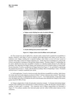
A cross-sectioned blood vessel shows stages of hemostasis. Top, VWF is the carrier protein for blood clotting factor VIII (FVIII). Under normal
conditions VWF does not interact with platelets or the blood vessel wall that is covered with endothelial cells. Middle left, following vascular
injury, VWF adheres to the exposed subendothelial matrix. Middle right, after VWF is uncoiled by local shear forces, platelets adhere to the
altered VWF and these platelets undergo activation and recruit other platelets to this injury site. Bottom left, the activated and aggregated
platelets alter their membrane phospholipids exposing phosphatidylserine, and this activated platelet surface binds clotting factors from
circulating blood and initiates blood clotting on this surface where fibrin is locally deposited. Bottom right, the combination of clotting and
platelet aggregation and adhesion forms a platelet-fibrin plug, which results in the cessation of bleeding. The extent of the clotting is carefully
regulated by natural anticoagulants. Subsequently, thrombolysis initiates tissue repair and ultimately the vessel may be re-endothelialized and
blood flow maintained.
Note: Used by permission of R.R. Montgomery.
Figure 1. VWF and Normal Hemostasis
128620_NIH_Text.qxp:Layout 1 1/4/08 6:12 PM Page 10
single amino acid residues. A database of VWF
mutations and polymorphisms has been compiled
for the International Society on Thrombosis and
Haemostasis (ISTH)
23,24
and is maintained for online
access at the University of Sheffield (f.
ac.uk/vwf/index.html). Mutations causing VWD
have been identified throughout the VWF gene.
In contrast to hemophilia A, in which a single major
gene rearrangement causes a large fraction of severe
disease, no such recurring mutation is common
in VWD. There is a good correlation between the
location of mutations in the VWF gene and the
subtype of VWD, as discussed in more detail in
“Classification of VWD Subtypes.” In selected
families, this information can facilitate the search
for VWF mutations by DNA sequencing.
Classification of VWD Subtypes
VWD is classified on the basis of criteria developed
b
y the VWF Subcommittee of the ISTH, first
published in 1994 and revised in 2006 (Table 4).
25,26
The classification was intended to be clinically
relevant to the treatment of VWD. Diagnostic
categories were defined that encompassed distinct
pathophysiologic mechanisms and correlated with
the response to treatment with DDAVP or blood
products. The classification was designed to be
conceptually independent of specific laboratory
testing procedures, although most of the VWD
subtypes could be assigned by using tests that were
widely available. The 1994 classification reserved
the designation of VWD for disorders caused by
mutations within the VWF gene,
25
but this criterion
11
Scientific Overview
The von Willebrand factor (VWF) protein sequence (amino acid 1–2813) is aligned with the cDNA sequence (nucleic acid 1–8439). The VWF
signal peptide is the first 22 aa, the propeptide (VWFpp) aa 23–763, and mature VWF aa 764–2800. Type 2 mutations are primarily located in
specific domains (regions) along the VWF protein. Types 2A, 2B, and 2M VWF mutations are primarily located within exon 28 that encodes for
the A1 and A2 domains of VWF. The two different types of 2A are those that have increased proteolysis (2A
2
) and those with abnormal multi-
mer synthesis (2A
1
). Type 2N mutations are located within the D’ and D3 domains. Ligands that bind to certain VWF domains are identified,
including FVIII, heparin, GPIb (platelet glycoprotein Ib complex), collagen, and GPIIb/IIIa (platelet glycoprotein IIb/IIIa complex that binds to the
RGD [arginine-glycine-aspartate] amino acid sequence in VWF).
Note: Used by permission of R.R. Montgomery.
Figure 2. Structure and Domains of VWF
128620_NIH_Text.qxp:Layout 1 1/4/08 6:12 PM Page 11
has been dropped from the 2006 classification
26
because in practice it is verifiable for only a small
fraction of patients.
VWD is classified into three major categories: partial
quantitative deficiency (type 1), qualitative deficiency
(type 2), and total deficiency (type 3). Type 2 VWD
is divided further into four variants (2A, 2B, 2M, 2N)
on the basis of details of the phenotype. Before the
publication of the 1994 revised classification of
VWD,
25
VWD subtypes were classified using Roman
numerals (types I, II, and III), generally corresponding
to types 1, 2, and 3 in the 1994 classification, and
within type II several subtypes existed (designated
by adding sequential letters of the alphabet; i.e.,
II-A through II-I). Most of the latter VWD variants
were amalgamated as type 2A in the 1994 classifica-
tion, with the exception of type 2B (formerly II-B)
for which a separate new classification was created.
In addition, a new subtype (2M) was created to
include variants with decreased platelet dependent
function (VWF:RCo) but no significant decrease of
higher molecular weight VWF multimers (which may
or may not have other aberrant structure), with “M”
representing “multimer.” Subtype 2N VWD was
defined, with “N” representing “Normandy” where
the first individuals were identified, with decreased
FVIII due to VWF defects of FVIII binding.
Type 1 VWD affects approximately 75 percent
of symptomatic persons who have VWD (see
Castaman et al., 2003 for a review).
27
Almost all of
the remaining persons are divided among the four
12
von Willebrand Disease
Typ e
1
2
2A
2B
2M
2N
3
Description
Partial quantitative deficiency of VWF
Qualitative VWF defect
Decreased VWF-dependent platelet
adhesion with selective deficiency of
high-molecular-weight multimers
Increased affinity for platelet GPIb
Decreased VWF-dependent platelet
adhesion without selective deficiency of
high-molecular-weight multimers
Markedly decreased binding affinity
for FVIII
Virtually complete deficiency of VWF
Table 4. Classification of VWD
Note: VWD types are defined as described in Sadler JE, Budde U,
Eikenboom JC, Favaloro EJ, Hill FG, Holmberg L, Ingerslev J, Lee CA,
Lillicrap D, Mannucci PM, et al. Update on the pathophysiology and
classification of von Willebrand disease: a report of the
Subcommittee on von Willebrand Factor. J Thromb Haemost 2006
Oct;4(10):2103–2114.
Type Inheritance Prevalence Bleeding Propensity
Type 1 Autosomal dominant Up to 1% Mild to moderate
Type 2A Autosomal dominant (or recessive) Uncommon Variable—usually moderate
Type 2B Autosomal dominant Uncommon Variable—usually moderate
Type 2M Autosomal dominant (or recessive) Uncommon Variable—usually moderate
Type 2N Autosomal recessive Uncommon Variable—usually moderate
Type 3 (Severe) Autosomal recessive Rare (1:250,000 to High (severe bleeding)
1:1,000,000)
Table 5. Inheritance, Prevalence, and Bleeding Propensity in Patients Who Have VWD
128620_NIH_Text.qxp:Layout 1 1/4/08 6:12 PM Page 12
type 2 variants, and the partitioning among them
varies considerably among centers. In France, for
example, patients’ distribution was reported to be
30 percent type 2A, 28 percent type 2B, 8 percent
type 2M (or unclassified), and 34 percent type 2N.
28
In Bonn, Germany, the distribution was reported to
be 74 percent type 2A, 10 percent type 2B, 13 percent
type 2M, and 3.5 percent type 2N.
29
Table 5 summa-
rizes information about inheritance, prevalence, and
bleeding propensity in persons who have different
types of VWD.
The prevalence of type 3 VWD in the population
is not known precisely but has been estimated
(per million population) as: 0.55 for Italy,
30
1.38
for North America,
31
3.12 for Sweden,
30
and 3.2
for Israel.
32
The prevalence may be as high as
6 per million where consanguinity is common.
1
Type 1 VWD
Type 1 VWD is found in persons who have partial
quantitative deficiency of VWF. The level of VWF
in plasma is low, and the remaining VWF mediates
platelet adhesion normally and binds FVIII normally.
Laboratory evaluation shows concordant decreases in
VWF protein concentration (VWF:Ag) and assays of
VWF function (VWF:RCo). Levels of blood clotting
FVIII usually parallel VWF and may be reduced
secondary to reduced VWF. Usually, in type 1 VWD,
the FVIII/VWF:Ag ratio is 1.5–2.0. In most persons
who have type 1 VWD, this results in FVIII being
normal, or mildly decreased, and not reduced as
much as the VWF. VWF multimer gels show no
significant decrease in large VWF multimers.
25
The
laboratory evaluation of VWD is discussed in the
“Diagnosis and Evaluation” section.
The spectrum of mutations occurring in VWD
type 1 has been described extensively in two major
studies.
33,34
Particularly severe, highly penetrant
forms of type 1 VWD may be caused by dominant
VWF mutations that interfere with the intracellular
transport of dimeric proVWF
35-39
or that promote the
rapid clearance of VWF from the circulation.
38,40,41
Persons who have such mutations usually have VWF
levels <20 IU/dL.
33,34
Most of the mutations charac-
terized to date cause single amino acid substitutions
in domain D3.
35–37,39,42
One mutation associated with
rapid clearance has been reported in domain D4.
38
Increased clearance of VWF from the circulation in
type 1 VWD may account for the exaggerated but
unexpectedly brief responses to DDAVP observed
in some patients. Consequently, better data on the
prevalence of increased clearance could affect the
approach to diagnosing type 1 VWD and the choice
of treatment for bleeding.
A diagnosis of type 1 VWD is harder to establish
when the VWF level is not markedly low but instead
is near the lower end of the normal range. Type 1
VWD lacks a qualitative criterion by which it can be
recognized and instead relies only on quantitative
decrements of protein concentration and function.
VWF levels in the healthy population span a wide
range of values. The mean level of plasma VWF
is 100 IU/dL, and approximately 95 percent of
plasma VWF levels lie between 50 and 200 IU/dL.
43,44
Because mild bleeding symptoms are very common
in the healthy population, the association of bleeding
symptoms with a moderately low VWF level may be
coincidental.
45
The conceptual and practical issues
associated with the evaluation of moderately low
VWF levels are discussed more completely later
in this section. (See “Type 1 VWD Versus Low VWF:
VWF Level as a Risk Factor for Bleeding.”)
Type 2 VWD
The clinical features of several type 2 VWD variants
are distinct from those of type 1 VWD, and they can
have strikingly distinct and specific therapeutic needs.
As a consequence, the medical care of patients who
have type 2 VWD benefits from the participation
of a hematologist who has expertise in hemostasis.
Bleeding symptoms in type 2 VWD are often thought
to be more severe than in type 1 VWD, although
this impression needs to be evaluated in suitable
clinical studies.
Type 2A VWD refers to qualitative variants in which
VWF-dependent platelet adhesion is decreased
because the proportion of large VWF multimers
is decreased. Levels of VWF:Ag and FVIII may be
normal or modestly decreased, but VWF function
is abnormal as shown by markedly decreased
VWF:RCo.
46
Type 2A VWD may be caused by
mutations that interfere with the assembly or
secretion of large multimers or by mutations that
increase the susceptibility of VWF multimers to
proteolytic degradation in the circulation.
47–49
The deficit of large multimers predisposes persons
to bleed.
13
Scientific Overview
128620_NIH_Text.qxp:Layout 1 1/4/08 6:12 PM Page 13
The location of type 2A VWD mutations sometimes
can be inferred from high-resolution VWF multimer
gels. For example, mutations that primarily reduce
multimer assembly lead to the secretion of multimers
that are too small to engage platelets effectively
and therefore are relatively resistant to proteolysis
by ADAMTS13. Homozygous mutations in the
propeptide impair multimer assembly in the Golgi
and give rise to a characteristic “clean” pattern of
small multimers that lack the satellite bands usually
associated with proteolysis (see “Diagnosis and
Evaluation”); this pattern was initially described
as “type IIC” VWD.
50–52
Heterozygous mutations
in the cystine knot (CK) domain can impair
dimerization of proVWF in the ER and cause a
recognizable multimer pattern originally referred
to as “type IID.”
53,54
A mixture of monomers and
dimers arrives in the Golgi, where the incorporation
of monomers at the end of a multimer prevents
further elongation. As a result, the secreted small
multimers contain minor species with an odd
number of subunits that appear as faint bands
between the usual species that contain an even
number of subunits. Heterozygous mutations in
cysteine residues of the D3 domain also can impair
multimer assembly, but these mutations often also
produce an indistinct or “smeary” multimer pattern
referred to as “type IIE.”
55,56
In contrast to mutations that primarily affect
multimer assembly, mutations within or near the
A2 domain of VWF cause type 2A VWD that is
associated with markedly increased proteolysis of
the VWF subunits
56
(see Figure 2, on page 11). These
mutations apparently interfere with the folding of the
A2 domain and make the Tyr1605–Met1606 bond
accessible to ADAMTS13 even in the absence of
increased fluid shear stress. Two subgroups of this
pattern have been distinguished: group I mutations
enhance proteolysis by ADAMTS13 and also impair
multimer assembly, whereas group II mutations
enhance proteolysis without decreasing the assembly
of large VWF multimers.
49
Computer modeling of
domain A2 suggests that group I mutations affect
both assembly and proteolysis, because group I
mutations have a more disruptive effect on the
folding of domain A2 than do group II mutations.
57
Type 2B VWD is caused by mutations that pathologi-
cally increase platelet–VWF binding, which leads to
the proteolytic degradation and depletion of large,
functional VWF multimers.
56,58
Circulating platelets
also are coated with mutant VWF, which may prevent
the platelets from adhering at sites of injury.
59
Although laboratory results for type 2B VWD may
be similar to those in type 2A or type 2M VWD,
patients who have type 2B VWD typically have
thrombocytopenia that is exacerbated by surgery,
pregnancy, or other stress.
60–62
The thrombocytope-
nia probably is caused by reversible sequestration of
VWF–platelet aggregates in the microcirculation.
These aggregates are dissolved by the action of
ADAMTS13 on VWF, causing the characteristic
decrease of large VWF multimers and the prominent
satellite banding pattern that indicates increased
proteolytic degradation.
63,64
The diagnosis of type 2B
VWD depends on finding abnormally increased
ristocetin induced platelet aggregation (RIPA) at low
concentrations of ristocetin.
Type 2B VWD mutations occur within or adjacent to
VWF domain A1,
23,55,65–68
which changes conforma-
tion when it binds to platelet GPIb.
69
The mutations
appear to enhance platelet binding by stabilizing the
bound conformation of domain A1.
Type 2M VWD includes variants with decreased
VWF-dependent platelet adhesion that is not caused
by the absence of high-molecular-weight VWF
multimers. Instead, type 2M VWD mutations reduce
the interaction of VWF with platelet GPIb or with
connective tissue and do not substantially impair
multimer assembly. Screening laboratory results in
type 2M VWD and type 2A VWD are similar, and the
distinction between them depends on multimer gel
electrophoresis.
67
Mutations in type 2M VWD have been identified
in domain A1 (see Figure 2 on page 11), where they
interfere with binding to platelet GPIb.
23,55,67,70–72
One family has been reported in which a mutation in
VWF domain A3 reduces VWF binding to collagen,
thereby reducing platelet adhesion and possibly
causing type 2M VWD.
73
Type 2N VWD is caused by VWF mutations that
impair binding to FVIII, lowering FVIII levels so that
type 2N VWD masquerades as an autosomal recessive
form of hemophilia A.
74–76
In typical cases, the FVIII
level is less than 10 percent, with a normal VWF:Ag
and VWF:RCo. Discrimination from hemophilia A
may require assays of FVIII–VWF binding.
77,78
Most mutations that cause type 2N VWD occur
within the FVIII binding site of VWF (see Figure 2
14
von Willebrand Disease
128620_NIH_Text.qxp:Layout 1 1/4/08 6:12 PM Page 14
on page 11), which lies between residues Ser764 and
Arg1035 and spans domain D’ and part of domain
D3.
23,79,80
The most common mutation, Arg854Gln,
has a relatively mild effect on FVIII binding and tends
to cause a less severe type 2N VWD phenotype.
77
Some mutations in the D3 domain C-terminal of
Arg1035 can reduce FVIII binding,
81–83
presumably
through an indirect effect on the structure or accessi-
bility of the binding site.
Type 3 VWD
Type 3 VWD is characterized by undetectable VWF
protein and activity, and FVIII levels usually are
very low (1–9 IU/dL).
84–86
Nonsense and frameshift
mutations commonly cause type 3 VWD, although
large deletions, splice-site mutations, and missense
mutations also can do so. Mutations are distributed
throughout the VWF gene, and most are unique to
the family in which they were first identified.
23,87,88
A small fraction of patients who have type 3 VWD
develop alloantibodies to VWF in response to the
transfusion of plasma products. These antibodies
have been reported in 2.6–9.5 percent of patients who
have type 3 VWD, as determined by physician surveys
or screening.
85,89
The true incidence is uncertain,
however, because of unavoidable selection bias in
these studies. Anti-VWF alloantibodies can inhibit
the hemostatic effect of blood-product therapy and
also may cause life-threatening allergic reactions.
85,90
Large deletions in the VWF gene may predispose
patients to this complication.
89
VWD Classification, General Issues
The principal difficulties in using the current VWD
classification concern how to define the boundaries
between the various subtypes through laboratory
testing. In addition, some mutations have pleiotropic
effects on VWF structure and function, and some
persons are compound heterozygous for mutations
that cause VWD by different mechanisms. This
heterogeneity can produce complex phenotypes
that are difficult to categorize. Clinical studies of
the relationship between VWD genotype and
clinical phenotype would be helpful to improve the
management of patients with the different subtypes
of VWD.
The distinction between quantitative (type 1) and
qualitative (type 2) defects depends on the ability
to recognize discrepancies among VWF assay
results,
80,91
as discussed in “Diagnosis and
Evaluation.” Similarly, distinguishing between type
2A and type 2M VWD requires multimer gel analysis.
Standards need to be established for using laboratory
tests to make these important distinctions.
The example of Vicenza VWD illustrates some of
these problems. Vicenza VWD was first described as
a variant of VWD in which the level of plasma VWF
is usually <15 IU/dL and the VWF multimers are
even larger than normal, like the ultralarge multimers
characteristic of platelet VWF.
92
The low level of
VWF in plasma in Vicenza VWD appears to be
explained by the effect of a specific mutation,
Arg1205His, that promotes clearance of VWF from
the circulation about fivefold more rapidly than
normal.
41
Because the newly synthesized multimers
have less opportunity to be cleaved by ADAMTS13
before they are cleared, accelerated clearance alone
may account for the increased multimer size in
Vicenza VWD.
93
Whether Vicenza VWD is classified
under type 1 VWD or type 2M VWD depends on
the interpretation of laboratory test results. The
abnormally large multimers and very low RIPA
values have led some investigators to prefer the
designation of type 2M VWD.
94
However, the
VWF:RCo/VWF: Ag ratio typically is normal, and
large VWF multimers are not decreased relative to
smaller multimers, so that other investigators have
classified Vicenza VWD under type 1 VWD.
41
Regardless of how this variant is classified, the
markedly shortened half-life of plasma VWF in
Vicenza VWD is a key fact that, depending on the
clinical circumstance, may dictate whether the patient
should receive treatment with DDAVP or FVIII/VWF
concentrates.
Type 1 VWD Versus Low VWF: VWF Level as
a Risk Factor for Bleeding
Persons who have very low VWF levels, <20 IU/dL,
ar
e likely to have VWF gene mutations, significant
bleeding symptoms, and a strongly positive family
history.
33,34,37,95–99
Diagnosing such persons as having
type 1 VWD seems appropriate because they may
benefit from changes in lifestyle and from specific
treatments to prevent or control bleeding.
Identification of affected family members also may be
useful, and genetic counseling is simplified when the
pattern of inheritance is straightforward.
15
Scientific Overview
128620_NIH_Text.qxp:Layout 1 1/4/08 6:12 PM Page 15