Tài liệu Báo cáo Y học: NMR-based determination of the binding epitope and conformational analysis of MUC-1 glycopeptides and peptides bound to the breast cancer-selective monoclonal antibody SM3 pptx
Bạn đang xem bản rút gọn của tài liệu. Xem và tải ngay bản đầy đủ của tài liệu tại đây (1.99 MB, 12 trang )
NMR-based determination of the binding epitope
and conformational analysis of MUC-1 glycopeptides and peptides
bound to the breast cancer-selective monoclonal antibody SM3
Heiko Mo¨ller
1
, Nida Serttas
1
, Hans Paulsen
1
, Joy M. Burchell
2
, Joyce Taylor-Papadimitriou
2
and Bernd Meyer
1
1
Institute of Organic Chemistry, University of Hamburg, Germany;
2
Imperial Cancer Research Fund Breast Cancer Biology Group,
Guy’s Hospital, London, UK
Mucin glycoproteins on breast cancer cells car ry shortened
carbohydrate chains. These partially deglycosylated mucin 1
(MUC-1) structures are recognized by the monoclonal
antibody SM3, which is being tested for its diagnostic utility.
We used NMR spectroscopy to analyze the binding mode
and t he binding epitope of peptide and glycopeptide antigens
to the SM3 antibody. The pentapeptide PDTRP and the
glycopentapeptide PDT( O-a-
D
-GalNAc)RP are known lig-
ands o f the monoclonal antibody. The 3D structures of the
ligands in the bound conformation were determined by an-
alyzing trNOESY build-up rates. The peptide was f ound to
adopt an extended conformation that fits into t he binding
pocket of the antibody. The binding epitopes of the ligands
were determined by saturation transfer difference (STD)
NMR spectroscopy. The peptide’s epitope is predominantly
located in the N-terminal PDT s egment whereas the C-ter-
minal R P segment has fewer interactions with the protein.
In contrast, the glycopeptide is interacting with SM3
utilizing all its amino ac ids. Pro1 shows the stronge st binding
effect that slightly decays towards Pro5. The GalNAc resi-
due interacts mainly via the N-acetyl residue while the other
protons show less interactions similar to that of Pro5. The
glycopeptide in the bound state also has an extended con-
formation o f t he peptide with th e carbohydrate oriented
towards the N-terminus. Docking studies showed that pep-
tide and glycopeptide fit t he binding pocket of the mAb SM3
very well.
Keywords: glycopeptide antibody complex; STD N MR;
breast cancer; MUC-1; binding epitope.
The extracellular part of the epithelial glycoprotein MUC-1
consists of tandem repeats of 20 amino acids
(PDTRPAPGSTAPPAHGVTSA, where the start of the
tandem repeat peptide sequence varies. We follow here the
definition by Gendler et al. who defined the start at PDTRP
[1]. Residues of peptides, that were elongated at the
N-terminus, are designated by an apostrophe, e.g. Ala20¢-
Pro1-Asp2-Thr3-Arg4-Pro5.) [2]. Each repeat can carry up
to five O-glycosyl c hains at S er and T hr residues t hat
account for the high carbohydrate content of the mucins [3].
Usually, 70–100 repeats are found in mucins. The clustering
of O-linked glycans on MUC-1 leads to an extended protein
core. Membrane-bound mucins extend several hundred
nanometers into the lumen and thus represent a first barrier
to the environment. They have important functions in cell-
cell recognition and shield the cell from microorganisms,
toxins and proteolytic attack [4].
Many diseases affect the p roduction of mucus. Both the
amount and the characteristics of the mucus can be altered.
In cystic fibrosis, for example, due to changes of the ionic
environment dramatic alterations in rheological properties
correlate with changes i n carbohydrate c omposition [2].
Modified oligosaccharides are also found in mucins of
patients with Crohn’s disease [2].
Epithelial c ells express the membrane-bo und MUC-1 at
their apical surface. In carcinomas, the localization at the
apical surface is lost. High concentrations of MUC-1 spread
out over the whole cell surface. This may protect the cells
against low pH and may interfere with immune surveillance
by causing steric h indrance of surface antigen presentation
[2,4].
In breast cancer, the MUC-1 glycoprotein is overex-
pressed and aberrantly glycosylated. Thus, in contrast to the
mucin produced by normal breast epithelial cells, which
carry core2 based structures [5], MUC1 from b reast c ancer
cells carries highly truncated, mainly core 1 based oligosac-
charide structures [6,7]. In some cases, the first sugar added,
N-acetyl galactosamine is not extended, or is sialylated to
form the cancer-related sialyl Tn epitope. Because of the
shorter side chains, the peptide core of the cancer mucin is
more exposed, and antibodies have been developed w hich
recognize epitopes exposed in the cancer mucin, which are
normally masked by large oligosaccharide s ide chains.
These antigenic peptide sequences therefore constitute
cancer-associated epitopes which are also found in the
Correspondence to B. Meyer, Institute of Organic Chemistry,
University of Hamburg, Martin-Luther-King-Platz 6,
20146 Hamburg, Germany.
Fax: + 49 (0)40 42838 2878, Tel.: + 49 (0)40 42838 5913,
E-mail:
Abbreviations: MUC-1, mucin 1 glycoprotein; SM3, breast cancer-
selective monoclonal antibody; STD NMR, saturation transfer
difference NMR; trNOE, transferred nuclear Overhauser enhance-
ment; SPR, surface plasmon resonance; SAR, structure activity
relationship; MD, molecular dynamics.
(Received 28 August 2001, revised 5 December 2001, accepted
14 January 2002)
Eur. J. Biochem. 269, 1444–1455 (2002) Ó FEBS 2002
short sugar side chains (e.g. T, ST a nd TF antigens) [8,9].
The monoclonal antibody SM3 was raised in mice against
partially deglycosylated human MUC-1. It shows a high
specificity to the MUC-1 of breast cancer cells. SM3 is being
tested for its diagnostic value [10,11] and also has a high
therapeutic potential.
The minimum peptide a ntigen epitope to SM3 w as
identified by the pepscan t echnique using ELISA detection.
Using heptapeptides the resulting binding epitope was
identified as Asp2-Thr3 [12], using octapeptides the resulting
epitope was Pro1-Asp2-Thr3-Arg4-Pro5 [13] a nd using
nona- and 20mer peptides the resulting epitope was
identified as Ala20¢-Pro1-Asp2-Thr3-Arg4-Pro5 and Pro1-
Asp2-Thr3, respectively [14,15]. For SM3 reacting with
pentamers and dimers of the MUC-1 tandem repeat the
binding constants were determined by surface plasmon
resonance to be K
d
¼ 6.25 · 10
)8
to 4.5 · 10
)7
M
[16].
Previous NMR studies of peptide a nd glycopeptide
fragments of MUC-1 containing the amino-acid sequence
PDTRP in the central part which were carried out in
solution without an antibody present reveal that this
sequence motif seems to adopt a knob-like or bent structure
[17] (J. D ojahn, C. Diot el, M. P aulsen and B . M eyer,
unpublished results). It was postulated that this knob-like
structure renders this region especially accessible to protein
interactions necessary for stimulation of immune responses.
Also in this context, the oligosaccharides attached to Thr3
are most accessible f or interaction w ith the cells of the
immune system.
It is not clear against w hat actual epitope SM3 was
developed. The antibody b inds more strongly to a MUC-1
that has only part of its carbohydrate chains removed [10].
It was later shown on a molecular level that a small
oligosaccharide attached t o Thr3 enhances binding affinity
of glycopeptides to the antibody [18].
Conventional pepscan analysis however, does not allow
easy analysis of the contribution of the carbohydrate
portion. To assess the involvement of carbohydrates in
antibody recognition of glycosylated structures numerous
glycopeptides would have to be synthesized and even this
approach would not directly reveal what part of the
oligosaccharides interacts with the protein.
Dokurno et al. published an X-ray structure ana-
lysis of S M3 complexed with t he MUC-1 peptide TSA
PDTRPAPGST [19]. At e ach end of the antigenic peptide
PDTRP two additional amino acids are resolved in the
X-ray crystal structure, while Thr18¢, Gly8-Ser9-Thr10 and
the side chain of Ser19¢ are disordered in the crystal. The
amino acids of the peptide sequence (S)APDTRPAP have
many interactions with the antibody’s surface. The covered
surface of the individual amino acids varies strongly. While
Ser19¢-Ala20¢, Thr3 and Pro5-Ala6 have r elatively small
contact areas to the protein, Pro1-Asp2 and Arg4 are much
more buried by the antibody.
NMR spectroscopy can be used to assess binding
properties of ligands under n ear physiological conditions
in solution by a variety of methods, e .g. trNOEs [20], STD
NMR [21–23], and SAR by NMR [24]. TrNOE spectra can
also be used to elucidate t he 3D structure of t he bound
ligand. Saturation transfer difference (STD) N MR is a
technique that can be used t o characterize a nd identify
binding [21–23]. It can a lso be used to id entify the binding
epitope of ligands to a protein receptor [21]. This feature can
be used to quickly identify the binding contribution from
either peptide or carbohydrate, especially in the case o f
glycopeptides. In contrast to conventional methods only
one substrate is necessary to obtain that information.
Here, we present the STD NMR epitope mapping and
trNOE-based conformational analysis of the MUC-1
peptide PDTRP and the MUC-1 glycopeptide PDT(O-a-
D
-GalNAc)RP (cf. Figure 1) bound to the monoclonal
antibody SM3.
MATERIALS AND METHODS
Chemicals
Chemicals for peptide synthesis were obtained from
PerSeptive Biosystems (Wiesbaden, Germany), acetonitrile
from Alfa (Karlsr uhe, G ermany), triisopropylsilane and
D
2
O from Sigma Aldrich (Steinheim, Germany), all other
chemicals of analytical grade were obtained f rom Merck
(Darmstadt, Germany). The glycopeptides [29] and the
monoclonal antibody SM3 [1] were prepared as described.
NMR Experiments
All spectra were recorded on Bruker DRX 500 spectrometer
with a t riple r esonance 5 mm inverse probe head. For
trNOE e xperiments with PDTRP t he sample contained
3.6 m g of SM3 (M
r
156 kDa, 23 nmol, 3 8 l
M
) and 270 lg
of PDTRP (M
r
583.64 gÆmol
)1
, 460 nmol, 760 l
M
)in
600 lLNaCl/P
i
(buffer concentration 20 m
M
,H
2
O/
D
2
O ¼ 9 : 1, 0.05% NaN
3
) at p H 7.0. This corresponds
to a ligand to protein ratio of 20 : 1. For STD experiments
the ligand t o protein ratio was raised to 200 : 1 (4.6 lmol,
7.6 m
M
PDTRP). TrNOE s tudies with the glycopeptide
were carried out with a sample containing 3 mg of SM3
(19.2 nmol, 32 l
M
) and 300 lgofPDT(O-a-
D
-GalNAc)RP
(384 nmol, 640 l
M
)in600lLNaCl/P
i
buffered solution
(buffer co ncentration 20 m
M
) a t pH 7.0. This corresponds
to a ligand to protein ratio of 20 : 1. For STD experiments
the ligand to protein ratio was raised to 150 : 1 (2.88 lmol,
4.8 m
M
PDT(O-a-
D
-GalNAc)RP).
Peptide or glycopeptide were added to the protein
solution using 22 m
M
stock s olutions. At the highest
excess, this resulted in a s ample dilution of 25% for the
peptide and 18% for the glycopeptide. As no titration
experiments were carried out, this d ilution was not import-
ant for the data analysis.
Solute e xchange w as achieved by ultrafiltration of the
156-kDa SM3 antibody with a Centricon (Millipore)
membrane having a cutoff value of 50 kDa.
Fig. 1. Pentapeptide and glycopentapeptide used in N MR studies and
completely glycosylated MUC-1 repetitive unit.
Ó FEBS 2002 NMR of MUC-1 glycopeptides (Eur. J. Biochem. 269) 1445
All spec tra were measured at 280 K. All chemical shifts
are referenced to the HDO signal at 4 .90 p.p.m. f or
1
H.
Water suppression was a chieved using the WATERGATE
sequence in all experiments. NMR chemical shifts of
peptide and glycopeptide are listed in Tables 1 and 2,
respectively. All spectra of samples containing protein
were recorded with a 30 ms spin lock pulse, or so called
T
1q
filter (cB
1
¼ 4680 Hz) after the p/2 pulse, which
eliminates the background protein resonances to facilitate
analysis. Interpretation of the spectra were carried out
with the
XWINNMR
(Bruker, v. 2.5) and the
AURELIA
program (Bruker, v. 2.1.5) on Silicon Graphics O
2
work-
stations. 1D STD NMR spectra were multiplied by an
exponential line b roadening function of 5 Hz prior to
Fourier transformation. The irradiation power in all STD
NMR experiments was set to 0.15 W. Selective presat-
uration of the protein was achieved by a train of 40
Gaussian shaped pulses of 50 ms length, each separated by
a 1 ms delay, leading to a total saturation time of 2.04 s.
The pulse scheme is as follows: relaxation delay, presat-
uration pulse train, (p/2), spin lock (where applicable),
acquisition. Subtraction of the 1D STD spectra was
performed internally via phase cycling after every scan to
minimize temperature and magnet instability artefacts.
The so called on resonance irradiation of the protein was
performed at a chemical shift of )2 ppm. Off resonance
irradiation was applied at 40 p.p.m., where no protein
signals are present. Between 256 and 1024 total scans were
collected, using 10 ppm spectral widths for the 1D STD
NMR spectra.
2D STD TOCSY spectra were recorded with 40 scans per
t
1
increment. A total of 256 t
1
increments were collected in
an interlaced mode for the on and off resonance spectra.
Prior to subtraction both spectra were processed and phased
identically. A MLEV (composite pulse decoupling used for
TOCSY spin lock) mixing time of 100 ms was applied in all
TOCSY spectra. The acquisition times for the 2D experi-
ments were typically around 22 h. 2D spectra were multi-
plied with a squared cosine bell function in all dimensions
and zero filled two times. The pulse sequence for the 2D
NOESY spectra included a filter to suppress zero quantum
coherence. The spectra we re recorded with mixing times of
50, 100, 150, 300 and 500 ms and 80 s cans for each of the
205 t
1
increments. The 2D ROESY spectrum was recorded
with a mixing time o f 300 ms and 80 scans for each of the
205 t
1
increments using a spin lock field of cB
1
¼ 1967 Hz
at 4.9 p.p.m.
Distance geometry calculations
The starting structures were generated with distance range
constraints obtained from the NOE d istances by adding or
subtracting 5% for upper and lower limit, respectively. The
conformation of PDTRP bound to SM3 was described by
13 distance range constraints (cf. Table 3). 500 structures
were calculated using the Redac strategy implemented in the
DYANA
package [26]. The conformation with lowest target
function was used for the following molecular dynamics
(MD) simulation. The structure of PDT(O-a-
D
-Gal-
NAc)RP in the binding site of SM3 was defined by 16
Table 1 .
1
H-NMR chemical shifts of PDTRP in p.p.m. Spectra were recorded at 280 K with HDO resonance at 4.9 p.p.m. Resonance s of protons
marked by – were not visible.
NH abb¢ cc¢ dd¢
Pro8 – 4.347 2.389 1.978 1.978 1.978 3.336 3.336
Asp9 – 4.666 2.707 2.543
Thr10 8.386 4.252 4.155 1.134
Arg11 8.484 4.574 1.798 1.724 1.629 1.629 3.160 3.160
Pro12 4.327 2.255 1.978 1.978 1.879 3.782 3.582
CONH 7.761
CONH¢ 7.043
Table 2 .
1
H-NMR chemical shifts of PDT(O-a-D-GalNAc)RP in p.p.m. Spectra were recorded at 280 K with HDO resonance at 4.9 p.p.m.
Resonances of protons marked by – were not visible.
NH abb¢ cc¢ dd¢
Pro8 – 4.347 2.397 1.983 1.983 1.983 3.368 3.317
Asp9 – 4.779 2.747 2.558
Thr10 8.914 4.445 4.297 1.217
Arg11 8.510 4.504 1.813 1.678 1.645 1.645 3.160 3.160
Pro12 4.295 2.270 1.990 1.990 1.879 3.705 3.591
CONH 7.788
CONH¢ 7.017
NHCH3123456a6b
GalNAc 7.803 1.975 4.775 4.016 3.850 3.914 3.972 3.715 3.691
1446 H. Mo
¨
ller et al. (Eur. J. Biochem. 269) Ó FEBS 2002
distance range constraints (cf. Table 4). The distance
geometry calculations were performed by an internal
algorithm in
SYBYL
(v. 6.3, Tripos). A total of 100 structures
were generated and energetically optimized. The lowest
energy conformation acted a s starting structure for the
following MD simulation.
MD simulations
Constrained MD simulations were carried out with the
SYBYL
program on Silicon Graphics Octane (R12000)
computers, using the Tripos force field. A harmonic
potential was employed at the edges of the distance range
constraints. The force field constants were set to 2 kcalÆ
(mol A
˚
2
)
)1
. Constraints to pseudoatoms, generated by
SYBYL
, w ere used for nonstereospecifically assigned methyl-
ene groups and methyl groups. The starting structures w ere
placed in water boxes (PDTRP: 9 31 water m olecules,
30 · 30 · 30 A
˚
3
, PDTRP/SM3 complex: 1708 water
molecules, 40 · 41 · 40 A
˚
3
,PDT(O-a-
D
-GalNAc)RP:
1152 water molecules, 33 · 33 · 33 A
˚
3
,PDT(O-a-
D
-
GalNAc)RP/SM3 complex: 2521 water molecules, 45 ·
48 · 42 A
˚
3
).
Before starting the MD simulation the box was energy
optimized over 200 steps. The constrained simulation was
performed at 300 K. The charges were calculated with the
Gasteiger Marsili method and a dielectric constant of four
was used. A cutoff radius of 8 A
˚
was used for the
nonbonded interactions. The initial velocities for the atoms
were taken from a Boltzmann distribution at 300 K and the
step size for the integration of Newton’s equation was 1 fs.
The c oupling to the temperature bath was set to 100 fs and
the nonbonded interactions were updated every 25 fs. The
MD simulations ran for 100 ps at constant volume and
temperature.
The final structures were e nergy minimized over 1000
steps a nd overlaid to the PDTRP fragment of the ligand of
the X-ray structure (RCSB PDB entry 1SM3). After small
manual corrections, t he ligands were docked into the
binding site of SM3 using the
FLEXIDOCK
module of t he
SYBYL
software package. The docking structures after
100 000 generations were subjected to final MD simulations
in the binding site of the antibody with flexible protein
residues in a perimeter of 10 A
˚
from the ligand.
RESULTS
The small PDTRP peptide and its glycosylated derivative
were used because larger p eptides did not show measurable
trNOE effects. This is most likely due to slow exchange
between the bound and the free state. For dimers and
pentamers of the MUC-1 tandem repeat [16], i.e. 40mer and
60mer peptides, the dissociation constant was determined to
K
d
¼ 10
)7
by SPR. At an on-rate of k
on
¼ 10
6
M
)1
Æs
)1
typical for antibody interactions one would have an off-rate
k
off
¼ 0.1 s
)1
, which is too slow for obtaining measurable
trNOE effects. The exact kinetic constants were not
published. More importantly, the larger peptides decom-
posed in the presence of the antibody within a few days
(N.Serttas,H.Mo
¨
ller, J.M. Burc hell, J. Taylor-Papadimi-
triou, B. Meyer and H. Paulsen, unpublished results). To
overcome these problems with large peptides, we used short
peptides to utilize their faster dissociation rates [25] and their
stability in the presence of SM3. With the pentapeptide and
glycopentapeptide we obtained strong STD effects and
weak trNOEs.
SM3 in complex with the peptide PDTRP
STD Experiments. In contrast to the larger peptides and
glycopeptides, the pentapeptide PDTRP is stable in the
presence of SM3 and possesses a favourable off-rate on the
NMR time scale to yield good trNOE spectra. Figure 2A
shows the 1D STD s pectrum (red) and a normal
1
H-spectrum (black) of the complex of PDTRP with
SM3. For comparison, the signals of the Pro1 b-methylene
protons are adjusted to have the same height. As evident
from Fig. 2, proton resonances of Pro1 and Asp2 have t he
highest intensities in the STD spectrum, signals of Thr3 are
of medium intensity, while the signals of Arg4 and Pro5
have the lowest intensity. The d-protons of Pro5 have only
Table 3. Constraints for PDTRP derived from trNOE build-up rates.
For distances b etween Protons of Asp2, Thr3, Arg4 (including intra-
residue contacts of Arg4) and between Thr3 and Pro5 the trNOE
build-up of t he b-protons of Asp2 was t aken as reference. For co ntacts
between Arg4 and Pro5 the d-protons of Pro5 acted as reference.
Proton pair Lower limit (A
˚
) Upper limit (A
˚
)
Asp2-a/Thr3-c 3.58 3.96
Asp2-a/Thr3-NH 2.35 2.59
Pro5-dd¢/Arg4-dd¢ 3.41 3.77
Pro5-dd¢/Arg4-cc¢ 2.76 3.05
Arg4-a/Arg4-dd¢ 3.10 3.42
Arg4-a/Arg4-NH 2.57 2.84
Arg4-a/Pro5-dd¢ 2.30 2.55
Arg4-cc¢/Arg4-NH 2.97 3.28
Arg4-NH/Arg4-bb¢ 2.75 3.04
Thr3-c/Thr3-a 3.02 3.34
Thr3-a/Thr3-NH 3.26 3.60
Thr3-a/Arg4-NH 3.38 3.74
Pro5-dd¢/Thr3-c 3.97 4.39
Table 4. Constraints for PDT(O-a-D-GalNAc)RP derived from
trNOE build-up r ates and trROESY data. * The NOEs ma rked with an
asterisk are overlapping and are assumed to have equal intensity.
Proton pair Lower limit (A
˚
) Upper limit (A
˚
)
Asp2-a/Thr3-NH* 2.35 2.60
GalNAc-H1/Thr3-NH* 2.35 2.60
Pro5-dd¢/Arg4-cc¢ 3.03 3.35
Arg4-a/Arg4-dd¢ 3.17 3.51
Arg4-a/Arg4-cc¢ 2.48 2.74
Arg4-a/Pro5-dd¢ 2.33 2.58
Arg4-bb¢/Arg4-dd¢ 2.99 3.30
Arg4-bb¢/Arg4-NH 2.83 3.12
Arg4-cc¢/Arg4-NH 2.56 2.83
Thr3-a/Thr3-c 2.46 2.72
Thr3-a/Thr3-NH 2.52 2.78
Thr3-a/Arg4-NH 2.44 2.69
Thr3-c/Thr3-NH 2.99 3.31
Thr3-c/GalNAc-H5 2.97 3.28
Thr3-NH/GalNAc-NH 3.13 3.46
GalNAc-NH/GalNAc-H3 2.65 2.93
Ó FEBS 2002 NMR of MUC-1 glycopeptides (Eur. J. Biochem. 269) 1447
25% relative intensity in the STD spectrum. Obviously,
Pro1 an d Asp2 get more saturation from th e protein than
the remaining residues of the ligand and therefore have
more and tighter contacts to the antibody’s s urface. The
mean STD i ntensities of each residue are summarized in
Fig. 2B. Here, it is evident that the mean intensities of
signals of Pro5 have only 40% intensity relative to t hose of
Pro1. Overall, there is a continuous drop in intensity from
the N-terminus to the C-terminus with a 50% value being
reached at Thr3.
By 2D STD TOCSY experiments one can usethei ncreased
dispersion for a more detailed epitope mapping. In Fig. 3 the
STD and normal TOCSY spectrum of the PDTRP/SM3
complex are shown. The peaks of Arg4 and Pro5 are so low
in intensity that they do not appear at the intensity cutoff
shown. Signals of Pro1, Asp2 and Thr3 are clearly visible
confirming the results from the 1D STD experiments. The
strongest signals are again cross peaks from Pro1.
trNOE experiments with PDTRP and SM3
The conformation of the peptide ligand PDTRP bound to
SM3 was obtained from transferred NOE spectra. In a
trNOESY spectrum of PDTRP in presence of SM3 (data
not shown) all c rosspeaks are of the same sign as the
diagonal signals and have relatively weak intensity. Thus,
these negative NOEs originate from the bound conforma-
tion. In absence of the antibody PDTRP s hows exclusively
positive NOEs. Most contacts are sequential or intraresidue
NOEs, which is in agreement with the elongated confor-
mation presented below. Pro5-d/Thr3-c is the only long
range interaction that can b e detected in the trNOE
spectrum.
The trNOE spectra were recorded as a function of
the m ixing time with intervals of 50, 100, 150, 300, and
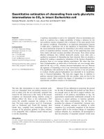
Fig. 2. STD data. (A) Superposition of a 1D STD spectrum (red) and
a r eference
1
H-spectrum (black) of PDTRP in complex with the
antibody SM3. The intensity is adjusted, so that the Pro1-b-meth ylene
signal is of same height in both spectra. Clearly visible are strong STD
effects for protons of Pro1 and Asp2 whereas Arg4 and Pro5 show
weaker sign als in the STD s pectru m. (B) Mean STD values (in percent)
of the protons of the individual amino acids calculated for each amino
acid of PDTRP from t he 1D spectrum.
Fig. 3 . TOCSY spectra. (A) 2D STD T OCSY and (B) conventio nal
TOCSY spectrum of PDTRP in complex with SM3. The strongest
STD signals originate from protons of Pro1, Asp2 and Thr3. The
signals of Arg4 and Pro5 visible in t he ref erence T OCSY (B) vanish
completely or have low intensity in the S TD experiment (A).
1448 H. Mo
¨
ller et al. (Eur. J. Biochem. 269) Ó FEBS 2002
500 ms. Shorter mixing times did not give spectra with an
interpretable signal/noise ratio. Inter proton d istances were
calculated from the extrapo lated slope at mixing time zero
using a biexponential fitting algorithm on the trNOE build-
up curves. The distance is obtained by comparing the
trNOE build-up of a n interesting proton pair with that of a
reference proton pair which has a known fixed d istance, i.e.
geminal protons. PDTRP offers three well resolved refer-
ence points, each of which forms a pair of geminal protons
with a proton/proton distance of 1.8 A
˚
:Asp2-b/b¢,Pro5-d/d¢
and the C-terminal carboxamide NH protons. As can be
seen from the three panels in Fig. 4, the initial slopes of the
build-up c urves o f the geminal proton pairs are very
different. The bu ild-up rate o f the b protons of Asp2 is
about 2.2-fold as big as t hat of the Pro5 d protons. This big
difference in NOEs converts to about 10% difference in the
corresponding distances because of the r
)6
dependence of
the NOEs on the distances. A s a result, using Asp2-b/b¢ as
reference the distance of Pro5-d/d¢ wascalculatedtobe
2.06 A
˚
while with Pro5-d/d¢ as reference the Asp2 methylene
protons should have a distance of 1.57 A
˚
.
There are two possible e xplanations for this behavior:
(a) the two segments of the peptide have a very different
rotational correlation time, i.e. have very different degrees of
freedom, or (b) the NOE between the Asp2 b protons is
relayed by a protein proton. Neither explanation can easily
be proven.
It is, however, unlikely that a transfer through protein
protons is responsible for the enhanced cross relaxation of
the Asp2 b protons. Assuming a binding mode as found in
the X-ray structure analysis the closest distance o f a protein
proton to the Asp2-b/b¢ proton is 2.7 and 3.4 A
˚
,respect-
ively. This relay proton contributes to the observed cross
relaxation rate of Asp2-b with b¢ with 2% only. All other
protons are further away and thus have less contributions.
The observed difference of more than 100% compared with
the cross relaxation rate of Pro5-d/d¢ can consequently not
be explained by a relay phenomenon. We find on the other
hand that the differences in segment flexibility are in perfect
agreement with the STD-based epitope mapping presented
above.
To accommodate these variations th roughout the mole-
cule we chose to reference distances to reference atom pairs
in the same segment, i.e. distances between protons of Asp2,
Fig. 4. trNOE build-up rates of PDTRP in presence of SM3 plotted as
percentage trNOE vs. mixing time (ms). The ligand to protein ratio is
20 : 1. The three curves r epresent geminal protons with a fixed d istance
of 1.8 A
˚
that are normally used as reference pairs. The build-up of the
trNOE between Asp 2-b and b¢ is much faster than t hat of the other two
reference points indicating differences of the rotational correlation
times of these proton pairs.
Fig. 5. PDTRP structures. (A) TrNOE -derived structure of PDT RP.
The constraints (black lines) lead to a good conformational definition
from Asp2 to Pro5. There were no NOE contact from Pro1 to Asp2
such that this segment was adjusted to fit the binding site of SM3.
(B) PDTRP (yellow) in the binding site of SM3 (atom colored surface)
(RCSB PDB entry 1SM3). This image shows the pe ptide ant ibod y
complex after 100 ps constrained MD simulation and minimiza tion
over 200 steps. Both the ligand and the binding site were kept flexible
during the simulation . (C) Sup erposition of PDTRP (red) with the
ligand of the X-ray structure analysis AAPDTRPAP (blue).
Ó FEBS 2002 NMR of MUC-1 glycopeptides (Eur. J. Biochem. 269) 1449
Thr3, Arg4 (including intra r esidue contacts of Arg4) and
between Thr3 and Pro5 were referenced to the b-protons of
Asp2. The d-protons of Pro5 were used as re ference for
contacts between Arg4 and Pro5. This approach inherently
carries the possibility of up to 10% error of the distances in
either segment. We also carried out structure calculations
with exclusively referencing on Asp2-b/b¢ or Pro5-d/d¢.This
produced similar conformations as in the mixed referencing
approach but with more constraint violations (data not
shown). The carboxamide protons were not used as a
reference pair because their initial slope was even lower than
that of Pro5-d/d¢ which is probably due to exchange with the
solvent. The final constraints that went into the distance
geometry/molecular dynamics simulation are summarized
in Table 3.
The calculations for the bound structures were per-
formed in several steps. (a) We generated c onformations
by distance geometry calculations using the constraints
from NOE experiments with the program
DYANA
[26]. (b)
The structures with the lowest target function were then
subjected to constrained MD simulations over 100 ps. (c)
The r esulting structures were superimposed on the peptide
from X-ray crystallography [19]. Due to t he relatively
small number of constraints we c ould not obtain a high
resolution structure. Conformations that could not be
fitted into the protein of the X-ray structu re analysis were
not followed further. (d) After small manual corrections
to avoid clashes with the protein, we docked the ligand
into the b inding site with the software tool
FLEXIDOCK
within the software package
SYBYL
. (e) We carried out
another constrained MD over 100 ps in the binding
pocket with ligand and protein flexible. All MD simula-
tions were carried out in water boxes.
Figure 5 A shows the resulting peptide conformation
with constraints depicted as lines. The peptide in the binding
Fig. 6. PDT(O-a-
D
-GalNAc)RP spectra. (A) Superposition of a 1D
STD spectrum ( red) and a reference
1
H-spectrum (black ) o f P DT( O-a-
D
-GalNAc)RP in complex with the antibody SM3. The inte nsity is
adjusted such that the Pro1-b-methylene signal is of same height in
both spectra. In this 1D experiment Asp2, Thr3, Arg4 and Pro5 have
signals of about the same intensity. P ro1 and the GalNAc N-acetyl
methyl group show stronger STD effects while the signals of the
GalNAc ring protons are of lower intensity. (B) Percent STD effects
calculated from the 1D spectrum of PDT(O-a-
D
-GalNAc)RP in
presence of SM3. Mean values are shown for the amino acids. STD
effects of GalNAc protons are presented in detail. Only the N-acetyl
methyl group obtains significant saturation at about the same level as
Pro1.
Fig. 7. 2D STD TOCSY (A) and a conventional TOCSY spectrum
(B) of PDT(O-a-D-GalNAc)RP in complex with SM3. The strongest
STD signals originate from protons of Pro1, Asp2 and Thr3. The
protons o f Arg4 and Pro5 give weak signals in the STD experiment.
Most of the GalNAc resonances disappear completely. Only the
N-acetyl methyl group shows a huge diagonal signal.
1450 H. Mo
¨
ller et al. (Eur. J. Biochem. 269) Ó FEBS 2002
site of SM3 is shown in Fig. 5B. For comparison of X-ray
and NMR structure a least square superposition of both
conformations can be seen in Fig. 5C. It i s obvious that the
NMR based structure determination of the bound confor-
mation of the pentapeptide agrees with that o btained in the
crystal. Probably because of fast e xchange with the solvent
H
2
O at a pH of 7.0 the amide proton of Asp2 was invisible.
The normal remedy for this is lowering the pH, which
cannot be used here because we wanted to preserve near
physiological conditions in the sample. As a consequence of
this exchange phenomenon there are no NOE contacts
between Pro1 a nd Asp2. This segment of the ligand is thus
ill defined and was manually adjusted to fit the X-ray
structure of the peptide.
SM3 in complex with the glycopeptide
PDT(
O
-a-
D
-GalNAc)RP
STD Experiments. MUC-1 peptides with O-glycosylation
at Thr3 show increased binding to SM3 [18]. As there is no
published X-ray structure of a glycopeptide binding to SM3
it is not known how a sugar moiety contributes to binding
energy and whether peptide and glycopeptide bind in a
similar way. From STD NMR spectra (cf. Figures 6 and 7)
it is obvious that the r ing protons of the GalNAc residue of
PDT(O-a-
D
-GalNAc)RP receive overall less saturation
than each of the amino acids. Only the N-acetyl methyl
group has a strong STD NMR signal. It is very unlikely that
this effect is due to different relaxation r ates of the methyl
group because we have shown earlier that carbohydrates
interacting with a protein through their ring protons do not
show an STD effect o n the N-acetyl methyl group [21]. The
mean of all STD values in each residue is presented in
Fig. 6B confirming strong interactions of Pro1 and the
GalNAc N-acetyl methyl group with the antibody. The
differences between amino acids are less pronounced than in
case of the unglycosylated peptide indicating that either the
glycopeptide is less flexible or that the binding contributions
are more evenly distributed within the glycopeptide. It has
been established in literature that the O-type glycosylation
in peptides introduces a stabilization of t hat particular
peptide fragment [27,28].
trNOE experiments with PDT(
O
-a-
D
-GalNAc)RP and SM3
Conformational analysis of the glycopeptide in the bound
state was not as straightforward as in the peptide case,
because the free glycopeptide g ives already negative NOEs
due to solvation of the GalNAc moiety which in turn
produces a relatively long correlation time. By comparing
build-up rates of the glycopeptide N OEs with and without
SM3 we could prove that we had i n fact real trNOEs. The
maximum o f the build-up curve moved from about 600 ms
without protein (data not shown) to 150 ms in presence of
SM3 (cf. Figure 8B). In the first attempt to calculate a
structure some constraint inconsistencies occurred. There-
fore, a trROESY spectrum was recorded (data not shown)
to identify cross peaks with a high fraction of spin diffusion.
PeaksthatvanishorevenchangesigninthetrROESYwere
subsequently not used for distance calculations. (cf. Fig-
ure 8A).
In contrast to PDTRP binding to SM3, large differences
in segment flexibility were not observed in the case of the
glycopeptide. As one can e stimate from t rNOE build-up
rates shown in Fig. 8B there are significantly less differences
in segment correlation time. This is evidence for a
conformational stabilization by the GalNAc moiety. The
structure calculation basically followed t he same scheme
presented above for the peptide. As the program
DYANA
cannot handle glycopeptides it was substituted by the
DG
algorithm of the
SYBYL
software package. Again, the amide
proton of Asp2 was invisible which led to an ill-defi ned
N-terminal part of the ligan d. The glycopeptide did not
show long range NOEs, th erefore only sequential and intra
residue contacts were used for constraint generation. The
glycopeptide structure which is the result of the
DG
calculation, constrained MD simulation of the ligand alone
and constrained MD simulation i n t he bind ing site of SM3
Fig. 8. trNOE data. (A) trNOESY of the glycopeptide PDT(O-a-
D
-GalNAc)RP in presence of SM3. Due to spin diffusion some peaks vanish in
the trROESY or have negative sign (marked by an arrow) and were subsequently not used for distance calculation. (B) trNOE b uild-up rates
plotted as percentage trNOE vs. mixing time ( ms). T he u pper three curves co me from geminal protons with a fixed distance of 1.8 A
˚
that are
normally used as reference pairs. In c ase of t he glycopeptide the difference between Asp2-b and Pro5-d is much smaller than for the peptide
indicating similar rotational correlation times of these proton pairs. The lower three diagrams show examples of build-ups of structurally relevant
NOE contacts.
Ó FEBS 2002 NMR of MUC-1 glycopeptides (Eur. J. Biochem. 269) 1451
is shown in Fig. 9. A superposition o f NMR glycopeptide
structure and X-ray peptide ligand can be seen in Fig. 9C.
DISCUSSION
PDTRP in complex with SM3
Using STD NMR spectra it is possible to perform a detailed
epitope mapping of the peptide bound to SM3. Pro1 gives
most intensive STD signals corresponding to a tight contact
to the protein. We see strong STD signals also for Asp2 and
Thr3. Arg4 and Pro5 are only weakly bound resulting in
smaller integrals.
The trNOE data suggests that there is a different
flexibility in the N-terminal and the C-terminal parts of
the molecule due to interactions with the protein. This is i n
full agreement with the STD determination of the binding
epitope that is loc ated on the N-terminal side of the
molecule with Pro1, Asp2 and Thr3 as the major interacting
residues. Pro5 of the peptide has a much shorter segment
correlation time compared to Asp2 which means more
flexibility and less contact to the protein. In the 3D structure
of PDTRP docked into the binding site of SM3 Pro1 a nd
Asp2 fill a deep cavity of the antibody while Arg4 and P ro5
have less contact to the surface of SM3 (c f. Figu re 5B).
Also, t he conformation of the peptide as determined from
the trNOE study fits p erfectly into the binding cavity of the
protein. During constrained MD simulation o f the peptide/
antibody complex in a water box the peptide remains in the
binding pocket and does not change its conformation
significantly.
The trNOE derived structure has a salt bridge between
the Asp2-carboxyl and the Arg4 guanidino group. Such a n
electrostatic interaction was also found by Fontenot et al.
[17] in the corresponding structure of the 60mer triple repeat
of the M UC1 pep tide. This salt bridge is not present in the
X-ray structure of the peptide SM3 complex. Crystal
contacts may however, be responsible for this because of
interactions of the arginine with glutamate 126 and
asparagine 128 at the bottom of the next protein molecule.
As the salt bridge was also found by Fontenot et al.[17]ina
solution structure we do not believe that it is induced by
binding.
The X-ray structure of a complex of a 13mer peptide
TSAPDTRPAPGST and the an tibody SM3 was deter-
mined by Dokurno et al. [19]. They report a significant
contribution of Arg4 to b inding by analyzing the surface of
the residues covered by the protein. They also see an
interaction of the C-terminal part RPAP with the surface
of the antibody. In t he shorter pep tide that we used we
found a high flexibility of the segment Arg4-Pro5 and
heavily reduced STD intensity for these two amino acids.
The binding of the 13mer peptide in the X-ray crystal
structure is, however, stabilized at the C -terminus by
significant interactions between the bound peptide and the
bottom of the next Fab segment in the crystal (cf. below
and F ig. 10).
Fig. 9. PDT(O-a-
D
-GalNAc)RP with trNOE-derived constraints
(black lines) (A), (B) PDT (O-a-
D
-GalNAc)RP (yellow, red) in the
binding site of SM3 (RCSB PDB entry 1SM3) (atom color), and (C)
Superposition of PDT(O-a-D- GalNAc)RP (red) with AAPDTRPAP
(blue) as found in the X-ray crystal s tructure analysis. In (A), the gly-
copeptide is we ll define d from A sp2 t o Pro5 . There w ere no N OE
contacts from Asp2 to Pro1 such that this segment was adjusted to fit
into the bin ding site of SM3. In (B), the glycopeptide antibody complex
is shown after 100 ps constrained MD simulation and minimization
over 200 steps. Both the ligand and the binding site were kept flexible
during the simulation.
1452 H. Mo
¨
ller et al. (Eur. J. Biochem. 269) Ó FEBS 2002
PDT(
O
-a-
D
-GalNAc)RP in complex with SM3
The amino acids of the glycopeptide give STD signals of
about equal intensity from Asp2 to Pro5 with only Pro1 and
the methyl group of the GalNAc residue being significantly
stronger. According to that the GalNAc ring has less
contact to the surface of SM3 than the other amino acids.
Looking at trNOE build-up rates the glycopeptide possesses
a uniform correlation time. This is in contrast to the
behavior of the peptide when bound to SM3 where we
found differing segmental correlation times. This is prob-
ably due to a conformational stabilization caused by the
GalNAc residue and/or by the binding contribution from
theGalNAcresidue.
Docking the glycopeptide to the antibody the sugar ring
has little contact to the protein surface. Only the N-acetyl
methyl group is positioned in c lose proximity to t he
antibody, which is in agreement with the STD NMR data
(cf. Figures 6,7 and 9). The o verall agreement between the
contacts obtained from docking the trNOE derived struc-
ture into the binding cavity of SM3 and the binding epitope
obtained from STD NMR is very good.
Comparison with the X-ray and previous NMR structures
As mentioned above, in the crystal cell the peptide shows
contacts with two Fab residues. Most of these contacts are
with the binding cavity in the Fv domain. However, there
are significant additional contacts to the bottom of the next
Fab (cf. Figure 10). The surface covered by the nonbinding
bottom portion of the next Fab is formed by the C-terminal
segment RPAP. The size o f the interaction surface between
the peptide and the non binding bottom of the Fab is 173 A
˚
2
which c orresponds to about 36% of the interaction of the
full peptide with the binding cavity of SM3. The sizes of the
surfaces were determined using distances between ligand
atoms and protein atoms of less than 3 A
˚
. These additional
interactions are solely due to crystal packing and are
certainly contributing to th e stabilization of the C-terminal
portion of the peptide as observed in the X-ray crystal
structure. It is unclear whether this segment of the peptide
would also show the same s table arrangement when in
solution the additional interactions are not present. In light
of the NMR data presented here it seems more likely that
there is no or little contribution to the binding of the peptide
from the C-terminal part.
It was postulated that the mucin forms a knob-like
structure that helps expose the immunogenic epitope
around the g lycosylation site at Thr3. This knob is mainly
built by the flanking peptide segments of DTR while the
DTR-motif itself has a more or less elongate d structure
[17] (J. Dojahn, C. Diotel, H. Paulsen and B. Meyer,
unpublished results). Our constraints are compatible
with an elongated conformation of the pentapeptide
PDTRP. However, as the knob becomes only evident at
the amino acids beyond the two flanking prolins, we
Fig. 10. X-ray crystal structure of SM3 in complex with TSAPDTRPAPGST (RCSB PDB e ntry 1SM3). Resolved residue s of the peptide ligand are
colored red (AAPDTRPAP). The binding site of the antibody is c olored black. Residues colo red blue a re parts o f a ne ighboring Fab fragm ent in th e
crystalcellthathaveadistanceof4A
˚
or less to the ligand. The C-terminal part of the pe ptide (RPAP) has clo se contacts to the n ext protein in the
crystal cell and is therefore stabilized on the surface o f SM3. It is unclear whether this part of the peptide represents the situation in solution.
Ó FEBS 2002 NMR of MUC-1 glycopeptides (Eur. J. Biochem. 269) 1453
cannot reject or confirm the presence of a knob-like
structure based on our data from the pentapeptide. Our
conclusion is that the peptide segment recognized by the
antibody SM3 is basically elongated while the adjacent
amino acids may still form the knob (cf. Fontenot et al.
[17]).
CONCLUSIONS
Here, we demonstrate that binding studies of peptides and
glycopeptides with large proteins can easily be performed
with STD NMR. Combined with trNOE information one
can obtain a 3D picture with the binding residues of the
ligand identified in their relative orientation in space and
thus efficiently optimize on the structure of the ligand for
further immunization studies.
In contrast to the X-ray structure analysis NMR
spectroscopy does not show artefacts from crystal packing.
Thus, NMR spectroscopy was able to obtain the informa-
tion on binding from a sample that is c lose to the natural
physiological environment.
ACKNOWLEDGEMENTS
This work was supported by the Deutsche Forschungsgemeinschaft
through Sonderforschungsbereich 470/B2, the Graduate College GRK
464 and a grant from the Fonds der chemischen Industrie (FCI) to
H. M.
REFERENCES
1. Gendler,S.J.,Burchell,J.M.,Duhig,T.,Lamport,D.,White,R.,
Parker, M. & Taylor-Papadimitriou, J. (1987) Cloning of partial
cDNA encoding differentiation and tumor-associated mucin
glycoproteins expressed by human mammary epithelium. Proc.
Natl Acad. Sci. USA 84, 6060–6064.
2. Strous, G.J. & Dekker, J. ( 1992) Mucin-type glycoproteins. Crit.
Rev. Biochem. Mol. Biol. 27, 57–92.
3. Muller, S., Goletz, S., Packer, N., Gooley, A., Lawso n, A.M. &
Hanisch, F.G. (1997) Localization of O-glycosylation sites on
glycopeptide fragments from lactation-associated MUC1. All
putative sites within the tandem repeat are glycosylation targets
in vivo. J. Biol. Chem. 272, 24780–24793.
4. Devine,P.L.&McKenzie,I.F.(1992)Mucins:structure,function,
and associations with malignancy. Bioessays 14 , 619–625.
5. Hanisch, F.G., Uhlenbruck, G., Peter-Katalinic, J., Egge, H.,
Dabrowski, J. & Dabrowski, U . (1989) Structu res of neutral
O-linked polylactosaminoglycans on human skim milk mucins.
A novel type of linearly extended poly-N-acetyllactosamine
backbones with Gal b eta (1–4) G lcNAc beta (1–6) r epeating units.
J. Biol. Chem. 264, 872–883.
6. Hull, S.R., Bright, A., Carraway, K.L., Abe, M ., Hayes, D.F. &
Kufe, D.W. (1989) Oligosaccharide d ifferences in t he DF3 sialo-
mucin antigen from normal human milk and the BT-20 human
breast carcinoma cell line. Cancer Commun. 1, 261–267.
7. Lloyd, K.O., Burchell, J., Kudryashov, V., Yin, B.W. & Taylor-
Papadimitriou, J. (1996) Comparison of O-linked carbohydrate
chains in MUC-1 mucin f rom normal breast epithelial cell lines
and breast carcinoma cell lines. Demonstration of simpler and
fewer glycan chains in tumor cells. J. Biol. Chem. 271, 33325–
33334.
8. Jerome, K.R., Barnd, D.L., Bendt, K.M., Boyer, C.M., Taylor-
Papadimitriou, J., McKenzie, I.F., Bast, R.C. & Finn, O.J. (1991)
Cytotoxic T-lymphocyt es derive d from p atients w ith breast a de-
nocarcinoma re cogn ize a n epito pe presen t o n t he pr otein co re of a
mucin m olecule preferentially expressed by malignant cells. Cancer
Res. 51, 2908–2916.
9. Baeckstrom, D., Nilsso n, O., Price, M.R., Lindholm, L . &
Hansson, G.C. (1993) Discrimination of MUC1 mucins from
other sialyl-Le (a)-carrying glycoproteins produced by colon
carcinoma cells using a novel monoclonal antibody. Cancer Res.
53, 755–761.
10. Burchell, J., Gendler, S., Taylor-Papadimitriou, J., Girling, A.,
Lewis, A., Millis, R. & Lamport, D. (1987) Development and
characterization o f b reast cancer reactive monoc lonal a ntib odies
directed to th e core p rotein of the human milk mucin. Cancer Res.
47, 5476–5482.
11. Biassoni, L., Granow ska, M., Carroll, M.J., M ather, S.J.,
Howell, R., Ellison, D., MacNeill, F.A., Wells, C.A.,
Carpenter, R. & Britton, K.E. (1998) 99mTc-labelled SM3 in the
preoperative evaluation of axillary lymph nodes and primary
breast cancer with change de tection s tatistical proc essing as an aid
to tumour detection. Br.J.Cancer77, 131–138.
12. Petrakou, E., Murray, A. & Price, M.R. (1998) Epitope mapping
of anti-MUC1 mucin protein co re m onoclonal a n tibodies.
Tumour Biol. 19, 21–29.
13. Burchell, J., Taylor-Papadimitriou, J., Boshell, M., Gendler, S. &
Duhig, T. (1989) A short sequence, within the amino acid tandem
repeat of a cancer- associated mucin, contains immunodominant
epitopes. Int. J. Cancer 44, 691–696.
14. Blockzjil, A., Nilsson, K. & Nilsson, O. (1998) Epitope
characterization of MUC1 antibodies. Tumour Biol. 19, 46–56.
15. Schol, D.J., M eulenbroek, M .F., Snijdewint, F.G., von
Mensdorff-Pouilly, S., Verstraeten, R.A., Murakami, F.,
Kenemans, P. & Hilgers, J. ( 1998) ÔEpitope fingerprintingÕ using
overlapping 20-mer peptides of the MUC1 tandem repeat
sequence . Tumour Biol. 19 , 35–45.
16.Karanikas,V.,Patton,K.,Jamieson,G.,Pietersz,G.&
McKenzie, I. (1998) Affinity of antibodies to MUC1 antigens.
Tumour Biol. 19, 71–78.
17. Fontenot, J.D., Mariappan, S.V., Catasti, P., Domenech, N.,
Finn, O.J. & Gupta, G. (1995) Structure of a tumor associated
antigen containing a tandemly repeated immunodominant
epitope. J. Biomol. Struct. Dyn. 13, 245–260.
18. Karsten, U., D iotel, C., Klich, G., Paulsen, H., Goletz, S.,
Muller, S. & Hanisch, F.G. (1998) Enhanced binding of anti-
bodies to the DTR motif of MUC1 tandem repeat peptide is
mediated by site-specific glycosylation. Cancer Res. 58, 2541 –2549.
19. Dokurno, P., Bates, P.A., Band, H .A., Stewart, L.M., Lally, J.M.,
Burchell, J.M., Taylor-Papadimitriou, J., Snary, D., Sternberg,
M.J. & F reemont, P.S. (1998) Crystal structure at 1.95 A
˚
resolu-
tion of the breast tumour-specific antibody SM3 complexed with
its peptide epitope reveals novel hypervariable loop recognition.
J. Mol. Biol. 284, 713–728.
20. Ni, F. (1994) Recent developments in transferred NOE methods.
Prog. NMR Spectrosc. 26, 517–606.
21. Mayer, M. & Meyer, B. (2001) Group epitope mapping by
saturation transfer difference NMR to id entify segments of a
ligand in direct contact with a protein receptor. J. Am. Chem. Soc.
123, 6108–6117.
22. Mayer, M. & Meyer, B. (1999) Characterization of ligand binding
by saturation transfer difference NMR spectroscopy. Angew.
Chem. Int. Ed. Engl. 38, 1784–1788.
23. Klein, J., Meinecke, R., Mayer, M. & Meyer, B. (1999) Detecting
binding affinity to immobilized receptor proteins in compound
librarie s by HR-M AS STD NMR. J. Am. Chem. Soc. 121, 5336–
5337.
24. Shuker, S.B., Hajduk, P.J., Meadows, R.P. & Fesik, S.W. (1996)
Discovering high-affinity ligands for proteins: SAR by NMR.
Science 274, 1531–1534.
25. Anglister, J. & Zilber, B. (1990) Antibodies against a peptide of
cholera toxin differing in cross- reactivity w ith the toxin differ in
1454 H. Mo
¨
ller et al. (Eur. J. Biochem. 269) Ó FEBS 2002
their specific interactions with the peptide as observed by 1H
NMR spectro scopy . Biochemistry 29, 921–928.
26. Gu
¨
nthert, P., Mumenthaler, C. & Wu
¨
thrich, K. (1997) Torsion
angle d ynamics for NMR structure calculation with the ne w
program D YANA. J. Mol. Biol. 273, 283–298.
27. Paulsen, H., Pollex-Kruger, A. & Sinnwell, V. (1991) Conforma-
tional analysis of N-terminal O-glycopeptide sequences of inter-
leukin-2. Carbohydr. Res. 214, 199–226.
28. Pieper, J., Ott, K.H. & Meyer, B. (1996) Stabilization of the T1
fragment of glycophorin A ( N) through interactions with N- and
O-linked glycans. Nat. Struct. Biol. 3, 228–232.
29. Klich, G., Paulsen, H., Meyer, B., Meldal, M. & Bock, K. (1997)
Synthesis and characterisation of highly glycosylated glyco-
peptides with Tn-antigenic structures corresponding to human
glycophorin AN. Carbohydr. Res. 299, 33–48.
Ó FEBS 2002 NMR of MUC-1 glycopeptides (Eur. J. Biochem. 269) 1455