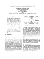
Tài liệu Báo cáo Y học: Kinetic study of sn-glycerol-1-phosphate dehydrogenase from the aerobic hyperthermophilic archaeon, Aeropyrum pernix K1 potx
Bạn đang xem bản rút gọn của tài liệu. Xem và tải ngay bản đầy đủ của tài liệu tại đây (552.86 KB, 8 trang )
Kinetic study of
sn
-glycerol-1-phosphate dehydrogenase
from the aerobic hyperthermophilic archaeon,
Aeropyrum pernix
K1
Jin-Suk Han
1
, Yoshitsugu Kosugi
2
, Hiroyasu Ishida
2
and Kazuhiko Ishikawa
1
1
National Institute of Advanced Industrial Science and Technology, Ikeda, Osaka, Japan;
2
National Institute of Advanced
Industrial Science and Technology, Tsukuba, Ibaraki, Japan
A gene h aving h igh s equence homology ( 45–49%) wit h th e
glycerol-1-phosphate dehydrogenase gene from Methano-
bacterium thermoautotrophicum was cloned from t he aero-
bic hyperthermophilic archaeon Aeropyrum pernix K1
(JCM 982 0). This gene expressed in Escherichia coli with
the pET vector syst em consists of 1113 nucleotides with an
ATG initiation codon and a TAG t ermination codon. The
molecular mass of t he purified enzyme was estimated to be
38 kDa by SDS/PAGE a nd 72.4 k Da by gel column
chromatography, indicating presence as a dimer. The
optimum reaction temperature of this enzyme was
observed to be 94–96 °C at near neutral p H. This enzyme
was subjected to two-substrate kinetic analysis. The
enzyme showed substrate specificity for NAD(P)H-
dependent dih ydroxyacetone phosphate reduction and
NAD
+
-dependent glycerol-1-phosphate (Gro1P)oxida-
tion. NADP
+
-dependent Gro1P oxidation was not
observed with this enzyme. For the production of Gro1P
in A. pernix cells, NADPH is the preferred coenzyme
rather than NADH. Gro1 P acted as a non competitive
inhibitor against dihydroxyacetone phosphate and
NAD(P)H. However, NAD(P)
+
acted as a competitive
inhibitor against NAD(P)H and as a noncompetitive
inhibitor against dihydroxyacetone phosphate. This kinetic
data indicates that the catalytic reaction by glycerol-
1-phosphate dehydrogenase f rom A. pernix follows a
ordered bi–bi mechanism.
Keywords: Aeropyrum pernix; a rchaea; g lycerol-1-phosphate
dehydrogenase; ordered bi–bi mechanism; hyperther-
mophile.
Archaea are a phylogenetically distinct group that diverged
from eubacteria and eukaryotes at an early stage in
evolution [1,2]. Archaea have several distinct features from
eubacteria and eukaryotes, including the unique stereo-
chemical backbones of phospholipids in their cellular
membrane. The core lipid of the phospholipids and
glycolipids in archaeal cells is sn-2,3-di-acylglycerol, which
has a polar head gro up in the sn-1 position . In contrast , the
major lipids of eukaryotic and bacterial cells mostly contain
sn-1,2-di-acylglycerol, which has a polar head group in the
sn-C-3 position [3]. Glycerol-1-phosphate (Gro1P)isthe
best substrate for the e nzymatic synthesis of 2,3-digeranyl-
geranyl-sn-glcerol-1-phosphate in the moderate thermophi-
lic (above 80 °C) Methanobacterium thermoautotrophicum
[4]. Therefore, Gro1P dehydrogenase is identified as the key
enzyme in the biosynthesis of archaeal enantiomeric polar
lipid structures, such a s the formation o f Gro1P from CO
2
and the subsequ ent formation o f t he ether lipid from Gro1P
in M. thermoautotrophicum [5,6]. The enzyme responsible
for Gro1P formation of archaea-specific glycerophosphate,
NAD(P)
+
-dependent sn-glycerol-1-phosphate deh ydrogen-
ase, was initially found in M. thermoautotrophicum [7].
Although several properties were investigated, there has
been no kinetic study of the mechanism of this enzyme.
Aeropyrum pernix K1 (JCM number 9820) is the first
aerobic h yperthermophilic archaea for which the complete
genome s equence h as been determined [8,9]. This archaeon’s
optimum growth temperature ranges from 90 to 105 °C.
Most of the proteins from A. pernix are expected to be
active at high temperature. The g lycerol dehydrogenase
gene in A. pernix K1 from the d atabase provided by
National Institute of Technology and Evaluation shows
high similarity with the genes of some archaeal Gro1P
dehydrogenases. To e xamine the function of the enzyme, we
have cloned a nd expressed Gro1P deh ydrogenase from
A. pernix using Escherichia coli.
MATERIALS AND METHODS
Strain and culture condition
A. pernix K1 (JCM number 9820) was obtained from the
Japan Collection of Microorganisms (Wako-shi, Japan).
The culture media c ontained 37.4 g of Bacto m arine broth
2216 (Difco) and 1.0 g of N a
2
S
2
O
3
ÆH
2
O in 1 L. The solution
of Na
2
S
2
O
3
ÆH
2
O was separately sterilized by filtration, and
aseptically added to t he medium. A. pernix was cultivated
for 48 h at 90 °C with shaking [8]. Genomic DNA was
isolated from the cultivated cell of A. pernix by the method
of Meade et al.[10].
Correspondence to K. Ishikawa, The Special Division for Human Life
Technology, National Institute of Advanced Industrial Science and
Technology (Kansai), 1-18-31, Midorigaoka, Ikeda, Osaka 563-8577,
Japan. Fax: + 8 1 727 51 9628, Tel.: + 81 727 51 9526,
E-mail:
Abbreviations:Gro1P, sn-glycerol-1-phosphate; Gro3P, sn-glycerol-
3-phosphate, Gro, glycerol.
Enzymes: glycerol-3-phosphate dehydrogenase (NAD) (EC 1.1.1.8);
glycerol de hydrogenase [NAD(P)] (EC 1.1.1.172); glycerol-1-phos-
phate dehydrogenase [NAD(P)] (EC 1.1.1.261).
(Received 5 October 2001, r evised 5 December 2001, accepted 7
December 2001)
Eur. J. Biochem. 269, 969–976 (2002) Ó FEBS 2002
Cloning and expression of the gene
Putative glycerol dehydrogenase gene (APE0519) from
A. pernix was cloned b y the method of Ishikawa et al.
[11]. The gene was amplified using PCR with two p rimers
containing unique restriction site. The upper primer (5¢-
CGTAAC TAAGACTCC GG
CATATGCTGTACCA
TAGCGT-3¢) contained an NdeI site as underlined. The
lower primer (5¢-AGGGGAAGAGAGGCA
GGATCCCT
AGC CAGACTATATA-3¢) contained a BamHI site as
underlined. PCR amplifications were performed at 94 °C
for 1 min, 61 °C for 2 min, and 70 °C for 3 min, for 35
cycles using V ent DNA polymerase. The a mplified gene was
hydrolyzed by the restriction enzymes a nd ligated to the
pET11a (Novagen, Madison, USA). The insert ed gene was
transformed u sing pET11a vector system in the host E. coli
BL21 (DE3) according to the manufacture’s instructions
(Novagen, Madison. USA), followed by sequence determi-
nation. Expression of the protein was induced by isopropyl
thio-b-
D
-galactoside induction according to a previou sly
reported method [11]. To verify t he identity with the
APE0519 sequence, DNA sequencing was carried out with
a L I-COR M odel L IC-4200(s)-2 S eq uencer (Aloka, Mitaka,
Tokyo, Japan). The concentration of t he protein was
determined with Coomassie protein assay reagent (Pierce
Chemical Company, Rockford, IL, USA) using bovine
serum albumin as the standard.
Purification of the Gro1
P
dehydrogenase from
E. coli
The transformant cells were h arvested b y centr ifugation a nd
frozen at )20 °C. The cells were disrupted with aluminium
oxide in 50 m
M
Tris/HCl buffer ( pH 8.0). After incubation
with DNase I (bovine pancreas, Sigma) for 3 0 min at 37 °C,
the crude extract was heated at 85 °C for 30 min and
centrifuged. The supernatant was dialyzed against 50 m
M
Tris/HCl buffer (pH 8.0) and the dialyzed sample was
purified by chromatography using a HiTrap Q column
(Pharmacia, U ppsala, Sweden), a H iLoad Phenyl Sepharose
column (Pharmacia), and a HiLoad Superdex column
(Pharmacia) according to the method described previously
[12]. Multiple alignment of amino-acid sequences was done
using the
CLUSTAL W
provided at .
The molecular mass o f purified enzyme w as determined by
SDS/PAGE electrophoresis using 10–15% gradient gel of
the Phast system (Pharmacia) and gel chromatography
using HiLoad Superdex column. The N-terminal amino-
acid sequence was analyzed using H P G1005 Protein
Sequencing System at the Takara S huzo Customer Service
Center (Kusatsu, Japan).
Assay of Gro1
P
dehydrogenase activity
The activity of Gro1P dehydrogenase was determined in
both directions, reduction and oxidation, spectrophoto-
metrically at 340 nm as d escribed by Nishihara & Koga [7].
The assay contained 50 m
M
Tris/HCl buffer (pH 7.0),
70 m
M
KCl, 2.1 m
M
dihydroxyacetone phosphate, and
0.32 m
M
NADH (0.32 m
M
NADPH) for the dihydroxy-
acetone phosphate reduction. The assay mixtu re for the
Gro1P oxidation direction contained 50 m
M
Tris/HCl
buffer (pH 7.0), 70 m
M
KCl, 10 m
M
Gro1P,and5.0m
M
NAD
+
(5.0 m
M
NADP
+
). The r eaction was performed at
65 °C in 1.5 mL cuvettes containing 1.2 mL reaction
mixture and initiated by the addition of 10 lLofenzyme
solution. Control reactions were carried out using the same
reaction mixture without enzyme. T he kinetic constants of
Gro1P dehydrogenase of A. pernix were o btained from
activity measurements, with substrate concentrations that
ranged from 0.1 · K
m
to 10 · K
m.
Each individual rate
measurement was run in triple and the kinetic mechanism
was determined by the damped nonlinear least-squares
method (Marquardt–Levenberg method) [13,14].
Materials
Gro1P was prepared by d ihydroxyacetone phosphate
reduction using the purified enzyme solution [15]. The
reaction mixtu re c ontained 4.2 m
M
dihydroxyacetone
phosphate, 2 .0 m
M
NADH, 50 m
M
Tris/HCl buffer
(pH 7 .0), and 50 lL purified enzyme solution. After the
Gro1P formation r eaction w as completed at 65 °Cfor6h,
Gro1P was purified by TLC chromatography [16] and its
concentration was measured by the phosphate analysis [17].
Glyceraldehyde phosphate, dihydroxyacetone phosphate,
sn-glycerol-2-phosphate, and dihydroxyacetone were pur-
chased from Sigma. NADH, NAD
+
,NADPH,and
NADP
+
were used the products of the O riental Yeast
Co. Ltd.
RESULTS AND DISCUSSION
Alignment of amino-acid sequence of various
dehydrogenases
The genome sequenced from A. pernix contained a putative
glycerol dehydrogenase gene t hat consisted of a 1113 bp
with an ATG initiation codon and a TAG termination
codon. This gene encoded a 39 351-Da polypeptide
consisting of 370 amino-acid residues. The deduced
amino-acid sequence of Gro1P dehydrogenase was used
for a similarity search in the protein resulting in strong
similarity with those of Gro1P dehydrogenases from
archaea. The results are s ummarized in Fig. 1. The sequence
identity for A. pernix Gro1P dehydrogenase to the Gro1P
dehydrogenase from Methanobacterium thermoautotrophi-
cum, Pyrococcus abyssi,andSulfolobus solfataricus was 45,
48, and 49%, respectively [6]. When compared to the
glycerol (Gro) dehydrogenase from E. coli, Schizosac-
charomyces pombe,andBacillus stearothermophilus,the
sequence identity was 19–20%. There was, however, no
similarity with those NAD(P)
+
-dependent sn-glycerol-
3-phosphate (Gro3P) dehydrogenases which provided
phospholipid back bones for bacteria. Many NAD(P)
+
-
dependent dehydrogenases have a similar folding pattern
described as an ÔADP-binding bab foldÕ [18]. The NAD
+
binding sites of dehydrogenase have a highly conserved
GXGXXG sequence, where X is any amino acid [19,20].
In contrast, some NADP
+
binding sites have an a lanine at
the position c orresponding to the third glycine residue of the
conserved t rio [21]. In A. pernix Gro1P dehydrogenase, the
NAD
+
binding site was found as conserved GXGXXG
sequence at position 113–117. Some representative
sequences of this conserved region a re shown Fig. 2. Based
on sequence alignment, the relative positions of the
conserved sequences are the same in the Gro1P and
970 J S. Han et al. (Eur. J. Biochem. 269) Ó FEBS 2002
Gro dehydrogenase families, suggesting a similar NAD
+
-
binding domain structure. On the other hand the relative
positions of the conserved sequences differ dramatically
between the Gro1P and Gro3P dehydrogenase families
indicating a structural difference. Based on sequence
homology, the gene product of APE0519 should be
classified as a Gro1P dehydrogenase with a closer structural
relationship to Gro dehydrogenases rather than Gro3 P
dehydrogenases [6].
Cloning of Gro1
P
dehydrogenase from
A. pernix
The Gro1P dehydrogenase gene from A. pernix was
amplified by PCR with unique two primers, inserted into
pET11a, with the constructed plasmid transformed into
BL21 (DE3). The sequence of the DNA inserted into the
host cell w as confirmed to have an identical s equence to the
APE0519 gene. The Gro1P dehydrogenase of A. pernix
was purified to homogeneity by a combination of ion
exchange, hydrophobic, and gel chromatography. The
purification procedure yielded approximately 4.4 mg of
protein at a purification factor of about 147 with specific
activity 3.22 lmolÆmin
)1
Æmg
)1
and recovery of 28%
(Table 1). Sequencing of the purified protein in solution
showed that the first seven N-terminal residues were Gly-
Leu-Tyr-Thr-Ser-Phe-His. With the exception that 17
residues of N-terminal were deleted, the amino-acid
sequence deduced was identical to that obtained in
database. The sequence of the deleted segment does not
seem to be a signal peptide [22]. The segment s eems to be
hydrolyzed during the process of purification. When tested
as a dehydrogenase (dihydroxyacetone phosphate reduc-
tion), Gro 1P dehydrogenase from A. pernix demonstrated
NADH- and NADPH-dependent activity. The purified
enzyme migrated as a single band on SDS/PAGE with
apparent molecular mass of 38 kDa. The deduced amino-
acid sequence of the open reading frame consisted of 367
amino acids with a molecular mass of 37 676 Da. The
molecular mass e stimated by gel chromatography (HiLoad
Superdex) w as approximately 72.4 kDa. This indicates that
Gro1P dehydrogenase from A. pernix formsadimeras
Fig. 2. Comparison of the amino-acid sequences of regions of
representative NAD(P)
+
-binding dehydrogenases. Conserved r esidues
thought t o be i mportant for enzyme binding a re marked w ith asterisks.
The box indicates conserved residues between the enzymes. Gro1P
DH, glycerol-1-phosphate dehydrogenase; Gro3P DH, glycerol-3-
phosphate dehydrogenase; G ro DH, glycerol dehydrogenase.
Fig. 1. Comparison of the amino-acid sequences of Gro1P dehydrogenase (A) and glycerol dehydrogenase (B). (A) Archaeal Gro1P dehydrogenase;
M. thermo, Methanobacterium thermoautotrophicum (370 amino acids), P. abyssi, Pyrococcus abyssi (346 am ino acids); S. solfa, Sulfolobus solfa-
taricus (351 amino acids); (B) Glycerol dehydrogenase fro m bacteria an d eukaryote; B. ster o, Bacillus stearothermophilus (370 amino acids); E. coli,
Escherichia c oli (380 amino acids); S. pombe, Schizosaccharomyces probe (450 ami no acids). The sequences have been aligned with dashes indicating
gaps. Asterisks i ndicate c onserved residues among four enzymes and an arrow i ndicates that the start point of amino acids in the purified enzyme.
Ó FEBS 2002 Glycerol-1-phosphate dehydrogenase from A. pernix (Eur. J. Biochem. 269) 971
opposed to that from M. thermoautotrophicum,which
exists as a homooctamer [7].
Substrate specificity and enzyme activity
The substrate specificity of Gro1P dehydrogenase was
examined using the purified enzyme. No activity w as
observed t oward glyceraldehyde phosphate, Gro3P,glycer-
ol-2-phosphate (Gro2P), Gr o, and dihydroxyacetone. This
enzyme efficiently catalyzed the NADH- and NADPH-
dependent dihydroxyacetone phosphate reduction, and also
the NAD
+
-dependent Gro1P oxidation (Table 2). The
oxidation rate of NADP
+
-dependent Gro1P was not
detected, indicating that the enzyme has no or very low
NADP
+
-dependent Gro1P oxidation a ctivity. The K
m
value for dihydroxyacetone phosphate was 19.4-fold less
than for Gro1P using NAD(H) as a coenzyme. This result
suggests t hat t he formation of Gro1P is the n atural direction
in the cell. The k
cat
of the dihydroxyacetone phosphate
reduction with NADH was h igher than that with NADPH.
The coenzyme NAD
+
was only used for the production
of dihydroxyacetone phosphate. T he G ro1P dehydrogenase
from A. pernix showed NAD(P)H-dependent dihydroxy-
acetone phosphate reduction and NAD
+
-dependent G ro1P
oxidation activities. In contrast, G ro1P dehydrogenase
from M. thermoautotrophicum was able to use both the
NAD(H) and NADP(H) coenzyme s for its oxidation/
reduction reactions [7].
General properties of Gro1
P
dehydrogenase
from
A. pernix
Maximal activity of Gro1P dehydrogenase was seen
between 94 and 96 °C at pH 7.0 (Fig. 3), which is in the
normal temperature range for growth of A. pernix [8].
Over 96 °C, enzyme activity decreased dramatically,
which seemed to be caused by irreversible denaturation
of the enzyme. With the exception of the temperature-
activity profile, the characteristics of the enzyme were
determined from initial velocity measurements in the
direction of the NADH-dependent dihydroxyacetone
phosphate reduction at 65 °C chosen as dihydroxyace-
tone phosphate and NADH were rapidly decomposed
over 70 °C [7]. The high growth temperature of A. pernix
may be linked to the higher optimum activity tempera-
ture of its Gro1P dehydrogenase [24]. The half-life of
activity was 30 min at the maximal activity temperature
(95 °C) and increased to 2 h at 90 °C (Fig. 4). The
enzyme activity of Gro1P dehydrogenase form M. ther-
moautotrophicum appeared to depend on the presence of
K
+
and Na
+
and showed maximum activity a t 70 m
M
of K
+
[7]. However, the purified enzyme from A. pernix
exhibited the highest levels of activity when assayed i n
metal free buffer after dialysis. Activity was decreased
to 86 and 80% by addition of 70 m
M
K
+
and Na
+
,
respectively. This result shows that the activity of Gro1P
dehydrogenase from A. pernix is affec ted differently by
the intracellular concentration of K
+
than M. thermo-
autotrophicum.
Kinetic analysis of Gro1
P
dehydrogenase
The above results show that thermophilic Gro1P dehydro-
genase catalyzes the following reaction:
DHAP þ NADðPÞH þ H
þ
¢Glycerol-1-phosphate
þ NADðPÞ
þ
Table 1. Purification table of Gro1P deh ydrogenase from A. pernix . The activity was measured in the direction of dihydroxyacetone p hosphate
reduction with t he standard assay mixture.
Purification step
Total activity
(units)
Protein
(mg)
Specific activity
(lmolÆmin
)1
Æmg
)1
)
Yield
(%)
Purification
factor
Cell extract 51.46 2287 0.023 100 1
Heat treatment 44.99 113.9 0.40 87 17.6
HiTrap-Q 23.89 36.66 0.65 46 29.1
HiLoad Phenyl Sepharose 19.67 15.77 1.26 38 55.8
HiLoad Superdex 14.28 4.434 3.22 28 147.2
Table 2 . Substrate s pecifi city of Gro1P dehydrogenase f rom A. pernix. T hese p arameters were estimated u sing nonlinear least-aquares method [ 23]
from experiments in which a fixed c onc entration of substrate or coenzyme and an appropriate range of c onc entration of the other reactant were
used. ND; not d etec ted.
Substrate K
m
(m
M
) k
cat
(min
)1
)
Dihydroxyacetone phosphate reduction
Dihydroxyacetone phosphate (0.32 m
M
NADH) 0.460 ± 0.127 154.25 ± 43.29
Dihydroxyacetone phosphate (0.32 m
M
NADPH) 0.290 ± 0.128 45.21 ± 12.82
NADH (4 m
M
dihydroxyacetone phosphate) 0.032 ± 0.005 143.96 ± 6.81
NADPH (4 m
M
dihydroxyacetone phosphate) 0.044 ± 0.022 43.29 ± 5.12
Gro1P oxidation
Gro1P (5 m
M
NAD) 8.92 ± 0.64 5.31 ± 0.11
Gro1P (5 m
M
NADP) ND ND
NAD (50 m
M
Gro1P) 1.57 ± 0.44 5.65 ± 0.45
NADP (50 m
M
Gro1P)NDND
972 J S. Han et al. (Eur. J. Biochem. 269) Ó FEBS 2002
Initial velocities of the forward reaction were analyzed by
varying the concentration of dihydroxyacetone phosphate
and NAD(P)H under nonsaturating conditions without
addition of reaction products. The reverse reaction using
Gro1P and NAD(P)
+
could not be carried out because the
backward rate was too low (see Table 2). The results of
initial velocity studies were plotted o n a Lineweaver–Burk
(double r eciprocal) plot (see A-1 and A-2 of Figs 5 and 6)
[25]. The result of Figs 5 and 6 indicate that the saturation
of the substrate was not reach ed u nder these conditions.
Double reciprocal plots using dihydroxyacetone phosphate
or NAD(P)H at various fixed levels of NAD(P)H or
dihydroxyacetone phosphate, respectively, resulted in a
family of lines with a common intersection to the left of the
ordinate. This result e xcludes an Ôequilibrium ordered b i–bi
mechanismÕ and indicates a sequential mechanism [26]. To
determine the binding order of substrates in a sequential
mechanism, we carried out the product inhibition studies in
which dihydroxyacetone phosphate or NAD(P)H was
varied at nonsaturating levels. From the L ineweaver-Burk
plots (see C-1 and C-2 of Figs 5 and 6), Gro1P acted as a
noncompetitive inhibitor at various levels of NAD(P)H a nd
dihydroxyacetone phosphate. Such an inhibition pattern
ruled out a simple Ôrapid equilibrium rand om bi–bi
mechanismÕ,aÔTheorell chance mechanismÕ,oraÔping-
pong mechanismÕ [27]. The coproduct NAD(P)
+
[9] was
found to be a noncompetitive inhibitor of the forward
reaction when dihydroxyacetone phosphate was varied at
the nonsaturated level of the coenzyme. H owever, i t w as not
clear whether NAD(P)
+
acted as a competitive or
noncompetitive inhibitor when NAD(P)H was varied at
the nonsaturated level of dihydroxyaceton e phosphate
because the family of lines did not share a common
intersection on the ordinate (see B-1 and B-2 of Figs 5 and
6). Within the range of experimental errors observed, this
enzyme probably works using an Ôordered bi–bi mechan-
ismÕ. T herefore, the experimental data was fitted to the
equation for an Ôord ered bi–bi mechanismÕ as follows [2].
V ¼
V
m
½A½B
K
ia
K
b
1 þ
½A
K
ia
þ
K
a
½B
K
ia
K
b
þ
½A½B
K
ia
K
b
þ
K
q
½P
K
p
K
iq
þ
½Q
K
iq
þ
½P½Q
K
p
K
iq
þ
K
q
½A½P
K
ia
K
p
K
iq
þ
K
a
½B½Q
K
ia
K
b
K
iq
þ
½A½B½P
K
ia
K
b
K
ip
þ
½B½P½Q
K
ib
K
p
K
iq
where [A], [B], [P] and [Q] are the concentrations of
NAD(P)H, dihydroxyacetone phosphate, Gro1P,and
NAD(P)
+
, re spectively. The kinetics c onstants K
a
(K
m
for
NAD(P)H), K
b
(K
m
for dihydroxyacetone phosphate), K
ia
(dissociation constant for NAD(P)H), and V
m
(maximal
velocity) values were determined from the initial velocity
studies ([P] ¼ [Q] ¼ 0) with a nonlinear least-squares
method [14]. The K
iq
(dissociation constant for NAD(P)
+
)
was obtained from the inhibitio n effect of NAD(P)
+
([P] ¼ 0). The K
ip
(dissociation constant for Gro1P)and
the K
p
/K
q
values were obtained from product inhibition
studies of Gro1P (Q ¼ 0). The K
p
and K
q
values are
simultaneously present in the above equation as interde-
pendent ratios. The experimental data was fitted to the
above equation initial value of K
q
setto1.Whenthe
Fig. 4. Effect o f heating on Gro1P dehydrogenase activity. Enzyme was
incubated in 100 m
M
Tris/HCl buffer (pH 8.0). Aliquots were removed
every hour and t he activity was measured in the standard assay mixture
at 65 °C. Residual activity is expressed on a logarithmic scale.
Fig. 3. Temperature dependence of specific activity for Gro1P
dehydrogenase. The e nzyme activity was measured in the d irection of
dihydroxyacetone ph osphate reduction in 50 m
M
Tris/HCl buffer,
pH 7.0, contai ning 70 m
M
KCl, 2 .1 m
M
dihydroxyacetone phosphate,
and 0.32 m
M
NADH for 5 min.
Ó FEBS 2002 Glycerol-1-phosphate dehydrogenase from A. pernix (Eur. J. Biochem. 269) 973
obtained values were plotted on a double reciprocal plot,
NAD(P)
+
acted as a competitive inhibitor against
NAD(P)H and a noncompetitive inhibitor against dihy-
droxyacetone phosphate, whereas Gro1P acted as a
noncompetitive inhibitor against NAD(P)H a nd dihydroxy-
acetone phosphate. This supports the conclusion that this
enzyme follows the ordered bi–bi mechanism. The final
fitted values were 99.7% and 99.1% with final standard
deviation of 0 .016 and 0 .010 using NADH and NADPH as
coenzyme, respectively. The combination of results from
initial velocity studies and inhibitio n patterns of p roducts,
suggest the reaction of Gro1P dehydrogenase is to be an
Ôordered bi–bi mechanism Õ. Estimated kinetic parameters of
the ordered bi–bi mechanism were summarized in Table 3.
The K
b
of NADPH (0.082 m
M
) was smaller than that of
NADH (0.278 m
M
) indicating that NADPH is the better
coenzyme for Gro1P production. The activity of this
enzyme was regulated by the product, Gro1P,and
NAD(P)
+
in contrast to the lack of p roduct inhibition of
theenzymefromM. thermoautotrophicum [7]. Although
inhibition by Gro1P was relatively low such that K
ip
against
NADH was 31.47 m
M
and that against NADPH was
12.1 m
M
, the inhibito ry effect could b e confirmed b y Figs 5
and 6 (C-1 and C-2). The observation that the NADP
+
-
dependent Gro1P oxidation a ctivity w as very low and the
above kinetic results mean that G ro1P can efficiently
control t he reduction reaction without decreasing the Gro1P
pool in the cell when NADPH is used as coenzyme. In
contrast, the Gro1P dehydrogenase from M. thermoauto-
trophicum was not affected by Gro1P concentration during
the production of Gro1P [7]. The i nhibition mechanism in
Gro1P dehydrogenase of A. pernix is different from that of
Fig. 5. Reciprocal plot of dihydroxyacetone
phosphate ( DHAP) reduction using NADH.
A-1, initial v elocity pattern with variable
concentrations of NADH and n onsaturating
fixed levels of dihydroxyacetone phosphate;
A-2, initial v elocity pattern with variable
concentrations of dihydroxyacetone phos-
phate a nd nonsaturating fi xed levels of
NADH; B-1, inhibition of dihydroxyacetone
phosphate reduction by NAD
+
at 2.1 m
M
dihydroxyacetone phosphate and varying
NADH concentration; B-2, in hibitio n of
dihydroxyacetone phosphate reduction by
NAD
+
at 0.32 m
M
NADH and varying
dihydroxyacetone phosphate concentration;
C-1, inhibition of dihydroxyac etone phos-
phate reduction by Gro1P at 2.1 m
M
dihy-
droxyacetone p hosphate and varying NADH
concentration; C-2, Inhibition of dihydrox-
yacetone phosphate reduction by Gro1P at
0.32 m
M
NADH and varying dihydrox-
yacetone phosphate concentration. The
enzyme activity was measured at 65 °Cin
50 m
M
Tris/HCl buffer (pH 7.0) containing
70 m
M
KCl and variable conce ntration of
substrates.
974 J S. Han et al. (Eur. J. Biochem. 269) Ó FEBS 2002
Fig. 6. Reciprocal plotting of dihydroxy-
acetone phosphate (DHAP) reduction using
NADPH. A-1, initial velocity pattern with
variable concentrations of NADPH and no n-
saturating fixed le vels of dihydroxyacetone
phosphate; A-2, in itial velocity pattern with
variable concentrations of d ihydroxyacetone
phosphate and nonsaturating fixed levels of
NADPH; B-1, inhibition o f dihydroxyacetone
phosphate reduction by NADP
+
at 2.1 m
M
dihydroxyacetone phosphate and varying
NADPH c oncentration; B-2, Inhibition of
dihydroxyacetone phosphate reduction by
NADP
+
at 0.48 m
M
NADPH a nd varying
dihydroxyacetone phosphate concentration;
C-1, inhibition of dihydroxyacetone p hos-
phate reduction by G ro1 P at 2.1 m
M
dihy-
droxyacetone phosphate a nd varying
NADPH concentration; C-2, inhibition of
dihydroxyacetone phosphate reduction by
Gro1P at 0.48 m
M
NADPH and varying
dihydroxyacetone phosphate concentration.
The enzyme a ctivity w as m e asured at 65 °Cin
50 m
M
Tris/HCl buffer (pH 7.0) containing
70 m
M
KCl and variable concentration of
substrates.
Table 3. Kinetic parameters for G ro1P dehydrogenase estimated by the o rdered bi–bi f unction. These parameters were calculated from F igs 5 an d 6
using t he Marquardt-Levenbery method [13,14]. k
cat
¼ turnover number, K
a
¼ K
m
for NAD(P)H, K
b
¼ K
m
for dihydroxyacetone phosphate,
K
ia
¼ dissociation constant for N AD(P)H, K
iq
¼ dissociation constant f or NAD(P)
+
, K
ip
¼ dissociation constant for Gro1 P.
Kinetic parameter
Estimated value
NADH NADPH
k
cat
(min
)1
) 149.55 ± 12.06 73.47 ± 7.91
K
a
(m
M
) 0.037 ± 0.014 0.159 ± 0.050
K
b
(m
M
) 0.278 ± 0.099 0.082 ± 0.097
K
ia
(m
M
) 0.020 ± 0.043 0.620 ± 0.245
K
iq
(m
M
) 0.331 ± 0.028 1.03 ± 0.128
K
ip
(m
M
) 31.5 ± 8.07 12.1 ± 2.74
K
p
/K
q
(—) 6.00 ± 0.49 2.68 ± 0.62
Final curve fitting (%) 99.7 99.1
Final SD of data (rms error) 0.016 0.010
Ó FEBS 2002 Glycerol-1-phosphate dehydrogenase from A. pernix (Eur. J. Biochem. 269) 975
M. thermoautotrophicum andalsoseemstobevery
important in the regulation of lipid biosynthe sis. The
Michaelis–Menten constant for G ro3P was over 50 m
M
in
Gro3P dehydrogenase from Saccharomyces cerevisiae,so
that the inhibitory effect of Gro3P was negligible in the
experimental data. The Gro3P dehydrogenase in E. coli
involved in lipid biosynthesis is regulated by allosteric
inhibition by the production of Gro3P; t his i s i mportant to
maintain a lo w intracellular pool of Gro3P and to regulate
lipid biosynthesis [28]. More detailed kinetic studies of
Gro1P dehydrogenase should provide more information
about how polar lipid biosynthesis in archaea d iffers from
that in bacteria.
ACKNOWLEDGEMENTS
This work was performed as part of the STA fellowship program
supported by the Japan Science and Technology C orporation.
REFERENCES
1. Nelson, K.H., Paulsen, I.T., Heidelberg, J.F. & Fraser, C.M.
(2000) Sta tus of genome projects for nonpathogenic bacteria and
archaea. N at. Biotechnol. 18 , 1049–1054.
2. Woese, C.R., Kandler, O. & Whee lis, M .L. (1990) Towards nat-
ural system of organisms: Proposal for the domains archaea,
bacteria, and eucarya. Proc. Natl Acad. Sci. 87, 4576–4579.
3. Zhang, D. & P oulter, C.D. (1993) Biosynthesis of archaebacterial
ether lipids. Formation of ether linkages by prenyltransferases.
J. Am. C he m. Soc. 115, 1 270–1277.
4. Zhang, D., Daniels, L. & Poulter, C.D. (1993) Biosynthesis of
archaebacterial ether m embranes. Formation of i soprene ethers by
a prenyl transfer. J. Am. Chem. Soc. 115, 1264–1265.
5. Nishihara, M. & Koga, Y. (1995) sn-glycerol-1-phosphate dehy-
drogenase in Methanobacterium t hermoautotrophicum: key enzyme
in biosynthesis of the enantiomeric glycerophosphate backbone of
ether p hospholipids o f archaebacteria. J. Biochem. 117, 933–935.
6. Koga, Y., Kyuragi, T. & Sone, N. (1998) Did archeal and bacterial
cell arise ind epen dently from noncellular precursors? A hypothesis
stating that advent of membrane phospholipid with enantiomer ic
glycerophosphate backbones caused the separation of the two
lines of descent. J. Mol. Evol. 46, 54–63.
7. Nishihara, M. & Koga, Y. (1997) Purification and properties of
sn-glycerol-1-phosphate dehydrogenase from Methanobacterium
thermoautotrophicum: Characterization of the biosynthetic enzyme
for the enatiomeric glycerophosphate backbone of ether polar
lipids of archaea. J. Biochem. 122, 572–576.
8. Sako,Y.,Nomura,N.,Uchida,A.,Ishida,Y.,Morri,H.,Koga,
Y., Hoaki, T. & Maruyama, T. (1996) Aeropyrum pernix General
Nov., sp. Nov., a novel aerobic hyperthermophilic archaeon
growing at t emperatures up to 100
o
C. In t. J. Syst. B acter iol . 46,
1070–1077.
9. Faguy, D.M. & Doolittle, W.F. (1999) Genomics: lessons f rom the
Aeropyrum pernix genome. Current Biol. 9, R883–R886.
10. Meade , H.M., Long, S.R., Ruvkun, G.B., Brown, S.E. & A u subel,
F.M. (1982) Physical and genetic characterization of symbiotic
and auxotro phic mu tants o f Rhizobium meliloti induced by
transposon Tn5 mutagenesis. J. Bacterial. 149, 1 14–122.
11. Ishikawa, K., Ishida, H., Koyama, Y., Kawarabayasi, Y.,
Kawahara, J., Matsui, E . & Matsui, I. (1998) Acylamino a cid-
releasing enzyme from the thermophilic archaeon Pyrococcus
horikoshii. J. Biol. Chem. 273, 1 7726–17731.
12. Ishikawa, K., Ishida, H., Matsui, I., Kawarabayasi, Y. & K ikuchi,
H. (2001) Novel bifunctional hyperthermostable carboxy-
peptidase/aminoacylase from Pyrococcus horikoshii OT3.
Appl. Environ. Microbiol. 67, 6 73–679.
13. Menke, W. (1989) Discrete i nverse the ory. In Geophysical Data
Analysis, Revised edn, pp. 143–160. Academic P ress, N ew York,
USA.
14. Press, W.H., T eukolsky, S.A., Vertterling, W .T. & Flannery, B.P.
(1992) Numerical Recipes in Fortran – the A rt of Scientific C om-
puting, 2nd edn. Cambridge University Press, USA.
15. Nishihara, M., Yamazaki, T., Oshima, T. & Koga, Y.
(1999) sn-Glycerol-1-phosphate-forming activities in Archaea:
Separation of archaeal phospholipid biosynthesis and glycerol
catabolism by glycerophosphate enantiomers. J. Bacteriol. 181,
1330–1333.
16. Nishihara, M. & Koga, Y. (1988) Quantitative conversion of
diether and tetraether phospholipids t o glycerophosphoesters by
dealkylation with boron trichloride: a tool for structural analysis
of archaebacterial lipids. J. Lipid R es. 29, 384–388.
17. Bartlett, G.R. (1959) Colorimetric assay methods for free and
phosphorylated glyceric acids. J. Biol. Chem. 234, 469–471.
18. Fan, F. & Plapp, B.V. ( 1999) Probing the affinity and specificity o f
yeast alcohol dehydrogenase I for coenzymes. Arch. Biochem.
Biophys. 367 , 240–249.
19. Wierenga, R.K., Terpsta, P. & Hol, G.J. (1986) Prediction of the
occurrence of the ADP-binding bab-fold in proteins, using an
amino a cid sequence fi ngerprint. J. Mol. Biol. 18 7 , 101–107.
20. Nagy, E., Henics, T., Eckert, M., Lightowlers, R. N., Kellermayer,
M. & Miseta, A. (2000) Identification o f the NAD
+
-binding fold
of glyceraldehyde-3-phosphate dehydrogenase as a novel RNA-
binding domain. Biochem. Biophys. Res. Commun. 275, 253–260.
21. Scrutton,N.S.,Berry,A.&Perham,R.N.(1990)Redesignofthe
coenzyme specificity of a dehydrogenase by protein engineering.
Nature 343, 3 8–43.
22. Marion, E.E.W. (1984) Complication of published signal
sequence s. Nucleic A cids Res. 12 , 5145–5164.
23. Sakoda, M . & Hiro mi, K. ( 1976) Determination of the be st-fit
values of kinetic parameters of the Michae lis–Menten equation by
the method of the le ast squares with the Taylor expansion.
J. Biochem. (Tokyo) 80, 547–555.
24. Yamano,S.,Sako,Y.,Momura,N.&Maruyama,T.(1999)A
cambialistic SOD in a strictly aerobic hyperthermophilic archae-
on, Aeropyrum pernix. J. Biochem. 126, 218–225.
25. Rudolph, F.B. & F romm, H .J. (1979) Plotting me thods f or ana-
lyzing enzyme rate data. In Methods i n Enzymology (Purich, D.L.,
ed.), pp. 138–158. Academic Press, San Diego, CA.
26. Cleland, W.W. (196 3b) The ki netics of enz yme-catalyzed reactions
with two or more substrates or products. 2. Inh ibition : nomen-
clature and the ory. Biochim. Biophys. Acta 67, 173–187.
27. Cai, J., Pietzsch, M., Theobald, U. & Rizzi, M. (1996) Fast pur-
ification and kinetics studies of the glycerol-3-phosphate dehy-
drogenase from the yeast Sacc haromyc es cerevisiae. J. Biote chnol.
49, 19–27.
28. Edger, J .R. & Bell, R.M. (19 78) Bi osynthesis i n Es cherichia coli of
sn-glycerol-3-phosphate, a precursor of phospholipid. Kinetic
characterization of wild type and feed-back-resistant form s of the
biosynthetic sn-glycerol-3-phosphate deh ydrogenase . J. Biol.
Chem. 253, 6354–6363.
976 J S. Han et al. (Eur. J. Biochem. 269) Ó FEBS 2002