Tài liệu Báo cáo Y học: Evidence that a eukaryotic-type serine/threonine protein kinase from Mycobacterium tuberculosis regulates morphological changes associated with cell division docx
Bạn đang xem bản rút gọn của tài liệu. Xem và tải ngay bản đầy đủ của tài liệu tại đây (239.09 KB, 8 trang )
PRIORITY PAPER
Evidence that a eukaryotic-type serine/threonine protein kinase
from
Mycobacterium tuberculosis
regulates morphological
changes associated with cell division
Rachna Chaba, Manoj Raje and Pradip K. Chakraborti
Institute of Microbial Technology, Chandigarh, India
A eukaryotic-type protein serine/threonine kinase, PknA,
was cloned from Mycobacterium t uberculosis strain H37Ra.
Sequencing of t he clone indicated 100% identity with the
published pknA sequence o f M. tuberculosis strain H37Rv.
PknA fused to maltose-binding protein was expressed in
Escherichia coli; it exhibited a molecular mass of % 97 kDa.
The fu sion protein was purified from the s oluble f raction b y
affinity chromatography using amylose resin. In vitro kinase
assays showed that the autophosphorylating ability o f PknA
is strictly magnesium/manganese-dependent, and sodium
orthovanadate can inhibit this activity. Phosphoamino-acid
analysis ind icated that PknA phosphorylates at serine and
threonine residues. PknA was also able to phosphorylate
exogenous substrates, such as myelin basic p rotein and his-
tone. A comparison of the n ucleotide-derived amino-acid
sequence of PknA with that of functionally characterized
prokaryotic serine/threonine kinases indicated its possible
involvement in cell d ivision/differentiation. Protein–protein
interaction studies revealed that PknA is capable o f phos-
phorylating at least a %56-kDa soluble p rotein from E. coli.
Scanning electron microscopy showed that constitutive
expression of this kinase resulted in elongation of E. coli
cells, supporting its regulatory role in cell division.
Keywords: a utophosphorylation; phosphorylation; PknA;
serine/ threonine kinase; signal transduction.
Signal-transduction pathways in both prokaryotes and
eukaryotes often utilize protein phosphorylation as a
molecular switch in regulating different cellular activities
such as adaptation and d ifferentiation. It is well known that
protein kinases play a cardinal role in the process. They are
grouped i nto t wo superfamilies, histidine (His) and serine/
threonine (Ser/Thr) o r t yrosine ( Tyr) kinases, based on their
sequence similarity and enzymatic specificity [1,2]. Signal
transduction in prokaryotes usually uses His kinases, which
autophosphorylate at histidine residues [2]. In eukaryotes,
such signalling pathways are mediated by Ser/Thr or Tyr
kinases, which autophosphorylate at serine/threonine or
tyrosine residues [1].
Interestingly, analysis of genome sequences revealed the
presence of putative genes encoding eukaryotic-type Ser/Thr
kinases in many bacterial species. A search of the Escheri-
chia coli genome also indicated the presenc e of sequences
exhibiting homology with eukaryotic-type Ser/Thr kinases,
but they h ave not been characterized bioc hemically or
functionally. Involvement of such kinases in regulating
growth and development has largely been d ocumented in
soil bacteria such as Myxococcus [3–6], Anabaena [7] and
Streptomyces [8,9]. In Yersinia p seudotuberculosis,YpkA
has been identified as the first secreto ry prokaryotic Ser/Thr
kinase involved in pathogenicity [10]. Besides these, eukary-
otic-type Ser/Thr kinase s have been implicated in virulence
in opportunistic pathogens s uch a s Pseudomonas aeruginosa
[11]. Thus a detailed study of these kinases, especially in
pathogenic bacteria, could produce important insights into
their contributions to signal transduction. This may help in
the design o f drug intervention strategies in a s ituation
where the emergence of drug-resistant strains of several
pathogenic bacteria has resulted in the rapid resurgence
of diseases thought to be near irradication. We focused
on tuberculosis, a disease caused by Mycobacterium
tuberculosis, which is responsible for considerable human
morbidity and mortality world wide [12].
In the M. tuberculosis genome, 11 putative eukaryotic-
type kinases have been reported [13]. Among these Ser/Thr
kinases, four (PknB, PknD, PknF, PknG) have been
biochemically characterized [14–16], but their bio logical
functions are not known. The M. tuberculosis genome
sequence further indicated t hat the gene for a putative Ser/
Thr kinase, pknA, is located adjacent to those encoding
bacterial morphogenic proteins. Interestingly, the p resence
of a Ser/Thr kinase at this location in the mycobacterial
genome is unique among prokaryotes [17]. We therefore
concentrated on PknA. In this paper, we report the cloning
and expression of PknA as a fusion with maltose-binding
protein (MBP). Characterization of the fusion protein
revealed th at it is capable of phosphorylating itself as well as
basic protein substrates not present i n M. tuberculosis.
Furthermore, we present strong evidence that the constitu-
tive expression o f this kinase causes elongation of cells in
E. coli , supporting a regulatory role for PknA in cell
division.
Correspondence to P. K. Chakraborti, Institute of Microbial
Technology, Sector 39A, Chand igarh 160 036, India.
Fax: + 91 172 690 585, Tel.: + 91 172 695 215,
E-mail: p
Abbreviations IPTG, isopropyl thio-b-
D
-galactoside;
MBP, maltose-binding protein.
(Received 16 November 2001, revised 3 January 2002, accepted
9 January 2002)
Eur. J. Biochem. 269, 1078–1085 (2002) Ó FEBS 2002
MATERIALS AND METHODS
Bacterial strains and vectors
M. tuberculosis strain H37Ra [18] used in this study was
grown at 37 °C using oleic acid/albumin/dextrose/catalase/
Tween-80/glycerol-supplemented Middle b rook 7H9 broth
or 7H10 agar. E. coli strains DH5a and TB1 were cultured
on Luria–Bertani agar or broth . Vectors such as pUC19 and
pMAL-c2X were obtained from commercial sources. The
Mycobacterium–E. c oli shuttle v ector, p19Kpro, was a gift
from D. B. Young and M. Blokpoel, Imperial College
School of Medicine at St Mary’s, London, UK.
PCR amplification, site-directed mutagenesis,
and construction of recombinant plasmids
Genomic DNA was isolated from M. tuberculosis strain
H37Ra a s described elsewhere [19] except that the sphero-
plast lysis step was carried out for 24 h at 37 °C with SDS
(4%) and proteinase K (500 lgÆmL
)1
). DNA thus obtained
was u sed f or PCR amplification of pknA. The Expand Long
Template PCR system (mixture of Pwo and Taq DNA
polymerases; Roche Molecular Biochemicals) was used for
this purpose. The forward (CC7: 5¢-CATATGAGCCCC
CGAGTTGG-3¢) and reverse (CC8: 5¢-TCATTGCGCTA
TCTCGTATCGG-3¢) primers were designed on the basis
of the published M. tuberculosis genome sequence [13] of
pknA (Rv0015c). Oligonucleotides used in this study were
custom-synthesized from IDT, Coralville, IN, USA. PCR
was carried out for 30 cycles (denaturation, 95 °Cfor30s
per cycle; annealing, 50 °C for 30 s per cycle; elongation,
68 °C for 2 min for fi rst 10 cycle s a nd then for the remaining
20 cycles the elongation step w as extended f or an additional
20 s in each cycle).
PCR was also used to generate the K42N ( replacement o f
lysine by asparagine at residue 42) point mutant of PknA.
Two f orward primers, CC58 (5¢-CACAGGAATTCCATA
TGAGCCCCCGAGTTGG-3¢), CC62 (5¢-GTGTTGCGG
TGAA
TGTGCTCAAGAGCG-3¢) and tw o reverse prim-
ers, CC61 (5¢-CTGCCCGGTGGGGGTGATCAAGA
TG-3¢), CC63 (5¢-CGCTCTTGAGCAC
ATTCACCGCA
ACAC-3¢), were synthesized. Base mismatches ( underlined
bases) for the desired mutations were incorporated in
primers CC62 and CC63. To generate the mutant, two sets
of primary and one set of secondary PCR reactions were
carried out as described elsewhere [20] using the gel-purifie d
pknA (% 1.3 kb) as template. Primary reactions were
carried out with primers CC58/CC63 and CC61/CC62,
while for secondary reactions, PCR primers CC58 and
CC61 were used. Thus, the K42N mutation was contained
within the amplified % 460-bp fragment of pknA, which has
a unique XhoI site in addition to the EcoR IandNdeI sites
incorporated in the primer CC58.
All manipulations with DNA were performed by stand-
ard methods [21]. Restriction/modifying enzymes and other
molecular biological reagents used in this study were
obtained from New England Biolabs. After PCR amplifi-
cation, pknA was t reated with K lenow, and the b lunt-ended
fragment was cloned at the SmaI site of pUC19 (pPknA).
Plasmid DNA was prepared after transform ation of pPknA
in E. coli strain DH5a and sequenced in an automated
sequencer (ABI; PE Applied Biosystems).
To monitor expression of PknA fused with MBP, E. coli
vector pMAL-c2X was used. After digestion of pPknA and
pMAL-c2X with NdeIandBamHI, respectively, they were
treated with K lenow to obtain blunt-ended f ragments. Both
these fragments were further r estriction-d igested with
HindIII, ligated and t ransformed in E. coli strain TB1 to
obtain clones c ontaining the plasmid (pMAL-PknA) bear-
ing in-frame fusion of % 1.3 kb pknA (confirmed b y junction
sequencing) at the 3¢ end of MBP. To express the K42N
mutant as an MBP fusion protein, a % 460-bp fragment of
mutated pk nA was digested with EcoRI/XhoI and substi-
tuted for the corresponding wild-type fragment in the
pMAL-PknA backbone. The resulting construct, pMAL-
K42N, was sequenced to confirm the mutation.
pknA or the K42N mutant w as also cloned in t he
Mycobacterium–E. c oli shuttle vector p19Kpro [22] to
obtain t he constitutive expression plasmids (p19Kpro-PknA
or p19Kpro-K42N). The strategy adopted was same a s for
construction of pMAL-PknA. To clone pknA in an
antisense o rientation, pPknA was initially digested with
NdeI and treated with Klenow to obtain a blunt-ended
fragment. After restriction digestion with BamHI, this
fragment was subsequently ligated to p19Kpro, which was
already digested with BamHI and EcoRV. The antisense
construct of pknA was designated p19Kpro-aPknA. All
three constructs, p19Kpro-PknA, p19Kpro-K42N and
p19Kpro-aPknA were transformed i n E. coli strain
DH5a. Clo nes carryin g the gene of interest were confirmed
at all steps by restriction analysis and Southern-blot
hybridization. The probe (PCR-amplified pknA)usedwas
radiolabelled by random priming with [a-
32
P]CTP (BRIT,
Hyderabad, India).
Expression of recombinant protein
pMAL-PknA or pMAL-K42N cultures were grown at
37 °C a nd induced with 0.3 m
M
isopropyl thio-b-
D
-galacto-
side (IPTG) at an A
600
of 0.5. Cells were harvested a fter 3 h,
lysates were prepared, and expression was monitored by
SDS/PAGE (8% gel) and C oomassie Brilliant Blue staining.
To find out the solubility of the expressed fusion protein,
after induction cells were suspended in lysis buffer and
sonicated. S upernatant and pellet fractions obtained after
sonication were subjected to SDS/PAGE. Finally, the
fusion protein was purified by affinity chromatography on
an amylose column according to the manufacturer’s
instructions (New England Biolabs). In a similar manner,
MBP–bgal fusion protein expressed b y pMAL-c2X was
also purified for its use as a control. To exam ine the
constitutive expression of the p rotein and its solubility,
overnight cultures (at 37 °C) of constructs in p19Kpro were
processed in the same way as pMAL-PknA except that
IPTG induction was not required.
Kinase assay
The ability of PknA or the K42N mutant, as a purified
fusion protein with MBP, to autophosphorylate and
phosphorylate exogenous substrates such as histone (from
calf thymus, type II-AS; Sigma) or myelin basic protein
(from bovine brain; Sigma) was determined in an in vitro
kinase assay. Aliquots (usually 800 ng to 6 lgin20lL
reaction volume) of fusion protein (MBP–PknA or
Ó FEBS 2002 Characterization of PknA from M. tuberculosis (Eur. J. Biochem. 269) 1079
MBP–K42N or MBP–bgal) were mixed with 1 · kinase
buffer (50 m
M
Tris/HCl, pH 7.5, 50 m
M
NaCl, 10 m
M
MnCl
2
), and the reaction was initiated by adding 2 lCi
[c-
32
P]ATP. After incubation at 24 °C f or 20 min, the
reaction was stopped by adding SDS sample buffer (30 m
M
Tris/HCl, pH 6 .8, 5% glycerol, 2.5% 2-mercaptoethanol,
1% SDS and 0.01% bromophenol blue). Samples were
boiled for 5 min and resolved by SDS/PAGE (8–12.5%
gels). Gels were stained w ith C oomassie Brilliant Blue, dried
in a g el dryer ( Bio-Rad) at 70 °C f or 2 h and finally exposed
to Kodak X -Omat/AR film. To monitor the effect of
bivalent cations, the 10 m
M
MnCl
2
in the 1 · kinase buffer
was substituted with 1, 10 or 100 m
M
Mn
2+
/Mg
2+
/Ca
2+
.
The autophosphorylating ability of the constitutively
expressed PknA was determined using p19Kpro-PknA-
transformed E. coli extract in a similar manner.
To identify proteins that interacted with PknA, MBP–
PknA (100 lg) was immobilized on amylose resin and
incubated in the presence of soluble protein extracts
(250 lg) prepared from E. coli strain DH5a for 10 h at
4 °C. Amylose beads were washed (4500 g for 5 min) four
times to remove unbound proteins. After suspension of
washed beads in TEN buffer (20 m
M
Tris/HCl, pH 7.5,
200 m
M
NaCl and 1 m
M
EDTA), aliquots (12 lL) were
used for phosphorylation assays.
Western blotting
Phosphoamino-acid analysis was carried out by Western
blotting. Purified fusion proteins or cell extracts (800 ng to
3 lg p rotein per slot) were resolved by SDS/PAGE (8% gel)
and t ransferred at 250 m A f or 45 min t o n itrocellulose
membran e (0.45 lm) in a mini-transblot apparatus (Bio-
Rad) using Tris/glycine/SDS buffer (48 m
M
Tris, 39 m
M
glycine, 0.037% SDS and 20% methanol, pH % 8.3).
Primary a ntibodies (anti-MBP, anti-phosphoserine, anti-
phosphothreonine and a nti-phosphotyrosine) used for dif-
ferent immunoblots were commercially available (New
England Biolabs, Santa Cruz Biotech and Sigma). Horse-
radish peroxidase-conjugated anti-(mouse IgG) Ig or a nti-
(rabbit IgG) Ig s econdary antibod y ( Roche Molecu lar
Biochemicals) was chosen depending on the primary
antibody used, and the blots were processed by the ECL
detection system (Amersham Pharmacia Biotech) f ollowing
the manufacturer’s recommended protocol.
Northern blotting
Total R NA was isolated from cultures harbouring p19Kpro
or p19Kpro-PknA plasmid by the hot phenol extraction
method [23]. For Northern-blot analysis, RNA samples
were electrophoresed on 1.2% agarose gel containing
formaldehyde and transferred to a nylon membrane. The
membrane was UV c ross-linked and then hybridized with
[a-
32
P]CTP-labelled pk nA as a probe following the s tandard
protocol [21].
Scanning electron microscopy
Overnight cultures (E. coli strain DH5a transformed with
p19Kpro, p19Kpro-PknA, p19Kpro-aPknA or p19Kpro-
K42N) were r einoculated such that initial A
600
was 0.05 a nd
grown f or a further 12 h. After harvesting, cells were
washed three times with ice-cold NaCl/P
i
. The cells were
then resusp ended i n N aCl/P
i
, adhered t o c overslips t hat h ad
been coated with 0.1% poly(
L
-lysine). Adherent cells were
washed with NaCl/P
i
and then dehydrated using an
ascending series of ethanol incubations (30 min each step).
Finally, cells on coverslips were i nfiltrated with t-butyl
alcohol and freeze-dried in a lyophilizer [24]. D ried samples
were sputter-coated with gold/palladium and then observed
under a scanning electron microscope.
Bioinformatic analysis
Nucleotide-derived amino-acid sequences were compared
with Ônr databaseÕ in the
PSI
-
BLAST
program using the mail
server at NIH. The multiple sequence alignments of the
retrieved sequences were carried out using the
CLUSTAL W
1.74 program [25]. The gap opening and e xtension penalties
of 10 and 0.05, respectively, were used during the align-
ments. The multiple sequence alignments for generating the
phylogenetic tree were performed by excluding highly
variable N-terminal and C-terminal stretches of the
sequences. The tree was constructed after 100 cycles of
bootstrapping using
PROTDIST
,
UPGMA
and
CONSENSE
pro-
grams, which are available a t the
PHYLIP
site [26], a nd was
drawn with
TREEVIEW
[27].
RESULTS AND DISCUSSION
Analysis of the M. tuberculosis genome sequence revealed
the presence of 11 eukaryotic-type Ser/Thr kinases [ 13].
However, so far the functions of such a large number of
regulatory proteins in this intracellular facultative pathogen
have not been elucidated. As the focus in the postgenomic
era has been characterization of individual genes deduced
from the genome for biological understanding of an
organism, we concentrated on one such homologue of
mycobacterial Ser/Thr kinases, pk nA. It is located adjacent
to genes encoding bacterial morphogenic proteins, which
seems to be unique among prokaryotes [17] and therefore
demands special attention.
We decided to amplify pknA from M. tuberculosis strain
H37Ra by PCR. The primers were designed from the
published M. tuberculosis H37Rv genome sequence [ 13] of
pknA (Rv0015c). PCR at an annealing temperature of 50 °C
with primers CC7 and CC8 and genomic DNA from
M. tuberculosis H37Ra resulted in amplification of the
expected % 1.3-kb fragment. Only reaction mixtures that
contained template DNA, primers and e nzymes sho wed the
amplification (data not shown). S equencing o f this % 1.3-kb
fragment (exactly 1293 bp or 431 amino acids) after cloning
in pUC19 indicated 100% identity at the nucleotid e level
with the published pknA sequence of the pathogenic strain,
H37Rv, of M. tuberculosis. This observation possibly
exclude its direct association in pathogenicity/virulence.
Southern-blot a nalysis using pknA as a probe revealed the
presence of a similar gene in Mycobacterium bovis BCG b ut
not in a saprophyte such as Mycobacterium smegmatis (data
not shown).
PknA fused with MBP was expressed after subcloning in
pMAL-c2X. SDS/PAGE analysis of the cell lysate prepared
from E. coli strain TB1 harbouring plasmid pMAL-PknA
indicated e xpression of at least three different bands (% 97 ,
% 70 and % 42 kDa) after IPTG induction (Fig. 1A,
1080 R. Chaba et al.(Eur. J. Biochem. 269) Ó FEBS 2002
compare lanes 2 and 3). All these induced proteins were
found in the soluble fraction (Fig. 1A, lane 4). Subsequent
affinity purification of the soluble proteins revealed binding
of only t he one of molecular mass 97.1 ± 1.3 kDa
(mean ± SD, n ¼ 4) on amylose resin (Fig. 1A, lane 5).
The expression was further confirmed b y Western-blot
analysis with the antibody to MBP (data not shown).
However, the molecular mass of the purified fusion protein
was higher t han that o f the one predicted from the s equence
(% 88.7 kDa). This anomalous migration is not unusual as
it has a lready been reported that the autophosphorylating
proteins may show slower mobility on SDS/PAGE analysis
[28]. In f act a kinase-de ficient v ariant of PknA was found to
run at 89.3 ± 6.8 kDa (mean ± SD, n ¼ 6) on SDS/
PAGE (Fig. 1B, upper panel; compare lanes 3 and 5).
Moreover, migration of a protein on SDS/PAGE has often
been correlated with t he number of proline r esidues present.
Interestingly, comparison o f the nucleotide-derived amino-
acid sequence of PknA revealed the proline content to be
10.4% of total molecular mass, w hich is comparable t o that
of othe r p roteins that showed s uch anomalous mobility [ 28].
The autophosphorylating ability of the fusion protein
was monitored b y incubating it with [c-
32
P]ATP in t he
presence of Mn
2+
, f ollowed by separation of reaction
products by SDS/PAGE. Finally, the labelled protein was
identified by autoradiography of dried gel. In vitro kinase
assays revealed that MBP–PknA fusion protein is capable
of phosphorylating in a concentration-dependent manner.
On the other hand, neither MBP nor MBP–K42N showed
any labelling (Fig. 1B). Thus, lysine at residue 42 in
subdomain II is essential for catalyzing t he phosphorylation
reaction. This result is in agreement with those for known
Ser/Thr kinases [3]. Autophosphorylation o f the % 97-kDa
band could not be seen when boiled protein was used in the
kinase assays (data not shown and also see below Fig. 2A,
lanes 3 and 7 or Fig. 2B, lane 5). Incorporation of c-
32
P
from ATP to the fusion protein occurred by 2 0 min (data
not shown).
To investigate whether bivalent cations have an effect on
the autophosphorylation of PknA, in vitro kinase assays
were carried out in the presence and absence of M g
2+
or
Mn
2+
. As s hown in F ig. 1C, phosphorylation is only
detectable in the presence of either Mg
2+
or Mn
2+
(compare lanes 1 and 2). Compared with a concentration
of 1 m
M
,10m
M
Mg
2+
produced an approximately fivefold
increase in autophosphorylation of PknA (Fig. 1C, upper
panel). The autophosphorylating ability of PknA was also
augmented u p t o a concentration o f 10 m
M
Mn
2+
(Fig. 1 C,
lower panel). However, both M g
2+
and Mn
2+
had an
inhibitory effect on enzyme activity at higher concentrations
(Fig. 1C). Interestingly, it seems that PknA is distinct from
one of its homologues, PknD, for which Mg
2+
did not
influence the enzyme activity [14]. Furthermore, bivalent
cations such as Ca
2+
in the p resence o f M n
2+
did not affect
autophosphorylation of P knA (data not shown), w hich is in
contrast with PknD, for which it did have an inhibitory
effect on the in vitro kinase activity [14].
The literature indicates that v anadate being a phosphate
analogue binds to a large number of phosphotransferases
and phosphohydrolases and thus specifically inhibits phos-
phoryl-transfer reactions [29]. The effect of sodium ortho-
vanadat e on in vitro protein phosphorylation was therefore
assessed. Preincubation (15 min at room temperature) of
vanadate (0.5–2.5 m
M
) with the fusion protein inhibited its
ability to incorporate c-
32
P (Fig. 1D). This inhibition by
vanadate is specific because another oxyanion, tungstate,
did not have any effect on phosphorylation of PknA (data
not shown).
The autophosphorylating amino acids in P knA were
identified by immunoblot analysis using s pecific antibodies
against phosphoserine and phosphothreonine. Both anti-
bodies recognized PknA, suggesting that the phosp horyl-
ated residues are serine and threonine (Fig. 1E, lanes 2 and
4). However, both antisera do not recognize PknA equally,
as phosphorylation of threonine was more than that of
serine (Fig. 1E, compare lanes 2 and 4). This observation
does not seem to be unusual as PknD, another Ser/Thr
kinase from M. tu berculosis, mainly phosphorylated at
Fig. 1. MBP–PknA fusion protein has autophosphorylating ability.
(A) Expression a nd purification of M BP–PknA fusion protein. P rotein
samples a t various st ages of p urification were subjected t o SDS/PAGE
(8% gel) followed by Coomassie Brilliant Blue staining. Lane 1,
molecular mass marker; lane 2, uninduced lysate; lane 3, induced
lysate;lane4,solublefraction;lane5,amyloseresin-purifiedfusion
protein. (B) In vitro kinase a ssay with the purified f usion p rotein; 6 lg
MBP–bgal control (lane 1), 800 n g ( lane 2) and 6 lg (lane 3) MBP–
PknA, 800 ng (lan e 4) and 6 lg (lane 5) MBP–K42N mutant protein
after C oomassie Brilliant Blue staining (upper panel) or c-
32
Plabelling
(lower panel). (C) Effect of bivalent cat ions on the a utoph osphoryla-
tion o f PknA. In vitro kinas e assays were carried out in t he presence of
0 (lane 1), 1 (lane 2), 10 (lane 3) and 100 (lane 4 ) m
M
Mg
2+
(upper
panel) or Mn
2+
(lower panel). (D) E ffect of s odium orthovanadate on
the enzyme activity. MBP–PknA fusion protein samples were pre-
incubated for 15 min a t room t emperature wit h 0 ( lane 1 ), 0.5 (lane 2),
1 (lane 3) and 2.5 (lane 4) m
M
sodium orthovanadate and then assayed
for phosphorylation activity. (E) Phosphoamino-acid analysis of
PknA. MBP–bgal control (lanes 1 and 3) and MB P–PknA fusion
protein (lanes 2 and 4) after Western-blot analysis with antibodies to
phosphothreonine (left panel) and phosph oserine (right panel).
Numbers denote size of the molecular mass standards.
Ó FEBS 2002 Characterization of PknA from M. tuberculosis (Eur. J. Biochem. 269) 1081
threonine [14]. On the other hand, no specific signal was
obtained in Western blots using antibody to phosphotyro-
sine (data not shown).
The ability of PknA to phosphorylate known exogenous
substrates was also e xamined. Purified MBP–PknA fusion
protein was added to reaction mixtures c ontaining
[c-
32
P]ATP and either histone or myelin basic protein. The
reaction products were subjected to SDS/PAGE (12.5%
gel), gels were dried, and labelled proteins w ere iden tified by
autoradiography. As shown in Fig. 2A, in addition to an
autophosphorylating band of MBP–PknA at % 97 kDa ,
substrate phosphorylation was also observed (lanes 4, 5, 8
and 9). In contrast, exogenous substrates alone showed
negligible phosphorylation (Fig. 2A, lanes 2 and 6 ). Ev en in
the presence of boiled fusion protein, phosphorylation of
histone/myelin basic protein could not be seen (Fig. 2A,
lanes 3 and 7).
To elucidate the possibility of its interaction with
unknown protein(s), the soluble fraction of cell lysates from
E. coli strain DH5a was incubated for 10 h a t 4 °Cwith
MBP–PknA fusion protein that was immobilized on
amylose resin. In vitro kinase assays with aliquots of the
resin after thorough washing indicated the phosphorylation
of a 56.36 ± 0.83 kDa (mean ± SD, n ¼ 3) protein in
addition to the % 97-kDa autophosphorylating MBP–PknA
(Fig. 2 B, lane 7). The MBP–PknA-immobilized amylose
resin when incubated with or without boiled lysate s howed
the phosphorylation of only the % 97-kDa fusion protein
(Fig. 2 B, lanes 4 and 6 ). This % 56-kDa band did not seem
to be an experimental artifact, because it was absent from
the controls (resin only, re sin with either lysate or MBP–
bgal and lysate) used in the assay. Furthermore, immobil-
ization of the boiled MBP–PknA on amylose resin followed
by incubation with the lysate neither showed auto-
phosphorylation of the fusion protein nor highlighted
Fig. 3. Dendrogram exhibiting the phylogenetic placement of PknA
from M. tuberculosis with respect to other bacterial Ser/Thr kinases with
known function. Criteria for t he selection of these bacterial S er/Thr
kinases and procedure for the generation of the phylogenetic tree are
described in the text. Abbreviations used: PknA.mtb, PknA from
M. tuberculosis [13]; Pkn1.mx, Pkn1 [3], P kn2.m x, P kn2 [4], Pkn5.mx,
Pkn5 [5], Pkn6.mx, Pkn6 [5] a nd Pkn9.mx, Pkn9 [6] f rom M. xanthus;
AfsK.sc, AfsK from Streptomyces coelicolor [8]; Pkg2.sg, Pkg2 from
Streptomyces granaticolor [9]; PpkA.pa, PpkA from P. aeruginosa [31];
PknA.ana, PknA from Anabaena [7]; YpkA.yp, YpkA from Y. pseu-
dotuberculosis [10].
Fig. 2. Substrate phosphorylation by PknA. (A) Phosphorylation of
exogenous substrates. In vitro kinase assays were carried out as des-
cribed in Materials and methods. Lane 1, MBP–PknA; lane 2, h istone
(50 lg);lane3,histone(50lg) with boiled MBP–PknA; lane 4, histone
(1 lg) with MBP–PknA; lane 5, histone (5 0 lg) with MBP–PknA; lane
6, myelin basic protein (50 lg);lane7,myelinbasicprotein(50lg)
with boiled MBP–PknA; lane 8, myelin basic protein (1 lg) with
MBP–PknA; lane 9 , myelin basic protein (50 lg) with MBP–PknA.
The positions of phosphorylated exogenou s substrates are indicated by
arrows. ( B) Phosphorylation o f soluble protein of E. coli by PknA.
MBP–bgal or MBP–PknA (100 lg) was i mmobilized on amylose r esin
and incubated with crude soluble protein extracts of E. coli strain
DH5a (250 lg) for 10 h at 4 °C. In vitro kinase assays were carried out
with aliquots (12 lL) of washed amylose beads su spended in buffer as
described in Materials and methods. Lane 1, resin only; lane 2, resin
incubated w ith crud e so luble protein extracts of E. coli;lane3,resin
incubated with MBP–bGal and crude soluble protein extracts of
E. coli; lane 4, r esin incubated with M BP–PknA; lane 5, r esin incu-
bated with boiled MBP–PknA and crud e soluble protein extracts of
E. coli; lane 6, resin incubated with MBP–PknA and boiled crude
soluble protein extracts of E. coli; lane 7 , resin incubated with MBP–
PknA a n d crude soluble protein extracts of E. coli. The p osition of the
% 56-kDa band is indicated by an arrow. The numbers den ote the size
of molecular mass markers.
1082 R. Chaba et al.(Eur. J. Biochem. 269) Ó FEBS 2002
phosphorylation of the % 56-kDa band (Fig. 2 B, lan e 5).
Thus our results indicate that at least a % 56-kDa soluble
protein of E. coli interacts with PknA.
Bacterial Ser/Thr kinases c haracterized so far have been
shown to be involved in different processes, namely regula-
tion of development, stress responses, and pathogenicity
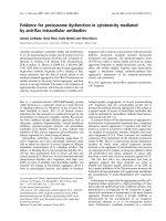
Fig. 4. Effect o f con stitutive expre ssion of PknA on t he mor phology of E. coli cells. (A) No rthern-blot analysis ind icating constitutive expression of
pknA in E. coli at the mRNA level. Tota l RNA was isolated from E. coli DH5a cells transformed with either p 19Kpro (lane 1) or p19Kpro-PknA
(lane 2), electrophoresed on 1.2% agarose gel containing formaldehyde, transferred on to a nylon membrane, and processed as described in the tex t.
Upper panel: the blot after hybridization using [a-
32
P]CTP-labelled pknA as the probe. Lower panel: the same blot after m eth ylene blu e st ain ing,
serving as a lo ad ing control. (B) Expression of the % 45-kDa PknA pr otein which is able to autophosphorylate. Soluble fractions of cru de lysates o f
E. coli DH5a cells transformed with e ither p19Kpro vector or p19Kpro-PknA were subjected t o SDS/PAGE and Coomassie Brilliant Blue staining
(left p anel). In vitro kinase assay was carried out with the same lysate as described in M aterials and m ethods (right p anel). Lane 1 , Molecular mass
marker; lanes 2 and 4, p19Kpro; lanes 3 and 5, p19Kpro-PknA. Numbers denote size of the molecular mass standards, and arrows indicate the
position of th e constitutively expressed PknA protein with autophosphorylating ability. (C) Phenotypic alteration of E. coli strain DH5a after
expression of PknA. The morphology of the cells was determined by scanning electron microscopy as described in the text. Panels a–d: E. coli DH5a
cells transformed w ith p19Kpro (a), p19Kpro-PknA (b), p19Kpro-aPknA (c), or p19Kpro-K42N (d). The bar in each panel indicates magnifi-
cation.
Ó FEBS 2002 Characterization of PknA from M. tuberculosis (Eur. J. Biochem. 269) 1083
[3–10,30,31]. To relate PknA to other bacterial Ser/Thr
kinases for which functions have already b een assign ed, we
carried out sequence database c omparisons using
BLAST
and
PSI-BLAST
programs. N ine different bacterial Ser/Thr kinase
sequences were retrieved through th ese searches; the
homology score varied from 80 to 162 with expected values
of between e
)15
and e
)39
. In contrast, YpkA, a Ser/Thr
kinase from Y. pseudotuberculosis known to be associated
with virulence [10], showed insignificant homology
(score ¼ 39.9, expected value ¼ 0.054). In a phylogenetic
tree generated by multiple s equence alignment of different
bacterial Ser/Thr kinases excluding highly variable
N-termini and C-termini, PknA is found to be very close
to Pkn1 and Pkn9 of Myxococcus xanthus (Fig . 3). As these
kinases, are involved in sporulation or cell division/differ-
entiation, it seems likely that PknA has similar functions.
In the M. tuberculosis genome, pknA (R v0015c) i s l ocated
adjacent to pbpA (Rv0016c) and rodA (Rv0017c) genes,
which encode putative morphogenic proteins belonging to
the SEDS (shape, elongation, division and s porulation)
family [32]. Members of this family of proteins have b een
reported to be present in all eubacteria in which a
constituent o f t he cell envelope is peptidoglycan. These
proteins are known t o be involved in c ontrolling cell shape
and peptidoglycan synthesis in bacteria such as Bacillus
subtilis [32] and E. coli [33]. Thus the presence o f a kinase at
this location in the genome suggests a regulatory role in
mycobacterial cell division.
Alteration in cell shape is the initial event in bacterial cell
division which involves ordered assembly of proteins
[34,35]. These proteins are fairly conserved among different
prokaryotes. This is evident from the fact that a % 56-kDa
soluble protein of E. coli interacted with the mycobacterial
PknA (Fig. 2 B). In a preliminary study, we observed that
pMAL-PknA-transformed cells of E. coli (strain TB1)
grown for 2–10 h after IPTG induction exhibited an
unusual elongation pattern compared with that of the cells
harbouring only the pMAL-c2X plasmid. To investigate
further the involvement of PknA in this process, we sought
to express t he pro tein constitutively in the E. coli host s train
DH5a using a low-copy vector. However, expression of
mycobacterial protein in E. coli is known to be difficult,
especially unde r the control of a heterologous promoter [36].
We therefore used a Mycobacterium–E. coli shuttle vec tor
p19Kpro, derived from p16R1 [22] containing a mycobac-
terial 19-kDa antigen promoter. These series of vectors are
known to elicit a low leve l of mycobacterial gene expression
in E. coli [36]. pknA was cloned in p19Kpro, and, after
transformation in E. coli, its expression was monitored at
the mRNA and protein levels. Northern-blot analysis of
total RNA extracted from cells transformed with either
p19Kpro (vector) or p19Kpro-PknA using pknA as a p robe
confirmed e xpression of the kinase at t he mRNA level
(Fig. 4 A, upper panel, compare lanes 1 and 2). The
constitutive expression of PknA at the protein level was
also evident from the expected % 45-kDa band on SDS/
PAGE after Coomassie Brilliant Blue staining (Fig. 4B, left
panel, compare lanes 2 and 3). The protein was found in the
soluble fraction. In vitro kinase assay of crude cell e xtracts
indicated autophosphorylating ability of the expressed
protein (Fig. 4B, right panel, compare lanes 4 and 5). The
effect of con stitutive expression of pknA on the phenotype
of the E. coli cells was evalu ated by scanning electron
microscopy. As s hown i n Fig. 4C, E. coli strain DH5a
transformedwithp19Kpro(panelÔaÕ) were normal rods of
size 1–2 lm. On the other hand, E. coli cells transformed
with p19Kpro-PknA (panel ÔbÕ) showed remarkable elong-
ation (more than 95% of the cells were in the range 60–
70 lm). Interestingly, E. coli transformed with either the
antisense construct, p19Kpro-aPknA (panel ÔcÕ)orthe
kinase-deficient mutant, p19Kpro-K42N (panel ÔdÕ)didnot
show such phenotypic alteration. Furthermore, cell elong-
ation did not seem to result in any toxicity from Ôout of
contextÕ expression of the mycobacterial gene as experi-
mental and control g rowth curves were similar (data not
shown). There are, in fact, examples of mycobacterial gene
expression using E. coli as a host [16]. Thus, a ll these lines of
evidence convincingly establish the participation of myco-
bacterial PknA in regulating morphological changes asso-
ciated with cell division.
Finally, our study in a heterologous setting has shown the
involvement of PknA in cell shape regulation; it is the first
report describing the functionality of any eukaryotic-type
Ser/Thr kinase from M. tuberculosis. Identification of the
natural substrate of PknA in mycobacteria would a id
progress towards its utilization as a drug target, which is a
top priority in this e ra of bac terial drug resistance.
ACKNOWLEDGEMENTS
We thank Dr Amit Ghosh, Director of the I nstitute of Microbial
Technology for providing u s with excellent laboratory facilitie s.
We acknowledge the gift of the Myco bacterium–E. coli shuttle vector,
p19Kpro, from Drs D. B. Young and M. Blokpoel, Imperial College
School of Medicine at St Mary’s, London, UK. We are grateful t o
Drs T . C hakrabarti, A . M ondal and S. Mande for helpful suggestions.
We thank Mr Jankey P rasad a nd Mr Anil Theophilus for excellent
technical assistance. R . C. is the recipien t of a Senior Re search
Fellowship from the Council of Scientific and Industrial Research, New
Delhi, India.
REFERENCES
1. Hunter, T. (1995) Protein kinases and phosphatases: the yin and
yang of protein p hosp horylation a nd signalling. Cell 80 , 2 25–236.
2. West, A.H. & Stock, A.M. (2001) Histidine kinases and response
regulator proteins in two-component signaling systems. Trends
Biochem. Sci. 26, 369–376.
3. Munoz Dorado, J., Inouye, S. & Inouye, M. (1991) A gene
encoding a protein serine/threonine kinase is required for normal
development of M. xanthus, a gram-negative bacterium. Cell 67,
995–1006.
4.Udo,H.,Munoz-Dorado,J.,Inouye,M.&Inouye,S.(1995)
Myxococcus xanthus, a Gram-negative bacterium, contains a
transmembrane protein serine/threonine kinase that blocks
the secretio n of b-lactamase b y p hosphorylatio n. Genes Dev. 9,
972–983.
5. Zhang, W ., Inouye, M . & Inouye, S . ( 1996) Reciprocal r egulation
of the d ifferentiation of Myxococcus xanthus by Pkn5 and Pkn6,
eukaryotic-like ser/thr protein kinases. Mol. Microbiol. 20,
435–447.
6. Hanlon, W.A., Inouye, M. & Inouye, S. (1997) Pkn9, a Ser/Thr
protein kinase involved in the development of Myxococcus
xanthus. Mol. Microbiol. 23, 459–471.
7. Zhang, C.C. (1993) A gene encoding a protein related to eukary-
otic protein kinases from the filamentou s heterocystous cyano -
bacterium Anabaena PCC 7120. Proc. Natl Acad. Sci. USA 90,
11840–11844.
1084 R. Chaba et al.(Eur. J. Biochem. 269) Ó FEBS 2002
8. Matsumoto, A., Hong, S.K., Ishizuka, H., Horinouchi, S. &
Beppu, T. (1994) Phosphorylation of t he AfsR protein involved i n
secondary metabolism in Streptomyces speciesbyaeukaryotic-
type protein kinase. Gene 146, 47–56.
9. Nadvornik, R., Vomastek, T., Janecek, J., Technikova, Z. &
Branny, P. (1999) Pkg2, a novel transmembrane protein ser/thr
kinase of Streptomyces granaticolor. J. Bacteriol. 181, 15–23.
10. Galyov, E.E., Hakansson, S., Forsberg, A. & Wolf-Watz, H.
(1993) A secreted protein kinase of Yersinia pseudotuberculosis is
an indispensable virulence determinant. Nature 361, 730–732.
11. Mukhopadhyay, S., Kapatral, V., Xu, W. & Chakrabarty, A.M.
(1999) Characterization of a Hank’s type serine/threonine kinase
and a serine/threonine phosphoprotein phosphatase in Pseudo-
monas aeruginosa. J. Bacteriol. 181, 6615–6622.
12. Bloom, B.R. & Murray, C.J. (1992) Tuberculosis: commentary on
a reemergent killer. Science 257, 1 055–1064.
13. Cole, S.T., Brosch, R., Parkhill, J., Garnier, T., Churcher, C.,
Harris, D., Gordon, S.V., Eiglmeier, K., Gas, S., Barry, C.E.
et al. (1998) Deciph ering the biolo gy of Mycobacterium tuber-
culosis from the complete genome sequence. Nature 393,
537–544.
14. Peirs,P.,DeWit,L.,Braibant,M.,Huygen,K.&Content,J.
(1997) A serine/threonine protein kinase from Mycobacterium
tuberculosis. Eur. J. Biochem. 244 , 604–612.
15. Av-Gay, Y., Jamil, S. & Drews, S.J. (1999) Expression and
characterization of the My cobacterium tuberculosis serine/threo-
nine protein kinase PknB. Infect. Immun. 67, 5676–5682.
16. Koul, A., Choidas, A., Tyagi, A.K., Drlica, K., S ingh, Y. &
Ullrich, A. (2001) Serine/threonine protein kinases PknF and
PknG of Myc obacterium tuberculosis: characterization and local i-
zation. Microbiology 147, 2307–2314.
17. Av-Gay, Y. & Everett, M. (2000) The eukaryotic-like ser/thr
protein kinases of Mycobacterium tuberculosis. Trends Microbiol.
8, 238–244.
18. Agrewala, J.N. & Mishra, G.C. (1995) A 38kDa antigen of
Mycobacterium tuberculosis predominantly induces the secretion
of interleuk in-2, interferon-gamma and IgG2a antibodies.
Microbiol. Immunol. 39, 801–808.
19. Banerjee, S.K., Bhatt, K., M isra, P. & Chakraborti, P.K. (2000)
Involvement of a natural transport system in the p rocess of efflux
mediated drug resistance in Mycobacterium smegmatis. Mol. Gen.
Genet. 262, 949–956.
20. Sarin, J., Aggarwal, S., Chaba, R., Varshney, G.C. & Chakra-
borti, P.K. (2001) B-subunit of phosphate-specific transporter
from Mycobacterium tuberculosis is a thermostable ATPase.
J. Biol. Chem. 276, 44590–44597.
21. Sambroo k, J., Fritsch, E.F. & Maniatis, T. (1989) Molecular
Cloning: a Laboratory Manual, 2nd edn. Cold Spring Harbor
Laboratory Press, Cold Spring Harbor, NY, USA.
22. Garbe, T.R., Barathi, J., Barnini, S., Zhang, Y., Abou-Zeid, C.,
Tang, D., Mukherjee, R. & Young, D.B. (1994) Transformation
of mycobacterial species u sing hygromycin r esistanc e as selectable
marker. Microbiology 140, 133–138.
23. Schmitt, M.E., Brown, T .A. & Trumpower, B.L. (1990) Rapid
and simple method for preparation of RNA from Saccharomyces
cerevisiae. Nucleic Acids Res. 18, 3091–3092.
24. Inoue, T. & Osatake, H. (1988) A ne w drying method of b iological
specimens for scan ning electron microscopy: the t-butyl alcohol
freeze-drying method. Arch. H istol. Cytol. 51, 53–59.
25. Thompson , J.D., Higgins, D.G. & Gibson, T.J. (1994) CLUSTAL
W: improving the sensitivity of progressive multiple sequence
alignment through sequence weighting, position-specific gap penal-
ties and weight matrix c hoice. Nucleic Acids Res. 22, 4673–4680.
26. Felsenstein, J. (1993) PHYLIP: Phylogeny Inference Package
v.3.5c. Department of G enetics, University of Washington,
Seattle, WA, USA.
27. Page, R.D.M. (1996) TREEVIEW: an application to display
phylogenetic trees on personal compu ters. Comput. Appl. Biosci.
12, 357–358.
28. Motley, S.T. & Lory, S. (1999) Functional characterization of a
serine/threonine kinase of Pseudomonas a eruginosa. Infect. Immun.
67, 5386–5394.
29. Maruta, S., Mitsuhashi, S., Yamada, M. & Ikebe, M. (1998) ADP/
vanadate mediated photocleavage of myosin light c hain kinase a t
the autoinhibitory region. J. Biochem. 124, 557–564.
30. Zhang, C.C. (1996) Bacterial signalling involving eukaryotic-type
protein kinases. Mol. Microbiol. 20, 9–15.
31. Wang, J., Li, C., Yang, H., Mushegian, A. & Jin, S. (1998) A novel
serine/threonine protein kinase homologue of Pseudomonas aeru-
ginosa is specifically inducible within the host infection s ite and is
required for full virulence in neutrop enic mice. J. Bacteriol. 180,
6764–6768.
32. Henriques, A.O., Glase r, P., Piggot, P.J. & Moran, C.P. Jr (1998)
Control of c ell shape and elongation by t he rodA gene in Bacillus
subtilis. Mol. Microbiol. 28, 235–247.
33. Begg, K.J. & Donachie, W.D. (1985) Cell shape and division in
Escherichia coli: experiments with shape and division mutants.
J. Bacteriol. 163, 615–622.
34. Lutkenhaus, J. & A ddinall, S. G. (1997) Bacterial cell division and
the Z ring. Annu. Rev. Biochem. 66, 93–116.
35. Daniel, R.A., Harry, E .J. & Errington, J. (2000) Role of penicillin-
binding protein PBP 2B in assembly and functioning of the divi-
sion machinery of Bacillus subtilis. Mol. Microbiol. 35, 299–311.
36. Garbe, T., Harris, D., Vordermeier, M., Lathigra, R., Ivanyi, J.
& Young, D. (1993) Expression of the Mycobacterium tuber-
culosis 19-kilodalton antigen in Mycobacterium smegmatis:
immunological analysis and evidence of glycosylation. Infect.
Immun. 61, 260–267.
Ó FEBS 2002 Characterization of PknA from M. tuberculosis (Eur. J. Biochem. 269) 1085