Tài liệu Báo cáo Y học: Mutations in the docking site for cytochrome c on the Paracoccus heme aa3 oxidase Electron entry and kinetic phases of the reaction pptx
Bạn đang xem bản rút gọn của tài liệu. Xem và tải ngay bản đầy đủ của tài liệu tại đây (281.55 KB, 9 trang )
Mutations in the docking site for cytochrome
c
on the
Paracoccus
heme
aa
3
oxidase
Electron entry and kinetic phases of the reaction
Viktoria Drosou
1
, Francesco Malatesta
2
and Bernd Ludwig
1
1
Molecular Genetics, Institute of Biochemistry, Johann-Wolfgang-Goethe Universita
¨
t, Frankfurt, Germany;
2
Department of Basic and Applied Biology, University of L’Aquila, Italy
Introducing site-directed mutations in surface-exposed
residues of subunit II of the heme aa
3
cytochrome c oxidase
of Paracoccus denitrificans, we analyze the kinetic para-
meters of electron transfer from reduced horse heart cyto-
chrome c. Specifically we address the following issues: (a)
which residues on oxidase contribute to the docking site for
cytochrome c, (b) is an aromatic side chain required for
electron entry from cytochrome c, and (c) what is the
molecular basis for the previously observed biphasic reaction
kinetics. From our data we conclude that tryptophan 121 on
subunit II is the sole entry point for electrons on their way to
the Cu
A
center and that its precise spatial arrangement, but
not its aromatic nature, is a prerequisite for efficient electron
transfer. With different reaction partners and experimental
conditions, biphasicity can always be induced and is critically
dependent on the ionic strength during the reaction. For an
alternative explanation to account for this phenomenon, we
find no evidence for a second cytochrome c binding site on
oxidase.
Keywords: Paracoccus denitrificans; cytochrome c oxidase;
docking site; electron transfer; biphasic kinetics.
Cytochrome c oxidase is the terminal complex of the
respiratory chains of mitochondria and many bacteria
[1–4]. It catalyzes the reduction of oxygen to water, coupling
the free energy of this reaction to the generation of a proton
gradient across the membrane.
During the redox reaction, an electron delivered from
cytochrome c is first transferred to Cu
A
, a binuclear copper
center located close to the surface of the large hydrophilic
domain of subunit II. It is then donated to heme a
embedded in subunit I, and subsequently to the heme a
3
Æ
Cu
B
center where oxygen reduction, and most likely the
redox coupling to proton pumping, take place.
While the mitochondrial enzyme comprises up to 13
different subunits in a dimeric complex, the oxidase of the
bacterium Paracoccus denitrificans consists of only four
subunits, with the three largest ones homologous to the
corresponding mitochondrial subunits.
Typically, many isolated oxidases are somewhat promis-
cuous towards their substrate molecules. Early studies
analyzing the surface properties of cytochromes c of
different origin revealed a basic cluster of mostly lysine
residues located around the heme crevice. Being responsible
for docking to their redox partners, an interaction between
cytochrome c and oxidase based on electrostatic forces was
described [5–7]. Experiments with monoclonal antibodies
directed against subunit II of cytochrome c oxidaseleadtoa
loss of activity [8] and supported the notion that the catalytic
binding site is located predominantly on subunit II. This
result was confirmed by chemical modifications and early
site-directed mutagenesis experiments [9,10], and is consis-
tent with the crystal structures of the eukaryotic and the
Paracoccus denitrificans oxidase showing clusters of negat-
ively charged residues on the surface of subunit II [11,12].
Previous studies on the binding of cytochrome c to the
Paracoccus oxidase were interpreted by a two-step model in
which electrostatic forces are responsible for an efficient
long-range docking, followed by the reorientation of the
redox partner driven by hydrophobic interactions [13].
Specifically, a set of four acidic residues exposed on subunit
II (D135, D178, and to a lesser extent, E126, D159; see
Fig. 1 and [13]) had been assumed to interact electrostat-
ically with the horse heart cytochrome c. While a pivotal
role in electron transfer from cytochrome c was assigned to
residue W121 on subunit II [14], this early study did not
address the question whether any other (aromatic) side
chains in this or the neighbouring position might be able to
support electron transfer to the Cu
A
site, thereby function-
ally replacing tryptophan 121.
As already observed previously, oxidase kinetics may
yield nonlinear Eadie–Hofstee plots (e.g. [15]). Two different
phases, denoted high and low affinity, are clearly discern-
ible, each being characterized by a set of individual kinetic
parameters. These biphasic steady-state kinetics become
monophasic at higher ionic strength, a phenomenon
discussed in terms of different binding sites (e.g. [15,16]).
or of conformational changes within the enzyme-substrate
complex related to the coupling of electron transfer with
proton pumping [17].
Correspondence to B. Ludwig, Molecular Genetics, Institute of
Biochemistry, Biozentrum, Marie-Curie-Strasse 9,
D-60439 Frankfurt, Germany.
Fax: + 49 69 798 29244, Tel.: + 49 69 798 29237,
E-mail:
Abbreviations:I,ionicstrength;c
552
-f: soluble fragment of the
Paracoccus denitrificans cytochrome c
552
, expressed and purified from
Escherichia coli.
(Received 4 February 2002, revised 12 April 2002,
accepted 2 May 2002)
Eur. J. Biochem. 269, 2980–2988 (2002) Ó FEBS 2002 doi:10.1046/j.1432-1033.2002.02979.x
Here we give a comprehensive description of the inter-
action domain for cytochrome c on subunit II of the
P. denitrificans oxidase, specifically addressing (a) the
electron entry point to oxidase and its possible alternatives,
(b) the size and extent of the acidic patch on subunit II, and
(c) ways of experimentally influencing the kinetic phases
during enzyme turnover.
MATERIALS AND METHODS
Mutagenesis and enzyme preparation
Site-directed mutagenesis in the ctaCgenewasper-
formed according to the Ôaltered siteÕ mutagenesis protocol
(Promega, Heidelberg). Complementation of the oxidase-
deficient deletion mutant ST4 was performed as described
previously [18]. Mutant strains were grown aerobically in
succinate medium [19] including streptomycin sulfate
(25 lgÆmL
)1
), membranes isolated according to [20] and
solubilized using n-dodecyl b-
D
-maltoside.
The four-subunit cytochrome c oxidase was purified by
conventional chromatographic protocol as described in [21];
the oxidase complexed with an antibody fragment (F
v
)was
isolated in a single chromatographic step as described
previously [22,23], and excess F
v
removed by gel filtration.
The two-subunit oxidase complex was prepared by
combining the standard purification in dodecyl maltoside
[21], however, running the gel filtration step in the presence
of Triton X-100 to dissociate subunits III and IV [24].
Briefly, the following steps were performed: The superna-
tant after ultracentrifugation was loaded on the first
column, a DEAE-Sepharose CL-6B (Pharmacia Biotech)
equilibrated with 20 m
M
potassium phosphate pH 8.0,
1m
M
EDTA and 0.5 gÆL
)1
dodecyl maltoside. For elution,
a linear gradient ranging from 100 to 600 m
M
NaCl was
used with this buffer. Fractions of highest heme/protein
ratio were pooled and concentrated in an Amicon cell (cut
off 30 kDa). Triton X-100 was added to a final concentra-
tion of 10% (w/v). The solution was stirred for 1 h at 4 °C,
applied to an Ultrogel AcA34 (IBF Biotechniques) gel
filtration column equilibrated and processed with the above
buffer with 2 gÆL
)1
Triton X-100 replacing the dodecyl
maltoside. To reintroduce this latter detergent for subse-
quent steps (and to avoid the detrimental effects of Triton
X-100 on activity [25]), pooled fractions were rechromato-
graphed on the first column equilibrated in 0.2 gÆL
)1
dodecyl maltoside, and eluted with a linear gradient from
100 to 400 m
M
NaCl. The oxidase fractions were analyzed
by SDS/PAGE to verify their subunit composition, con-
centrated and stored at )80 °C.
Steady-state kinetics and determination of the ionic
strength dependence
Cytochrome oxidase activity was measured with a Kontron
Uvikon 941 spectrophotometer at 25 °Cin20m
M
Tris/
HCl, pH 7.5, 1 m
M
EDTA, 0.2 gÆL
)1
n-dodecyl b-
D
-malto-
side. The different ionic strength conditions were adjusted
by adding KCl. Ferrocytochrome c (horse heart, Sigma)
was prepared by reduction with dithionite and excess
reductant removed by Sephadex G25 chromatography.
Concentrations were varied between 0.5 and 40 l
M
,and
oxidation followed at 550 nm after adding the purified
enzyme (40 p
M
up to 400 p
M
). Determination the ionic-
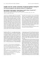
Fig. 1. The presumed docking site for cyto-
chrome c on subunit II of the P. de nitrificans
oxidase. The periplasmically oriented hydro-
philic domain housing the homodimeric Cu
A
site (blue spheres) is depicted, omitting most of
the two transmembrane helices (bottom).
Selected side chains shown in detail were
mutated; the residue crucial for electron entry,
W121, is highlighted in yellow, along with
other residues important for docking. The
figure was prepared on the basis of the pub-
lished coordinates (pdb1ar1), using the
SWISS
PDB VIEWER/POV RAY
program [31].
Ó FEBS 2002 Cytochrome c docking site (Eur. J. Biochem. 269) 2981
strength (I) dependence was performed in the same buffer at
20 l
M
cytochrome c, with the ionic strength adjusted to
values between 1.8 m
M
and 296 m
M
by the addition of KCl.
The buffer for I ¼ 1.8 m
M
was 2.5 m
M
Tris/HCl, pH 7.5,
0.2 gÆL
)1
dodecyl maltoside.
Stopped-flow kinetics
The presteady-state kinetics were followed on a ther-
mostated Applied Photophysics DX.117 MV stopped-
flow apparatus at 20 °Cin20m
M
potassium phosphate
pH 7.6, 1 m
M
EDTA, 0.2 g L
)1
dodecyl maltoside. The
ionic strength was 140 m
M
, adjusted with KCl. Cytochrome
oxidase (4–6 l
M
) was incubated with 5 m
M
KCN at 4 °C
for at least 6 h. Cytochrome c concentrations were varied in
the range of 2–32 l
M
; after mixing, the reaction was
followed at 550 nm (oxidation of cytochrome c) and/or
605 nm (reduction of heme a), and biphasic time courses
were obtained. Three independent measurements were done
for each donor concentration, and the average was fitted to
the sum of two exponentials. The observed rate constant
from the fast phase of this double-exponential decay was
plotted against the cytochrome c concentration, and the
apparent bimolecular rate constant k
on
calculated from the
slope.
RESULTS
The large, periplasmically oriented hydrophilic domain of
subunit II of the heme aa
3
oxidase represents the major
interaction site for cytochrome c. We constructed a set of
mutants in surface-exposed residues to further identify
amino acid side chains involved in the docking of
cytochrome c, or in the presumed electron entry from
cytochrome c to the Cu
A
center. Positions subjected to
mutagenesis in this and two previous studies [13,14] are
summarized in Fig. 1. All mutant enzymes, and several
preparations of oxidase differing in polypeptide composi-
tion, were assayed for their kinetic parameters under
different ionic strength conditions.
Ionic strength dependence of the turnover number
The reaction of cytochrome c oxidase shows a strict
dependence of the turnover number on ionic strength. We
measured the oxidation of 20 l
M
horse heart cytochrome c
by the various oxidase preparations under steady-state
conditions, all yielding bell-shaped curves. While the
optimum ionic strength for the wild-type enzyme was found
to be 56 m
M
(Fig. 2; see also [13]), those for the two-subunit
enzyme (Fig. 2) and all mutants in positions 121 and 122
(W121F, W121Y, W121Q/Y122Q, W121F/Y122F, W121G
and W121Y/Y122W) were decreased to 36 m
M
.Ofthe
remaining mutants, two showed wild-type behaviour
(H119N, N160D), whereas all others were shifted to
46 m
M
(data not shown).
Turnover and presteady-state kinetics
We measured steady-state kinetics for all the subunit II
mutants at their optimum ionic strength as determined
above, using the reduced horse heart cytochrome c.The
46 m
M
ionic strength group was assayed both at 36 and at
56 m
M
, to allow for an unequivocal assignment to either a
hyperbolic or nonhyperbolic Michaelis–Menten kinetic
regime. Tables 1 and 2 list the relevant parameters, K
m
and k
cat
, for the different ionic strength conditions: the K
m
value is taken from the so-called high-affinity phase, and the
k
cat
value is derived from the low-affinity phase.
Positions W121 and Y122. A comprehensive set of single
mutants in each of the two positions, or of double mutants,
was generated (Table 1). While K
m
values for all complexes
do not deviate from that of wild-type by more than a factor
of 1.6, the catalytic activity of any mutant in the W121
position is drastically diminished. Residual activities for the
two nonaromatic replacements (Q, G) are between 1 and
2%. Additional single mutations in the 121 position which
exchange the tryptophan for two other aromatic residues
(F, Y) show almost the same distinct loss of electron transfer
activity, with residual k
cat
values around 3–5% compared to
wild-type. When the neighbouring Y122 residue is changed
to a phenylalanine, the kinetic behaviour is that of wild-
type, and a glutamine in this position only reduces activity
to 50%. We conclude that Y122 is not involved in the
Fig. 2. Ionic strength dependence of the turnover number for the isolated
four- and two-subunit Paracoccus heme aa
3
oxidase complex. The
spectrophotometric assay was performed under steady-state condi-
tions with 20 l
M
horse heart cytochrome c;forfurtherdetails,see
Materials and methods. 4 su, four-subunit; 2 su, two-subunit complex.
Table 1. Steady-state and stopped-flow parameters of horse heart
cytochrome c oxidation by Paracoccus oxidase mutated in selected
exposed aromatic residues of subunit II. The K
m
value was taken from
the high-affinity phase at I ¼ 36 m
M
.Thek
cat
value was taken from
the low-affinity phase at I ¼ 36 m
M
. NR, no rate measurable.
Mutant position K
m
(l
M
) k
cat
(s
)1
) k
on
· 10
6
(
M
)1
Æs
)1
)
Wild-type oxidase 1.4 669 3.7
W121Q 1.9 11 NR
W121G 1.1 6 NR
W121F 1.5 31 0.05
W121Y 1.3 22 0.03
Y122Q 2.2 333 0.4
Y122F 1.2 660 4.6
W121Q/Y122Q 1.7 9 NR
W121Y/Y122W 1.2 7 0.13
W121F/Y122F 1.5 7 NR
Y226F 1.3 626 2.4
2982 V. Drosou et al.(Eur. J. Biochem. 269) Ó FEBS 2002
electron transfer from cytochrome c to any large extent, nor
is this position involved in maintaining the low residual
activity when the W121 residue is mutated. Double mutants
in both positions are not further diminished in activity
compared to W121 single mutations (see Table 1). Further
mutations (Y226F, H119N) in residues previously consid-
ered as potential alternative entry points for electrons from
cytochrome c (see Discussion) showed no deviations from
wild-type in their kinetic properties.
To exclude the possibility that diminished electron
transfer activities in turnover experiments might be due to
changes in redox properties of the first acceptor in oxidase,
Cu
A
, we measured relevant redox steps in the W121F
mutant, confirming that the redox potential for Cu
A
is in the
wild-type range (P. Hellwig, Institut fu
¨
r Biophysik, Johann-
Wolfgang, Goethe Universita
¨
t, Frankfurt, Germany,
personal communication).
Focussing on the parameters in Table 2 we found an
increase of K
m
for mutants H119I/Q120I, D146N, E140Q
and P196G measured at 56 m
M
. Comparing these values
with those measured at 36 m
M
, again we found increased
K
m
values for these mutants and also for E142Q. k
cat
as the
parameter for maximum turnover is decreased. Mutants
H119N and N160D reveal wild-type values; these positions
do not seem to be involved in cytochrome c binding.
We also assayed the oxidase mutants under presteady-
state conditions, to ensure that the observed effects indeed
relate to the early phases of electron entry. Using the
cyanide-inhibited enzyme, the reaction sequence is limited to
the transfer of the first two electrons reaching the Cu
A
/heme
a redox couple. To shift cytochrome c oxidation kinetics
into the time resolution of a stopped-flow apparatus, the
reaction was followed at 140 m
M
ionic strength (see
Materials and methods) and recorded at 550 nm (oxidation
of cytochrome c) and at 605 nm (reduction of heme a). The
observed time course was described by a sum of two
exponentials. The fitted pseudo-first order rate constant was
plotted against the cytochrome c concentration after mix-
ing. From the slope of this linear plot the bimolecular rate
constants k
on
were calculated for the wild-type and mutant
enzymes (Tables 1 and 2). This analysis reflects and con-
firms the k
cat
values obtained from turnover experiments.
Some of the mutants showed extremely slow reduction
behaviour, and bimolecular rates could not be determined
(see Table 1). The k
on
values for the mutants Y122F,
Y226F, N160D, H119N and H119I/Q120I are in the same
range as the wild-type oxidase while the other mutants show
a significantly decreased k
on
value (see Tables 1 and 2).
Kinetic differences between the two-subunit
and the four-subunit wild-type and mutant complexes
To assess kinetic properties of both forms under identical
detergent conditions, we prepared cytochrome c oxidase
lacking both subunits III and IV by replacing the detergent
in one of the chromatographic steps of the standard
purification procedure (see Materials and methods): prior
to gel filtration, the partially purified material was incubated
with a large excess of Triton X-100, known to dissociate the
oxidase and leave an enzymatically active two-subunit
complex [24]. After gel filtration in Triton, dodecyl malto-
side was reintroduced in the final step of column purification
to exclude known detergent effects in the subsequent
analysis [25].
Ionic strength dependency of the maximum turnover
number was shifted from 56 m
M
for the four-subunit
complex to 36 m
M
for the two-subunit preparation.
Figure 2 also demonstrates that both complexes display a
basically similar line shape, and turnover numbers are in
close agreement at 20 l
M
cytochrome c. This behaviour is
taken as a first evidence that the periplasmically oriented
regions of one or both of the two ancillary subunits may
contribute to some extent to the interaction domain for the
substrate (see also Discussion).
Kinetic parameters for both complexes under several
ionic strength conditions are listed in Table 3. Comparing
K
m
and k
cat
each at optimum ionic strength for both forms,
it is evident that k
cat
is lower by a factor of three for the two-
subunit enzyme, while its K
m
is diminished twofold. The
overall specificity constant (k
cat
/K
m
) of this two-subunit
complex therefore remains in the same range, explaining in
part its comparable activity at a given substrate concentra-
Table 2. Oxidation of horse heart cytochrome c by wild-type oxidase and subunit II mutants under turnover and pre-steady state conditions at different
ionic strengths. TM1, triple mutant (E126Q, D135N, D178N) in subunit II. ND, not determined.
I ¼ 36 m
M
a
I ¼ 56 m
M
I ¼ 140 m
M
Mutation K
m
(l
M
) k
cat
(s
)1
) K
m
(l
M
) k
cat
(s
)1
) k
on
· 10
6
(
M
)1
Æs
)1
)
b
Wild-type oxidase 1.4 669 5.9 1031 3.7
E142Q 3.2 270 4.7 588 1.1
D146N 2.7 239 10.2 357 2.7
E140Q 5.6 277 7.0 250 2.1
D135N
c
2.1 167 12.1 104 0.3
D178N
c
2.0 213 15.0 313 2.3
TM1
c
7.9 25 ND ND ND
P196G 1.1 345 10.4 714 1.5
H119I/Q120I ND ND 9.8 303 2.6
H119N ND ND 5.1 896 4.3
N160D ND ND 6.4 909 2.8
a
Mutants as published in [13] were re-analyzed side by side, and are presented for a complete overview.
b
K
m
value taken from the high-
affinity and k
cat
from the low-affinity phase.
c
Pre-steady-state kinetics of cytochrome c oxidation measured by stopped-flow, see Materials
and methods for details.
Ó FEBS 2002 Cytochrome c docking site (Eur. J. Biochem. 269) 2983
tion (Fig. 2). Analyzing lower ionic strength datasets for
both preparations, the general trend persists that k
cat
is
below that of the four-subunit enzyme, while K
m
values
approach each other (see Table 3).
Shifts from mono- to biphasic behaviour are observed for
both the four-subunit and the two-subunit oxidase on going
from higher ionic strength to lower values. Figure 3
exemplifies this transition to nonlinear kinetic behaviour
for the two-subunit complex when [I] is diminished in steps
from 56 to 15 m
M
. Eadie–Hofstee plots yield clear breaks
for the two lower salt conditions (Fig. 3B). These transition
points are listed in Table 3 (last column) for selected
preparations/mutants (see also below). Also this criterion
distinguishes the two-subunit variant from the four-subunit
complex, where the transition occurs already at 36 m
M
,
clearly indicating that biphasic kinetics are not due to the
presence of subunits III and IV.
Biphasic behaviour of the triple mutant TM1 (subunit II:
E126Q, D135N, D178N) is evident when the four-subunit
complex is assayed: while other mutants containing single
acidic residue replacements followed biphasic kinetics at
I ¼ 36 m
M
(not detailed), TM1 was monophasic at
I ¼ 36 m
M
. Nevertheless, on further decreasing ionic
strength, biphasic kinetics were again observed with a
transition point at around 15 m
M
(see Table 3). The same
holds true when the TM1 preparation was stripped of its
subunits III and IV: the resulting two-subunit mutant
complex displayed biphasic kinetics at 15 m
M
ionic
strength.
Further criteria for manipulating the kinetic phases
of reaction
Transitions from mono- to biphasic reaction conditions,
depending on ionic strength variation, can be induced by
other means as well. Specific F
v
fragments, derived from
Table 3. Kinetic parameters and biphasic transitions under different ionic strength conditions for selected oxidase preparations. ND, not
determined.
I ¼ 7.4 m
M
I ¼ 14.8 m
M
I ¼ 26 m
M
I ¼ 36 m
M
I ¼ 56 m
M
Biphasicity
Oxidase preparation
K
m
(l
M
)
k
cat
(s
)1
)
a
K
m
(l
M
)
k
cat
(s
)1
)
K
m
(l
M
)
k
cat
(s
)1
)
K
m
(l
M
)
k
cat
(s
)1
)
K
m
(l
M
)
k
cat
(s
)1
)
transition at
I(m
M
)
b
Four-subunit oxidase ND ND 0.6 434 0.9 555 1.4 669 5.9 1031 36
Four-subunit oxidase ND ND 0.3 63 1.6 154 3.6 338 10.5 400 26
purified with F
v
Four-subunit oxidase ND ND 0.5 88 1.1 270 4.1 384 ND ND 26
+ specific F
v
added
Four-subunit oxidase ND ND ND ND ND ND 1.6 555 ND ND 36
+ control F
v
added
Two-subunit oxidase ND ND 0.8 254 0.85 263 2.9 288 15.1 336 26
Two-subunit oxidase 1.6 220 3.8 243 ND ND ND ND ND ND 7.4
+ specific F
v
added
Four-subunit TM1
c
ND ND 0.56 8 1.6 20 7.9 25 ND ND 14.8
Two-subunit TM1
c
ND ND 7 30 ND ND ND ND ND ND 14.8
Four-subunit oxidase ND ND 2.8 1000 28.5 474 52.5 100 ND ND 26
vs. c
552
–f
d
a
The K
m
value is taken from the high-affinity phase and k
cat
from the low-affinity phase, whenever kinetics are biphasic.
b
On lowering the
ionic strength, transition from monophasic to biphasic kinetics is observed at specified ionic strength (I).
c
Triple mutant TM1 (E126Q,
D135N, D178N) in subunit II.
d
Data taken from V. Drosou & B. Ludwig, unpublished results.
Fig. 3. Eadie–Hofstee plots (A and B) representing horse heart cyto-
chrome c oxidation by the two-subunit oxidase at different ionic strength
conditions. Steady-state kinetics were determined spectrophotometri-
cally at 25 °C.
2984 V. Drosou et al.(Eur. J. Biochem. 269) Ó FEBS 2002
monoclonal IgG directed against a subunit II epitope [22],
may be added in a 3 : 1 molar excess to purified oxidase
both as a four- or a two-subunit complex. Alternatively, F
v
may be used to affinity-purify the four-subunit oxidase from
solubilized membranes, yielding a stable 1 : 1 complex
which was instrumental in the structure determination of
the P. denitrificans oxidase [11]. Table 3 indicates that in all
cases the F
v
fragment, present with or added to the enzyme,
induced a decrease in the transition point to biphasic
kinetics. To some extent, individual effects appear to be
additive when following this shift from the four-subunit to
the two-subunit enzyme, and to the F
v
-complexed oxidase
lacking the two smallest subunits.
In a control reaction employing an unspecific F
v
protein
not recognizing any oxidase epitope [26], wild-type beha-
viour ensued. It should also been noted that under true
biphasic conditions (26 m
M
), kinetic parameters for the
F
v
-complexed oxidase point at a somewhat diminished
overall catalytic efficiency of this enzyme form (see Table 3),
although, with the exception of the F
v
-complexed two-
subunit oxidase, the high affinity K
m
values are comparable.
While all the above mentioned experiments were per-
formed with the commercially available horse heart cyto-
chrome c, the heterologous expression of a soluble c-type
cytochrome fragment, c
552
-f, will allow to probe this
bacterial oxidase with its homologous electron donor
derived from P. denitrificans [27–30]. With regard to
reaction kinetics with the four-subunit oxidase complex,
this soluble bacterial cytochrome fragment is a competent
donor to oxidase (V. Drosou & B. Ludwig, unpublished
results), and more importantly it is characterized by biphasic
Eadie–Hofstee plots once the ionic strength drops to 26 m
M
or below (see Table 3, last row).
DISCUSSION
Extent of the acidic patch on subunit II involved
in the cytochrome
c
docking reaction
A two-step model has been proposed to describe the docking
of the membrane-embedded oxidase with its soluble sub-
strate cytochrome c. In a first step governed by long–range
electrostatic interaction mediated by oppositely charged
surfaces on either protein, a preorientation of both redox
partners is obtained, which is followed by a fine-tuning
mediated by hydrophobic surfaces to aquire a docking
conformation for optimal electron transfer [14,32]. A strong,
positive surface potential for the mitochondrial electron
donor, cytochrome c, is evident, while several acidic residues
have been suggested to participate in docking on a negatively
charged patch located mostly on subunit II above the first
electron acceptor in oxidase, the Cu
A
center (see introduc-
tion). A bell-shaped dependency of the turnover number on
ionic strength of the assay medium (see also Fig. 2) has been
taken as initial experimental evidence that protein surface
charges get progressively shielded by increasing the ionic
strength of the medium. Under turnover conditions, an
optimal salt concentration results from a compromise of the
association and the dissociation rates for cytochrome c.
From a previous mutagenesis study [13], a partial
contribution of a few acidic residues on subunits I and III
to the acidic docking site on the periplasmic face of the
P. denitrificans oxidase appeared likely. Making use of the
fact that this bacterial enzyme can be isolated both as a
four- and a two-subunit complex without major kinetic
defects (see Fig. 2, and below), a distinct decrease (by
20 m
M
) in the ionic strength maximum for the two-
subunit wild-type complex confirms the contribution of
additional charge(s) located on the two further subunits of
the native oxidase.
In focussing on the main interaction domain on subunit
II, we introduced additional mutations in exposed residues
in the relevant area above the Cu
A
site (see Fig. 1 and
Table 2), to estimate the extent of the acidic region
responsible for cytochrome c docking. While no direct
structural information is at hand for the docked complex,
the interaction domain for cytochrome c on the cyto-
chrome bc
1
complex of yeast turned out to be confined to a
few residues only [33].
Both the mutants H119N and N160D (Table 2) show
wild-type characteristics, along with an ionic strength
optimum at 56 m
M
. For H119N this is not surprising since
no charge change results. In position N160 an additional
negative charge was introduced, but available kinetic
parameters suggest that this mutant, despite its higher
negative surface potential, does not provide a more potent
docking site for its substrate. This observation may be
explained by the fact that this residue is located too far out
from the actual electron entry site W121 (see below).
Mutants E140Q, E142Q, D146N, and P196G all show a
shift in the ionic strength optimum to 46 m
M
, providing first
evidence that these residues are involved in substrate
binding. They were characterized at 56 m
M
and at 36 m
M
;
in the latter condition, clear biphasic kinetics were recorded
(see below). Mutants D135N and D178N and the triple
mutant TM1 have already been characterized as bona fide
docking mutants in the past [13] but were re-analyzed side by
side with the other mutants generated here. Three of these
positions were changed from an acidic side chain into the
corresponding amide derivative, yielding unequivocal evi-
dence for their contribution in the cytochrome c oxidation
reaction. Compared to wild-type, they show some changes
in K
m
,butatthesametimealsoink
cat
. When biphasic
reactions are obtained at 36 m
M
ionic strength, the tendency
increases for a more pronounced rise in the K
m
value, but a
concomitant loss in k
cat
cannot be overlooked under this
condition either. This decrease of turnover numbers may
partly be explained by the fact that for the purpose of a
uniform comparison, these mutants were measured at
36 m
M
and 56 m
M
whereas the individual optimal ionic
strength was found to be at 46 m
M
in some cases.
When presteady-state kinetics are measured at 140 m
M
ionic strength for this set of mutants, it is evident that a
parallel trend, even though not always in a quantitative
manner, is seen for E142, D146N, and E140Q (Table 2).
The double mutant H119I/Q120I should lead to an
increase of the hydrophobic free energy, and its K
m
value is
increased (along with a decrease of k
cat
), which means that
substrate binding is influenced. Since the single mutant
H119N showed wild-type behaviour, the position Q120 is
most likely responsible for the observed effects.
The interpretation of the low-affinity K
m
values (not
given) is not straightforward since the explanation for this
phase is still hypothetical (see below). However, the same
general trend for both the high-affinity and the low-affinity
K
m
values is observed.
Ó FEBS 2002 Cytochrome c docking site (Eur. J. Biochem. 269) 2985
Taken together with our earlier data [13], this study now
defines an extensive area of exposed acidic residues on
subunit II which are involved in the initial docking (see
above) of the horse heart cytochrome c. In viewing down
the axis from W121 to the Cu
A
center as in Fig. 1, a lobe of
three carboxylate side groups (D146, E140, D159), with a
minor contribution from E142, extends to the edge of the
presumed interaction site. A more central region, closer to
W121, is made up of residues D135, E126, and D178 (as
modified together in the triple mutant TM1). Further
residues important for interaction in this latter lobe include
Q120, and possibly P196. This docking site model includes
the four homologous positions of acidic residues considered
most effective also in the Rhodobacter spheroides heme aa
3
oxidase [34].
Experiments replacing the mitochondrial cytochrome c
with a fragment of the homologous bacterial electron
donor, cytochrome c
552
of P. denitrificans [27,28] confirm
that all of the above mentioned residues on oxidase are also
involved in this docking reaction, while some additional
ones appear specific for the bacterial donor protein (for
details, see V. Drosou & B. Ludwig, unpublished results).
From this we conclude that the surface area on oxidase,
covered by the bacterial cytochrome c, is at least as large as
that for the mitochondrial protein.
Specificity of the electron entry site into oxidase
Previous mutagenesis data on the P. denitrificans [14] and
on the Rh. spheroides [34] oxidase clearly indicated that the
tryptophan at position 121 is of crucial importance for
electron transfer from cytochrome c to the Cu
A
center in
oxidase. Being located approx. 5 A
˚
above the metal
center, it is followed in sequence by another aromatic side
chain, Y122. Table 1 summarizes the kinetic effects of
single mutations in either residue, and of several double
mutants, indicating that in no case any major changes on
K
m
, resp., on affinity towards the substrate, occur.
However, whenever a W121 mutation is introduced, k
cat
is drastically diminished to a few percent residual activity
for aromatic side chain replacements, and even lower for
aliphatic ones. On the contrary, exchanges in the neigh-
bouring aromatic residue, Y122, only lead to moderate or
no activity changes at all. Double mutants like the
W121Q/Y122Q do not fall below the single W121Q
activity, i.e. its (already low) residual electron transfer
activity is not maintained by the neighbouring tyrosine,
while the W121F/Y122F mutant activity may be viewed as
a commitment of the tyrosine residue to support the
(somewhat higher) residual activity of the W121F single
mutant. Pre-steady-state kinetics again fully support the
turnover data, showing that for some cases a bimolecular
rate in the electron transfer reaction is no longer meas-
urable (see Table 1).
We conclude that a tryptophan is strictly required in this
position to accept electrons from cytochrome c, most likely
for steric reasons, since virtually no other residue, not even
another aromate, neither in this position nor an adjacent
position, is apt for maintaining this role. This statement
seems to hold true for further alternative positions suggested
from computational docking studies (L. Dutton, Johnson
Foundation, Philadelphia, PA, USA, personal communi-
cation) as potential entry site: mutations in an exposed
tyrosine (Y226F; see Table 1) and in a histidine (H119N;
Table 2) show wild-type kinetics.
Biphasic steady-state kinetics
Non-linear kinetics have been observed for cytochrome c
oxidation (see introduction) in many experimental systems.
Generally speaking, higher ionic strength conditions result
in monophasic plots in a typical Eadie–Hofstee presenta-
tion, whereas experiments at lower ionic strength may lead
to biphasic kinetics. This effect is exemplified in Fig. 3 for
the isolated two-subunit oxidase complex in going from
I ¼ 56 to I ¼ 15 m
M
ionic strength assay conditions, where
the transition to biphasicity occurs at 26 m
M
.Basedonthis
observation, we further examine the bacterial oxidase and
specify a number of widely differing conditions (see Table 3)
to manipulate this transition point from mono- to biphasic
behaviour.
Subunit composition of the oxidase complex, as already
discussed above in terms of ionic strength optimum of
cytochrome c oxidation, is an experimental criterion for
differentiation: the two-subunit complex reaction becomes
biphasic at a lower salt concentrations when compared to
the four-subunit enzyme (see Table 3).
Loss of charged (acidic) side chains, either in many single
mutations or in the triple mutant TM1 (Table 3), down-
shifts the transition considerably, also in the context of the
above subunit criterion.
Binding of F
v
to the subunit II epitope has a profound
effect on the transition. As outlined in Table 3, this cannot
be explained by the purification method since this effect
occurs both for a F
v
(affinity chromatography protocol)
preparation as well as for a conventionally isolated enzyme
incubated with a threefold molar excess of F
v
prior to the
kinetic measurement. Moreover, the effect is specific for the
particular epitope/antibody, and cannot be mimicked by
addition of a F
v
antibody preparation lacking any oxidase
affinity. This kinetic phenomenon is difficult to rationalize
since the epitope is located on a site of subunit II, opposite
of the presumed docking area for cytochrome c [11], and a
direct competition with substrate therefore appears unlikely.
We also note that both the K
m
and the k
cat
of oxidase are
appreciably perturbed under most conditions when the
specific F
v
is present. At least two possible explanations may
be given at this point, either a slight conformational
ÔfreezingÕ effect due to the tight F
v
binding, or a general
disturbance of the surface potential of the hydrophilic
region of this subunit.
Different donor molecules do cause such shifts as well.
Comparing the standard horse heart cytochrome c with the
homologous bacterial donor, c
552
(employed as a soluble
fragment; Table 3, last line), the transition point is lowered
for the four-subunit complex reacting with the Paracoccus
donor.
From the above collection of examples (which are largely
descriptive in nature), it is evident that so far we cannot find
any in vitro conditions under which cytochrome c oxidation
proceeds in a strictly monophasic manner, apart from
increasing ionic strength. Whenever the ionic strength in
the assay medium is adequately reduced, nonhyperbolic
Michaelis–Menten kinetics can be obtained. However, in
this investigation we have been able to exclude that this
general feature depends on (a) the presence of subunits III
2986 V. Drosou et al.(Eur. J. Biochem. 269) Ó FEBS 2002
andIVintheParacoccus enzyme, and by inference on the
presence of cytoplasmically coded subunits of the mito-
chondrial enzyme as well, and (b) on differences in
purification strategies. We also have no evidence that
biphasicity is a consequence of a potential second binding
site for cytochrome c, as recently again suggested for the
mitochondrial enzyme on the basis of crosslinking experi-
ments [35] and theoretical considerations [36], which,
however, are in contradiction to early evidence, obtained
on spectroscopic grounds [37], favouring a single functional
binding site: Several attempts to eliminate a hypothetical
second site have been made here for the bacterial enzyme by
stripping subunits III and IV off the native complex, and by
further destroying a large part of the acidic lobe(s) of the
docking site of subunit II in the TM1 mutant. Nevertheless,
even the latter construct, as a severely crippled two-subunit
complex, displays biphasic kinetics.
Inspecting all the above data, it appears that the
transition point (to biphasic behaviour), as a general trend,
lies below the ionic strength value for the turnover
maximum. We may speculate that the kinetic phenomenon
of biphasicity is simply caused, in mechanistic terms, by
steric interference between oxidized cytochrome c (with a
sluggish off-rate to dissociate from the enzyme), and the
next incoming reduced cytochrome c molecule, both com-
peting for the docking site under turnover conditions [15]. In
this context it is interesting to note that even for a covalently
linked cytochrome c domain, as present, e.g. in the caa
3
oxidase of B. subtilis, biphasic reaction kinetics have been
reported in the ascorbate/tetramethyl-p-phenylenediamine
assay [38]. Thus, the observed low ionic strength nonhy-
perbolic Michaelis–Menten kinetics may not be solely due
to changes in the initial ferrocytochrome c concentration,
and rather are an intrinsic enzymic property ensuing from
the mechanistic details of the cytochrome oxidase reaction.
ACKNOWLEDGEMENTS
We are grateful to Maurizio Brunori and Oliver Richter for helpful
criticism, to Andrea Hermann and Hans-Werner Mu
¨
ller for excellent
technical assistance, and thank Petra Hellwig for help with the Cu
A
redox potential determination. This work was supported by Deutsche
Forschungsgemeinschaft (SFB 472) and Fonds der Chemischen
Industrie, by Conferenza dei Rettori delle Universita
`
Italiane, and
Deutscher Akademischer Austauschdienst (DAAD Vigoni Program).
REFERENCES
1. Trumpower, B.L. & Gennis, R.B. (1994) Energy transduction by
cytochrome complexes in mitochondrial and bacterial respiration:
the enzymology of coupling electron transfer reactions to trans-
membrane proton translocation. Annu.Rev.Biochem.63, 675–
716.
2. deGier,J W.L.,Lu
¨
bben, M., Reijnders, W.N.M., Tipker, C.A.,
Slotboom, D J., van Spanning, R.J.M., Stouthamer, A.H. &
van der Oost, J. (1994) The terminal oxidases of Paracoccus
denitrificans. Mol. Microbiol. 13, 183–196.
3. Michel, H., Behr, J., Harrenga, A. & Kannt, A. (1998) Cyto-
chrome c oxidase: structure and spectroscopy. Annu.Rev.Biomol.
Struct. 27, 329–356.
4. Ludwig, B., Bender, E., Arnold, S., Hu
¨
ttemann,M.,Lee,I.&
Kadenbach, B. (2001) Cytochrome c oxidaseandtheregulationof
oxidative phosphorylation. Chembiochem. 2, 392–403.
5. Staudenmeyer, N., Ng, S., Smith, M.B. & Millet, F. (1977) Effects
of specific trifluoroacatylation of individual cytochrome c lysines
on the reaction with cytochrome oxidase. Biochemistry 16, 600–
604.
6. Ferguson-Miller, S., Brautigan, D. & Margoliash, E. (1978)
Definition of cytochrome c binding domains by chemical mod-
ification. J. Biol. Chem. 253, 149–159.
7. Rieder, R. & Bosshard, H.R. (1980) Comparison of the binding
sites on cytochrome c for cytochrome c oxidase, cytochrome bc
1
and cytochrome c
1
. J. Biol. Chem. 255, 4732–4739.
8. Taha, T.S.M. & Ferguson-Miller, S. (1992) Interaction of cyto-
chrome c with cytochrome c oxidase studied by monoclonal
antibodies and a protein modifying reagent. Biochemistry 31,
9090–9097.
9. Bisson, R., Steffens, G.C.M., Capaldi, R.A. & Buse, G. (1982)
Mapping of the cytochrome c binding site on cytochrome c oxi-
dase. FEBS Lett. 144, 359–363.
10. Lappalainen, P., Watmough, N.J., Greenwood, C. & Saraste, M.
(1995) Electron transfer between cytochrome c and the isolated
Cu
A
domain: Identification of substrate-binding residues in cyto-
chrome c oxidase. Biochemistry 34, 5824–5830.
11. Iwata, S., Ostermeier, C., Ludwig, B. & Michel, H. (1995) Struc-
ture at 2.8 A
˚
resolution of cytochrome c oxidase from Paracoccus
denitrificans. Nature 376, 660–669.
12. Tsukihara, T., Aoyama, H., Yamashita, E., Tomizaki, T.,
Yamaguchi, H., Shinzawa-Itoh, K., Nakashima, R., Yaono, R. &
Yoshikawa, S. (1996) The whole structure of the 13-subunit oxi-
dized cytochrome c oxidase at 2.8 A
˚
. Science 272, 1136–1144.
13. Witt,H.,Malatesta,F.,Nicoletti,F.,Brunori,M.&Ludwig,B.
(1998) Cytochrome c binding site on cytochrome c oxidase in
Paracoccus denitrificans. Eur. J.Biochem. 251, 367–373.
14. Witt,H.,Malatesta,F.,Nicoletti,F.,Brunori,M.&Ludwig,B.
(1998) Tryptophan 121 of subunit II is the electron entry site to
cytochrome c oxidase in Paracoccus denitrificans – involvement of
a hydrophobic patch in the docking reaction. J. Biol. Chem. 273,
5132–5136.
15. Ferguson-Miller, S., Brautigan, D. & Margoliash, E. (1976) Cor-
relation of the kinetics of electron transfer activity of various
eukaryotic cytochromes c with binding to mitochondrial cyto-
chrome c oxidase. J.Biol. Chem. 251, 1104–1115.
16. Speck, S.H., Dye, D. & Margoliash, E. (1984) Single catalytic site
model for the oxidation of ferrocytochrome c by mitochondrial
cytochrome c oxidase. Proc. Natl Acad. Sci. USA 81, 347–351.
17. Brzezinski, P. & Malmstro
¨
m, B.G. (1986) Electron-transport-dri-
ven proton pumps display nonhyperbolic kinetics: simulation of
the steady-state kinetics of cytochrome c oxidase. Proc. Natl Acad.
Sci. USA 83, 4282–4286.
18. Witt, H., Zickermann, V. & Ludwig, B. (1995) Site-directed
mutagenesis of cytochrome c oxidase reveals two acidic residues
involved in the binding of cytochrome c. Biochim. Biophys. Acta
1230, 74–76.
19. Ludwig, B. (1986) Cytochrome c oxidase from Paracoccus deni-
trificans. Methods Enzymol. 126, 153–159.
20. Gerhus, E., Steinru
¨
cke, P. & Ludwig, B. (1990) Paracoccus deni-
trificans cytochrome c
1
gene replacement mutants. J. Bacteriol.
172, 2392–2400.
21. Hendler, R.W., Pardhasaradi, K., Reynafarje, B. & Ludwig, B.
(1991) Comparison of energy-transducing capabilities of two- and
three-subunit cytochromes aa
3
from Paracoccus denitrificans and
the 13-subunit beef heart enzyme. Biophys. J. 60, 415–423.
22. Kleymann, G., Ostermeier, C., Ludwig, B., Skerra, A. & Michel,
H. (1995) Engineered F
v
fragments as a tool for the one-step
purification of integral multisubunit membrane protein com-
plexes. Biotechnology N.Y. 13, 155–160.
23. Pfitzner, U., Odenwald, A., Ostermann, T., Weingard, L., Ludwig,
B. & Richter, O M.H. (1998) Cytochrome c oxidase (heme aa
3
)
Ó FEBS 2002 Cytochrome c docking site (Eur. J. Biochem. 269) 2987
from Paracoccus denitrificans: analysis of mutations in putative
proton channels of subunit I. J. Bioenerg. Biomembr. 30, 89–97.
24. Ludwig, B. & Schatz, G. (1980) A two-subunit cytochrome c
oxidase (cytochrome aa
3
)fromParacoccus denitrificans. Proc.
Natl Acad. Sci. USA 77, 196–200.
25. Nicoletti,F.,Witt,H.,Ludwig,B.,Brunori,M.&Malatesta,F.
(1998) Paracoccus denitrificans cytochrome c oxidase: a kinetic
study on the two- and four-subunit complexes. Biochim. Biophys.
Acta 1365, 393–403.
26. Ostermann, T. (2000) Herstellung von Fv-Antiko
¨
rperfragmenten als
Hilfsmittel zur Reinigung und Kokristallisation membranesta
¨
ndiger
Chinoloxidasen von Escherichia coli und Paracoccus denitrificans.
PhD Thesis, University of Frankfurt, Germany.
27. Turba, A., Jetzek, M. & Ludwig, B. (1995) Purification of the
cytochrome c
552
of Paracoccus denitrificans and sequence analysis
of the gene. Eur. J. Biochem. 231, 259–265.
28. Reincke, B., Tho
¨
ny-Meyer, L., Dannehl, C., Odenwald, A.,
Aidim, M., Witt, H., Ru
¨
terjans, H. & Ludwig, B. (1999) Hetero-
logous expression of soluble fragments of cytochrome c
552
acting
as electron donor to the Paracoccus denitrificans cytochrome c
oxidase. Biochim. Biophys. Acta 411, 114–120.
29. Reincke, B., Pe
´
rez, C., Pristovsek, P., Lu
¨
cke, C., Ludwig, C., Lo
¨
hr,
F.,Rogov,V.,Ludwig,B.&Ru
¨
terjans, H. (2001) Solution struc-
ture and dynamics of the functional domain of Paracoccus deni-
trificans c
552
in both redox states. Biochemistry 40, 12312–12320.
30. Harrenga, A., Reincke, B., Ru
¨
terjans, H., Ludwig, B. & Michel,
H. (2000) Structure of the soluble domain of cytochrome c
552
from
Paracoccus denitrificans in the oxidized and reduced states. J. Mol.
Biol. 295, 667–678.
31. Povray, E. (1994) Schwan Ray Tracing for the Macintosh CD.
Waite Group Press, Corte Madera, CA, USA.
32. McLendon, G. & Hake, R. (1992) Interprotein electron transfer.
Chem. Rev. 92, 481–490.
33. Lange, C. & Hunte, C. (2002) Crystal structure of the yeast
cytochrome bc
1
complex with its bound substrate cytochrome c.
Proc. Natl Acad. Sci. USA 99, 2800–2805.
34. Zhen, Y., Hoganson, C.W., Babcock, G.T. & Ferguson-Miller, S.
(1999) Definition of the interaction domain for cytochrome c on
cytochrome c oxidase. I. Biochemical, spectral, and kinetic char-
acterization of surface mutants in subunit II of Rhodobacter
spheroides cytochrome aa
3
. J. Biol. Chem. 274, 38032–38041.
35. Sampson, V. & Alleyne, T. (2001) Cytochrome c/cytochrome c
oxidase interaction. Direct structural evidence for conformational
changes during enzyme turnover. Eur. J. Biochem. 268, 6534–6544.
36. Roberts, V.A. & Pique, M.E. (1999) Definition of the interaction
domain for cytochrome c on cytochrome c oxidase. III. Prediction
of the docked complex by a complete, systematic search. J. Biol.
Chem. 274, 38051–38060.
37. Michel, B. & Bosshard, H.R. (1984) Spectroscopic analysis of the
interaction beween cytochrome c and cytochrome c oxidase.
J.Biol. Chem. 259, 10085–10091.
38. Assempour, M., Lim, D. & Hill, B.C. (1998) Electron transfer
kinetics during the reduction and turnover of the cytochrome caa
3
complex from Bacillus subtilis. Biochemistry 37, 9991–9998.
2988 V. Drosou et al.(Eur. J. Biochem. 269) Ó FEBS 2002