Tài liệu môn bệnh cây: bệnh HRGMT sahana n banakar, et al 8p
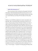
Bạn đang xem bản rút gọn của tài liệu. Xem và tải ngay bản đầy đủ của tài liệu tại đây (422.6 KB, 8 trang )
Int.J.Curr.Microbiol.App.Sci (2017) 6(3): 1146-1153
International Journal of Current Microbiology and Applied Sciences
ISSN: 2319-7706 Volume 6 Number 3 (2017) pp. 1146-1153
Journal homepage:
Original Research Article
/>
Morphological and Cultural Studies of Sclerotium rolfsii Sacc.
causing Foot Rot Disease of Tomato
Sahana N. Banakar*, V.B. Sanath Kumar and A.G. Thejesha
Department of Plant Pathology, College of agriculture, V. C Farm Mandya,
UAS, Bengaluru, India
*Corresponding author
ABSTRACT
Keywords
Sclerotium rolfsii,
Foot rot,
Morphological
variability,
Cultural
variability.
Article Info
Accepted:
20 February 2017
Available Online:
10 March 2017
Morphological and cultural variability of S. rolfsii infecting tomato were
studied on different solid and liquid media based on radial growth,
topography, sclerotial number, diameter of sclerotia, test weight of
sclerotial bodies and dry mycelial weight. Significant variability with
reference to mycelial and sclerotial characters was observed on different
media. This investigation revealed that the maximum mycelial growth was
observed in Potato Dextrose Agar (9 cm) and more test weight (262 mg) of
sclerotial bodies was recorded in Sabouraud’s dextrose agar. Cultural
studies showed the maximum dry mycelial weight of fungus in potato
dextrose broth (750 mg) followed by oat meal broth (663 mg).
Introduction
Tomato (Lycopersicon esculentum L.) is an
important nutritive rich and warm season
vegetable crop grown throughout the world.
In the world, tomato is cultivated in an area of
4.8 million hectare with an annual production
of 161.8 million tonnes (Anonymous, 2012).
In India, it occupies an area of about 0.88
million hectares with the production of 18.22
million tonnes (Anonymous, 2013). In
Karnataka it occupies an area of 0.05 million
hectare with a production of 1.76 million
tonnes (Anonymous, 2013). It suffers from
number of fungal, bacterial, nematode and
many
viral
diseases.
Among
the
phytopathogenic fungi, disease caused by
Sclerotium rolfsii, a soil borne fungi which
causes foot rot or collar rot of tomato is
gaining a serious status. It is known to be
pathogenic on nearly 500 plant species. The
disease is also referred as Sclerotium blight,
Sclerotium wilt, southern blight, southern
stem rot and white mold which cause 55-95%
mortality of the crop at seedling stage under
condusive conditions (Gurha and Dubey,
1982). S. rolfsii Sacc. is widely distributed in
tropics, subtropics and also in warmer parts of
temperate zone of the world. In India, it is
wide spread in almost all the states and
causing economic losses in many crops. The
numerous reports from tropical and sub
tropical areas of the world, coupled with the
large number of hosts attacked by it indicate
1146
Int.J.Curr.Microbiol.App.Sci (2017) 6(3): 1146-1153
that, economic losses are substantial every
year due to infection of S. rolfsii (Aycock,
1966).
sclerotial) and shape of sclerotia on individual
media were also recorded.
Cultural variability
Although,
extreme
variations
in
morphological characteristics have been
noticed in worldwide collections of the
pathogen (Koech et al., 1994 and Kumar et
al., 1995) not much is known about the
variations on different media. The objective
of the present investigation was to study the
variations regard to morphology of mycelium
and sclerotia viz., colour, shape and size,
number and test weight of sclerotia on
different solid and liquid media.
Materials and Methods
Isolation of the pathogen
Isolation of the fungus was done by following
standard tissue isolation technique from
infected tomato plants. The fungal growth on
the infected tissue was aseptically transferred
to the PDA plates for growth and purification.
Pure culture of the pathogen was transferred
to PDA slants for further studies.
Morphological variability
The study comprises of 8 solid and liquid
media (Table 1). Fifteen ml of each sterilised
medium was poured into Petri plates.
Mycelial disc from seven day old culture of
the S. rolfsii was placed at the centre of the
plate. Three replications were maintained at
room temperature (27±1°C) for three days
and colony characters viz., diameter,
pigmentation, radial growth and type of
margin were recorded. To get matured
sclerotial bodies, the cultures were further
incubated up to thirty days. On each medium,
diameter of ten sclerotial bodies per
replication was recorded with the help of
screw gauge and observations were
statistically analysed. The total number of
sclerotia produced per plate, test weight (100
Twenty ml each of the liquid media was taken
in 100ml conical flasks and sterilized in an
autoclave at 121.6°C and 15 PSI for 15 min.
Five mm discs of seven day old culture were
used for inoculation to each flask. Inoculated
flasks were incubated at 27±1°C for 10 days.
Each treatment was replicated thrice. After
the incubation period, the mycelial mat was
filtered through Whatman No. 42 filter paper
discs. The mycelial mat on the filter paper
was dried to a constant weight in an electrical
oven at 50°C, cooled in desiccators and
weighed immediately on an analytical
electrical balance. The weight of dry
mycelium in each replication was recorded
and the data were statistically analyzed.
Results and Discussion
Morphological characters of S. rolfsii on eight
different solid media based on the characters
like radial growth, mycelial characteristics,
colony colour, shape and number of sclerotial
bodies per plate and test weight of sclerotial
bodies were studied.
The result revealed that the variation in
colony diameter (Table 2 and Plate 1) on
different media was found significant after 72
h of incubation. In Potato dextrose agar
(PDA) maximum colony diameter (9.0 cm)
was recorded after 48 hours of incubation
whereas on oat meal agar maximum colony
diameter (9.0 cm) was recorded after 72 hours
of incubation. However, these two media
were on par with each other. The minimum
colony diameter (3.4 cm) was recorded on
Sabouraud’s Dextrose Agar (SDA) followed
by Nutrient agar (4.8 cm). There was
significant difference between media and time
interval.
1147
Int.J.Curr.Microbiol.App.Sci (2017) 6(3): 1146-1153
With respect to colony colour, three types of
colours were observed on different solid
media (Table 3). Among the media viz., PDA,
Kirchoff’s agar, SDA, Hunsen’s agar and
tomato leaf extract showed pure white
colonies, where as Richard’s agar and nutrient
agar showed dull white coloured colony. On
oat meal agar cottony white coloured colony
was observed. With regard to growth pattern,
S. rolfsii showed compact growth on PDA
and oat meal agar was recorded. On
Kirchoff’s agar and Hunsen’s agar, it showed
filamentous growth and on SDA and tomato
leaf extract it was fluffy. However on nutrient
agar colony growth was cloudy type. On most
of the media mycelial margin was smooth
except SDA and Kirchoff’s agar which
showed filamentous and serrated margin
respectively. With respect to distribution of
mycelial growth, S. rolfsii showed thick
mycelial growth on PDA, oat meal agar and
tomato leaf extract. However in Kirchoff’s
agar, SDA, Hunsen’s agar it showed thin
mycelia growth whereas in Richard’s agar
and nutrient agar showed irregular mycelial
growth.
The sclerotial initiation started on eight days
after incubation on tomato leaf extract and it
was on 10th day in case of PDA. On Hunsen’s
agar sclerotial initiated after 13th day. Media
viz., Kirchoff’s agar and nutrient agar on 18th
day, whereas in oat meal agar and SDA on
20th day after incubation (Table 4). However
there was no sclerotial body production was
recorded in Richard’s agar (Plate 2). With
regard to position of sclerotia, on three media
i.e. PDA, oat meal agar and tomato leaf
extract, sclerotia were distributed uniformly
all over the plates. But in case of Kirchoff’s
agar, SDA, Hunsen’s agar and nutrient agar
sclerotial bodies were concentrated at the
edges or periphery of the Petri plate.
With respect to shape of sclerotia, on most of
the media S. rolfsii produced round sclerotial
except SDA and tomato leaf extract in which
it had produced irregular sclerotial bodies.
Among all the media tested four types of
colours were observed with respect to
sclerotial colour. Sclerotial bodies were
brown in colour on PDA, Hunsen’s agar and
tomato leaf extract where as dark brown
colour was observed on oat meal agar and
SDA. Light brown and very light brown
colours were observed on Kirchoff’s agar and
nutrient agar respectively.
Table.1 List of media used to study the growth characters of Sclerotium rolfsii
causing tomato foot rot disease
SI.
No
1
2
3
4
5
6
7
8
Solid media
Potato dextrose agar (PDA)
Oat meal agar
Kirchoff’s agar
Sabouraud’s dextrose agar
(SDA)
Hunsen’s agar
Richard’s agar
Nutrient agar
Tomato leaf extract
Liquid broth
Potato dextrose broth
Oat meal broth
Kirchoff’s broth
Sabouraud’s dextrose
broth
Hunsen’s broth
Richard’s broth
Nutrient broth
Tomato leaf extract
broth
1148
Type of media
Semi-synthetic
Semi-synthetic
Synthetic
Synthetic
Synthetic
Synthetic
Synthetic
Semi-synthetic
Int.J.Curr.Microbiol.App.Sci (2017) 6(3): 1146-1153
Table.2 Growth of S. rolfsii on different solid media
Treat
ments
T1
T2
T3
T4
T5
T6
T7
T8
Media
Mean colony diameter(cm) (hours after
incubation)
24
48
72
Mean
3.8
9.0
9.0
7.22
3.2
8.2
9.0
6.80
2.4
3.8
5.1
3.76
2.5
2.9
3.4
2.99
3.5
7.2
8.6
6.43
2.3
4.2
5.0
3.88
1.7
3.1
4.8
3.20
2.5
5.9
8.0
5.46
0.076
0.213
0.015
0.228
0.638
0.0495
Potato dextrose agar (PDA)
Oat meal agar
Kirchoff’s agar
Sabourauds dextrose agar (SDA)
Hunsen’s agar
Richard’s agar
Nutrient agar
Tomato leaf extract
SEm±
C.D at 1%
Table.3 Colony characters of S. rolfsii on different solid media
Mycelia
margin
Growth
pattern
Distribution
of mycelia
growth
Treatments
Media
Mycelia
colour
T1
Potato dextrose
agar
Pure white
Smooth
Compact
Thick
T2
Oat meal agar
Cottony
white
Smooth
Compact
Thick
T3
Kirchoff’s agar
Pure white
Smooth
Filamentous
Thin
T4
Sabouraud’s
dextrose agar
Pure white
Filamentous
Fluffy
Thin
T5
Hunsen’s agar
Pure white
Smooth
Filamentous
Thin
T6
Richard’s agar
Dull white
Serrated
Filamentous
Irregular
Dull white
Smooth
Cloudy
Irregular
Pure white
Smooth
Fluffy
Thick
T7
T8
Nutrient agar
agar
Tomato leaf
extract
1149
Int.J.Curr.Microbiol.App.Sci (2017) 6(3): 1146-1153
Table.4 Sclerotial characters of S. rolfsii on different solid media
Treatments
MEDIA
Days to
sclerotial
initiation
Distribution
over media
Shape
Colour
and
texture
Diameter
(mm)
Test
weight
(mg) (100
sclerotial
bodies)
T1
Potato
dextrose agar
10th
All over
Round
Brown
1.50
238
212
T2
Oat meal agar
20th
All over
Round
1.70
131
205
18th
Periphery
Round
1.60
91
59
20th
Periphery
Irregular
1.50
262
60
T3
T4
Kirchoff’s
agar
Sabouraud’s
dextrose agar
Dark
Brown
Light
Brown
Dark
Brown
No.
per
plate
T5
Hunsen’s agar
13th
Periphery
Round
Brown
1.50
92
48
T6
Richard’s agar
*-
-
-
-
-
-
-
T7
Nutrient agar
18th
Periphery
Round
V.light
Brown
0.11
56
45
T8
Tomato leaf
extract
8th
All over
Irregular
Brown
0.12
121
198
SEm±
0.221
1.38
1.00
C.D at 1%
0.662
4.15
4.131
Note: *- Sclerotial bodies were not produced
1150
Int.J.Curr.Microbiol.App.Sci (2017) 6(3): 1146-1153
Table.5 Cultural chararacters (dry mycelia weight, no. of sclerotia/flask, colour of sclerotia)
of S. rolfsii on different liquid media
S.No
Name of the broth
1
2
3
4
5
6
7
8
Potato dextrose broth
Oat meal broth
Kirchoff’s broth
Sabouraud’s broth
Hunsen’s broth
Richard’s broth
Nutrient broth
Tomato leaf extract broth
SEm±
C.D at 1%
Dry mycelia
weight (mg)
750
663
435
203
365
244
10
550
6.444
19.310
No. of
sclerotia/flask
350
310
96
120
67
0
15
295
3.880
11.632
Colour of
sclerotia
Brown
Dark Brown
Light Brown
Dark Brown
Brown
No sclerotia
V.light Brown
Brown
Plate.1 Growth of Sclerotium rolfsii on different solid media
Legend:
T1: Potato dextrose agar; T2: Oat meal agar; T3: Kirchoff’s agar; T4: Sabourauds dextrose agar;
T5: Hunsen’s agar; T6: Richard’s agar; T7: Nutrient agar agar; T8: Tomato leaf extract
1151
Int.J.Curr.Microbiol.App.Sci (2017) 6(3): 1146-1153
Plate.2 Production of sclerotial bodies of Sclerotium rolfsii on different solid media
Legend:
T1: Potato dextrose agar; T2: Oat meal agar; T3: Kirchoff’s agar; T4: Sabourauds dextrose agar; T5: Hunsen’s agar;
T6: Richard’s agar; T7: Nutrient agar agar; T8: Tomato leaf extract
Plate.3 Growth of Sclerotium rolfsii on liquid media
Legend:
1: Potato dextrose broth; 2: Oat meal broth; 3: Kirchoff’s broth; 4: Sabourauds dextrose broth; 5: Hunsen’s broth;
6: Richard’s broth; 7: Nutrient broth; 8: Tomato leaf broth
The variations in size of sclerotial bodies of
different media were recorded. On oat meal agar
biggest sclerotial bodies of 1.7 mm diameter were
produced followed by Kirchoff’s agar (1.6 mm).
Smaller size of sclerotial bodies were recorded on
nutrient agar (0.11 mm diameter) followed by
tomato leaf extract (0.12 mm diameter).
The test weight (weight of 100 sclerotial bodies)
of sclerotial bodies on all the solid media varied
significantly. Maximum test weight of sclerotial
bodies was recorded in SDA (262 mg) followed
by PDA (238 mg). Minimum test weight was
recorded in nutrient agar medium (56 mg). The
density of sclerotial bodies per Petri plate revealed
that there was a significant variation among the
different solid media. On PDA more densely
populated sclerotial bodies (212 no./plate) were
recorded which was followed by oat meal agar
(205 no./plate). However no sclerotial body
1152
Int.J.Curr.Microbiol.App.Sci (2017) 6(3): 1146-1153
production was recorded in Richard’s agar.
Cultural characters like dry mycelia weight,
number of sclerotia/flask, colour of sclerotia) of S.
rolfsii were studied on eight different liquid media
(Table 5 and Plate 3). The results showed that the
synthetic media produce less mycelial weight and
number of sclerotia/flask, compared to semi
synthetic media. Maximum dry mycelial weight
of fungus was obtained in potato dextrose broth
(750 mg) followed by oat meal broth (663 mg)
and host leaf extract (550 mg). Among the
synthetic media tested, maximum mycelial weight
of the fungus was recorded in Kirchoff’s broth
(435 mg) followed by Hunsen’s broth (365 mg)
and Richard’s broth (244 mg). Least mycelial dry
weight of the fungus was recorded in nutrient
broth (10 mg).
Among the semi synthetic media, highest number
of sclerotial bodies were produced in potato
dextrose broth (350 no./flask) and among the
synthetic media, Sabouraud’s broth (120
no./flask) produced more sclerotial bodies. Less
sclerotial bodies were recorded in nutrient broth
(15 no./flask). The present study is in line with
many workers (Lingaraju (1977), Prabhu (2003),
Basamma (2008) and Manu (2012) who had
reported that the morphology of mycelium,
number sclerotiaand morphology of sclerotia
produced, varied in different media depends on
nutritional factors. The present finding strongly
suggest that the there is a morphological and
cultural variation on different solid and liquid
media.
References
Anonymous. 2012. FAOSTAT: Production crop
2012 data.
Anonymous. 2013. National Horticulture
Mission, 3rd January to 12th January,
pp.10.
Anonymous. 2013. Indian horticulture database.
MOA. 1-301. pp.
Aycock, R., 1966. Stem rot and other diseases
caused by S. rolfsii. North Carolina
Agricultural
Experimental
Station,
Technical Bull., 174: 202.
Basamma. 2008. Integrated management of
Sclerotium wilt of potato caused by
Sclerotium rolfsii Sacc. M. Sc. (Agri.)
Thesis, Univ. Agric. Sci., Dharwad, 113.
pp.
Dominique, B. 2012. A colour handbook of
tomato diseases. Identification, biology and
control. Manson publishing Ltd., 688. pp.
Gurha, S.N. and Dubey, R.S. 1982. Occurrence of
possible sources of resistance in chickpea
(Cicer arietinum L.) against Sclerotium
rolfsii Sacc. Madras Agric. J., 70: 63-64.
Jabbar Sab. 2013. Studies on collar rot of chick
pea caused by Sclerotium rolfsii Sacc. M.
Sc. (Agri.) Thesis, Univ. Agric. Sci.,
Bengaluru.
Koech, J.K., Lewis, B. and Mithem, R. 1994.
Pathogenic variation in isolates of
Plasmodiophora brassicae on Brassica spp.
Indian J. Pl. Patho., 12: 5-9.
Kumar, S., Thind, T.S. and Mohanchander. 1995.
Morphogenic and pathogenic variations in
Gloeosporium amoelophagum. Indian
Phytopathol., 48: 331-334.
Lingaraju, S. 1977. Studies on Sclerotium rolfsii
Sacc. with respect to the survival in soil. M.
Sc. (Agri.) Thesis, Univ. Agric. Sci.,
Bangalore, 61. pp.
Manu, T.G. 2012. Studies on Sclerotium rolfsii
(Sacc.) causing foot rot disease on finger
millet M.Sc. (Agri.) Thesis, Univ. Agric.
Sci., Bangalore, 76. pp.
Prabhu, H.V. 2003. Studies on collar rot of
soybean caused Sclerotium rolfsii Sacc. M.
Sc. (Agri.) Thesis, Univ. of Agric. Sci.,
Dharwad.
How to cite this article:
Sahana N. Banakar, V.B. Sanath Kumar and Thejesha, A.G. 2017. Morphological and Cultural Studies
of Sclerotium rolfsii Sacc. causing Foot Rot Disease of Tomato. Int.J.Curr.Microbiol.App.Sci. 6(3):
1146-1153. doi: />
1153