LC-NMR and Other Hyphenated NMR Techniques doc
Bạn đang xem bản rút gọn của tài liệu. Xem và tải ngay bản đầy đủ của tài liệu tại đây (2.68 MB, 232 trang )
LC-NMR and Other
Hyphenated NMR
Techniques
LC-NMR and Other
Hyphenated NMR
Techniques
Overview and Applications
Maria Victoria Silva Elipe
Amgen, Inc.
Copyright Ó 2012 by John Wiley & Sons, Inc. All rights reserved.
Published by John Wiley & Sons, Inc., Hoboken, New Jersey.
Published simultaneously in Canada.
No part of this publication may be reproduced, stored in a retrieval system, or transmitted in any form
or by any means, electronic, mechanical, photocopying, recording, scanning, or otherwise, except as
permitted under Section 107 or 108 of the 1976 United States Copyright Act, without either the prior
written permission of the Publisher, or authorization through payment of the appropriate per-copy fee
to the Copyright Clearance Center, Inc., 222 Rosewood Drive, Danvers, MA 01923, (978) 750-8400,
fax (978) 750-4470, or on the web at www.copyright.com. Requests to the Publisher for permission
should be addressed to the Permissions Department, John Wiley & Sons, Inc., 111 River Street, Hoboken,
NJ 07030, (201) 748-6011, fax (201) 748-6008, or online at />Limit of Liability/Disclaimer of Warranty: While the publisher and author have used their best
efforts in preparing this book, they make no representations or warranties with respect to the accuracy
or completeness of the contents of this book and specifically disclaim any implied warranties of
merchantability or fitness for a particular purpose. No warranty may be created or extended by sales
representatives or written sales materials. The advice and strategies contained herein may not be
suitable for your situation. You should consult with a professional where appropriate. Neither the
publisher nor author shall be liable for any loss of profit or any other commercial damages, including
but not limited to special, incidental, consequential, or other damages.
For general information on our other products and services or for technical support, please contact
our Customer Care Department within the United States at (800) 762-2974, outside the United States
at (317) 572-3993 or fax (317) 572-4002.
Wiley also publishes its books in a variety of electronic formats. Some content that appears in print
may not be available in electronic formats. For more information about Wiley products, visit our web
site at www.wiley.com.
Library of Congress Cataloging-in-Publication Data:
Silva Elipe, Maria Victoria, 1963-
LC-NMR and other hyphenated NMR techniques : overview and applications /
Maria Victoria Silva Elipe.
p. cm.
Includes bibliographical references and index.
ISBN 978-0-470-54834-9 (hardback)
1. Nuclear magnetic resonance spectroscopy–Industrial applications.
2. Organic compounds–Analysis. 3. Drug development. I. Title.
QD96.N8S54 2012
543’.66–dc23
2011018343
Printed in the United States of America
10987654321
To my parents, Joaquin Silva Garcia and Maria Elipe Ruiz,
for their love, dedication, and memories that will last a lifetime.
To my husband, Regnar Llego Madarang, and my children,
Eva Silva Madarang and Regnar Silva Madarang, for their love.
Contents
Preface xi
Abbreviations, Symbols, and Units xv
1. Basic Concepts of NMR Spectroscopy 1
1.1 Introduction / 1
1.2 Basic Knowledge Regarding the Physics of NMR
Spectroscopy / 2
1.3 Basic Parameters for NMR Interpretation / 7
1.3.1 Chemical Shift / 8
1.3.2 Spin–Spin Coupling Constants / 13
1.3.3 Spin Systems / 20
1.3.4 Signal Intensities / 21
1.3.5 Bond Correlations / 23
1.3.6 Spatial Correlations / 27
1.3.7 Other Topics / 30
1.4 Conclusions / 35
References / 36
2. Historical Development of NMR and LC-NMR 39
2.1 Introduction / 39
2.2 Historical Development of NMR / 39
vii
2.3 Historical Development of LC-NMR / 46
2.4 Historical Development of Other Analytical
Techniques Hyphenated with NMR / 49
2.5 Current Trends / 52
References / 52
3. Basic Technical Aspects and Operation of LC-NMR
and LC-MS-NMR 59
3.1 Introduction / 59
3.2 Technical Considerations Regarding LC-NMR / 59
3.2.1 Solvent Compatibility / 60
3.2.2 Solvent Suppression / 61
3.2.3 NMR Flow Cell / 62
3.2.4 LC-NMR Sensitivity / 64
3.3 Technical Considerations Regarding
LC-MS-NMR / 65
3.3.1 Deuterated Solvents / 66
3.4 Modes of Operation of LC-NMR / 66
3.4.1 On-Flow Mode / 67
3.4.2 Stop-Flow Mode / 67
3.4.3 Time-Sliced Mode / 77
3.4.4 Loop Collection Mode / 77
3.5 Modes of Operation of LC-MS-NMR / 77
3.5.1 On-Flow Mode / 80
3.5.2 Stop-Flow Mode / 87
3.6 Other Modes of Operation / 87
3.7 Challenging Considerations / 89
3.7.1 Air Bubbles / 89
3.7.2 Carryover with and Without an
Autosampler / 90
3.7.3 Sample Solubility and Precipitation / 90
3.7.4 Flow Cell and System Cleaning / 91
3.7.5 Flow Rate and Magnetic Susceptibility / 91
3.7.6 Quantitation / 92
3.8 Conclusions / 92
References / 93
viii CONTENTS
4. Applications of LC-NMR 95
4.1 Introduction / 95
4.2 Applications of LC-NMR / 96
4.2.1 Natural Products / 96
4.2.2 Drug Metabolism / 102
4.2.3 Drug Discovery / 108
4.2.4 Impurity Characterization / 111
4.2.5 Degradation Products / 112
4.2.6 Food Analysis / 115
4.2.7 Polymers / 118
4.2.8 Metabolomics and Metabonomics / 118
4.2.9 Isomers, Tautomers, and Chiral Compounds / 119
4.2.10 Others Areas / 120
4.3 Conclusions and Future Trends / 120
References / 121
5. Applications of LC-MS-NMR 131
5.1 Introduction / 131
5.2 Applications of LC-MS-NMR / 132
5.2.1 Natural Products / 132
5.2.2 Drug Metabolism / 134
5.2.3 Drug Discovery and Development / 135
5.2.4 Metabolomics and Metabonomics / 136
5.2.5 Others Areas / 139
5.3 Conclusions and Future Trends / 139
References / 140
6. Hyphenation of NMR with Other Analytical
Separation Techniques 143
6.1 Introduction / 143
6.2 GC-NMR / 144
6.3 GPC-NMR / 144
6.4 SEC-NMR / 145
6.5 SFC-NMR / 146
6.6 SFE-NMR / 147
6.7 CE-NMR / 147
CONTENTS ix
6.8 CEC-NMR / 149
6.9 CZE-NMR / 150
6.10 cITP-NMR / 150
6.11 CapLC-NMR / 152
6.12 SPE-NMR / 154
6.13 SPE-MS-NMR / 159
6.14 Conclusions and Future Trends / 167
References / 168
7. Special Topics and Applications Related to LC-NMR 179
7.1 Introduction / 179
7.2 Off-Line Versus Online NMR for Structural Elucidation / 180
7.2.1 Cases Solved Off-Line / 180
7.2.2 Cases Solved Online / 186
7.3 Analysis of Chiral Molecules by NMR / 188
7.3.1 Classical Approach: Off-Line / 189
7.3.2 Nonclassical Approach: Online / 190
7.4 Monitoring Chemical Reactions In Situ / 190
7.4.1 Classical Approach: Off-Line / 191
7.4.2 Nonclassical Approach: Online / 194
7.5 Analysis of Mixtures Off-Line, Online, and by
Other NMR Methodologies / 196
7.5.1 Traditional Analysis of Mixtures by
Off-Line HPLC and NMR / 196
7.5.2 Online NMR Analysis of Mixtures / 203
7.5.3 Other NMR Methodologies That Mimic
LC-NMR Separation / 208
7.6 Current Trends / 210
References / 211
Index 217
x CONTENTS
Preface
Since the subject of nuclear magnetic resonance (NMR) was awarded its first
Nobel Prize in 1952 due to its successful detection by Bloch and Purcell in
1945, the technology and its applications have developed tremendously. The
first two decades were focused on technical developments of instrumentation
and methodologies to apply to the structure determination of compounds.
During the late 1970s, several research groups developed modifications of
NMR probes to convert them to an online mode for the analysis of sample
mixtures. However, with the hardware and software technology available at
that time, it was difficult to hyphenate high-performance liquid chromato-
graphy (HPLC) and NMR to perform those analyses. During the past two
decades, interest in sample mixture analysis and screening methods has been
the driver for the latest developments and applications of hyphenated
analytical techniques with NMR. Improvements in solvent suppression NMR
techniques have facilitated the coupling to NMR of HPLC with reversed-
phase columns, for what is known today as LC-NMR. Further technological
developments have also supported the hyphenation of mass spectrometry
(MS) to LC-NMR as LC-MS-NMR. In addition, other analytical separation
techniques have been hyphenated to NMR. However, the only ones commer-
cially available and commonly used are capillary HPLC (capLC) as capLC-
NMR and solid-phase extraction (SPE) as SPE-NMR, including SPE
hyphenated to MS-NMR as SPE-MS-NMR. Many laboratories in industry
and academia have NMR as a hyphenated technique as part of their instru-
mentation to solve structural problems. This technology has become an
important option for complex analysis.
xi
The aim of this book is to provide an overview of the applications of
hyphenated NMR techniques in industry and academia. The book is focused
on understanding the pros and cons of NMR as a hyphenated and a non-
hyphenated technique for the structural determination analysis of samples as
organic materials. The purpose of the basic overview of the main concepts for
structural elucidation by NMR and technical issues for online NMR is to
facilitate an understanding of the pros and cons of the technique. A major
emphasis of the book is on the application of hyphenated NMR in industry and
academia. For completeness, the book has a chapter dedicated to the historical
development of hyphenated NMR techniques, and another chapter focused on
a comparison of other methodologies used to analyze sample mixtures.
The book begins with a description of basic NMR concepts for the
structural elucidation of organic compounds, the historical development of
NMR and hyphenated NMR in the structural elucidation world, followed by
applications of hyphenated NMR as LC-NMR and LC-MS-NMR in industry
and academia, such as to natural products, degradation products, impurity
characterization, drug metabolism, food analysis, drug discovery, polymers,
and others. Another chapter is dedicated to other analytical separation
techniques hyphenated with NMR and MS-NMR, with special emphasis on
capillary capLC and SPE due to be available commercially, and their
applications compared to the other hyphenated NMR techniques. A special
chapter is directed at understanding the applications of NMR online and off-
line for structure elucidation, chiral analysis, in situ reaction monitoring, and
analysis of sample mixtures by other NMR methodologies.
The audience for this book includesscientists in industry and academia who
work and analyze complex sample mixtures in the areas of organic chemistry,
medicinal chemistry, process chemistry, analytical chemistry, drug metabo-
lism, separation science, natural products, chemical engineering, and others.
In addition, the book contains the fundamentals of NMR and applications of
hyphenated NMR techniques for college instructors to use as a complemen-
tary textbook for undergraduate and, especially, for graduate courses. The
book is an excellent source of information and references for NMR basics,
especially for applications of hyphenated NMR in industry and academia. The
book also contains updated information on the latest advancements and
applications of LC-NMR and other analytical techniques hyphenated with
NMR focused on structural elucidation as of early 2011. The approach is based
on explaining the basic pros and cons of the technique in a practical way, to
make it easier for nonexperts in the field to understand the technology.
Examples are provided, illustrated with figures and detailed explanations.
Other books targeting those concepts have been used as reference material.
Previous to this book, I wrote some review articles and a book chapter.
I gratefully acknowledge Elsevier for permitting me to use materials from one
xii PREFACE
of the review articles [M.V. Silva Elipe, Advantages and Disadvantages of
Nuclear Magnetic Resonance Spectroscopy as Hyphenated Technique, Anal.
Chim. Acta 497 (2003), 1–25]. My sincere gratitude to Dr. Ray Bakhtiar (Drug
Metabolism of MRL at Rahway) and Dr. Byron H. Arison (currently retired
but previously at Drug Metabolism of MRL at Rahway) for their interest,
support, and encouragement through constructive discussions, and to
D. Knapp and U. Parikh (Medicinal Chemistry of MRL at Rahway) for
technical help in online connection of radioactivity and MS detectors to an
LC-NMR system. I am especially thankful to my father, Joaquin Silva Garcia,
and my mother, Maria Elipe Ruiz, for their encouragement, love, and
dedication to their children (the author and her siblings, Pedro Luis Silva
Elipe and Maria Eva Silva Elipe). Last but not least, I thank my husband,
Regnar Llego Madarang, for his support, and my children, Eva Silva
Madarang and Regnar Silva Madarang, for their patience and support. There
are not enough words to express my appreciation.
M
ARIA VICTORIA SILVA ELIPE
Thousand Oaks, California
PREFACE xiii
Abbreviations, Symbols,
and Units
ACN acetonitrile
ACN-d3 deuterated acetonitrile
API atmospheric pressure ionization, active principal ingredient
APCI atmospheric pressure chemical ionization
B applied magnetic field along x or y axis
B
eff
effective magnetic field
bd broad doublet
B
0
applied magnetic field along z axis
bs broad singlet
bt broad triplet
C degree Celsius or centigrade
capLC capillary liquid chromatography
CAT computer of averaging transients
CD circular dichroism
CD
3
CN deuterated acetonitrile
CD
3
OD deuterated methanol
CE capillary electrophoresis
CEC capillary electrochromatography
CHPLC capillary high-performace liquid chromatography
CI chemical ionization
cIPT capillary isotachophoresis
cm centimeter
COSY correlation spectroscopy
CW continuous wave
xv
CYP cytochrome P450 enzyme
CZE capillary zone electrophoresis
d doublet
D deuterium
1D one dimension
2D two dimensions
Da dalton
dd doublet of doublets
ddd doublet of doublet of doublets
DEPT distortionless enhancement by polarization transfer
DEPT-135 distortionless enhancement by polarization transfer
at 135
angle
DEPTQ distortionless enhancement by polarization
transfer, including the detection of quaternary nuclei
DI direct injection
DMSO-d6 dimethyl-d6 sulfoxide
DNP dynamic nuclear polarization
D
2
O deuterated water or deuterium oxide
DOSY diffusion-ordered spectroscopy
DQF double quantum filter
dt doublet of triples
E energy
EOF electroosmotic flow
EPR electron paramagnetic resonance
ERETIC electronic reference to access in vivo concentrations
ESI electrospray ionization
FIA flow injection analysis
FID free induction decay
FT Fourier transform
GC gas chromatography
GHz gigahertz
GPC gel permeation chromatography
GSH glutathione
h Planck’s constant
HETCOR heteronuclear correlation spectroscopy
HMBC heteronuclear multiple bond correlation
HMQC heteronuclear multiple quantum correlation
HOD residual water from deuterated solvents
HPLC high-performance liquid chromatography
HSQC heteronuclear single quantum coherence
Hz hertz
I nuclear spin
xvi ABBREVIATIONS, SYMBOLS, AND UNITS
ICH International Conference of Harmonisation
of Technical Requirements
INADEQUATE incredible natural abundance double quantum
transfer experiment
INEPT intensive nuclei enhanced by polarization transfer
IR infrared
J coupling constant
k Boltzmann constant
K kelvin
LC liquid chromatography
LOD limit of detection
mL microliter
mL milliliter
m meter; multiplet
mm millimeter
M molar; molecular ion
mM millimolar
mM micromolar
ms millisecond
MEK methyl ethyl ketone
MHz megahertz
MS mass spectrometry
MSPD matrix solid-phase dispersion
MW molecular weight
m/z mass over charge
NMR nuclear magnetic resonance
NOE nuclear Overhauser effect
NOESY nuclear Overhauser effect spectroscopy
oct octet
PAT process analytical technology
PCA principal components analysis
pCEC pressured capillary electrochromatography
PEEK poly(ether ether ketone)
PKDM pharmacokinetics drug metabolism
ppm part per million
q quartet
qNMR quantitation NMR
qui quintet
RDC residual dipolar coupling
RF radio frequency
ROE rotating frame Overhauser effect
ROESY rotating frame Overhauser effect spectroscopy
ABBREVIATIONS, SYMBOLS, AND UNITS xvii
RT room temperature
s seconds; singlet
SEC size-exclusion chromatography
SFE supercritical fluid extraction
SFC supercritical fluid chromatography
S/N signal-to-noise ratio
SPE solid-phase extraction
spt septet
sxt sextet
t triplet
T temperature, tesla
T
1
spin-lattice or longitudinal relaxation time
T
2
spin-spin or transverse relaxation time
td triplet of doublets
TIC total ion chromatogram
TMS tetramethylsilane
TOCSY total correlation spectroscopy
UF ultrafast
UV ultraviolet
WET water suppression enhanced through T
1
effects
g gyromagnetic ratio
d chemical shift
n frequency
s shielding constant
t
c
correlation time
o
0
Lamour frequency
xviii ABBREVIATIONS, SYMBOLS, AND UNITS
1
Basic Concepts of
NMR Spectroscopy
1.1. INTRODUCTION
Nuclear magnetic resonance, known widely as NMR spectroscopy, is a
powerful technique applied extensively to the structural elucidation of organic
compounds. Since its discovery in the early twentieth century, NMR has been
in wide use while evolving to what it is today. Understanding the basic
concepts in interpreting NMR spectra is fundamental for the structural
analysis of organic compounds. In this chapter we introduce the reader to
the basic concepts of NMR data interpretation. The major topics discussed
provide information on the chemical structures of organic compounds, and the
connectivities and correlations of atoms through bonds and space. Under-
standing how to interpret NMR data from hyphenated and nonhyphenated
NMR instruments is essential. This chapter is not intended to explain the
theory of NMR with mathematical equations and algorithms, as these can be
found elsewhere [1–4]. In addition, more detailed information from a practical
perspective with less focus on mathematical algorithms is available in the
literature [5–16].
LC-NMR and Other Hyphenated NMR Techniques: Overview and Applications, First Edition.
Maria Victoria Silva Elipe.
Ó 2012 John Wiley & Sons, Inc. Published 2012 by John Wiley & Sons, Inc.
1
1.2. BASIC KNOWLEDGE REGARDING THE PHYSICS
OF NMR SPECTROSCOPY
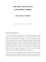
Spectroscopy studies the interaction of matter with electromagnetic radiation,
resulting in absorption or emission of energy. When energy is in the radio-
frequency (RF) region (>10
6
to 10
8
Hz), nonionizing radiation energy is used
to quantize the energy levels of spin nuclei (Figure 1-1). Nuclear magnetic
resonance is an absorption spectroscopy that involves the magnetic properties
of atomic nuclei. Under a magnetic field in the RF region, nuclei with
magnetic moments can have different energy levels, and those absorption
energy transitions can be measured by NMR. Following are the basic rules
regarding nuclei magnetic moments and their nuclear spin based on their
nuclear properties.
.
Nuclei with an odd mass number have half-integer nuclear spin I
(e.g., I ¼1/2 for
1
H,
13
C,
15
N,
31
P,
19
F,
29
Si; I ¼3/2 for
23
Na,
11
B;
I ¼5/2 for
17
O).
.
Nuclei with an even mass and an even atomic number have zero nuclear
spin I (e.g., I ¼0 for
12
C,
16
O).
.
Nuclei with an even mass number and an odd atomic number have integer
nuclear spin I (e.g., I ¼1 for
2
H,
14
N; I ¼3 for
10
B).
Table 1-1 displays the nuclear spin properties of the most common nuclei
studied in the field of organic molecules. Under a magnetic field, a nucleus
with nuclear spin will present a concrete number of energy levels. The number
Frequency (Hz)
10
18
10
16
10
14
10
12
10
10
10
8
10
6
x-ray
Ionizing Radiation
(Bonds Break)
Nonionizing Radiation
(Heating)
UV
Visible
IR
Microwave
RF
Electron
Transitions
NMR/MRI
Nuclear Spin
Transitions
Decreasing Energ
y
FIGURE 1-1. Electromagnetic radiation energy spectrum as frequency (Hz).
2 BASIC CONCEPTS OF NMR SPECTROSCOPY
of levels depends on the magnetic moment of each nucleus and follows the rule
2I þ1, where I is the nuclear spin number [e.g., for I ¼1/2, two is the number
of energy levels [2(1/2) þ1 ¼2] with an a spin state, or I
1
¼þ1/2, for the low
energy level and a b spin state or, I
2
¼À1/2, for the high energy level, for
nuclei with a positive gyromagnetic ratio, as indicated below]. For nuclear
spin I ¼1/2 (e.g.,
1
H and
13
C), each nucleus can be displayed as a magnet
randomly oriented in any direction (Figure 1-2a). Under the magnetic field,
those magnets in the sample have two orientations, aligned or opposite to the
direction of the applied magnetic field (Figure 1-2b). The distance between the
energy levels depends on the strength of the magnetic field applied and the
gyromagnetic ratio for the particular nucleus. For nuclei with I ¼1/2 and a
negative gyromagnetic ratio, such as
15
N and
29
Si, b is the lower spin state
(I
1
¼À1/2) and a is the higher spin state (I
2
¼þ1/2). The difference in energy
(DE) for the transition to occur is
DE ¼ hn ¼
hgB
0
2p
where n is the frequency of the transition, g is the gyromagnetic ratio intrinsic
per nucleus (see Table 1-1 for some examples), B
0
is the magnetic field
applied, and h is Planck’s constant. Figure 1-3 depicts the energy-level
separation for a nucleus with half-integer nuclear spin (I ¼1/2; e.g.,
1
H
and
13
C) pointing to the different energy when a magnetic field strength
of 300 MHz (7.05 T) or 600 MHz (14.10 T) is applied to the nucleus.
TABLE 1-1. Properties of Some Common Nuclei in Organic Molecules
Nucleus Spin I
Natural
Abundance (%)
Sensitivity
(Relative to
1
H)
Gyromagnetic
Ratio g
(10
7
rad T
À1
s
À1
]
Frequency
(MHz) at
B
0
¼2.3488 T
1
H 1/2 99.985 1 26.7519 100.0
2
H 1 0.015 9.65 Â10
À3
4.1066 15.351
10
B 3 19.58 1.99 Â10
À2
2.8747 10.746
11
B 3/2 80.42 0.17 8.5847 32.084
12
C 0 98.9
13
C 1/2 1.108 1.59 Â10
À2
6.7283 25.144
14
N 1 99.63 1.01 Â10
À3
1.9338 7.224
15
N 1/2 0.37 1.04 Â10
À3
À2.7126 10.133
16
O 0 99.96
17
O 5/2 0.037 2.91 Â10
À2
À3.6280 13.557
19
F 1/2 100 0.83 25.1815 94.077
23
Na 3/2 100 9.25 Â10
À2
7.0704 26.451
29
Si 1/2 4.70 7.84 Â10
À3
À5.3190 19.865
31
P 1/2 100 6.63 Â10
À2
10.8394 40.481
Source: Data from references 6 and 24.
BASIC KNOWLEDGE REGARDING THE PHYSICS OF NMR SPECTROSCOPY
3
N
S
N
S
N
S
N
S
N
S
N
S
N
S
N
S
N
S
N
S
N
S
N
S
(a)
N
N
B
0
S
N
S
S
N
S
N
S
N
S
N
S
N
S
N
S
N
S
N
S
N
S
(
b
)
FIGURE 1-2. Orientation of the nuclear spins as simple magnets for I ¼1/2 in the absence of an external
magnetic field (except for the Earth’s magnetic field) (a) or the presence (b) of an external magnetic field
(different from the Earth’s magnetic field).
4 BASIC CONCEPTS OF NMR SPECTROSCOPY
Conventionally, the frequency unit megahertz is used for proton
1
Hto
denominate the strength of the magnetic field instead of the magnetic field
unit tesla. Unfortunately, NMR spectroscopy is a low-sensitivity technique
because the energy difference between the levels (DE) of the nuclear spin
states is much less than the thermal energy (kT, where k is the Boltzmann
constant and T is the temperature) at normal or room temperature (around
25
C). This energy difference is also an indication of the small difference in
the population of nuclei between the two spin states. The slight excess of
population in the lower-energy state is in agreement with the Boltzmann
distribution. The energy difference is proportional to the magnetic field
applied (B
0
); therefore, increasing the strength of the magnetic field increases
the population difference of the spin states and the sensitivity (Figure 1-3). The
distribution of nuclei between two spin states is given by the Boltzmann
equation,
N
a
N
b
¼ e
ÀDE=kT
% 1 À
DE
kT
¼ 1 À
hgB
0
2pkT
where N
a
and N
b
are the number of nuclei in the ground state a and the excited
state b. For the case of a magnetic field of 60 MHz (1.41 T) and 300 K (27
C),
the population ratio for
1
HisN
a
/N
b
%0.9999904, and for a magnetic field of
300 MHz (7.05 T), N
a
/N
b
%0.99995. Figure 1-2b is a simplistic representation
of the small excess in the population of nuclei in the lower energy level for
nuclei aligned with the external applied magnetic field. Overall, with the small
difference in energy level, energy transitions of nuclear spins can occur with a
B
0
(Magnetic Field)
I = -1/2
β
spin state
(higher energy level)
I = 1/2
α
spin state
(lower energy level)
Energy
Δ
E = h × 300 MHz
Δ
E = h × 600 MHz
7.05 T
14.10 T
FIGURE 1-3. Graphical relationship between magnetic field (B
0
) and frequency (n) for nuclei with
nuclear spin I ¼1/2 and positive gyromagnetic ratio (e.g.,
1
H,
13
C,
31
P,
19
F) NMR absorptions. For nuclei
with I ¼1/2 and negative gyromagnetic ratio such as
15
N and
29
Si, b is the lower spin state and a is the higher
spin state. The graph is not to scale.
BASIC KNOWLEDGE REGARDING THE PHYSICS OF NMR SPECTROSCOPY 5
second RF pulse applied to nuclei with a magnetic moment. From a practical
perspective, acquiring multiple scans improves the signal-to-noise (S/N) ratio
of a spectrum by the square root of the number of scans (n):
S
N
¼
ffiffiffi
n
p
Therefore, the number of scans has to increase fourfold to double the
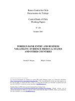
signal-to-noise ratio. When nuclei are under an external magnetic field, they
precess along the axis of the field, conventionally the z-axis. Figure 1-4 is a
pictorial representation of the two energy levels of a nucleus with I ¼1/2 (e.g.,
1
H and
13
C) under a magnetic field based on its orientation. The nucleus is
considered a micromagnet that under a magnetic field can rotate clockwise or
counterclockwise. Those two rotations represent the two energy levels of the
half-integer nuclear spin. For nuclei with I ¼1/2 and a negative gyromagnetic
ratio, such as
15
N and
29
Si (see Table 1-1), the energy levels are reversed, with
b for the lower spin state (I ¼À1/2) and a for the higher spin state (I ¼1/2).
Having a negative gyromagnetic ratio also decreases the population ratio
between the two nuclear spin levels, which contributes to making the nuclei
less sensitive.
This book focuses primarily on nuclei with a nuclear spin of half-integer
I ¼1/2 (e.g.,
1
H,
13
C,
15
N,
19
F,
31
P), the major nuclei present in organic
B
0
= 0
B
0
≠ 0
I = -1/2
I = 1/2
B
0
Precession along the z axis
π
γ
ν
2
B
0
h
hE ==Δ
S
S
N
N
FIGURE 1-4. Classical representation of the energy levels and precession of the two spinstatesfornuclei
with half-integer nuclear spin (I ¼1/2; e.g.,
1
H,
13
C,
31
P,
19
F). For nuclei with I ¼1/2 and negative
gyromagnetic ratio such as
15
N and
29
Si, b is the lower spin state (I ¼À1/2) and a is the higher spin state
(I ¼1/2).
6 BASIC CONCEPTS OF NMR SPECTROSCOPY
compounds. Under a magnetic field, a nucleus with a half-integer nuclear spin
will rotate or precess along the axis of the applied magnetic field (normally the
z-axis) at the Lamour frequency n ¼gB
0
, where n is the Lamour frequency, g is
the gyromagnetic ratio intrinsic per nucleus, and B
0
is the external magnetic
field (Figure 1-4). Transitions occur when a second RF pulse perpendicular to
the external magnetic field is applied to the sample within a short time frame.
Those transitions are detected by a detector in the xy-plane and converted to an
appropriate format for interpretation.
As in any other type of spectroscopy, after radiation to reach a high energy
level over a period of time, spin nuclei will return to the ground state through
different mechanisms. The NMR signal is termed free induction decay (FID)
because it is free of the influence of the particular RF field, it is induced in the
coil, and it decays back to equilibrium. Magnetization is the sum of all the
magnetic moments in the sample for a particular nucleus (e.g.,
1
H). In NMR,
there are two mechanisms that bring the magnetization to the ground state:
spin–lattice or longitudinal (along the z-axis) relaxation (T
1
) and spin–spin
or transverse (perpendicular to the z-axis and in the xy-plane) relaxation (T
2
). T
1
is the time to transfer energy from the magnetization going from the excited to
the ground state in the z-axis, and T
2
is the time to transfer energy within the
precessing nucleus (e.g.,
1
H) in the xy-plane to the ground state through
dephasing, line broadening, and consequently, loss of signal. More details
on those relaxation mechanisms may be found in the literature [1–12,15–17]. T
1
and T
2
values are influenced by the size of the molecule based on molecular
weight and affect the quality of the NMR signal. T
1
and T
2
are in the same range
(T
1
%T
2
) for small molecules (e.g., methanol) that tumble fast in solution. T
1
is
greater than T
2
(T
1
> T
2
) for large molecules (e.g., proteins, polymers) that
tumble more slowly in solution. T
1
is much greater than T
2
(T
1
)T
2
) for solid or
crystalline samples in the solid state, which makes the NMR lines much broader
than they are in liquids, affecting the resolution due to short T
2
’s [1–12,15–17].
1.3. BASIC PARAMETERS FOR NMR INTERPRETATION
The NMR spectra of half-integer nuclei (I ¼1/2; e.g.,
1
H,
13
C,
15
N,
31
P, and
19
F) are the focus of this book and provide structural information on how the
atoms are connected to each other through bonds and relative orientations in
space. The parameters containing structural information are chemical shifts,
spin–spin coupling constants, spin systems, signal intensities, and bond and
spatial correlations: the most commonly used parameters for the structural
elucidation of organic compounds. These parameters are described
below with examples illustrating their significance in the world of structural
elucidation in organic chemistry.
BASIC PARAMETERS FOR NMR INTERPRETATION 7
1.3.1. Chemical Shift
Since the discovery of NMR, technology has played an important role in its
development. From the early electromagnets and permanent magnets to the
current superconducting magnets, the magnetic field of NMR instruments
has increased from the first 40-MHz instruments to the 900-MHz instruments
available at present. At the time of this writing, 1-GHz NMR instruments are
almost ready to become available commercially. The most common NMR
instruments used in the field of small molecules are between 300 and
600 MHz. Occasionally, 800-MHz NMR instruments are used when sample
quantity is limited. By using greater magnetic field instruments with
cryogenic probes, increased sensitivity can be achieved. Data from a sample
acquired using instruments that have different magnetic fields are difficult to
compare on the scale of frequency units because the energy absorption is
different with different magnetic fields. Over the years it became obvious
that uniformity of scale was needed for NMR spectra, regardless of the
strength of the magnetic field of the instrument used to acquire the data.
Historically, tetramethylsilane (TMS) was selected as a reference compound
because it is chemically inert, volatile [boiling point (bp) 27
C], symmet-
rical, and soluble in most organic solvents. There were a variety of
approaches, but over the years, chemical shift in parts per million (ppm)
has been shown the success of the best uniformity on the NMR scale. The
chemical shift (d) is defined as
d ¼
n
sample
À n
standard ðTMSÞ
n
spectrometer
 10
6
where n
sample
is the resonance frequency of the protons of the sample,
n
standard(TMS)
is the resonance frequency of TMS as the standard, and
n
spectrometer
is the frequency of the spectrometer where the data are acquired
(e.g., 600-MHz instrument). For TMS, the chemical shift of the methyl
protons is assigned conventionally to 0.00 ppm, based on the definition of
chemical shifts. Figures 1-5 and 1-6 depict the spectra of a 3-vinylphenol
compound simulated at frequencies of 60 to 600 MHz in the frequency
domain and on the chemical shift scale, respectively. Obviously, compar-
ison of the data for any molecule in the frequency domain becomes
difficult, but it is straightforward on the chemical shift scale, which has
become the standard scale for NMR spectroscopy. Another observation
from Figures 1-5 and 1-6 is that increasing sensitivity and resolution are
achieved by increasing the magnetic field. At higher magnetic fields,
analysis of the NMR data becomes less cumbersome than analysis at
lower fields. For 3-vinylphenol, all the protons have a distinct resonance
8 BASIC CONCEPTS OF NMR SPECTROSCOPY
100 MHz
60 MHz
4000 3600 3200 2800 2400
f1
(
Hz
)
2000 1600 1200 800 600 400
1
2
3
4
300 MHz
600 MHz
OH
FIGURE 1-5. Simulated
1
H NMR spectra of 3-vinylphenol at magnetic fields of 60, 100, 300, and
600 MHz, respectively on the frequency scale (in hertz) from bottom to top.
5.05.15.25.35.45.55.65.75.85.96.06.16.26.36.46.56.66.76.86.97.07.17.27.37.4
f1 (ppm)
1
2
3
4
60 MHz
100 MHz
300 MHz
600 MHz
OH
FIGURE 1-6. Simulated
1
H NMR spectra of 3-vinylphenol at magnetic fields of 60, 100, 300, and
600 MHz on the chemical shift scale (ppm) from bottom to top, respectively.
BASIC PARAMETERS FOR NMR INTERPRETATION 9
frequency. However, in the case of a molecule with equivalent protons, such
as the protons of a methyl group, the equivalent protons will resonate at the
same frequency.
Table 1-2 shows the typical ranges of
1
H and
13
C chemical shifts for
common functional groups in organic compounds. As shown in the table,
carbons resonate at a greater range of frequencies than that of protons and
spread their signals over a wider scale (ca. 0 to 13 ppm for
1
H and 0 to 220 ppm
for
13
C), which decreases signal overlapping and facilitates assignments. For
any nucleus, the location of the chemical shift signals depends on the local
influence of the electronic layer surrounding the functional groups. When an
external magnetic field is applied, the electrons surrounding a nucleus (e.g.,
1
H,
13
C) will circulate perpendicular to the field, creating an induced magnetic
field opposed to the applied external magnetic field in the area of the nucleus.
Therefore, the nucleus is under a weaker magnetic field than the externally
applied field and is said to be shielded. This is the principle of the diamagnetic
TABLE 1-2. Typical Ranges of
1
H and
13
C Chemical Shifts for Common Functional Groups
in Organic Compounds
Functional Group
1
H(d/ppm)
13
C(d/ppm)
TMS (tetramethylsilane) 0.0 0.0
Primary alkyl (RCH
3
) 0.7–1.3 0–30
Secondary alkyl (R
2
CH
2
) 1.2–1.5 15–45
Tertiary alkyl (R
3
CH) 1.4–1.7 30–50
Quaternary alkyl (R
4
C) 35–70
Allylic (ÀC¼CÀCH) 1.6–2.3
Vinylic/alkene (R
2
C¼CH) 4.5–6.5 80–145
Acetylenic/alkyne (RÀCXCH) 2.5–3.0 70–90
Benzylic methyl (ArCH
3
) 2.2–2.8 20–65
Aromatic (CH of Ar) 6.0–9.0 110–170
Alkyl fluoride (FÀCH) 4.1–4.7 70–80
Alkyl chloride (FÀCH) 3.6–3.8 25–50
Alkyl bromide (BrÀCH) 3.4–3.6 10–40
Alkyl iodide (IÀCH) 3.1–3.3 0–40
Alcohol/ether (OÀCH) 3.1–4.0 50–90
Aldehyde (O¼CÀH) 9.0–10.0 180–210
Carboxylic acid (O¼CÀOH) 10.0–13.0 170–180
Carbonyl ketone (R
2
C¼O) 190–220
Carbonyl ester/amide (RÀCOOR, RÀCONR
2
) 150–180
Alcohols (RÀOH) 2.5–5.0 (variable)
Phenols (ArÀOH) 5.0–10.0 (variable)
Amines (RÀNH
2
,R
2
ÀNH) 0.5–4.0 (variable)
Amines (ArÀNH
2
,Ar
2
ÀNH) 6.0–9.0 (variable)
Ammoniums (RÀNH
3
þ
,R
2
ÀNH
2
þ
,R
3
ÀNH
þ
) 2.5–5.0 (variable)
Ammoniums (ArÀNH
3
þ
,Ar
2
ÀNH
2
þ
,Ar
3
ÀNH
þ
) 6.0–9.0 (variable)
Source: Data from references 18 and 24.
10 BASIC CONCEPTS OF NMR SPECTROSCOPY