Lab Exercises in Organismal and Molecular Microbiology_2 doc
Bạn đang xem bản rút gọn của tài liệu. Xem và tải ngay bản đầy đủ của tài liệu tại đây (28.95 MB, 160 trang )
Alexander−Strete−Niles:
Lab Exercises in
Organismal and Molecular
Microbiology
VI. Controlling the Risk and
Spread of Bacterial
Infections
27. Killing Bacteria with
High Temperature
© The McGraw−Hill
Companies, 2003
200
Killing Bacteria with
High Temperature
EXERCISE
27
tive cells of E. coli, if present. Likewise, species of Sal-
monella, such as S. enteritidis and S. typhimurium,are
associated with eating undercooked chicken and eggs,
causing salmonellosis. The thorough grilling or baking of
chicken and eggs to a temperature of 80°C or above
should kill all vegetative cells of Salmonella, if present.
Using Wet Heat in the Kitchen
Boiling water has been used for a long time around the
home in cooking and disinfecting items, such as baby
bottles and canning jars. Drinking water may also
require boiling on occasion. For example, whenever
water flow is interrupted in water lines by a rupture or
drop in pressure, there is a chance of bacterial con-
taminants entering the water supply. In these cases, city
officials may advise people to boil their water prior to
use. This eliminates the risk of contracting a water-
borne infection until normal service is restored.
In summary, when properly used, heat is an effec-
tive household tool to eliminate the risk of bacterial
infection. This exercise will demonstrate the killing
power of wet heat.
Table 27.1 Types of Heat Used to Kill Bacteria
Type of heat Examples Effect on cells Uses
Dry
Incineration Oxidizes cell components Used to sterilize laboratory loops and
needles; used to destroy waste and
infectious materials
Hot-air oven Oxidizes cell components Used to sterilize laboratory glassware;
used in home cooking
Wet Boiling water Coagulates cell proteins Used in home disinfection and cooking
Autoclave/pressure Coagulates cell proteins Autoclave used to sterilize laboratory
cooker media; pressure cooker used in
home cooking/canning
Pasteurization Coagulates cell proteins Used to disinfect liquids (e.g., milk) to
increase shelf life and kill pathogens
Fractional sterilization Coagulates cell proteins Used to sterilize heat-sensitive
instruments and chemicals
Background
Dry and Wet (Moist) Heat
Heat is one of the most effective methods used to kill bac-
teria. Heat is generally divided into dry and wet (moist)
heat (table 27.1). Dry heat, which includes incineration
and the hot-air oven, kills bacteria by oxidizing compo-
nents of the cell. Wet (moist) heat, which includes boiling
water, autoclave/pressure cooker, pasteurization, and frac-
tional sterilization, kills bacteria by coagulating proteins
in the cell, including essential enzymes and cell structures.
Using Dry Heat in the Kitchen
Dry heat is used for grilling on the stovetop or baking
in the oven. When properly used, dry heat in the kitchen
can effectively eliminate the risk of contracting certain
types of bacterial diseases.
Pathogenic strains of Escherichia coli, such as the
0157:H7 strain, cause diarrhea, and can be contracted by
eating undercooked hamburger. Cooking hamburger meat
to a temperature of 80°C or above should kill all vegeta-
Alexander−Strete−Niles:
Lab Exercises in
Organismal and Molecular
Microbiology
VI. Controlling the Risk and
Spread of Bacterial
Infections
27. Killing Bacteria with
High Temperature
© The McGraw−Hill
Companies, 2003
Killing Bacteria with High Temperature E
XERCISE
27 201
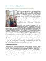
Figure 27.1 Experimental setup for heating broth tubes
inoculated with Escherichia coli.
burner to a position beneath the tripod to heat the
water. Examine figure 27.1 to see this experimental
setup without the 16 inoculated tubes.
5. During heating, remove one tube at every 5°C
interval, beginning at 25°C. Label each tube with
the temperature at which it was removed, and
place it in the test tube rack with the control tube.
When the water reaches 100°C, remove the last
tube, and turn off the Bunsen burner.
6. Place the test tube rack with the 17 tubes in a
35°C incubator.
Caution: Use care when
disposing of the hot water!
Caution: Do not pipette by
mouth.
Materials
Culture (24-hour in tryptic soy broth)
Escherichia coli
Media
Tryptic soy broth tubes (18): 16!150 mm
tubes containing 5 ml broth per tube, capped
Equipment
Incubator (35°C)
Miscellaneous supplies
Beaker (1 liter)
Bunsen burner and striker
Pipette (1 ml, sterile); pipette bulb
Test tube rack
Thermometer (°C)
Tripod with ceramic-lined wire mesh
Wax pencil
Procedure
First Session: Inoculation
and Heating of Broth Tubes
1. Place a pipette bulb onto a 1 ml sterile pipette and
fill the pipette with the broth culture of E. coli.
This should be sufficient culture to inoculate 17
of the 18 broth tubes.
2. Aseptically transfer 1 drop of culture to each of
17 broth tubes. Note: Insert the pipette into the
tube close to the surface of the liquid, and aim
the drop directly into the liquid. A drop deposited
on the side of the glass may not reach the broth,
resulting in a false negative.
3. Thoroughly mix the drop into the broth. Place one
of the inoculated tubes in a test tube rack. Label this
tube the control. Place the remaining 16 inoculated
tubes in the 1 liter beaker, and fill the beaker with
tap water to a level above the broth. Now carefully
insert the thermometer in the uninoculated broth
tube, and place the tube in the water.
4. Place the beaker on the wire mesh platform
mounted on the tripod. Move a lighted Bunsen
Alexander−Strete−Niles:
Lab Exercises in
Organismal and Molecular
Microbiology
VI. Controlling the Risk and
Spread of Bacterial
Infections
27. Killing Bacteria with
High Temperature
© The McGraw−Hill
Companies, 2003
202 S
ECTION
VI Controlling the Risk and Spread of Bacterial Infections
Second Session: Examination
of Broth Tubes
1. After 48 hours, examine each tube for growth. If
viable cells remained after heating, they will have
multiplied into millions of cells, turning the broth
cloudy or turbid. In this case, you will not be able
to see through the liquid. Score these tubes as (;)
for growth, indicating that the temperature wasn’t
sufficient to kill all vegetative cells. If all
vegetative cells were killed after heating, none
will have been left to multiply, leaving the broth
clear. In this case, you will be able to see through
the liquid. Score these tubes as (:) for growth,
indicating that the temperature was sufficient to
kill all vegetative cells. Record your score for
each tube in the laboratory report.
2. Continue scoring tubes as (;) or (:) using the
criteria in step 1 until all tubes have been scored.
Evaluate the results of your experiment as related
to the use of heat in your home.
Alexander−Strete−Niles:
Lab Exercises in
Organismal and Molecular
Microbiology
VI. Controlling the Risk and
Spread of Bacterial
Infections
27. Killing Bacteria with
High Temperature
© The McGraw−Hill
Companies, 2003
EXERCISE
27
L
ABORATORY
R
EPORT
N
AME
D
ATE
L
AB
S
ECTION
203
Killing Bacteria with High Temperature
1. In the following table, record your scores for each tube; use a (;) for tubes with cloudy, turbid growth;
use a (:) for tubes with clear broth.
Broth turbid (T) Heat killed all
Temperature (°C) or clear (C)? Growth (;) or (:)? vegetative cells?
25
30
35
40
45
50
55
60
65
70
75
80
85
90
95
100
2. According to your results in this experiment, what is the minimum temperature required to kill all vege-
tative cells of E. coli? What application might this have for cooking your hamburger meat at home?
3. If you received a notice from city officials to boil your water before use, would boiling kill E. coli and
other vegetative bacterial cells if they were present? Explain.
Alexander−Strete−Niles:
Lab Exercises in
Organismal and Molecular
Microbiology
VI. Controlling the Risk and
Spread of Bacterial
Infections
28. Skin Disinfection:
Evaluating Antiseptics and
Hand Sanitizers
© The McGraw−Hill
Companies, 2003
205
Skin Disinfection: Evaluating Antiseptics
and Hand Sanitizers
EXERCISE
28
method, outlined in figure 28.1. In this method, filter
paper disks are dipped into an antiseptic and then placed
on an agar plate that has been inoculated with a bacte-
rial culture. The plate is then incubated to allow bac-
terial growth. After growth, plates are examined for
zones of inhibition around the chemical-soaked disks,
indicating chemical effectiveness. In this exercise, you
will use the filter paper method to examine the effec-
tiveness of antiseptics commonly applied to the skin.
Evaluating Hand Sanitizers
Bacteria are numerous on the hands, and represent both
members of the normal flora and transients picked up
from the environment. While the normal flora is typi-
cally not harmful, transients can be disease-causing
agents. One of the simplest and most effective ways
to eliminate these transient disease-causing agents is to
wash your hands. Hungarian physician Ignaz Semmel-
weis advocated hand washing as a means of preventing
disease transmission in the mid-1800s. This simple task
is still recommended today by health-care specialists as
one of the most effective means of preventing infection.
Table 28.1 Commonly Used Antiseptics
Chemical agent Effect on cells Commercial uses
Alcohol (ethyl or isopropyl) Dehydrates the cell; alters cell Skin cleansing and degerming
membrane; denatures cell proteins agent; skin antiseptic
Benzalkonium chloride Alters cell membrane Skin antiseptics
Cetylpyridinium chloride Alters cell membrane Mouthwashes
Hexachlorophene Alters cell membrane; denatures Soaps and skin antiseptics
cell proteins
Hydrogen peroxide Oxidizes cell components Skin antiseptic
Mercurochrome or Denatures cell proteins Skin antiseptic
Merthiolate
Tincture of iodine Denatures cell proteins Skin antiseptic
Triclosan Alters cell membrane; denatures Antibacterial soaps
cell proteins
Background
A variety of chemical agents display antimicrobial activ-
ity against bacteria. One category of antimicrobial chem-
ical agents, the antibiotics, was examined in Exercise 25.
Two other categories of chemical agents commonly used
in the household are antiseptics and disinfectants. Anti-
septics are chemicals safe enough to be applied to the
skin; they are used to prevent wound infections and to dis-
infect skin. Some commonly used antiseptics and their
effects on bacterial cells are presented in table 28.1.
The effectiveness of these skin-applied chemical
agents will be examined in this exercise. Disinfectants
are chemicals considered too harsh to be applied to the
skin, and are only used on inanimate surfaces. Disin-
fectants will be evaluated in Exercise 29.
Evaluating Antiseptics: The Filter
Paper Method
Antiseptics are commonly used on the skin to prevent
wound infections. One of the ways to determine the
effectiveness of antiseptics is to use the filter paper
Alexander−Strete−Niles:
Lab Exercises in
Organismal and Molecular
Microbiology
VI. Controlling the Risk and
Spread of Bacterial
Infections
28. Skin Disinfection:
Evaluating Antiseptics and
Hand Sanitizers
© The McGraw−Hill
Companies, 2003
206 S
ECTION
VI Controlling the Risk and Spread of Bacterial Infections
(a) Obtain a sterile disk using sterile forceps, and dip the
disk halfway into antiseptic to allow the disk to soak up
the chemical.
(b) Place the chemical-soaked disk on an inoculated plate.
Repeat for three other antiseptics.
Zones of inhibition
(c) After incubation, examine plates for zones of
inhibition, indicative of antiseptic effectiveness.
Figure 28.1 The filter paper method for evaluating antiseptics.
Pseudomonas aeruginosa
Staphylococcus aureus
All agents in red are BSL2 bacteria.
Media
Tryptic soy agar (TSA) plates
Tryptic soy broth tubes
Chemicals and reagents
Antiseptics
Alcohol, ethyl or isopropyl
Benzalkonium chloride (found in
skin antiseptics)
Today, using a hand sanitizer is a popular way to
clean the hands. These products are popular because they
can be used to disinfect the hands while away from home
or when soap, water, or towels are not available. These gel
products are dispensed from plastic bottles onto the hands.
The hands are then rubbed together until dry. The active
ingredient in these products is 62% ethyl alcohol.
This exercise will also evaluate the effectiveness
of hand sanitizers in removing bacteria from the hands.
Materials
Cultures (24-hour in tryptic soy broth)
Bacillus cereus
Escherichia coli
Alexander−Strete−Niles:
Lab Exercises in
Organismal and Molecular
Microbiology
VI. Controlling the Risk and
Spread of Bacterial
Infections
28. Skin Disinfection:
Evaluating Antiseptics and
Hand Sanitizers
© The McGraw−Hill
Companies, 2003
Skin Disinfection: Evaluating Antiseptics and Hand Sanitizers E
XERCISE
28 207
Cetylpyridinium chloride (found
in mouthwashes)
Hexachlorophene (found in soaps
and skin antiseptics)
Hydrogen peroxide
Mercurochrome or Merthiolate
Tincture of iodine
Triclosan (found in antibacterial hand soaps)
Ethanol, 70%
Hand sanitizer (active ingredient,
62% ethyl alcohol)
Equipment
Incubator (35°C)
Miscellaneous supplies
Beaker, 250 ml
Bunsen burner and striker
Cotton-tipped swabs, sterile
Filter paper disks, sterile, in a petri dish
Forceps
Wax pencil
Procedure
First Session
Evaluating Antiseptics: The Filter
Paper Method
1. Dip a cotton-tipped swab into one of the four
cultures, and use it to inoculate a tryptic soy agar
plate using the procedure outlined in Exercise 25
(see figure 25.2). Note: A lawn of bacterial
growth is necessary for this method, as it was for
antibiotic testing in Exercise 25. Repeat this
inoculation procedure for a second plate using the
same culture. Label each plate with a wax pencil.
2. Repeat step 1 for the remaining three cultures.
You should now have a total of eight plates, two
for each culture. After inoculation, allow all
plates to dry for 15 minutes before proceeding to
the next step.
3. Pour some 70% ethanol into a 250 ml beaker.
b. Now pick up a sterile disk with the forceps,
and insert it halfway into a drop of the
antiseptic poured into a beaker or a petri dish.
Let the disk soak up the chemical; when
thoroughly soaked, lift the disk and place it on
an inoculated plate.
c. After placement, tap the disk lightly to make
sure it is secure.
Repeat steps a–c until you have placed
this antiseptic on a plate for each culture.
Proceed to the next antiseptic until you have
placed four disks on a plate for each culture.
Place the remaining four antiseptics on the
second plate, for a total of eight antiseptics
per culture. Note: Place the disks as far apart
as possible, and mark the antiseptic on the
bottom of the plate.
4. When all disks are in place, put your plates into
a 35ÚC incubator.
Evaluating Hand Sanitizers
1. Dip a cotton-tipped swab into a tube of tryptic
soy broth to wet the cotton. Rub lightly on the
inside of the tube to remove excess liquid.
2. Swab the left hand as follows: Begin at the top of
the first finger (nearest the thumb) and swab
down to the base of the thumb; roll the swab, and
come back up to the fingertip; repeat this two
more times to cover this area of the finger and
palm (figure 28.2). Use this swab to inoculate a
tryptic soy agar plate. Swab the entire surface of
the plate, turn 90Ú, and swab the entire surface
again. Be sure to rotate the swab as you go to
deposit all the bacteria lifted from the hand.
Label this plate “Before, Replicate 1.”
3. Repeat step 2 for the third finger of the left hand,
swabbing the finger and palm as before with a
fresh swab, and then transferring the bacteria
lifted to a second tryptic soy agar plate. Label
this plate “Before, Replicate 2.”
4. Take the hand sanitizer, and place a thumbnail-
sized amount in the palm of the left hand. Rub
the palms of both hands together, covering all
inside surfaces of the hands with sanitizer.
Continue rubbing until the gel has disappeared
and the hands are dry.
Caution: Keep the alcohol away
from the flame!
a. Dip your forceps into the alcohol, and pass them
over a Bunsen burner flame to sterilize them.
Alexander−Strete−Niles:
Lab Exercises in
Organismal and Molecular
Microbiology
VI. Controlling the Risk and
Spread of Bacterial
Infections
28. Skin Disinfection:
Evaluating Antiseptics and
Hand Sanitizers
© The McGraw−Hill
Companies, 2003
208 S
ECTION
VI Controlling the Risk and Spread of Bacterial Infections
5. After sanitizer treatment, take a fresh swab, and
wet it in broth as before. Swab the second finger,
starting at the tip and moving downward to the
base of the palm. Rotate the swab, and move
upward to the fingertip. Repeat this down-and-up
process two more times as before (figure 28.2).
Inoculate a third tryptic soy agar plate as before.
Label this plate “After, Replicate 1.”
6. Using a fresh swab, repeat the swabbing
procedure in step 5 for the fourth finger
(smallest). Inoculate a fourth tryptic soy agar
plate as before, and label it “After, Replicate 2.”
7. Place these four plates in a 35°C incubator with
the antiseptic plates.
Second Session
Examining Antiseptic Plates
1. After 48–72 hours, examine the culture plates
containing antiseptic disks. Examine the growth
around the disks.
2. For each disk, look for a zone of inhibition. As
for antibiotics, these areas indicate the
effectiveness of a chemical agent in preventing
growth. However, in this case, the diameter of the
zone may not equate to a degree of effectiveness,
since chemicals vary in their volatility and
diffusion through the agar. Therefore, record only
a (;) for a zone of inhibition around a disk
indicating susceptibility. Record a (:) for no zone
of inhibition, indicating resistance.
3. Complete your observation of all disks for the
four cultures, recording a (;) or (:) in the
laboratory report.
Examining Hand Sanitizer Plates
1. After 48–72 hours, examine the plates inoculated
with the swabs of your left hand. Separate these
into “before” and “after” plates.
2. Count the total number of colonies on the two
replicate “before” plates and the total number of
colonies on the two replicate “after” plates. Record
these numbers in your laboratory report. Calculate
a “before” average and an “after” average.
3. Record the percentage of bacteria killed by the
hand sanitizer.
3
x
(e)
After,
Replicate 2
(c)
(d)
After,
Replicate 1
(b)
Washing with hand sanitizer
Before,
Replicate 2
Before,
Replicate 1
(a)
3
x
3
x
3
x
Figure 28.2 Testing the effectiveness of hand sanitizers.
Alexander−Strete−Niles:
Lab Exercises in
Organismal and Molecular
Microbiology
VI. Controlling the Risk and
Spread of Bacterial
Infections
28. Skin Disinfection:
Evaluating Antiseptics and
Hand Sanitizers
© The McGraw−Hill
Companies, 2003
EXERCISE
28
L
ABORATORY
R
EPORT
N
AME
D
ATE
L
AB
S
ECTION
Skin Disinfection: Evaluating Antiseptics and Hand Sanitizers
Antiseptics
1. In the following table, record your results for antiseptic plates. Record a (;) for the presence of a zone of
inhibition around the disk. Record a (:) for no zone of inhibition.
209
Culture
Antiseptic Bacillus Escherichia Pseudomonas Staphylococcus
cereus coli aeruginosa aureus
Benzalkonium chloride
Cetylpyridinium chloride
Ethanol (70%)
Hexachlorophene
Hydrogen peroxide
Isopropyl alcohol
Mercurochrome or Merthiolate
Tincture of iodine
Triclosan
2. Which antiseptic(s), if any, had the widest spectrum of activity? How would this trait make this a useful
antiseptic? Explain.
Alexander−Strete−Niles:
Lab Exercises in
Organismal and Molecular
Microbiology
VI. Controlling the Risk and
Spread of Bacterial
Infections
28. Skin Disinfection:
Evaluating Antiseptics and
Hand Sanitizers
© The McGraw−Hill
Companies, 2003
210 S
ECTION
VI Controlling the Risk and Spread of Bacterial Infections
2. Calculate the average percent reduction of bacteria on the hand: %
3. Did the hand sanitizer remove the large majority of bacteria from your hand? Based on these results,
would you buy this product for use when away from home? When would it be useful?
Hand Sanitizer
1. In the following table, record your results for the hand sanitizer. Record the total number of colonies on
the two “before” plates and the total number of colonies on the two “after” plates.
Total number of colonies
Replicate Before hand sanitizer After hand sanitizer
1
2
Average
Alexander−Strete−Niles:
Lab Exercises in
Organismal and Molecular
Microbiology
VI. Controlling the Risk and
Spread of Bacterial
Infections
29. Cleaning Countertops
with Disinfectants
© The McGraw−Hill
Companies, 2003
211
Cleaning Countertops with Disinfectants
EXERCISE
29
Materials
Media
Tryptic soy agar plates
Tryptic soy broth tubes
Chemicals and reagents
Disinfectants, commercially available (those
listed in table 29.1 or others that contain the
same chemicals)
Equipment
Incubator (35°C)
Miscellaneous supplies
Adhesive tape
Bottles, spray-dispenser type
Cotton-tipped swabs, sterile
Paper towels
Ruler, metric
Wax pencil
Background
Antimicrobial chemical agents are important in the con-
trol of microorganisms. Exercise 25 examined the effec-
tiveness of antibiotics, while Exercise 28 evaluated the
effectiveness of antiseptics. A third category of chem-
ical agents, disinfectants, are considered too harsh for
use on or in the human body; however, they are useful
on inanimate surfaces. Some of the chemical agents
commonly used in disinfectants are listed in table 29.1.
Disinfectants are widely used around the house to
remove bacteria from surfaces. Surfaces that require
disinfection at home include the kitchen sink and coun-
tertops, bathroom sink and countertops, toilet, shower,
and bathtub. Similar surfaces that require periodic dis-
infection are also found in public facilities and at work.
Keeping these surfaces clean and low in bacterial num-
bers is one of the most effective means of controlling
the occurrence and spread of infectious agents.
In this exercise, you will evaluate the effectiveness
of several commercially available disinfectants con-
taining the chemical compounds listed in table 29.1.
Table 29.1 Chemical Agents Commonly Used in Disinfectants
Chemical agent Effect on cells Commercial uses
Sodium hypochlorite Oxidizes cell components Surface disinfectants and bleach
Orthophenylphenol Denatures cell proteins Surface disinfectants, such as
Lysol
Alkyldimethylbenzyl Alters cell membrane Surface disinfectants, such as
ammonium chloride Formula 409
Pine oil Alters cell membrane; Surface disinfectants, such as
denatures cell proteins Pine-Sol
Alexander−Strete−Niles:
Lab Exercises in
Organismal and Molecular
Microbiology
VI. Controlling the Risk and
Spread of Bacterial
Infections
29. Cleaning Countertops
with Disinfectants
© The McGraw−Hill
Companies, 2003
212 S
ECTION
VI Controlling the Risk and Spread of Bacterial Infections
Countertop area (3,600 cm
2
)
B: After cleaning with disinfectant, swab each B area with
another swab. Inoculate a second tryptic soy agar plate.
A: Before cleaning, swab each A area with a wet, cotton-tipped
swab. Inoculate a tryptic soy agar plate.
60 cm
60 cm
BA
10 cm
10 cm
Disinfectant 4
BA
10 cm
10 cm
Disinfectant 3
BA
10 cm
10 cm
Disinfectant 2
BA
10 cm
10 cm
Disinfectant 1
Figure 29.1 Procedure for testing the effectiveness
of disinfectants.
4. Dip a sterile, cotton-tipped swab into tryptic soy
broth. Use it to swab the 100 cm
2
area denoted as
A, Disinfectant 1. Swab the entire 100 cm
2
area
twice, the second time at a 90° angle to the first.
Use the swab to inoculate a tryptic soy agar plate.
Rub the swab over the entire surface of the plate,
rolling the swab as you do so. Rotate the plate
90° and swab again. Label this plate “A,
Disinfectant 1.”
5. Take disinfectant 1, and clean the entire
disinfectant 1 test area. Do not spray into any
of the other areas. Prepare the disinfectant per
the directions on the container, mixing the
disinfectant with water in a spray-type dispenser.
In this way, the disinfectant can be thoroughly
sprayed over the entire surface before wiping
with a paper towel. Be sure to wipe the surface
dry. Do not wipe into any of the other areas.
6. Dip a fresh cotton-tipped swab in sterile broth,
and use it to swab the 100 cm
2
area denoted as B,
Disinfectant 1. Again, be sure to swab the entire
100 cm
2
area twice. Use this swab to inoculate a
second tryptic soy agar plate as before. Label this
plate “B, Disinfectant 1.”
7. Repeat steps 4–6 to complete the sampling of
each A and B area for disinfectants 2, 3, and 4.
When finished, you should have inoculated a total
of eight tryptic soy agar plates.
8. After completing your sampling of the first
surface, repeat steps 2–7 for the second surface.
You should have inoculated another eight tryptic
soy agar plates for this surface, giving you a total
of 16 plates for the two surfaces.
9. Place all plates into a 35°C incubator.
Second Session: Examination of Plates
1. After 48–72 hours, examine your plates. Sort
the plates by surface cleaned, disinfectant used,
and before cleaning (A) and after cleaning
(B). Count the total number of bacterial
colonies on each plate, and fill in your results
in the laboratory report.
2. Calculate the percent decrease in the bacteria on
each cleaned surface for each disinfectant.
Procedure
First Session: Inoculation of Plates
1. Select two surfaces to be cleaned. A laboratory
countertop and a bathroom or kitchen
countertop are recommended. If a bathroom
or kitchen is unavailable, select a second
laboratory countertop.
2. Mark off a 3,600 cm
2
area of the first surface to
be cleaned. Use four 60 cm pieces of adhesive
tape to mark the edges of this area. Also place a
piece of adhesive tape in the center of this area.
The center piece of tape will help delineate four
areas within the 3,600 cm
2
area: an upper left
area, upper right area, lower left area, and lower
right area. Designate these four areas as test areas
for disinfectants 1, 2, 3, and 4, respectively
(figure 29.1).
3. In each test area, use pieces of adhesive tape
10 cm long to mark the edges of two adjacent
100 cm
2
areas, one designated A, before cleaning
with disinfectant, and the other designated B,
after cleaning with disinfectant (figure 29.1).
Alexander−Strete−Niles:
Lab Exercises in
Organismal and Molecular
Microbiology
VI. Controlling the Risk and
Spread of Bacterial
Infections
29. Cleaning Countertops
with Disinfectants
© The McGraw−Hill
Companies, 2003
EXERCISE
29
L
ABORATORY
R
EPORT
N
AME
D
ATE
L
AB
S
ECTION
213
Cleaning Countertops with Disinfectants
1. Record the number of colonies on your plates.
a. Laboratory countertop (first surface)
Disinfectant Before (A) After (B) Percent Decrease
1=
2=
3=
4=
b. Second surface
Disinfectant Before (A) After (B) Percent Decrease
1=
2=
3=
4=
2. Explain the difference between disinfection and sterilization. Which of these terms applies to the action
of the chemicals used in this exercise?
3. Do these chemical agents work effectively to remove bacteria from surfaces? Were there any that seemed
to work best?
4. Based on your results, do you think the use of these chemicals around the home is justified? If so, when
and where would you use these products?
Alexander−Strete−Niles:
Lab Exercises in
Organismal and Molecular
Microbiology
VI. Controlling the Risk and
Spread of Bacterial
Infections
30. Bacteriological
Examination of Drinking
Water Using the MPN
Method
© The McGraw−Hill
Companies, 2003
215
Bacteriological Examination of Drinking Water
Using the MPN Method
EXERCISE
30
Completed test:
Select typical coliform
colonies; inoculate lactose
broth and agar slant;
incubate 24 hours.
Acid and gas not produced:
Negative completed test—
original isolate not
coliform; water potable
Coliform group present:
Positive completed test—
water nonpotable
Confirmed test:
Streak from lactose broth
onto eosin methylene blue
(EMB) plates; incubate
24 hours.
Presumptive test:
Inoculate lactose broth;
incubate 24–48 hours.
Colonies not coliform:
Negative confirmed test—
water potable
Acid and gas not produced:
Negative presumptive
test— water potable
Typical coliform colonies:
dark centers, metallic sheen
Positive confirmed test
Acid and gas produced:
Positive presumptive test
Gram-negative
rods present;
no spores
present
Acid and
gas
produced
Agar
slant
Lactose
broth
Figure 30.1 The MPN method used to detect coliforms
in drinking water.
Background
Coliforms, Indicators
of Fecal Contamination
Water is routinely tested to ensure that it is safe for drink-
ing. A widely used indicator of the suitability of drink-
ing water is coliform bacteria. Coliforms are
Gram-negative, non-endospore-forming rods that are fac-
ultatively anaerobic and produce acid and gas from lac-
tose within 48 hours at 35°C. The key indicator organism
in this group is Escherichia coli, which is not normally
present in soil and water, but present in large numbers
in the intestines and feces, and capable of long-term sur-
vival in the environment. Therefore, the presence of
E. coli is indicative of human or animal fecal waste. Water
contaminated with fecal material, as determined by the
presence of coliforms, is considered nonpotable, mean-
ing unsuitable for drinking. Water that is coliform-free
is considered potable and safe for drinking.
Human fecal waste may also carry intestinal
pathogens, such as Salmonella typhi, the cause of
typhoid fever; Salmonella typhimurium, the cause of
salmonellosis; Vibrio cholerae, the cause of cholera;
and Shigella sonnei, the cause of shigellosis. Each of
these intestinal pathogens is transmitted by fecal con-
tamination of drinking water. However, their presence
is difficult to detect since they do not typically occur
in large numbers and do not survive long in soil and
water. As a consequence, coliforms, especially E. coli,
are used as the indicator of fecal contamination.
Testing Water for Coliforms
One of the methods used to detect coliforms in drinking
water is the most probable number (MPN) method.
This method, outlined in figure 30.1, consists of three
parts: (1) a presumptive test; (2) a confirmed test; and
(3) a completed test.
In the presumptive test, three series of five tubes
each, or 15 tubes total, are inoculated with a water sam-
ple. Each tube contains 10 ml of lactose broth and a
durham tube. Each tube in the first series of five tubes
Alexander−Strete−Niles:
Lab Exercises in
Organismal and Molecular
Microbiology
VI. Controlling the Risk and
Spread of Bacterial
Infections
30. Bacteriological
Examination of Drinking
Water Using the MPN
Method
© The McGraw−Hill
Companies, 2003
216 S
ECTION
VI Controlling the Risk and Spread of Bacterial Infections
receives 10 ml of sample; each tube in the second series
of five tubes receives 1 ml of sample; and each tube in
the third series of five tubes receives 0.1 ml of sample.
After 24 hours incubation at 35°C, tubes are examined
for the presence of acid and gas, products of lactose fer-
mentation. A positive tube, which has turned yellow and
has a gas bubble in the durham tube, is depicted in
figure 30.2a. Also depicted is a negative tube, which is
unchanged in color and has no gas bubble in the durham
tube (figure 30.2b). After 48 hours of incubation, nega-
tive tubes are examined again for a delayed positive reac-
tion. All tubes after 48 hours are denoted as either (+)
or (–), and a most probable number is assigned accord-
ing to the index shown in table 30.1. If only one tube
scores positive, this is considered a positive presump-
tive test-that is, it presumes that coliforms are present.
However, their presence must be confirmed in the next
part. If all tubes score negative, this is considered a neg-
ative presumptive test. In this case, the water is consid-
ered free of coliforms and, therefore, potable.
In the confirmed test, all positive tubes from the
highest dilution of sample are streaked onto eosin
methylene blue (EMB) agar (table 30.2). This agar
selects for and differentiates coliform bacteria. E. coli
is especially easy to differentiate since it produces a dis-
tinctive green, metallic sheen on this agar. The presence
of colonies on EMB with this characteristic is consid-
ered a positive confirmed test—that is, it confirms the
presence of coliforms. However, their presence must be
further substantiated by the completed test described
next. The absence of colonies on EMB with this char-
acteristic is considered a negative confirmed test, and
the water is considered absent of coliforms and potable.
Red
No gas bubble
in durham tube
Yellow
Gas bubble
in durham tube
(b)(a)
Figure 30.2 Lactose broth. (a) Positive tube.
(b) Negative tube.
In the completed test, colonies from EMB with a
green, metallic sheen are transferred to a lactose broth
tube and a nutrient agar slant. If acid and gas are produced
in the lactose broth tube within 24 hours and a Gram stain
detects a Gram-negative rod, this is considered a posi-
tive completed test, meaning that the confirmation of col-
iforms in the water is complete. The water is considered
contaminated with coliforms and unsafe to drink.
In this exercise, you will use the MPN method to
examine the bacteriological quality of three water sam-
ples: sewage, surface water, and tap water.
Materials
Water samples
Sewage
Sewage may contain pathogens
Surface water (from pond, lake, or stream)
Tap water
Media
Eosin methylene blue (EMB) plates
Lactose broth tubes: each with 10 ml broth
and a durham tube, both double-strength
and single-strength
Nutrient agar slant
Chemicals and reagents
Gram-stain reagents
Equipment
Incubator (35°C)
Light microscope
Miscellaneous supplies
Bunsen burner and striker
Inoculating loop
Immersion oil
Lens paper
Microscope slides
Pipettes, 10 ml and 1 ml, sterile; pipette bulb
Test tube racks
Wax pencil
Procedure
First Session: Inoculation
of Lactose Broth Tubes
1. Take 15 lactose tubes, five double-strength and 10
single-strength, and align into three rows of five in
a test tube rack. Place the five double-strength
Alexander−Strete−Niles:
Lab Exercises in
Organismal and Molecular
Microbiology
VI. Controlling the Risk and
Spread of Bacterial
Infections
30. Bacteriological
Examination of Drinking
Water Using the MPN
Method
© The McGraw−Hill
Companies, 2003
Bacteriological Examination of Drinking Water Using the MPN Method E
XERCISE
30 217
Table 30.1 MPN Index and 95% Confidence Limits for Various Combinations of Positive Results
When Five Tubes Are Used per Dilution (10 ml, 1.0 ml, 0.1 ml)
95% confidence 95% confidence
limits limits
Combination of MPN index/ Combination of MPN index/
positives 100 ml Lower Upper positives 100 ml Lower Upper
0 0 0 <2 — — 4-3-0 27 12 67
0-0-1 3 1.0 10 4-3-1 33 15 77
0-1-0 3 1.0 10 4-4-0 34 16 80
0-2-0 4 1.0 13 5-0-0 23 9.0 86
1-0-0 2 1.0 11 5-0-1 30 10 110
1-0-1 4 1.0 15 5-0-2 40 20 140
1-1-0 4 1.0 15 5-1-0 30 10 120
1-1-1 6 2.0 18 5-1-1 50 10 150
1-2-0 6 2.0 18 5-1-2 60 30 180
2-0-0 4 1.0 17 5-2-0 50 20 170
2-0-1 7 2.0 20 5-2-1 70 30 210
2-1-0 7 2.0 21 5-2-2 90 40 250
2-1-1 9 3.0 24 5-3-0 80 30 250
2-2-0 9 3.0 25 5-3-1 110 40 300
2-3-0 12 5.0 29 5-3-2 140 60 360
3-0-0 8 3.0 24 5-3-3 170 80 410
3-0-1 11 4.0 29 5-4-0 130 50 390
3-1-0 11 4.0 29 5-4-1 170 70 480
3-1-1 14 6.0 35 5-4-2 220 100 580
3-2-0 14 6.0 35 5-4-3 280 120 690
3-2-3 17 7.0 40 5-4-4 350 160 820
4-0-0 13 5.0 38 5-5-0 240 100 940
4-0-1 17 7.0 45 5-5-1 300 100 1,300
4-1-0 17 7.0 46 5-5-2 500 200 2,000
4-1-1 21 9.0 55 5-5-3 900 300 2,900
4-1-2 26 12 63 5-5-4 1,600 600 5,300
4-2-0 22 9.0 56 5-5-5 ≥ 1,600 — —
4-2-1 26 12 65
Source: Standard Methods for the Examination of Water and Wastewater. 18th edition. Copyright 1992 by the American Public Health Associ-
ation, the American Water Works Association, and the Water Environment Federation. Reprinted with permission.
tubes in the front row. In a similar manner, arrange
15 tubes for each of the other two samples, for a
total of 45 tubes. Number the tubes in each row
1 to 5; also designate the sample type and sample
amount added: 10 ml (front row), 1 ml (middle
row), or 0.1 ml (back row).
2. Place a pipette bulb onto a 10 ml pipette.
Caution: Do not pipette
by mouth!
Alexander−Strete−Niles:
Lab Exercises in
Organismal and Molecular
Microbiology
VI. Controlling the Risk and
Spread of Bacterial
Infections
30. Bacteriological
Examination of Drinking
Water Using the MPN
Method
© The McGraw−Hill
Companies, 2003
218 S
ECTION
VI Controlling the Risk and Spread of Bacterial Infections
Peptone 10 g
Lactose 5 g
Sucrose 5 g
Dipotassium phosphate 2 g
Eosin Y 0.4 g
Methylene blue 0.065 g
Agar 13.5 g
Distilled water 1,000 ml
Final pH 7.2
Source: The Difco Manual. Eleventh Edition. Difco Laboratories.
Table 30.2 Composition of Eosin
Methylene Blue (EMB) Agar
Second Session: Examination of
Lactose Broth Tubes (Presumptive Test)
1. After 24–48 hours, examine each tube for the
presence of acid and gas. Record tubes with a
yellow color and gas as (+) in the laboratory
report. Record tubes without a color change or
gas as (–). Use the (+) and (–) results to calculate
an MPN for each sample (table 30.1).
2. For samples with a positive presumptive test (i.e.,
one or more tubes with a yellow color and gas),
continue to the confirmed test by streak–plating
positive tubes of the highest dilution onto EMB
agar plates. Place these plates in a 35°C incubator.
Third Session: Examination of EMB
Agar Plates (Confirmed Test)
1. After 24–48 hours, examine each EMB plate for
the presence of colonies with a green, metallic
sheen. The presence of these colonies represents
a positive confirmed test, while their absence
represents a negative confirmed test.
2. If one or more samples have coliform colonies,
continue to the completed test by selecting a green,
metallic sheen colony from an EMB plate and
using it to inoculate a lactose broth tube and a
nutrient agar slant. Place these in a 35°C incubator.
Fourth Session: Examination
of Lactose Broth Tube and
Gram Stain (Completed Test)
1. After 24 hours, examine the lactose broth tube for
acid and gas. If positive, do a Gram stain from
the nutrient agar slant to determine if the culture
is a Gram-negative rod. If lactose-positive and a
Gram-negative rod, the confirmation of coliforms
in the sample is complete.
2. Based on your results, determine the potability of
each water sample.
Caution: Do not pipette
by mouth!
The tubes in the second row each receive 1 ml
of sample, while those in the third row each
receive 0.1 ml. Be sure to change pipettes
between each sample.
Place all pipettes that were used on the
sewage sample in a disinfectant solution or
in some other waste container designated by
your laboratory instructor.
4. After completing the inoculation of all tubes,
place the test tube racks in a 35°C incubator.
Add 10 ml of the first sample to each of the five
tubes in the front row. Do the same for the
second and third samples. Use a fresh 10 ml
pipette for each sample.
3. After all the tubes in the front row have been
inoculated, use a 1 ml pipette with bulb to
inoculate the second and third row of tubes
for each sample.
Alexander−Strete−Niles:
Lab Exercises in
Organismal and Molecular
Microbiology
VI. Controlling the Risk and
Spread of Bacterial
Infections
30. Bacteriological
Examination of Drinking
Water Using the MPN
Method
© The McGraw−Hill
Companies, 2003
EXERCISE
30
L
ABORATORY
R
EPORT
N
AME
D
ATE
L
AB
S
ECTION
219
Bacteriological Examination of Drinking Water
Using the MPN Method
1. Results for water sample #1:
a. Presumptive test
(+) or (–)
Sample Number of
added Tube 1 Tube 2 Tube 3 Tube 4 Tube 5 positive tubes
10 ml
1 ml
0.1 ml
Combination of positives=
MPN index/100 ml=
Presumptive test: positive or negative?
b. Confirmed test
Number of tubes of highest dilution streaked onto EMB plates
Number of these plates with green, metallic-sheen colonies
Confirmed test: positive or negative?
c. Completed test
Number of green, metallic-sheen colonies selected from EMB plates
Number of these colonies that produced acid and gas from lactose and were Gram-negative rods
Completed test: positive or negative?
d. Conclusion: Water potable or nonpotable?
Alexander−Strete−Niles:
Lab Exercises in
Organismal and Molecular
Microbiology
VI. Controlling the Risk and
Spread of Bacterial
Infections
30. Bacteriological
Examination of Drinking
Water Using the MPN
Method
© The McGraw−Hill
Companies, 2003
2. Results for water sample #2:
a. Presumptive test
(+) or (–)
220 S
ECTION
VI Controlling the Risk and Spread of Bacterial Infections
Sample Number of
added Tube 1 Tube 2 Tube 3 Tube 4 Tube 5 positive tubes
10 ml
1 ml
0.1 ml
Combination of positives=
MPN index/100 ml=
Presumptive test: positive or negative?
b. Confirmed test
Number of tubes of highest dilution streaked onto EMB plates
Number of these plates with green, metallic-sheen colonies
Confirmed test: positive or negative?
c. Completed test
Number of green, metallic-sheen colonies selected from EMB plates
Number of these colonies that produced acid and gas from lactose and were Gram-negative rods
Completed test: positive or negative?
d. Conclusion: Water potable or nonpotable?
3. Results for water sample #3:
a. Presumptive test
(+) or (–)
Sample Number of
added Tube 1 Tube 2 Tube 3 Tube 4 Tube 5 positive tubes
10 ml
1 ml
0.1 ml
Combination of positives=
MPN index/100 ml=
Presumptive test: positive or negative?
Alexander−Strete−Niles:
Lab Exercises in
Organismal and Molecular
Microbiology
VI. Controlling the Risk and
Spread of Bacterial
Infections
30. Bacteriological
Examination of Drinking
Water Using the MPN
Method
© The McGraw−Hill
Companies, 2003
b. Confirmed test
Number of tubes of highest dilution streaked onto EMB plates
Number of these plates with green, metallic-sheen colonies
Confirmed test: positive or negative?
c. Completed test
Number of green, metallic-sheen colonies selected from EMB plates
Number of these colonies that produced acid and gas from lactose and were Gram-negative rods
Completed test: positive or negative?
d. Conclusion: Water potable or nonpotable?
4. What are coliforms? Why is their presence in drinking water routinely monitored?
5. What action should be taken if coliforms are detected in drinking water?
6. Answer the following questions based on these photographs:
A water sample yielded these results for the presumptive test (left) and the confirmed test (right).
Collectively, what do these results indicate?
What would be the next step?
Bacteriological Examination of Drinking Water Using the MPN Method E
XERCISE
30 221
Alexander−Strete−Niles:
Lab Exercises in
Organismal and Molecular
Microbiology
VII. Bacterial Genetics 31. Bacterial DNA Isolation
and Southern Analysis
© The McGraw−Hill
Companies, 2003
224
Bacterial DNA Isolation
and Southern Analysis
EXERCISE
31
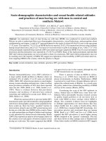
sisting of a single, circular, double-stranded DNA chro-
mosome, is 4,639,221 base pairs long and contains
4,403 genes. A partial genetic map of the E. coli K12
chromosome is shown in figure 31.1.
Background
The sequence of the genome of one strain of Escherchia
coli, K12, was completed in 1997 by researchers at the
University of Wisconsin, Madison. The genome, con-
thrA,B,C
araD,A,B,C
leuB,A
tonA
metD
proA,B
argF
lacA,Y,Z,O,P
tsx
purE
lip
galK,T,E
att
bio,A,B,F
,C,D
uvrB
serC
pyrD
pyrC
purB
att
80
t
rpA,B,C,D,E
man
tyrS
p
heS
argS
u
vrC
cheB,A
hisG
,
D,C,B
,H,A,
F, I
,
E
n
alA(gyrA)
purF
pt
sl
cysA
pheA
t
yrA
recA
argA
recB
lysA
serA
metC
argG
a
rgR
m
alA
xyl
p
yrE
dnaA
oriC
i
lvG,E,D,A,C
rhaD,A,B,C
metB
a
rgE,C,B,H
thiA,B,C
malB
dnaB
uvrA
p
urA
pyrB
v
alS
p
il
dnaC
Figure 31.1 Genetic map of E. coli K12 with the locations of selected genes. E. coli K12 strains
are used for fundamental work in biochemistry, genetics, and biotechnology, acting as carriers of
genes encoding therapeutic proteins.
Alexander−Strete−Niles:
Lab Exercises in
Organismal and Molecular
Microbiology
VII. Bacterial Genetics 31. Bacterial DNA Isolation
and Southern Analysis
© The McGraw−Hill
Companies, 2003
Bacterial DNA Isolation and Southern Analysis E
XERCISE
31 225
In preparation for analysis, the DNA must be iso-
lated from a pure culture of the bacteria. The isolation
involves lysing the cells, degrading cellular RNA and
protein with enzymes, and separating cellular debris
from the DNA through extraction with an organic sol-
vent. The DNA is then cut into fragments with a restric-
tion endonuclease, an enzyme that cuts through
double-stranded DNA at a particular recognition
sequence, (see also Exercise 33 and table 33.1). The
restriction enzyme EcoRI, for example, cuts DNA
wherever it contains the sequence,
-GAATTC-
-CTTAAG-
Therefore, cutting a series of DNA samples from
the same source with EcoRI will always generate the
same set of restriction fragments. These fragments can
be separated by size using gel electrophoresis.
However, cellular DNAs are so long (here, over
4 million base pairs) that when they are cut with a
restriction enzyme and the fragments are separated on
a typical electrophoresis gel, no clear restriction pattern
can be seen. Only a smear of DNA representing frag-
ments of just about every possible size is visible (figure
31.2). Think of this DNA smear as a ladder that has so
many rungs so close together that you cannot distin-
guish one rung from the next, or as a barcode that is
solid black—there is no information there. Southern
blotting allows the detection of a discrete region of
the DNA, revealing a restriction pattern of just that part
of the genome (figure 31.3). Southern blotting is also
often employed to generate DNA fingerprints (see
Exercise 36).
In this exercise, you will isolate DNA from bacte-
ria for restriction analysis (figure 31.3 a–c). If time per-
mits, you may proceed with a Southern blot over the
next few lab sessions (figure 31.3 d–i) in order to iden-
tify the restriction pattern of the bacterial gene lacZ. The
lacZ gene encodes the enzyme b-galactosidase.
Size marker
(base pairs)
(b)
321
Size marker
(base pairs)
2,027
2,322
4,361
6,557
9,416
23,130
2,027
2,322
4,361
6,557
9,416
23,130
(a)
321
Figure 31.2 Agarose gel electrophoresis of DNA isolated from E. coli. The 0.8%
agarose gels have been stained with (a) methylene blue or (b) ethidium bromide.
Both gels contain the following samples: bacteriophage lambda DNA cut with the
restriction enzyme HindIII (size marker, lane 1), E. coli DNA cut with the restriction
enzyme EcoRI (lane 2), and E. coli DNA that has not been cut with a restriction
enzyme (lane 3). The fragments (bands) in lane 1 are distinct because the lambda
genome is only about 49,000 base pairs long, and the enzyme cut the DNA into
discernible fragments. The E. coli DNA restriction fragment lengths in lane 2 are
indistinguishable from one another by this method, and appear as a smear.
Alexander−Strete−Niles:
Lab Exercises in
Organismal and Molecular
Microbiology
VII. Bacterial Genetics 31. Bacterial DNA Isolation
and Southern Analysis
© The McGraw−Hill
Companies, 2003
226 S
ECTION
VII Bacterial Genetics
(i) Development/detection: Restriction fragments
that have hybridized with probe appear as a
pattern on the membrane (or on the film if the
label was a radioisotope).
(h) Washing: Probe that is not extensively base-
paired to the immobilized DNA is washed away;
probe that is nonspecifically bound is removed.
(g) Hybridization: The membrane is submerged in a
solution containing many molecules of a specific
single-stranded DNA "probe," labeled in some
way for later detection. The probe DNA forms
base pairs with target DNA molecules on the
membrane.
(f) DNA immobilization: The membrane is baked to
irreversibly bind the DNA to the membrane.
(e) DNA transfer (blotting): DNA is transferred
from the gel to the surface of a membrane, such
as nitrocellulose. The method of transfer shown
here is called capillary blotting.
(d) DNA denaturation: The DNA fragments in the
gel are made single-stranded.
(c) Agarose gel electrophoresis: The restriction
fragments are separated by size; the distance
migrated by a fragment during electrophoresis is
inversely proportional to its size.
(b) Restriction enzyme digestion: The large fragments
of DNA are cut at specific sites with a restriction
enzyme, generating restriction fragments
characteristic of the organism.
(a) Isolation of DNA from tissues, cells, or viruses:
The DNA is mechanically sheared during this
procedure, generating large fragments.
(+)(−)
Shorter
fragments
Well
Longer
fragments
Bake
80º
ss DNAds DNA
NaOH
Weight Dry paper
Membrane
Gel
Sponge Salt solution
Hybridization
solution containing
labeled probe
molecules
Membrane
Alexander−Strete−Niles:
Lab Exercises in
Organismal and Molecular
Microbiology
VII. Bacterial Genetics 31. Bacterial DNA Isolation
and Southern Analysis
© The McGraw−Hill
Companies, 2003
Bacterial DNA Isolation and Southern Analysis E
XERCISE
31 227
Figure 31.3
(opposite page) Overview of Southern blotting and hybridization. With the com-
pletion of the Southern technique, what was once visible only as a smear of DNA fragments on
a gel now becomes a distinct pattern of specific restriction fragments on a membrane.
Phenol, equilibrated with 0.5 mM Tris, pH 8.0
Chloroform (chloroform:isoamyl
alcohol, 24:1)
3.0 M sodium acetate
Isopropanol
70% ethanol
Distilled water, autoclaved
Restriction enzyme and control reaction mixes
(table 31.1)
Equipment
37°C bacterial incubator with shaker platform
Microwave oven
Water bath or heat block at 37°C
Water bath or heat block at 50°C
Miscellaneous supplies
Laboratory marker
Latex gloves (when handling DNA; to protect
DNA from deoxyribonucleases on hands)
Ice
Microfuge tubes
Pasteur pipettes/bulb
1.0 ml serological pipette/pipettor
Micropipettors/tips (1–10 ml, 10–100 ml,
100–1,000 ml)
Table 31.1 Components of the Restriction Enzyme Mix and the Control Mix. Add 10 ml of each
mix to the corresponding reaction and control tubes. Store mixes on ice.
EcoRI No enzyme
control
Use 10 ml Use 10 ml
restriction mix. no enzyme control mix.
10µ restriction buffer 3 ml3 ml
Sterile distilled water 6 ml7 ml
EcoRI (10–20 units/ml) 1 ml 0 ml
Total mix volume 10 ml 10 ml
Total reaction volume
with 20 ml bacterial DNA 30 ml30ml
Restriction
mix
components
Materials
First Session: Bacterial DNA Isolation
and Restriction Digestion
Cultures
E. coli B and S. marcescens, each grown
overnight in 2 ml LB broth and
then inoculated into 50 ml fresh LB
for log growth
Media
LB broth: 10 g bacto-tryptone, 5 g yeast
extract, 10 g NaCl per liter
Reagents
TNE (10 mM Tris, pH 8.0, 10 mM NaCl,
0.1 mM EDTA), autoclaved
TE (10 mM Tris, pH 8.0, 0.1 mM EDTA),
autoclaved
HTE (50 mM Tris, pH 8.0, 20 mM EDTA),
autoclaved
2% sarcosyl (N-lauroyl sarcosine) in HTE
RNase on ice (pancreatic RNase A, 10 mg/ml,
in TE, preheated to 80°C for 10 minutes to
inactivate DNases)
Pronase on ice (10 mg/ml, in TNE, preheated
to 37°C for 15 minutes to inactivate DNases)
Alexander−Strete−Niles:
Lab Exercises in
Organismal and Molecular
Microbiology
VII. Bacterial Genetics 31. Bacterial DNA Isolation
and Southern Analysis
© The McGraw−Hill
Companies, 2003
228 S
ECTION
VII Bacterial Genetics
Second Session: Agarose Gel
Electrophoresis, Staining,
and Southern Transfer
Reagents
0.8 % agarose gel prepared with TBE: Tris-
Borate-EDTA (108 g Tris-base, 55 g boric
acid, 40 ml 0.5 M EDTA, pH 8.0, per liter)
DNA standard, lambda-HindIII, 1 mg per 30 ml
TBE; one per gel
DNA sample loading buffer (tracking dyes):
0.25% bromphenol blue, 0.25% xylene
cyanol, 30% glycerol in distilled water
DNA Blue InstaStain™
Denaturing solution (0.5 N NaOH, 1.5 M NaCl)
Neutralization solution (0.5 M Tris, pH 7.5,
1.5 M NaCl)
20µ SSC (3 M NaCl, 0.3 M sodium citrate),
diluted to 10µ SSC
Equipment
Horizontal gel electrophoresis system and
power source
Kitchen sponge (one per gel, for Southern
transfer)
Miscellaneous supplies
Micropipettors/tips (1–10 ml, 10–100 ml)
125 ml Erlenmeyer flask
Laboratory marker
Latex gloves (when handling DNA samples)
1.0 ml microcentrifuge tubes
Weigh boat or shallow dish (for staining)
Optitran BA-S supported nitrocellulose
membranes
3MM chromatography paper
Third Session: Probe Preparation and
Southern Hybridization
Reagents
DIG-High Prime DNA Labeling and Detection
Starter Kit I (table 31.2)
Probe DNA: pBLU digested with HindIII
(1 mg in 16 ml distilled, autoclaved water).
One probe for every 2 membranes.
20µ SSC (3 M NaCl, 0.3 M sodium citrate),
diluted to 2µ SSC
Equipment
Oven set at 80°C
Oven set at 42°C with a rocker platform
covered with bench-coat absorbent paper
Water bath set at 42°C
Boiling water bath or heat block set at 100°C
Note: Wear gloves from this
point on.
Miscellaneous supplies
Micropipettors/tips (1–10 ml, 10–100 ml)
50 ml conical tubes
Fourth Session: Washing
and Blot Development
Reagents
20µ SSC (3 M NaCl, 0.3 M sodium citrate),
diluted to 2µ SSC
2µ SSC, 0.1% SDS
0.5µ SSC, 0.1% SDS
Equipment
Oven set at 42°C with a rocker platform
covered with bench-coat absorbent paper
Water bath set at 42°C
Water bath or oven at 68°C
Bench top rocker or shaker platform
Miscellaneous supplies
3MM chromatography paper
Large weigh dishes
Procedure
First Session: Bacterial DNA Isolation
and Restriction Digestion
Yesterday, each E. coli strain was inoculated into 2 ml
of LB for overnight growth at 37°C with shaking. Ear-
lier today, each 2 ml culture was transferred into
50 ml of fresh broth in 125 ml flasks and incubated at
37°C with shaking.
1. Remove a flask of bacteria from the 37°C
incubator (the culture is expected to be in the log
phase of growth), and pipette 1 ml of it into a
microfuge tube. Centrifuge the sample in a
microfuge at full speed (14,000 RPM) for
15 seconds. Decant the supernatant into a waste
receptacle, and let the liquid drain off onto a
tissue. Dispose of the tissue in a biohazard bag.
2. Resuspend the cell pellet in 0.3 ml HTE, mixing
until there are no remaining cell clumps.
3. Add 0.35 ml 2% sarcosyl in HTE. Mix well by
capping and inverting the tube. Note that the
liquid is quite cloudy. Once lysis is complete
(after step 4), the liquid will be less cloudy.