Handbook of Clinical Neurology Vol. 82_2 pdf
Bạn đang xem bản rút gọn của tài liệu. Xem và tải ngay bản đầy đủ của tài liệu tại đây (25.43 MB, 218 trang )
Handbook of Clinical Neurology, Vol. 82 (3rd series)
Motor Neuron Disorders and Related Diseases
A.A. Eisen, P.J. Shaw, Editors
© 2007 Elsevier B.V. All rights reserved
Chapter 11
Monomelic amyotrophy of upper or lower limbs
M. GOURIE-DEVI*
Institute of Human Behaviour and Allied Sciences, and Department of Clinical Neurophysiology,
Sir Ganga Ram Hospital, New Delhi, India
11.1. Introduction
Monomelic amyotrophy in which neurogenic atrophy is
restricted to one limb is a heterogenous disorder,
involving one upper or lower limb. Insidious onset of
atrophy and weakness, presumed to be due to anterior
horn cell involvement, starting in the second or third
decade with male preponderance and sporadic occur-
rence are the characteristic features. Progression is slow
and followed by stabilization within a few years, result-
ing in a benign outcome. Cranial nerves, pyramidal,
sensory, cerebellar and extrapyramidal systems are not
involved.
Hirayama et al. (1959) from Japan reported atro-
phy of a single upper limb and labeled it as “juvenile
muscular atrophy of unilateral upper extremity.”
Prabhakar et al. (1981) from India reported atrophy of
muscles of one lower limb and described it as “wasted
leg syndrome.” Since either one upper or lower limb is
affected, Gourie-Devi et al. (1984a, 1986) suggested
the eponym “monomelic amyotrophy” (MMA) as a
more appropriate term. The authors further suggested
that upper limb MMA may be called “brachial mono-
melic amyotrophy” to differentiate it from MMA of
a lower limb, which may now be called “crural
monomelic amyotrophy” (Gourie-Devi and Nalini,
2003). Focal amyotrophy has been described under
a variety of descriptive names, which refer to the
limb involved, the site of muscles affected and the
benign and non-progressive course of the disease
(Table 11.1).
11.1.1. Monomelic amyotrophy of upper limb
More than 300 cases have been reported from Japan
(Hirayama et al., 1963; Hashimoto et al., 1976;
Sobue et al., 1978; Hirayama, 2000a). The atrophy was
distal and segmental, confined to one upper limb, but
electromyographic abnormalities were noted in some
patients in the non-atrophic upper limb. From India also
more than 200 cases (including a personal series of 89
cases) have been reported of single upper limb atrophy,
a large proportion of them with distal muscle involve-
ment and a few with proximal muscle involvement
(Singh et al., 1980; Gourie-Devi et al., 1984a,b, 1987a;
Virmani and Mohan, 1985; Misra and Kalita, 1995;
Pradhan and Gupta, 1997; Saha et al., 1997; Khandelwal
et al., 2004; Misra et al., 2005).
Reports from many other countries including
Sri Lanka (Peiris et al., 1989), Korea (Kim et al., 1994),
Hong Kong (Chan et al., 1991), Taiwan (Kao et al.,
1993a) and Malaysia (Tan, 1985) reaffirm the frequency
of MMA in Asia. Initially there were few reports from
Western countries, mostly isolated cases or a small
number of patients, but with increasing awareness more
publications have appeared in the literature (Pilgaard,
1968; Compernolle, 1973; Engel, 1977; Adornato et al.,
1978; De Visser et al., 1988). Large series of cases, notably
from France and Brazil, have been published (Serratrice
et al., 1987; De Freitas and Nascimento, 2000).
Hirayama et al. (1963) referred to 10 cases reported
by Marie and Foix in 1912, of isolated non-progressive
atrophy of small muscles of hand, older age at onset of
the disease in the fifth to eighth decades in eight cases
and second decade in two cases. The autopsy findings
in four of these patients are discussed later (§ 11.12).
11.1.2. Monomelic amyotrophy of lower limb
Monomelic amyotrophy of a lower limb is less frequent
than MMA of an upper limb. More than 130 cases
(including a personal series of 36 cases) have been
*Correspondence to: M. Gourie-Devi, Flat 9, Doctors Apartments, Vasundhara Enclave, New Delhi – 110096, India. E-mail:
, , Tel: +91-11-22618573, Fax: +91-11-22599227.
Ch11-N51894 9/8/06 10:39 AM Page 207
reported from India (Prabhakar et al., 1981; Gourie-
Devi et al., 1984a,b, 1987a; Virmani and Mohan, 1985;
Chopra et al., 1987; Saha et al., 1997) and more than
40 cases from Western countries (Riggs et al., 1984;
Serratrice et al., 1987; Uncini et al., 1992; De Freitas
and Nascimento, 2000; Felice et al., 2003). It is note-
worthy that, although numerous cases of MMA of an
upper limb are described from Japan, there is only one
isolated report of two cases of MMA of a lower limb
(Hamano et al., 1999).
11.2. Prevalence and geographic distribution
Monomelic amyotrophy constituted 8–29% of all motor
neuron diseases in different series reported from India
(Gourie-Devi et al., 1984a, 1987a; Saha et al., 1997).
The estimated prevalence rate of MMA was 0.9, of
upper limb 0.5 and lower limb 0.4 per 100,000 popula-
tion (Gourie-Devi et al., 1984a; Gourie-Devi, 2004),
based on the ratio of cases of monomelic amyotrophy
to amyotrophic lateral sclerosis, as suggested by
Kurtzke (1962), the prevalence rate of ALS having been
determined to be 4 per 100,000 population (Gourie-
Devi et al., 1984a, 1995). The geographic distribution
of MMA of upper and lower limb in Asia and other
countries is shown in Tables 11.2 and 11.3.
11.3. Classification
Monomelic amyotrophy can be classified based on the
limb involved and the site of muscles affected:
Type 1. Monomelic amyotrophy of upper limb.
Distal: Hand and forearm muscles.
Proximal: Shoulder girdle and arm muscles.
Global: Entire limb.
Type 2. Monomelic amyotrophy of lower limb.
Distal: Leg and foot muscles.
Proximal: Pelvic girdle and thigh muscles.
Global: Entire limb.
In the majority of cases in both type 1 and type 2, the
atrophy is confined to a single limb with electromyo-
graphic abnormalities in the contralateral limb in
some patients. In type 1, spread to the contralateral
limb with atrophy and weakness may occur in 10 to
30%, but significant asymmetry is a distinctive fea-
ture, the initially involved limb being more severely
affected (Gourie-Devi et al., 1984a; Sobue et al.,
1978). In contrast, in type 2, atrophy is usually
restricted to a single lower limb (Prabhakar et al.,
1981; Gourie-Devi et al., 1984a; Virmani and Mohan,
1985; Serratrice et al., 1987) with rare instances of
spread to the opposite limb (Kim et al., 1994; Felice
et al., 2003).
208
M. GOURIE-DEVI
Table 11.1
Eponyms used for single limb atrophy
A. Upper and lower limb
Monomelic amyotrophy (Gourie-Devi et al., 1984a).
Benign focal amyotrophy (Adornato et al., 1978; Riggs et al., 1984).
Monomelic spinal muscular atrophy (De Visser et al., 1988).
Spinal monomelic amyotrophy (Serratrice, 1991).
Benign monomelic amyotrophy (De Freitas and Nascimento, 2000).
B. Upper limb
Juvenile muscular atrophy of unilateral upper extremity (Hirayama et al., 1959).
Juvenile non progressive muscular atrophy localized to hand and forearm (Hashimoto et al., 1976).
Juvenile type of distal and segmental muscular atrophy of upper extremities (Sobue et al., 1978).
Juvenile muscular atrophy localized to arms (Singh et al., 1980).
Juvenile lower cervical spinal muscular atrophy (Kao et al., 1993a).
Juvenile amyotrophy of distal upper extremity (Biondi et al., 1989).
Non-familial spinal segmental muscular atrophy in juvenile and young subjects (Virmani and Mohan, 1985).
Non-progressive juvenile spinal muscular atrophy of the distal upper limb (Hirayama’s disease) (Hirayama, 1991).
Juvenile asymmetric segmental spinal muscular atrophy (Pradhan and Gupta, 1997).
Brachial monomelic amyotrophy (Gourie-Devi and Nalini, 2003).
C. Lower limb
Wasted leg syndrome (Prabhakar et al., 1981).
Benign monomelic amyotrophy of lower limb (Uncini et al., 1992).
Benign calf amyotrophy (Felice et al., 2003).
Crual monomelic amyotrophy (Gourie-Devi, 2004).
Ch11-N51894 9/8/06 10:39 AM Page 208
11.4. Clinical features
The age of onset in the majority (90%) varies from 15 to
35 years with a median age of 20 years in MMA of
upper limb and slightly older in MMA of lower limb
with a median age of 25 years (Hirayama et al., 1963;
Sobue et al., 1978; Gourie-Devi et al., 1984a). In excep-
tional cases the age at onset can be as early as 2 years
and as late as 84 years, the older age at onset being more
often noted in MMA of lower limb (Sobue et al., 1978;
Serratrice et al., 1987; Felice et al., 2003). However,
because the condition is so insidious in onset it can be
difficult to determine the age at onset. There is remark-
able gender preference, with men outnumbering women
with a ratio varying from 3:1 to 20:1, with more men
affected in MMA of lower limb compared to MMA of
upper limb (Hirayama et al., 1963; Sobue et al., 1978;
Prabhakar et al., 1981; Gourie-Devi et al., 1984a;
Virmani and Mohan, 1985). The duration of illness at
first consultation may vary from a few months to as long
as 15 years, with a mean duration of 2.5 to 4.5 years
(Hirayama et al., 1963; Prabhakar et al., 1981; Gourie-
Devi et al., 1984a; De Freitas and Nascimento, 2000).
11.4.1. Clinical features of MMA of upper limb
In monomelic amyotrophy of upper limb, the common
initial symptoms are weakness and atrophy in the major-
ity, followed by tremulousnesss of fingers. Coarse,
intermittent nonrhythmic tremors of fingers present
at rest, accentuated by outstretching of hands and on
MONOMELIC AMYOTROPHY OF UPPER OR LOWER LIMBS
209
Table 11.2
Geographic distribution of monomelic amyotrophy of upper limb
A. Countries in Asia
India: Singh et al., 1978; Gourie-Devi et al., 1984a; Virmani and Mohan, 1985; Misra and Kalita, 1995;
Pradhan and Gupta, 1997; Saha et al., 1997; Nalini et al., 2004; Khandelwal et al., 2004.
Hong Kong: Chan et al., 1991.
Israel: Neufeld et al., 1991.
Japan: Hirayama et al., 1963; Hashimoto et al., 1976; Sobue et al., 1978; Mukai et al., 1985; Iwasaki et al., 1987;
Kikuchi et al., 1987; Konno et al., 1997; Kohno et al., 1998.
Korea: Kim et al., 1994.
Malaysia: Tan, 1985.
Sri Lanka: Peiris et al., 1989.
Taiwan: Kao et al., 1993a.
Turkey: Gucuyener et al., 1991.
B. Countries outside Asia
Australia: Kiernan et al., 1999.
Belgium: Robberecht el al., 1997.
Brazil: De Freitas and Nascimento, 2000.
Canada: Oryema et al., 1990.
Denmark: Pilgaard, 1968.
France: Serratrice et al., 1987; Chaine et al., 1988; Biondi et al., 1989.
Germany: Schlegal et al., 1987; Schroder et al., 1999.
Italy: Barontini et al., 1991; Di Guglielmo et al., 1996; Polo et al., 2003.
Netherlands: Compernolle, 1973; Thijsse and Spaans, 1983; De Visser et al., 1988.
Poland: Drozdowski et al., 1998.
Switzerland: Kaeser et al., 1983.
USA: Engel, 1977; Adornato et al., 1978; Metcalf et al., 1987; Tandan et al., 1990; Liu and Specht, 1993;
Donofrio, 1994; Rowin et al., 2001.
Table 11.3
Geographic distribution of monomelic amyotrophy of
lower limb
A. Countries in Asia
India: Prabhakar et al., 1981; Gourie-Devi et al.,
1984a; Virmani and Mohan, 1985;
Saha et al., 1997.
Japan: Hamano et al., 1999.
Korea: Kim et al., 1994.
B. Countries outside Asia
Austria: Willeit et al., 2001.
Brazil: De Freitas and Nascimento, 2000.
France: Nedelec et al., 1987; Serratrice et al., 1987.
Germany: Munchau and Rosenkranz, 2000.
Italy: Uncini et al., 1992; Di Muzio et al., 1994;
Di Guglielmo et al., 1996.
Netherlands: De Visser et al., 1988.
Spain: Martinez et al., 1990.
USA: Riggs et al., 1984; Felice et al., 2003.
Ch11-N51894 9/8/06 10:39 AM Page 209
voluntary action is present in 60 to 80% of patients
(Hirayama et al., 1963; Gourie-Devi et al., 1984a). This
feature has been observed in spinal muscular atrophy
and the descriptive term minipolymyoclonus has been
coined (Spiro, 1970). Minipolymyoclonus needs to be
distinguished from tremors, which are generally rhyth-
mic, and from fasciculations. Discharges by motor neu-
rons innervating large territory of muscle are implicated
in the causal mechanisms of these tremor-like move-
ments, but probably not specific, and may be seen in
hand weakness from most neuromuscular disorders.
Fasciculations are commonly observed in atrophic
muscles and also in the unaffected muscles in a few
patients. Hirayama (1972) described “cold paresis,” an
interesting phenomenon of aggravation of weakness on
exposure to cold. Some of them also complain of stiff-
ness of hands on dipping the hands in cold water, how-
ever there was no clinical or electromyographic
evidence of myotonia (Gourie-Devi et al., 1984a).
In MMA of upper limb the distal muscles of hand and
forearm are affected in more than 50% of patients, prox-
imal muscles of shoulder and upper arm in 5–10% and
diffuse involvement in 40% with the distal muscles more
severely affected than proximal muscles. Small muscles
of the hand, flexors and extensors of the wrist, chiefly
C7-T1 spinal segments, are the most severely affected
muscles (Figs. 11.1–11.3). Relative sparing of brachiora-
dialis muscle among surrounding atrophic muscles
(Fig. 11.2) is a characteristic feature of this disease
(Hirayama et al., 1963). In the diffuse form with involve-
ment of an entire upper limb, the additional muscles atro-
phied are biceps, triceps, deltoid and scapular muscles
(Compernolle, 1973; Thijsse, 1983; Gourie-Devi et al.,
1984a). Unilateral atrophy of scapulohumeral muscles in
C5–C6 myotomes (Fig. 11.4) was described by Kaeser
(1983) from Switzerland and similar cases were
observed by others (Gourie-Devi et al., 1984a; Virmani
and Mohan, 1985; Amir et al. 1987; De Visser et al.,
1988; Kao et al., 1993a). The pattern of muscles affected
in our series of 89 patients (Gourie-Devi and Nalini,
unpublished observations) is shown in Figure 11.5.
11.4.2. Clinical features of MMA of lower limb
In MMA of lower limb, atrophy of the limb was noted by
the patient because of pain on walking, and in nearly a
third of the patients it was incidentally observed by a
family member, friend or physician during consultation
for unrelated illness (Prabhakar et al., 1981; Gourie-Devi
et al., 1984a). Under these circumstances the precise age
at onset and duration of illness may not be accurate.
Muscle cramps and fasciculations have been observed in
20 to 30% of patients. Unilateral pes cavus may be a
presenting feature (De Freitas and Nascimento, 2000).
Unlike as in postpoliomyelitis progressive muscular
atrophy there is no shortening of limb.
210
M. GOURIE-DEVI
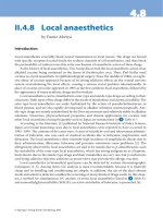
Fig. 11.1. Mild atrophy of flexors of forearm of right upper limb best seen in semiprone position.
Ch11-N51894 9/8/06 10:39 AM Page 210
MONOMELIC AMYOTROPHY OF UPPER OR LOWER LIMBS
211
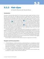
Fig. 11.2. Atrophy of flexor and extensor muscles of right forearm with sparing of brachioradialis muscle and mild wasting of
hand muscles.
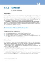
Fig. 11.3. Severe atrophy of thenar, hypothenar and interossei,
particularly first dorsal interosseous muscle of right hand.
Fig. 11.4. Severe wasting of left shoulder and upper arm
muscles with normal forearm muscles.
Ch11-N51894 9/8/06 10:39 AM Page 211
In the distal form, which accounts for 20% of cases,
with predominant calf muscle atrophy, inability to stand
on tiptoe is a presenting feature (Felice et al., 2003).
Anterior and posterior crural muscles are most com-
monly affected (Fig. 11.6), while intrinsic foot muscles
are infrequently involved (Prabhakar et al., 1981;
Gourie-Devi et al., 1984a; Virmani and Mohan, 1985; De
Visser et al., 1988; Uncini et al., 1992; Hamano et al.,
1999; De Freitas and Nascimento, 2000; Felice et al.,
2003). In the proximal type, isolated atrophy of quadri-
ceps (Fig. 11.7) may occur (Prabhakar et al., 1981;
Gourie-Devi et al., 1984a) or may be involved along with
hamstring muscles (Prabhakar et al., 1981; Gourie-Devi
et al., 1984a; Riggs et al., 1984; Virmani and Mohan,
1985). The commonest type is involvement of the entire
limb with atrophy of proximal and distal muscles and has
been observed in 70% of patients (Prabhakar et al., 1981;
Gourie-Devi et al., 1984a; Virmani and Mohan, 1985;
Hamano et al., 1999). The pattern of muscle involvement
in our series of 36 cases (Gourie-Devi and Nalini, unpub-
lished data) is shown in Figure 11.8.
11.4.3. Other clinical features
The tendon reflexes in the affected limb in both type 1
and 2 are usually absent or sluggish. In some patients
they are normal and brisk reflexes are rare, but plantar
response is invariably flexor. In the unaffected homolo-
gous limb and other limbs, the reflexes were generally
normal and infrequently sluggish. Although subjective
symptoms of numbness have been reported, no objec-
tive sensory deficit has been documented. Excessive
sweating and coldness of affected limb is a frequent
feature. Cognitive function, cranial nerves, pyramidal,
extrapyramidal and cerebellar systems are not involved.
There is no evidence of other neurological disorders in
the affected subject or their family members.
11.5. Associated factors and antecedent events
Febrile illness, vaccination, exposure to toxic sub-
stances and electric shock preceding the illness have
212
M. GOURIE-DEVI
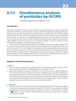
31
12
12
43
37
69
19
62
62
81
56
94
69
94
94
94
Scapular
Latissmus dorsi
Pectoralis Major
Deltoid
Biceps Brachialis
Triceps
Brachioradialis
Supinator
Pronator
Wrist Flexors
Wrist Extensors
Finger Flexors
Finger Extensors
Thenar
Hypothenar
Interossei
Fig. 11.5. Pattern of muscle atrophy and weakness in 89
patients of monomelic amyotrophy of upper limb (Gourie-
Devi and Nalini, unpublished data).
Fig. 11.6. Atrophy of calf muscles of right leg.
Ch11-N51894 9/8/06 10:39 AM Page 212
not been observed in the majority of patients
(Hirayama et al., 1963; Gourie-Devi et al., 1984a;
Virmani and Mohan, 1985, Peiris et al., 1989). In rare
instances poliomyelitis in childhood has been reported
(Gourie-Devi et al., 1984a; Peiris et al., 1989; Gourie-
Devi, 1996). Mechanical trauma including injuries or
surgery have been recorded preceding the onset of neu-
rological symptoms by many months to years and in
some of them atrophy occurred in the previously
injured limb (Sobue et al., 1978; Gourie-Devi et al.,
1993; Paradiso, 1997). In a case control study which
examined the risk factors in 21 cases and 63 age and
gender matched control subjects, strenuous physical
activity was observed to be a significant associated
factor (Gourie-Devi et al., 1993). Occupations involv-
ing heavy manual exertion and participation in com-
petitive sports have also been recorded in patients with
MMA (Hashimoto et al., 1976; Prabhakar et al., 1981;
Biondi et al., 1989).
11.6. Familial monomelic amyotrophy
Familial occurrence of MMA is extremely rare. Gourie-
Devi et al. (1984a) did not detect muscle weakness,
wasting or sluggish tendon reflexes in 48 siblings and
parents of 17 patients. A total of 15 families have been
reported so far from countries in Asia, Europe and USA
(Table 11.4). Two brothers were affected in each of six
families, father and son in four, mother and son in two
families, sister and brother, identical twin brothers and
two half brothers in one family each. In 13 families
the upper limb was involved and in two families lower
limb was affected. The age at onset was in the second or
MONOMELIC AMYOTROPHY OF UPPER OR LOWER LIMBS
213
27
45
81
45
100
37
36
81
Glutei
Adductors
Quadriceps
Hamstring
Anterior Crural
Peronell
Posterior Crural
Foot Muscles
Fig. 11.8. Pattern of muscle atrophy and weakness in 36
patients with monomelic amyotrophy of lower limb (Gourie-
Devi and Nalini, unpublished data).
Fig. 11.7. Atrophy of thigh muscles of right lower limb with
preserved calf muscles.
Ch11-N51894 9/8/06 10:39 AM Page 213
third decade in 13 families, first decade in one family
(Gucuyener et al., 1991) and fifth decade and beyond in
one family (Serratrice et al., 1987). There were 25 males
and three females with a M:F ratio of 8.3:1. These
reports suggest autosomal recessive inheritance in some
families and autosomal dominant inheritance with vari-
able expression in others (De Visser et al., 1991;
Robberecht et al., 1997; Nalini et al., 2004). Occurrence
of disease predominantly in males and two half brothers
may indicate X-linked recessive inheritance which
needs to be further examined (Nedelec et al., 1987;
Misra et al., 2005).
Only a few genetic studies have been done. In one
family in two affected brothers, five exons of superox-
ide dismutase 1 (SOD 1) gene were normal and the
SOD activity in patients’ RBC was comparable to the
values in control subjects (Robberecht et al., 1997).
Subsequently, Mezei et al. (1999) describe a family
with a D90A SOD1 mutation in which the father of the
proband has clinical features typical of lower limb
monomelic amyotrophy. DNA analysis revealed him to
be heterozygous for D90A mutation. Survival motor
neuron gene (SMN) deletion in the region of 5q13 has
been demonstrated to be associated with phenotypic
expression of spinal muscular atrophy (SMA) (Lefebvre
et al., 1995) and for confirmatory diagnosis of SMA,
SMN1 and SMN2 gene deletion study is advocated
(Scheffer et al., 2001). It has also been shown that dele-
tions in SMN gene occur in adult onset SMA (Brahe
et al., 1995). Since MMA has been considered as a
focal form of SMA, studies have been done to examine
the deletion of SMN gene. Recent reports from Italy,
USA and India show that MMA of upper and lower
limb are not associated with deletions in exons 7 and
8 of the SMN gene (Di Guglielmo et al., 1996; Felice et
al., 2003; Misra et al., 2005). Mutation of mitochondr-
ial DNA, the 7472 insC in the gene coding the tRNA
Ser (UCN), has been reported from Italy in a patient
with monomelic amyotrophy and sensorineural hearing
loss in the patient, his mother and an elder sister (Fetoni
et al., 2004). Association of lower motor neuron involve-
ment with mt DNA mutation needs further elucidation.
11.7. Secondary monomelic amyotrophy
Monomelic amyotrophy may be secondary to demon-
strable causes including irradiation, atopy and human
immunodeficiency virus (HIV) infection. Lower motor
neuron syndrome may develop months to years after
irradiation for malignant disorders encompassing the
spinal cord. In most cases paraparesis has been reported
but rarely cases with monomelic amyotrophy have
been documented (Lamy et al., 1991; Jackson,
1992; Serratrice et al., 1993). The period between
radiotherapy and development of MMA ranged from 9
to 17 years. It is possible that radiotherapy damaged a
critical number of motor neurons and the compensatory
efforts of surviving motor neurons in reinnervation of
muscles could not be maintained over many years,
leading to focal atrophy (Jackson, 1992). However,
radiation necrosis more commonly affects the plexus
and proximal nerves.
Asthmatic amyotrophy, a polio-like syndrome, is
characterized by an asymmetrical lower motor neuron
paralysis following an acute episode of asthma (Hopkins,
1974; Batley and Johnson, 1991). Importance of atopy,
airways allergy in precipitating ‘circulatory insuffi-
ciency’ and its causal linkage to acute myelitis and to the
214
M. GOURIE-DEVI
Table 11.4
Familial case of monomelic amyotrophy
Author (year) Country Families Affected Limb
Igata et al. (1966) Japan 1 Father–son UL
Hirayama (1972) Japan 3 Brothers (2) UL
Sobue et al. (1978) Japan 1 Father–son UL
Hirayama et al. (1987) Japan 1 Brothers (2) UL
Schlegel et al. (1987) Germany 1 Father–son UL
Serratrice et al. (1987) France 1 Mother–son LL
Nedelec et al. (1987) France 1 Brothers (2) LL
Tandan et al. (1990) USA 1 Identical twin-brother UL
Gucuyener et al. (1991) Turkey 1 Sister–brother UL
Misra and Kalita (1995) India 1 Brothers (2) UL
Robberecht et al. (1997) Belgium 1 Brothers (2) UL
Nalini et al. (2004) India 1 Mother–son UL
Misra et al. (2005) India 1 Brothers (2) UL
Figure in parenthesis indicates number of affected members.
Ch11-N51894 9/8/06 10:39 AM Page 214
chronic disorder of monomelic amyotrophy has been
suggested (Kira et al., 1998; Horiuichi et al., 2000; Kira
and Ochi, 2001).
In HIV infection, several neurological disorders are
described, but motor neuron disease has been very rarely
reported (Huang et al., 1993; Moulignier et al., 2001).
A significant proportion of these patients were young,
the initial presentation was monomelic amyotrophy with
subacute progression to other limbs and involvement of
corticospinal tracts. The striking response to antiretro-
viral therapy convincingly establishes the etiological
relationship between HIV and motor neuron disease,
in these select patients (Jubelt and Berger, 2001;
Moulignier et al., 2001).
11.8. Investigations
11.8.1. Laboratory tests
Routine blood and cerebrospinal fluid analysis is usu-
ally normal, but a mild rise of CSF protein has been seen
in a few patients (Hirayama et al., 1963; Gourie-Devi
et al., 1984a). A slight increase in serum creatine kinase
level, just above the normal range, has been reported in
occasional patients (Gourie-Devi et al., 1984a).
Antibodies to viruses such as polio, Coxackie B, Echo,
influenza A and B, adeno and herpes simplex were not
detected in CSF (Sobue et al., 1978; Virmani and
Mohan, 1985). Lower serum neutralizing antibody titers
for poliovirus were found in patients compared to con-
trols suggesting that patients with MMA may be
immunologically unresponsive to a neutralizing epitope
of poliovirus (Kao et al., 1993b). Intrathecal immuno-
globulin synthesis was not detected and ganglioside
antibodies, particularly anti-GM 1 antibodies, were not
detected (Willeit et al., 2001).
11.8.2. Muscle biopsy
Variable findings of normal to small groups of angu-
lated muscle fibers, group atrophy, nuclear clumping,
fiber type grouping to end stage disease with diffuse
fatty infiltration and prominent increase in connective
tissue, all features suggestive of neurogenic atrophy in
the affected limb, have been noted in various studies
(Hirayama et al., 1963; Prabhakar et al., 1981; Gourie-
Devi et al., 1984a; Kao and Tsai, 1994; Kim et al.,
1994). Necrotic fibers with central nuclei, basophilic
fibers with large vesicular nuclei indicating secondary
myopathic changes, were observed in a few patients
(Prabhakar et al., 1981; Gourie-Devi et al., 1984a).
Subclinical diffuse involvement of anterior horn cells
was supported by evidence of mild muscle fiber type
grouping in the unaffected limb (Uncini et al., 1992).
Sural nerve biopsy did not show any abnormality
(Gourie-Devi et al., 1984a; Kim et al., 1994).
11.8.3. Electrophysiology
11.8.3.1. Electromyography
Needle electromyography shows fibrillations or positive
sharp waves, long duration, large amplitude polyphasic
potentials with poor recruitment indicating both active
denervation and chronic reinnervation, respectively, in
the atrophic muscles of the affected limb in MMA of
upper or lower limbs (Hirayama et al., 1963; Sobue
et al., 1978; Prabhakar et al., 1981; Gourie-Devi et al.,
1984a; Serratrice et al., 1987; Peiris et al., 1989; Kao
et al., 1993a; Kim et al., 1994; Khandelwal et al., 2004;
Misra et al., 2005). Active denervation, a consistent fea-
ture in the majority of cases, irrespective of the duration
of illness ranging from few months to 5 or more years,
was not seen in the patients who had attained a station-
ary course after an initial phase of progression (Kao
et al., 1993c; Misra and Kalita, 1995; Gourie-Devi and
Nalini, 2003). Rarely fibrillations or positive sharp
waves have been observed in a clinically stationary
phase of many years, suggesting a subclinical progres-
sion (Kao et al., 1993c).
In the clinically unaffected muscles of the involved
limb chronic reinnervative changes have been reported in
25 to 50% of patients with amyotrophy of upper
limb (Gourie-Devi et al., 1984a; De Visser et al., 1988;
Hirayama, 2000a), however no abnormalities have been
reported by other authors (Virmani and Mohan, 1985;
Kim et al., 1994; Misra et al., 2005). It is important to
note that the relatively well preserved brachioradialis
muscle usually does not show any EMG abnormalities
(Hirayama et al., 1963; Gourie-Devi et al., 1984a; Misra
and Kalita, 1995), with few exceptions (Sobue et al.,
1978). In the contralateral unaffected upper limb, the
homologous muscles show denervation and chronic rein-
nervation in 7–88% of patients (Hirayama et al., 1963;
Hashimoto et al., 1976; Sobue et al., 1978; Singh et al.,
1980; Gourie-Devi et al., 1984a; De Visser et al., 1988;
Gourie-Devi and Nalini, 2003; Khandelwal et al., 2004;
Misra et al., 2005) but were found to be normal by some
authors (Virmani and Mohan, 1985). In the lower limbs
which are clinically never affected, EMG abnormalities
have not been demonstrated in the vast majority of
patients (Hirayama et al., 1963; Hashimoto et al., 1976;
Singh et al., 1980; Sobue et al., 1978; Gourie-Devi et al.,
1984a; Willeit et al., 2001; Gourie-Devi and Nalini,
2003) with rare exceptions of mild chronic denervation
(De Freitas and Nascimento, 2000).
In MMA of lower limb, denervation and chronic
reinnervation have also been noted in the clinically
unaffected muscles of the atrophic limb but very rarely
in the contralateral lower limb (Prabhakar et al., 1981;
Gourie-Devi et al., 1984a; Riggs et al., 1984; Virmani
and Mohan, 1985; Uncini et al., 1992; Munchau and
Rosenkranz, 2000; Felice et al., 2003). The upper limbs
in this group do not show any abnormalities.
MONOMELIC AMYOTROPHY OF UPPER OR LOWER LIMBS
215
Ch11-N51894 9/8/06 10:39 AM Page 215
Electromyography did not reveal any evidence of
myotonic discharges, particularly in the context of
appearance of stiffness of hands on exposure to cold
(Gourie-Devi et al., 1984a). Aggravation of weakness
of fingers induced by exposure to cold has been attrib-
uted to impairment of muscle membrane conduction
since high frequency repetitive nerve stimulation
showed waning of amplitude of compound muscle
action potentials (Kijima et al., 2002). Only in a single
case of MMA of upper limb were myokymic discharges
observed (De Visser et al., 1988).
Lower cervical paraspinal muscles (C8-T1) involve-
ment on electromyography was not observed in MMA of
upper limb, although active denervation and chronic
reinnervation could be demonstrated in the muscles of
C7-T1 myotomes in the affected upper limbs, independ-
ent of the clinical stage of the disease or the duration of
illness (Kao et al., 1993c). In contrast, paraspinal muscle
involvement, an early and consistent sign demonstrable
by EMG in amyotrophic lateral sclerosis (Kuncl et al.,
1988), can help in differentiating ALS from monomelic
amyotrophy, particularly when the initial feature is single
limb involvement (Kao et al., 1993c).
Single fiber EMG done in a few patients showed
increased fiber density and jitter with occasional block-
ing in the affected limb, indicating unstable neuromus-
cular transmission due to new regeneration (Thijsse and
Spaans, 1983). During the stage of stabilization of the
disease, there is further increase of fiber density, but
jitter decreases suggesting maturation of reinnervation
(Hirayama, 2000a).
11.8.3.2. Nerve conduction
Motor conduction studies are usually normal in patients
with mild to moderate atrophy of muscles (Hirayama
et al., 1963; Sobue et al., 1978; Singh et al., 1980;
Gourie-Devi et al., 1984a; Virmani and Mohan, 1985;
De Visser et al., 1988; Peiris et al., 1989). Slight slow-
ing of motor conduction velocity may be observed con-
sistent with loss of fast conducting axons and the
compound muscle action potential amplitude is reduced
(Kim et al., 1994) and occasionally motor distal latency
may be prolonged (Tan, 1985). Conduction block has
not been demonstrated in amyotrophy of upper or lower
limb (Kim et al., 1994; Misra and Kalita, 1995; Gourie-
Devi and Nalini, 2001; Willeit et al., 2001; Khandelwal
et al., 2004). Sensory conduction studies are normal in
all patients.
F-wave latency and H-reflex are within normal
limits (Uncini et al., 1992; Kao et al., 1993c; Misra and
Kalita, 1995; Willeit et al., 2001) with few exceptions
of slight increase in latency and low persistence of
F-wave (Kuwabara et al., 1999).
11.8.3.3. Evoked potentials
Somatosensory evoked potentials (SEP) from upper and
lower limbs are normal in amplitude and latency (Kao
et al., 1993c; Pradhan and Gupta, 1997; Willeit et al.,
2001). Conflicting results show decrease of amplitude
of Erb’s point potentials and N13 spinal responses but
with normal latencies and normal N20 potential (Polo
et al., 2003). There was no correlation of these abnor-
malities with the clinical features. However, SEPS were
found to be normal following tibial nerve stimulation.
11.8.3.4. Central motor conduction
Central motor conduction time (CMCT) determined by
electrical stimulation of cortex or by transcranial mag-
netic stimulation was normal in all patients, providing
evidence that in MMA upper motor neuron is not
involved (Misra and Kalita, 1995; Khandelwal et al.,
2004). Contrary to these findings, slight but significant
prolongation of CMCT has been observed in some
patients (Polo et al., 2003). Cortical threshold intensity
(TI) which reflects a balance of cortical and spinal
excitability was also found to be normal (Khandelwal
et al., 2004). In motor neuron disease the CMCT and TI
have been found to be abnormal confirming upper
motor neuron involvement (Triggs et al., 1999), while
in MMA there is no evidence of pyramidal tract dys-
function. The absence of upper motor neuron involve-
ment in MMA has also been substantiated by normal
H/M ratio, vibratory inhibition and reciprocal inhibi-
tion of soleus H reflex (Misra and Kalita, 1995).
11.8.3.5. Dynamic electrophysiology
Dynamic electrophysiological studies showed increased
latency and decreased amplitude of motor evoked poten-
tials after transcranial magnetic stimulation, decrease in
F-wave persistence and decrease of amplitude of N13
somatosensory evoked potential during neck flexion
(Shizukawa et al., 1994; Kuwabara et al., 1999;
Restuccia et al., 2003).
11.8.4. Autonomic function tests and sympathetic
skin response
Increased sweating of hands and cyanosis of fingers
have been observed in nearly 50% of patients with
MMA of upper limb (Hirayama et al., 1963; Gourie-
Devi et al., 1984a). Decreased skin temperature in
distal portion of upper limb, plethysmographic abnor-
malities indicative of vasomotor dysfunction and con-
firmation of hyperhidrosis by sweat tests have been
documented (Hirayama, 1991).
A recent study of sympathetic skin response (SSR)
in MMA showed that SSR latency in the affected upper
216
M. GOURIE-DEVI
Ch11-N51894 9/8/06 10:39 AM Page 216
limb was significantly prolonged compared to controls
confirming the involvement of sympathetic nervous
system (Gourie-Devi and Nalini, 2001). Interestingly,
increase in latency was seen in the contralateral unaf-
fected upper limb but not in lower limbs. The abnor-
malities of SSR did not strictly correlate with clinical
symptoms of autonomic dysfunction in the atrophic
limb. Prolonged SSR latency may indicate subclinical
involvement of sympathetic nervous system in unaf-
fected upper limb (Shahani et al., 1984). These obser-
vations coupled with the pathological finding of
decrease in number of nerve cells in the inferior cervi-
cal sympathetic ganglion, suggest lesion in the efferent
sympathetic pathway at this level (Hirayama et al.,
1987; Gourie-Devi and Nalini, 2001).
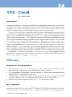
11.8.5. Imaging
11.8.5.1. Imaging of muscles
CT and MRI of muscles in monomelic amyotrophy
provide valuable information about the selectivity
of muscle affected (Fig. 11.9), delineate the sequence
of muscle involvement and enable correlation with dis-
ease duration. Imaging can disclose affected deep mus-
cles of thigh and leg, particularly in early stages or with
mild changes, when clinical and electrophysiological
examination fails to detect the involvement. In the distal
form of MMA of lower limb, gastrocnemius followed
by soleus are involved and in later stages muscles of
anterior compartment, particularly tibialis anterior are
affected (Hamano et al., 1999). In the thigh, quadriceps,
semimembranosus, semitendinosus and biceps femoris
are sequentially involved. (De Visser et al., 1988; Di
Muzio et al., 1994; Munchau and Rosenkranz, 2000).
Involvement of periphery of muscles, selective and
bilateral symmetric pattern without significant atrophy
of muscles, the distinctive features of myopathy, distin-
guish it from neurogenic disorder (Bulcke et al., 1979;
De Visser and Verbeeten, 1985; Schwartz et al., 1988;
Termote et al., 1980). Early stages of ALS with evidence
of involvement of a single limb may be differentiated from
MMA by the demonstration of selective muscle atrophy
on imaging in the latter disorder (Di Muzio et al., 1994).
11.8.5.2. Imaging of spinal cord
In MMA of upper limb earlier studies had reported
straight neck due to obliteration of cervical lordosis on
radiographs (Hashimoto et al., 1976) and, recently, CT
myelography and MRI have demonstrated focal cord
atrophy (Fig. 11.10) at lower cervical level with maxi-
mal changes at C5–C6 level, corresponding to segmen-
tal distribution of weakness (Matsumura et al., 1984;
Mukai et al., 1985; Metcalf et al., 1987; Biondi et al.,
MONOMELIC AMYOTROPHY OF UPPER OR LOWER LIMBS
217
A
B
Fig. 11.9. CT of (A) right thigh shows
severe atrophy of vastus lateralis, vastus
medialis, biceps femoris with mild atro-
phy of all other muscles and (B) left
thigh is normal.
Ch11-N51894 9/8/06 10:39 AM Page 217
1989; Gourie-Devi et al., 1992; Kao et al., 1993a;
Pradhan and Gupta, 1997; Misra et al., 2005). The atro-
phy was more marked on the side of the affected limb in
patients with atrophy and weakness restricted to one
upper limb while EMG changes were bilateral, but were
more severe on the affected side. In others, focal and
unilateral atrophy of the lower cervical cord limited to
the anterior horn region has been reported (Biondi et al.,
1989; Gourie-Devi et al., 1992). High intensity signals
localized to the anterior and lateral horns of the gray
matter on T2 weighted images (Fig. 11.11) have been
reported (Pradhan and Gupta, 1997; Chan et al., 1998;
Schroder et al., 1999; Willeit et al., 2001).
In MMA of lower limb, however, atrophy of lower
thoracic or lumbar cord was not observed and there was
no evidence of lumbar canal stenosis (Gourie-Devi
et al., 1992; Kim et al., 1994).
Rarely syringomyelia may present with only atrophy
and weakness of hand muscles without sensory changes
(Mukai et al., 1984). Therefore in MMA delayed scans
on CT myelography or MRI is mandatory to exclude
cavity (Gourie-Devi et al., 1992).
11.8.5.3. Dynamic imaging of spinal cord
Forward displacement of cervical dorsal sac and spinal
cord along with flattening of lower cervical cord has been
demonstrated with dynamic conventional myelography,
218
M. GOURIE-DEVI
Fig. 11.10. CT myelography shows cord atrophy at C5 to C7
levels with more severe changes on right side, ipsilateral to
the atrophic limb.
A
B
Fig. 11.11. T2-weighted image shows hyperintense signal in
spinal cord, (A) extending from C3 to C7 and (B) localized to
anterior and lateral horns of gray matter.
Ch11-N51894 9/8/06 10:39 AM Page 218
CT myelography and MRI, during neck flexion (Mukai
et al., 1985; Iwasaki et al., 1987; Toma and Shiozawa,
1995; Pradhan and Gupta, 1997; Hirayama and
Tokumaru, 2000). The posterior dura mater also moved
forward obliterating subarachnoid space leaving a large
posterior epidural space with prominent epidural venous
plexus. In normal subjects and in ALS the cord moved
forward with slight flattening of cervical cord, but there
was no displacement of the posterior dura mater
(Pradhan and Gupta, 1997). It has been shown that cer-
vical dorsal roots are short and asymmetric in patients
while they are slack in normal subjects (Toma and
Shiozawa,1995). It is postulated that the growth of cer-
vical roots does not keep pace with growth spurts in
adolescence. This fact may be responsible for over-
stretching and forward displacement of cord (Pradhan
and Gupta, 1997; Toma and Shiozawa, 1995; Hirayama
and Tokumaru, 2000). Interestingly, a recent report
provides evidence that the cervical spinal cord was
stretched even in the neutral position in patients due to a
disproportion between cervical spine and shorter cervi-
cal spinal cord (Kohno et al., 1998). Contrary to these
observations, dynamic imaging in neutral and maximum
flexion of neck in the patients, significant compression
of cervical spinal cord, forward displacement of dural
space and prominent epidural veins were not observed
(Schroder et al., 1999; De Freitas and Nascimento, 2000;
Willeit et al., 2001). The posterior subarachnoid space
and epidural space were normal. All these findings were
similar to the observations in healthy control subjects.
11.9. Diagnosis
Insidious onset of atrophy and weakness restricted to a
single limb in the second or third decade, male prepon-
derance, absence of sensory and upper motor neuron
signs, slow progression for 2 to 5 years followed by sta-
bilization, are all distinctive clinical features of
monomelic amyotrophy. Extrapyramidal, cerebellar
and cognitive functions are preserved. Normal CPK
levels, electromyographic features of neurogenic pat-
tern, normal nerve conduction studies and absence of
conduction block provide confirmation of localization
of lesion to anterior horn cells. Imaging of spinal cord
to exclude mass lesions, syringomyelia and vascular
lesions is mandatory. An algorithm (Fig. 11.12) pro-
vides a practical approach to diagnosis of monomelic
amyotrophy.
11.10. Differential diagnosis
Before considering the diagnosis of MMA of upper
limb, a number of disorders which “mimic” this disease
(Table 11.5) have to be excluded by appropriate and
relevant investigations. The presence of sensory involve-
ment, upper motor signs and imaging findings provide
evidence for structural lesions of spinal cord. In rare
instances, sensory deficit may be absent in syringomyelia
with only lower motor neuron signs in one or both upper
limbs, making it mandatory to do imaging of spinal cord
in MMA (Mukai et al., 1984). Cauda equina lesions can
also be excluded by imaging studies.
Spinal muscular atrophy, especially the distal form,
characteristically is bilateral with symmetric involve-
ment of upper or lower limbs, slowly progressive course
and positive family history in many cases with autoso-
mal recessive or dominant inheritance (McLeod and
Prineas, 1971; O’Sullivan and McLeod, 1978; Harding
and Thomas, 1980). In some patients, in the early stages,
distal SMA may be unilateral resembling MMA
(O’Sullivan and McLeod, 1978; Harding et al., 1983;
Peiris et al., 1989). In juvenile spinal muscular atrophy
(Kugelberg–Welander disease), the proximal limb
muscles are affected and the atrophy and weakness are
bilateral. In chronic neurogenic quadriceps amyotrophy,
considered a forme fruste of Kugelberg–Welander
disease, the atrophy of quadriceps muscles is bilateral
with occasional involvement of pelvic girdle and EMG
shows generalized involvement of unaffected muscles of
upper and lower limbs (Furukawa et al., 1977; Tetsuo
et al., 1977).
Early stage of ALS with single limb involvement can
be misdiagnosed as monomelic amyotrophy. Selective
involvement of muscles in MMA demonstrable on
imaging may be useful in distinguishing the two disor-
ders (Di Muzio et al., 1994). Spread to other limbs usu-
ally within 3 years, the presence of pyramidal signs and
inexorable progression to develop bulbar palsy charac-
terize ALS.
The age at onset in Madras motor neuron disease
(MMND) described from India is similar to MMA,
however high incidence of cranial nerve palsies (facial,
bulbar and tongue muscles), sensorineural deafness,
bilateral atrophy of the limbs and pyramidal signs have
been described in MMND (Meenakshisundaram et al.,
1970; Jagannathan and Kumaresan, 1987; Gourie-Devi
and Suresh, 1988). In this context a single case report
from Italy, of a young man from South India with
MMA, after a stationary phase of 11 years, developed
fresh neurological features, suggestive of Madras
MND, is of interest (Massa et al., 1998).
The criteria for “late progression of poliomyelitis”
suggested by Mulder et al. (1972) are (a) a credible
history of poliomyelitis, (b) partial recovery of function,
(c) a minimum 10-year period of stabilization of this
recovery from acute poliomyelitis, and (d) the subse-
quent development of progressive muscle weakness.
New weakness or atrophy can involve either the
MONOMELIC AMYOTROPHY OF UPPER OR LOWER LIMBS
219
Ch11-N51894 9/8/06 10:39 AM Page 219
previously affected or unaffected muscles (Dalakas et
al., 1986; Gourie-Devi, 1996, 2001). History of polio-
myelitis in childhood has not been documented in
large series of patients of MMA (Hirayama et al., 1963;
Sobue et al., 1978; Prabhakar et al., 1981; Gourie-Devi
et al., 1984a; Virmani and Mohan, 1985; Serratrice
et al., 1987) or shortening of limb, a common feature in
postpoliomyelitis progressive muscular atrophy, has not
been reported (Gourie-Devi, 1996).
Radiculopathy, plexopathy (thoracic outlet syn-
drome) entrapment neuropathy, multifocal motor neu-
ropathy, focal demyelinating neuropathy (Thomas et al.,
1996) and mononeuropathy multiplex can manifest as
focal atrophy of one limb. Electromyography and nerve
conduction studies provide confirmation of diagnosis.
Special mention needs to be made of multifocal motor
neuropathy (MMN) with the characteristic features of
pattern of muscle involvement in peripheral nerve distri-
bution, association with GM1 antibodies in 50–80% of
patients and conduction block of one or more nerves in
proximal or distal segments (Parry and Clark, 1988;
Pestronk et al., 1990; Visser et al., 2002). In recent years
multifocal motor neuropathy with evidence of demyeli-
nation but without conduction block and multifocal
motor axonopathy without conduction block have been
recognized as distinct forms and potentially treatable
disorders, which need to be distinguished from MMA
(Katz et al., 1997, 2002; Pakiam and Parry, 1998).
Rare cases of focal inflammatory polymyositis, fas-
cioscapulohumeral dystrophy and distal muscular dys-
trophy can be differentiated by elevated CPK, EMG and
muscle histopathologic findings (Lederman et al., 1984;
Serratrice, 1991; Takemitsu et al., 1993; Uncini et al.,
2002). Congenital hypoplasia of one limb in which all
tissues are affected and congenital unilateral absence of
pectoralis muscle (Poland’s syndrome) have to be differ-
entiated from MMA (Gourie-Devi and Mehta, 1981;
Serratrice, 1991).
220
M. GOURIE-DEVI
CPK-NORMAL
EMG-NEUROGENIC
NORMAL NERVE
CONDUCTION
FOLLOW UP
FOR 2 TO 5 YEARS
RESTRICTED TO
ONE LIMB
NERVE
CONDUCTION
ABNORMAL
NO
YES
PYRAMIDAL SIGNS ( BRISK DTR + EXTENSOR PLA
NTAR )
ATROPHY / WEAKNESS OF UNILATERAL LIMB
AMYOTROPHIC
LATERAL
SCLEROSIS
MONOMELIC
AMYOTROPHY
ELEVATED CPK
MYOPATHIC EMG
MUSCLE BIOPSY
INFLAMMATORY
FOCAL
POLYMYOSITIS
SPREAD
GENERALIZED
POLYMYOSITIS
SPREAD TO
OPPOSITE LIMB
SYMMETRIC
ASYMMETRIC
SPINAL
MUSCULAR
ATROPHY
SPINAL CORD
LESION
MMN, NERVE /
PLEXUS LESION
EMG
NEUROGENIC
CLINICAL
EVIDENCE OF
UMN AND LMN
SIGNS IN THREE
REGIONS
IMAGING
POSITIVE
Fig. 11.12. Algorithm for approach to a patient with single limb atrophy.
Ch11-N51894 9/8/06 10:39 AM Page 220
11.11. Course and prognosis
Following an insidious onset or an accidental observation
of atrophy and weakness of one limb, there is usually a
slow progression over a period of 2 to 5 years followed
by stabilization (Hashimoto et al., 1976; Sobue et al.,
1978; Singh et al., 1980; Gourie-Devi et al., 1984a; Virmani
and Mohan, 1985; Serratrice et al., 1987). In a few
patients the period of progression may be beyond 5 years
(Kao et al., 1993a; Gourie-Devi and Nalini, 2003). The
indicators of progression of the disease were worsening
atrophy and weakness of the initially affected muscles, or
involvement of new muscles in the affected limb or
spread to the contralateral limb. These observations were
essentially based on cross-sectional studies and very few
long term prospective studies have been reported (Peiris
et al., 1989; Barontini et al., 1991; Liu and Specht, 1993;
Massa et al., 1998; Rowin et al., 2001; Gourie-Devi and
Nalini, 2003). Peiris et al. (1989) and Gourie-Devi and
Nalini (2003), in a large series of patients with long term
follow-up of clinical status and EMG, observed that there
was clinical arrest of the disease within 5 years in 75% to
80%. In 5–7%, the disease had progressed up to 8 years,
followed by a stationary phase (Gourie-Devi and Nalini,
2003). Slight atrophy and tremors of the contralateral
upper limb was present in 16% (seven of 44 patients) at
presentation and during the follow-up period another 2%
(one patient) developed the disease in the opposite limb
(Gourie-Devi and Nalini, 2003). These authors also
reported that in 44 patients during a mean follow-up
period of 9.7 years (range 2.5 to 23 years), after stabiliza-
tion of the disease there was no evidence of late progres-
sion with appearance of new symptoms in the affected
upper limb, the contralateral upper limb and there was no
spread to involve the lower limbs. There are, however,
isolated case reports of involvement of contralateral
upper limb after a quiescent period ranging from 10 to
40 years (Hirayama et al., 1987; Serratrice, 1991). Late
clinical progression to involve one or both lower limbs is
indeed an extremely rare occurrence and has been
reported only in five patients (Thijsse and Spaans, 1983;
Liu and Specht, 1993; Massa et al., 1998; Rowin et al.,
2001). In many large series of patients, however, involve-
ment of lower limbs in MMA of upper limb or involvement
of upper limbs in MMA of lower limbs have not been doc-
umented (Hirayama et al., 1963; Sobue et al., 1978; Singh
et al., 1980; Gourie-Devi et al., 1984a; Virmani and
Mohan, 1985; Peiris et al., 1989; De Freitas and
Nascimento, 2000). It is also reassuring that MMA, in
general, and specifically patients with brisk reflexes, did
not evolve to ALS during a long follow-up mean period
of 12.9 years (range 8.6 to 20) and a mean duration of
illness of 14.9 years (range 6 to 22) (Gourie-Devi and
Nalini, 2003). There have been no deaths due to the
disease and, in the two autopsy cases, the cause of death
was due to unrelated disorders (Hirayama et al., 1987;
Araki et al., 1989).
11.11.1. Disability and quality of life
Adequate attention has not been focused on the prob-
lem of disability experienced by the patients and the
consequent impact on quality of life. Difficulty in writ-
ing, feeding, dressing due to atrophy and weakness of
intrinsic hand muscles were further aggravated by
tremors and on exposure to cold.
The disability was graded as mild in 68%, moderate
in 23%, severe in 4% and there was no disability in 5%
(Gourie-Devi and Nalini, 2003). Few patients with sig-
nificant disability were compelled to transfer activities
from atrophic limb to unaffected side. In the MMA of
lower limb, except for mild difficulty in walking and
running, there was no significant disability (Gourie-
Devi et al., 1984a).
11.12. Pathology
The earliest pathological description of spinal cord in
elderly patients above 70 years of age with clinical fea-
tures resembling MMA is by Marie and Foix (1912).
MONOMELIC AMYOTROPHY OF UPPER OR LOWER LIMBS
221
Table 11.5
Disorders which mimic monomelic amyotrophy
Spinal cord lesions
Syringomyelia
Intramedullary tumors
Cervical disc prolapse
Arteriovenous malformation
Compressive lesions
Cauda equina lesion
Anterior horn cell disorders/motor neuron disease
Distal spinal muscular atrophy
Amyotrophic lateral sclerosis
Madras motor neuron disease
Postpolio progressive muscular atrophy
Radiculopathy
Plexopathy – Brachial, Lumbar
Neuropathy
Entrapment neuropathy
Multifocal motor neuropathy
Focal demyelinating neuropathy
Muscle disorders
Focal inflammatory myopathy
Fascioscapulo humeral dystrophy
Distal muscular dystrophy
Ch11-N51894 9/8/06 10:39 AM Page 221
Softening of anterior horn of spinal cord corresponding
to the side of the involved limb led to the nomenclature
of tephromalacia (Tephra = ashes). The posterior horn
and white matter were well preserved. The ischemic
changes were attributed to syphilitic arteritis or arte-
riosclerosis with occlusion of spinal arteries. More
recently two cases with clinical and autopsy findings
have been reported from Japan (Hirayama et al., 1987;
Araki et al., 1989). The changes in spinal cord were seen
essentially at levels of C7–C8 with extension to C5 to
T1. Atrophy of spinal cord at C7–C8 levels, thinning of
C7 to T1 anterior roots, marked shrinkage of anterior
horns, decrease of large and small nerve cells, chroma-
tolysis, lipofuscin accumulation, occasional basophilic
inclusions in the remaining neurons and mild astroglio-
sis were the salient observations. There was no evidence
of vascular or inflammatory changes. Loss of myeli-
nated fibers in the anterior roots and decrease in number
of nerve cells in cervical sympathetic ganglia were the
other significant findings. The posterior horn and poste-
rior roots were normal. Based on the pathological
findings, circulatory insufficiency leading to focal cervi-
cal ischemic poliomyelopathy has been suggested
(Hirayama et al., 1987; Hirayama, 2000b).
11.13. Etiopathogenesis
In the etiopathogenesis, various hypotheses have been
considered, but the precise mechanism underlying this
disorder remains uncertain. Latent infection with
viruses having a selective propensity to induce damage
of anterior horn cells like poliomyelitis and other
enteroviruses, which may remain dormant with reactiva-
tion appears to be an attractive hypothesis, but there is
no evidence to support this contention, since antibodies
to these viruses have not been found in serum and cere-
brospinal fluid (Sobue et al., 1978; Virmani and Mohan,
1985; Kao et al., 1993b). Since the earlier studies were
based on detection of neutralizing antibodies, there is a
case for re-examining this concept using recent tech-
nique of reverse transcriptase-PCR to detect enteroviral
sequences in CSF samples (Julien et al., 1999). Since
the criteria defined by Mulder et al. (1972) for ‘late
progression of poliomyelitis’ (dealt in the earlier sec-
tion) are not satisfied, MMA stands out quite distinct
from postpoliomyelitis progressive muscular atrophy
(Prabhakar et al., 1981; Gourie-Devi et al., 1984a;
Virmani and Mohan, 1985; Uncini et al., 1992; Gourie-
Devi, 1996; De Freitas and Nascimento, 2000). It has
been suggested that mechanical injury and heavy physi-
cal activity in occupation and sports, which are associ-
ated risk factors, may cause progressive loss of anterior
horn cells due to vascular lesion of spinal cord segments
corresponding to the limb used (Hirayama et al., 1963;
Prabhakar et al., 1981; Gourie-Devi et al., 1993; Saha
et al., 1997). Similarly predominance of right hand
involvement has also been attributed to excessive use of
the limb used (Hirayama, 1972; Hashimoto et al., 1976).
If an injury mechanism is responsible for focal anterior
horn cell lesion, the damage would not be confined only
to anterior horn but should also involve sensory path-
ways, which are spared in MMA (Polo et al., 2003).
Imaging studies showing focal atrophy and stretch-
ing of cervical cord with forward displacement of dural
sac during flexion of neck resulting in traction, com-
pression and vascular insufficiency in the anterior spinal
artery territory leading to “flexion myelopathy” has been
considered in the pathogenesis (Mukai et al., 1987;
Pradhan and Gupta, 1997; Hirayama and Tokumaru,
2000), further supported by autopsy findings suggestive
of ischemic myelopathy (Hirayama et al., 1987;
Hirayama, 2000b). A careful appraisal of pathological
findings reveals that there is no convincing evidence of
ischemia or vascular abnormality (Misra and Kalita,
1995; Robberect et al., 1997). Failure to consistently
demonstrate forward displacement of dural sac, selective
focal atrophy of one limb and the absence of sensory
deficit and upper motor neuron signs, do not support the
hypothesis of ischemic myelopathy (Misra and Kalita,
1995; Schroder et al., 1999; De Freitas and Nascimento,
2000; Willeit et al., 2001). Vascular insufficiency or
direct compression of spinal cord does not appear to be
a likely possibility in the pathogenesis also of MMA of
lower limb, since the pattern of muscle atrophy does not
conform to vascular territory or somatotopic representa-
tion of muscles in the ventral gray matter of lumbar
spinal cord (Sharrard, 1955; Munchau and Rosenkranz,
2000) and imaging does not show spinal cord atrophy
(Gourie-Devi et al., 1992; De Freitas and Nascimento,
2000; Felice et al., 2003).
Monomelic amyotrophy has been considered as a
variant of spinal muscular atrophy that remains focal for
many years (Pearce and Harriman, 1966; McLeod and
Prineas, 1971; Riggs et al., 1984; De Visser et al., 1991).
Absence of deletion of exons 7 and 8 of spinal motor
neuron gene suggests that MMA is genetically a sepa-
rate entity from spinal muscular atrophy (Di Guglielmo
et al., 1996; Misra et al., 2005). Further, abnormality of
SOD1 gene found in familial ALS was not detected in
this order (Robberecht et al., 1997). In view of the male
preponderance, X linked inheritance has been sug-
gested, which needs further investigation (Misra et al.,
2005). Ethnic predisposition to development of disease
is suggested by predominance of cases reported from
Asian countries, particularly Japan and India, possibly
implicating a shared environment (Tan, 1985).
The relationship with motor neuron disease has been
discussed and MMA has been considered as a focal and
222
M. GOURIE-DEVI
Ch11-N51894 9/8/06 10:39 AM Page 222
benign form of motor neuron disease (Gourie-Devi et al.,
1984a; Riggs et al., 1984; Rowland, 1998). Loss of
motor neurons with gliosis, accumulation of lipofuscin
and basophilic inclusions without overt signs of ischemia
reported by Hirayama et al. (1987) while refuting the
vascular hypothesis suggests an intrinsic motor neuron
disease (Robberecht et al., 1997; Schroder et al., 1999).
11.14. Treatment
Coarse tremors of the hands interfering with fine activ-
ities leading to considerable disability can be partially
ameliorated by propranolol. Based on the hypothesis of
flexion myelopathy, cervical collar has been recom-
mended (Hirayama, 2000a). Follow up studies of 26
patients showed that the duration of progression was
significantly less compared to control patients.
Duraplasty in combination with posterior spinal fusion
or anterior stabilization of lower cervical vertebrae, in a
few patients, has shown promising results (Konno et al.,
1997; Hirayama, 2000a). Since MMA is a self-limiting
disease with spontaneous arrest, the results of use of
cervical collar and surgery should be validated in a
larger series of patients. Since the precise pathogenesis
of MMA remains unresolved and dynamic imaging had
not uniformly demonstrated displacement of spinal
cord in all patients, forceful arguments against surgery
in a benign, self-limiting disease have been put forth by
Schroder et al. (1999) and Willeit et al. (2001).
References
Adornato BT, Engel WK, Kucera J, Bertorini TE (1978).
Benign focal amyotrophy. Neurology 28: 399.
Amir D, Magora A, Vatine JJ (1987). Proximal monomelic
amyotrophy of the upper limb. Arch Phys Med Rehabil 68:
450–451.
Araki K, Ueda Y, Michinaka C, Takamasu M, Takino T,
Konishi H (1989). An autopsy case of juvenile muscular
atrophy of unilateral upper extremity (Hirayama’s disease).
J Jpn Soc Intern Med 78: 674–675.
Barontini F, Maurri S, Cincotta M (1991). Benign focal amy-
otrophy: a longitudinal study (13–15 years) in 3 cases.
Rev Neurol 61: 233–241.
Batley R, Johnson EW (1991). Asthmatic amyotrophy. Three
cases. Am J Phys Med Rehab 70: 332–334.
Biondi A, Dormont D, Weitzner I Jr, Bouche P, Chaine P,
Bories J (1989). MR imaging of the cervical cord in juvenile
amyotrophy of distal upper extremity. AJNR 10: 263–268.
Brahe C, Servidei S, Zappata S, Ricci E, Tonali P, Neri G
(1995). Genetic homogeneity between childhood-onset
and adult-onset of autosomal recessive spinal muscular
atrophy. Lancet 346: 741–742.
Bulcke JA, Termote L, Palmers Y, Crolla D (1979). Computed
tomography of the human skeletal muscular system.
Neuroradiology 17: 127–136.
Chaine P, Bouche P, Leger JM, Dormont D, Cathala HP
(1988). Progressive muscular atrophy localized in the
hand. Monomelic form of motor neuron disease? Rev
Neurol 144: 759–763.
Chan CJ, Chen CM, Wu CL, Rol S, Chen ST, Lee TH (1998).
Hirayama disease: MR diagnosis. AJNR 19: 365–368.
Chan YW, Kay R, Schwartz MS (1991). Juvenile distal spinal
muscular atrophy of upper extremities in Chinese males:
single fibre electromyographic study of arms and legs.
J Neurol Neurosurg Psychiatry 54: 165–166.
Chopra JS, Prabhakar S, Singh AP, Banerjee AK (1987).
Pattern of motor neuron disease in North India and wasted
leg syndrome. In: Gourie-Devi M (Ed.) Motor Neuron
Disease: Global Clinical Pattern and International
Research. Oxford & IBH, New Delhi, pp. 147–163.
Compernolle T (1973). A case of juvenile muscular atrophy
confined to one upper limb. Eur Neurol 10: 237–242.
Dalakas MC, Elder G, Hallett M, Ravitas J, Baker M,
Papadopoulos N, Albrecht P, Sever J (1986). A long term
follow up study of patients with postpoliomyelitis neuro-
muscular symptoms. N Eng J Med 314: 959–963.
De Freitas MRG, Nascimento OJM (2000). Benign
monomelic amyotrophy: A study of twenty one cases. Arq
Neuropsiquiatr 58: 808–813.
De Visser M, Verbeeten BJ Jr (1985). Computed tomography
of the skeletal musculature in Becker-type muscular dys-
trophy and benign infantile spinal muscular atrophy.
Muscle Nerve 8: 435–444.
De Visser M, De Visser BWO, Verbeeten B Jr (1988).
Electromyographic and computed tomographic findings
in five patients with monomelic spinal muscular atrophy.
Eur Neurol 28: 135–138.
De Visser M, Bolhuis PA, Barth PG (1991). Differential diag-
nosis of spinal muscular atrophies and other disorders of
motor neurons with infantile or juvenile onset. In: de Jong
JMBV (Ed.) Diseases of the Motor System, Handbook of
Clinical Neurology, Vol. 15 (59). Elsevier, Amsterdam,
pp. 367–382.
Di Guglielmo G, Brahe C, Di Muzio A, Uncini A (1996).
Benign monomelic amyotrophies of upper and lower limb
are not associated to deletions of survival motor neuron
gene. J Neurol Sci 141: 111–113.
Di Muzio A, Pizzi CD, Lugaresi A, Ragno M, Uncini A
(1994). Benign monomelic amyotrophy of lower limb: a
rare entity with a characteristic muscular CT. J Neurol Sci
126: 153–161.
Donofrio PD (1994). AAEM case report #28: Monomelic
amyotrophy. Muscle Nerve 17: 1129–1134.
Drozdowski W, Baniukiewiez E, Lewonowska M (1998).
Juvenile monomelic amyotrophy: Hirayama disease.
Neurol Neurochir Pol 32: 943–950.
Engel WK (1977). Motor neuron disorders. In: Goldensohn ES,
Appel SH (Eds.) Scientific Approaches to Clinical Neurology.
Vol 2. Lea & Febiger, Philadelphia, pp. 1322–1346.
Felice KJ, Whitaker CH, Grunnet ML (2003). Benign calf
amyotrophy. Clinicopathologic study of 8 patients. Arch
Neurol 60: 1415–1420.
Fetoni V, Briem E, Carrara F, Mora M, Zeviani M (2004).
Monomelic amyotrophy associated with the 7472insC
MONOMELIC AMYOTROPHY OF UPPER OR LOWER LIMBS
223
Ch11-N51894 9/8/06 10:39 AM Page 223
mutation in the mtDNA tRNA
Ser(UCN)
gene. Neuromuscul
Disord 14: 723–726.
Furukawa T, Akagami N, Maruyama S (1977). Chronic neu-
rogenic quadriceps amyotrophy. Ann Neurol 2: 528–530.
Gourie-Devi M (1996). Poliomyelitis and other anterior horn
cell disorders. In: Shakir RA, Newman PK, Poser CM (Eds.)
Tropical Neurology. WB Saunders, London, pp. 95–121.
Gourie-Devi M (2001). Common anterior horn cell disorders
in India. In: Das AK (Ed.) Postgraduate Medicine, Vol. 15.
The Association of Physicians of India, Mumbai,
pp. 375–383.
Gourie-Devi M (2004). Monomelic amyotrophy seen in
Asian patients. J Clin Neurosci 11 (suppl 1): S82.
Gourie-Devi M, Mehta BC (1981). Congenital absence of
pectoralis muscle – Poland’s syndrome. Neurology India
29: 71–74.
Gourie-Devi M, Suresh TG (1988). Madras pattern of motor
neuron disease in South India. J Neurol Neurosurg
Psychiatry 51: 773–777.
Gourie-Devi M, Nalini A (2001). Sympathetic skin response
in monomelic amyotrophy. Acta Neurol Scand 104:
162–166.
Gourie-Devi M, Nalini A (2003). Long-term follow-up of
44 patients with brachial monomelic amyotrophy. Acta
Neurol Scand 107: 215–220.
Gourie-Devi M, Suresh, TG, Shankar, SK (1984a).
Monomelic amyotrophy. Arch Neurol 41: 388–394.
Gourie-Devi M, Suresh TG, Shankar SK (1984b). Monomelic
amyotrophy – atypical and benign form of motor neuron
disease in India. In: Chen KM, Yase Y (Eds.) Amyotrophic
Lateral Sclerosis in Asia and Oceania. Shyan-Fu Chou,
National Taiwan University, Taipei, pp. 101–124.
Gourie-Devi M, Suresh TG, Shankar SK (1986). Benign focal
amyotrophy or monomelic amyotrophy. Arch Neurol 43:
1222.
Gourie-Devi M, Suresh TG, Shankar SK (1987a). Pattern of
motor neuron disease in South India and monomelic
amyotrophy (a benign atypical form). In: Gourie-Devi M
(Ed.) Motor & Neuron Disease: Global Clinical Patterns
and International Research. Oxford & IBH, New Delhi,
pp. 171–190.
Gourie-Devi M, Rao VN, Prakashi R (1987b).
Neuroepidemiological study in semi-urban and rural areas
in South India: Pattern of neurological disorders including
motor neuron disease. In: Gourie-Devi M (Ed.) Motor
Neuron Disease: Global Clinical Patterns and International
Research. Oxford & IBH, New Delhi, pp. 11–21.
Gourie-Devi M, Rao CJ, Suresh TG (1992). Computed tomo-
graphic myelography in monomelic amyotrophy. J Trop
Geograph Neurol 2: 32–37.
Gourie-Devi M, Gururaj G, Vasisth S, Subbakrishna, DK
(1993). Risk factors in monomelic amyotrophy – A case
control study. NIMHANS J 11: 79–87.
Gourie-Devi M, Gururaj G, Satishchandra P (1995).
Neuroepidemiology in India. A perspective. In: Rose FC
(Ed.) Recent Advances in Tropical Neurology. Elsevier,
Amsterdam, pp. 17–30.
Gucuyener K, Aysun S, Topaloglu H, Inan L, Varli K (1991).
Monomelic amyotrophy in siblings. Pediatr Neurol 7:
220–222.
Hamano T, Mutoh T, Hirayama M, Ito K, Kimura M, Aita T,
Kiyosawa K, Ohtaki T, Kuriyama M (1999). MRI findings
of benign monomelic amyotrophy of lower limb. J Neurol
Sci 165: 184–187.
Harding AE, Thomas PK (1980). Hereditary distal spinal
muscular atrophy: A report on 34 cases and a review of the
literature. J Neurol Sci 45: 337–348.
Harding AE, Bradbury PG, Murray NMF (1983). Chronic
asymmetrical spinal muscular atrophy. J Neurol Sci 59:
69–83.
Hashimoto O, Asada M, Ohta M, Kuroiwa Y (1976). Clinical
observation of juvenile non-progressive muscular
atrophy localized in hand and forearm. J Neurol 211:
105–110.
Hirayama K (1972). Juvenile non-progressive muscular atro-
phy localized in the hand and forearm – observation in 38
cases. Clin Neurol 12: 313–324.
Hirayama K (1991). Non-progressive juvenile spinal muscu-
lar atrophy of the distal upper limb (Hirayama’s disease).
In: de Jong JMBV (Ed.) Diseases of the Motor System.
Handbook of Clinical Neurology, Vol.15 (59). Elsevier,
Amsterdam, pp. 107–120.
Hirayama K (2000a). Juvenile muscular atrophy of distal
upper extremity (Hirayama disease). Intern Med 39:
283–290.
Hirayama K (2000b). Juvenile muscular atrophy of distal
upper extremity (Hirayama disease): focal cervical
ischemic poliomyelopathy. Neuropathology 20: S91–S94.
Hirayama K, Tokumaru Y (2000). Cervical dural sac and
spinal cord in juvenile muscular atrophy of distal upper
extremity. Neurology 54: 1922–1926.
Hirayama K, Toyokura Y, Tsubaki T (1959). Juvenile muscu-
lar atrophy of unilateral upper extremity: a new clinical
entity. Psychiatr Neurol Jpn 61: 2190–2197 (Abstract in
English).
Hirayama K, Tsubaki T, Toyakura Y, Okinaka S (1963).
Juvenile muscular atrophy of unilateral upper extremity.
Neurology 13: 373–380.
Hirayama K, Tomonaga M, Kitano K, Yamada T, Kojima S,
Arai K (1987). Focal cervical poliopathy causing juvenile
muscular atrophy of distal upper extremity: a pathological
study. J Neurol Neurosurg Psychiatry 50: 285–290.
Hopkins IJ (1974). A new syndrome: poliomyelitis-like ill-
ness associated with acute asthma in childhood. Aust
Paediatr J 10: 273–276.
Horiuchi I, Yamasaki K, Osoegawa M, Ohyagi Y, Okayama A,
Kurokawa T, Yamada T, Kira J (2000). Acute myelitis after
asthma attacks with onset after puberty. J Neurol
Neurosurg Psychiatry 68: 665–668.
Huang PP, Chin R, Song S, Lasoff S (1993). Lower motor
neuron dysfunction associated with human immunodefi-
ciency virus infection. Arch Neurol 50: 1328–1330.
Igata A, Tsukagoshi H, Toyokura Y (1966). Familial muscu-
lar atrophy of distal upper extremities with benign progno-
sis. Clin Neurol 6: 243–244.
Iwasaki Y, Tashiro K, Kikuchi S, Kitagawa M, Isu T, Abe H
(1987). Cervical flexion myelopathy: a ‘tight dural canal
mechanism’. J Neurosurg 66: 935–937.
Jackson M (1992). Post radiation monomelic amyotrophy.
J Neurol Neurosurg Psychiatry 54: 629.
224
M. GOURIE-DEVI
Ch11-N51894 9/8/06 10:39 AM Page 224
Jagannathan K, Kumaresan G (1987). Madras pattern of
motor neuron disease. In: Gourie-Devi M (Ed.) Motor
Neuron Disease. Global Clinical Patterns and International
Research. Oxford & IBH, New Delhi, pp. 191–193.
Jubelt B, Berger JR (2001). Does viral disease underlie ALS?
Lessons from the AIDS pandemic. Neurology 57: 945–946.
Julien J, Lepare-Goffart I, Lina B, Fuchs F, Foray S, Janatova
I, Aymard M, Kopecka H (1999). Postpolio syndrome:
poliovirus persistence is involved in the pathogenesis.
J Neurol 246: 472–476.
Kaeser HE, Feinstein R, Tackmann W (1983). Unilateral
scapulohumeral muscular atrophy. Eur Neurol 22: 70–77.
Kao K, Tsai C (1994). Muscle biopsy in juvenile distal spinal
muscular atrophy. Eur Neurol 34: 103–106.
Kao K, Wu Z, Chern C (1993a). Juvenile lower cervical
spinal muscular atrophy in Taiwan: report of 27 Chinese
cases. Neuroepidemiology 12: 331–335.
Kao K, Liu W, Wang S, Chern C (1993b). Lack of serum neu-
tralizing antibody against poliovirus in patients with juve-
nile distal spinal muscular atrophy of upper extremities.
Brain Dev 15: 219–221.
Kao K, Lin K, Chern C, Wu Z, Tsai C, Liao K (1993c). Lack
of cervical paraspinal muscle involvement in juvenile
distal spinal muscular atrophy: an electromyographic
study on 15 cases. J Neurol 240: 284–286.
Katz JS, Wolfe GI, Bryan WW, Jackson CE, Amato AA,
Barohn RJ (1997). Electrophysiologic findings in multifo-
cal motor neuropathy. Neurology 48: 700–707.
Katz JS, Barohn RJ, Kojan S, Wolfe GI, Nations SP,
Saperstein DS, Amato AA (2002). Axonal multifocal
motor neuropathy without conduction block or other fea-
tures of demyelination. Neurology 58: 615–620.
Khandelwal D, Bhatia M, Singh S, Shukla G, Goyal V,
Srivastava T, Behari M (2004). Threshold intensity and
central motor conduction time in patients with monomelic
amyotrophy: a transcranial magnetic stimulation evalua-
tion. Electromyogr Clin Neurophysiol 44: 357–360.
Kiernan MC, Lethlean AK, Blum PW (1999). Monomelic
amyotrophy: non-progressive atrophy of the upper limb. J
Clin Neurosci 6: 353–355.
Kijima M, Hirayama K, Nakajima Y (2002).
Symptomatological and electrophysiological study on
cold paresis in juvenile muscular atrophy of distal upper
extremity (Hirayama’s disease). Rinsho Shinkeigaku 42:
841–848 (Abstract in English).
Kikuchi S, Tashiro K, Kitagawa M, Iwasaki Y, Abe H (1987).
A mechanism of juvenile muscular atrophy localized in
the hand and forearm (Hirayama’s disease) – flexion
myelopathy with tight dural canal in flexion. Clin Neurol
27: 412–419 (Abstract in English).
Kim JY, Lee KW, Roh JK, Chi JG, Lee SB (1994). A clinical
study of benign focal amyotrophy. J Korean Med Sci 9:
145–154.
Kira J, Kawano Y, Yamasaki K, Tobimatsu S (1998). Acute
myelitis with hyperIgEaemia and mite antigen specific
IgE: atopic myelitis. J Neurol Neurosurg Psychiatry 64:
676–679.
Kira J, Ochi H (2001). Juvenile muscular atrophy of the distal
upper limb (Hirayama disease) associated with atopy.
J Neurol Neurosurg Psychiatry 1: 212–215.
Kohno M, Takahashi H, Yagishita A, Tanabe H (1998).
‘Disproportion theory’ of the cervical spine and spinal
cord in patients with juvenile cervical flexion myelopathy.
A study comparing cervical magnetic resonance images
with those of normal controls. Surg Neurol 50: 421–430.
Konno K, Goto S, Murakami M, Mochizuki M, Motegi H,
Moriya H (1997). Juvenile amyotrophy of the distal upper
extremity: pathologic findings of the dura mater and sur-
gical management. Spine 22: 486–492.
Kuncl RW, Cornblath DR, Griffin JW (1988). Assessment of
thoracic paraspinal muscles in the diagnosis of ALS.
Muscle Nerve 11: 484–492.
Kurtzke JF (1962). Comments on the epidemiology of amy-
otrophic lateral sclerosis (ALS). In: Norris FH, Kurland
LT (Eds.) Motor Neuron Diseases: Contemporary
Neurology Symposia, Vol. 2. Grune & Stratton, New York,
pp. 84–85.
Kuwabara S, Nakajima M, Hattori T, Hirayama K (1999).
Electrophysiology of juvenile muscular atrophy of unilat-
eral upper limb (Hirayama’s disease). Rinsho Shinkeigaku
39: 508–512 (Abstract in English).
Lamy C, Mas JL, Varet B, Ziegler M, De Recondo J (1991).
Postradiation lower motor neuron syndrome presenting as
monomelic amyotrophy. J Neurol Neurosurg Psychiatry
54: 648–649.
Lederman RJ, Salanga VD, Wilbourn AJ, Hanson MR,
Dudley AW (1984). Focal inflammatory myopathy.
Muscle Nerve 7: 142–146.
Lefebvre S, Burglen L, Reboullet S, Clermont O, Burlet P,
Viollet L, Benichou B, Cruaud C, Millasseau P, Zeviani M
(1995). Identification and characterization of a spinal
muscular atrophy-determining gene. Cell 80: 155–165.
Liu GT, Specht LA (1993). Progressive juvenile segmental
spinal muscular atrophy. Pediatr Neurol 9: 54–56.
Marie P, Foix C (1912). L’atrophie isolee non progressive des
petites muscles de la main. Nouv Iconogr Salpetr 25:
353–363, 425–454.
Martinez MJM, Garcia de la Rocha ML, Araguz MA (1990).
Monomelic segmental amyotrophy: a Spanish case involv-
ing the leg. Rev Neurol 146: 443–445.
Massa R, Scalise, Iani C, Palmieri MG, Bernardi G (1998).
Delayed focal involvement of upper motor neurons in the
Madras pattern of motor neuron disease.
Electroencephalogr Clin Neurophysiol 109: 523–526.
Matsumura K, Inoue K, Yagishita A (1984). Metrizamide CT
myelography of Hirayama’s disease with local atrophy of
the lower cervical spinal cord. Rinsho Shinkeigaku 24:
848–852.
McLeod JG, Prineas JW (1971). Distal type of chronic spinal
muscular atrophy. Clinical, electrophysiological and
pathological studies. Brain 94: 703–714.
Meenakshisundaram E, Jagannathan K, Ramamurthy B
(1970). Clinical pattern of motor neurone disease seen in
younger age groups in Madras. Neurology India 18 (Suppl
I): 109–112.
Metcalf JC, Wood JB, Bertorini TE (1987). Benign focal
amyotrophy: metrizamide CT evidence of cord atrophy.
Case report. Muscle Nerve 10: 338–345.
Mezei M, Andersen PM, Stewart H, Weber M, Eisen A (1999).
Motor system abnormalities in heterozygous relatives of a
MONOMELIC AMYOTROPHY OF UPPER OR LOWER LIMBS
225
Ch11-N51894 9/8/06 10:39 AM Page 225
D90A homozygous CuZn-SOD ALS patient of Finnish
extraction. J Neurol Sci 169: 49–55.
Misra UK, Kalita J (1995). Central motor conduction in
Hirayama disease. Electroencephalogr Clin Neurophysiol
97: 73–76.
Misra UK, Kalita J, Mishra VN, Kesari A, Mittal B (2005).
A clinical, magnetic resonance imaging and survival
motor neuron gene deletion study of Hirayama disease.
Arch Neurol 62: 120–123.
Moulignier A, Moulonguet A, Pialoux G, Rosenbaum W
(2001). Reversible ALS-like disorder in HIV infection.
Neurology 57: 995–1001.
Mukai E, Sobue I, Takahashi A, Murakami K, Goto S (1984).
Demonstration of central cavity in juvenile type of distal
and segmental muscular atrophy of upper extremities. Clin
Neurol 24: 562–566 (Abstract in English).
Mukai E, Sobue I, Muto T, Takahashi A, Goto S (1985).
Abnormal radiological findings on juvenile-type distal
and segmental muscular atrophy of upper extremities. Clin
Neurol 25: 620–626 (Abstract in English).
Mukai E, Matsuo T, Muto T, Takahashi A, Sobue I (1987).
Magnetic resonance imaging of juvenile-type distal and
segmental muscular atrophy of upper extremities. Clin
Neurol 27: 99–107 (Abstract in English).
Mulder DW, Rosenbaum RA, Layton DO Jr (1972). Late
progression of poliomyelitis or forme fruste amyorophic
lateral sclerosis. Mayo Clinic Proc 47: 756–761.
Munchau A, Rosenkranz T (2000). Benign monomelic amy-
otrophy of the lower limb – cases report and brief review
of the literature. Eur Neurol 43: 238–240.
Nalini A, Lokesh L, Ratnavalli E (2004). Familial monomelic
amyotrophy: a case report from India. J Neurol Sci 220:
95–98.
Nedelec C, Dubas F, Truelle JL, Pouplard F, Delestre F,
Penisson-Besnier I (1987). Familial distal progressive
spinal amyotrophy with asymmetry of the lower limbs.
Rev Neurol 143: 765–767.
Neufeld MY, Inzelberg R, Nisipeanu P, Korczyn AD (1991).
Juvenile segmental muscular atrophy. Funct Neurol 6:
405–410.
O’Sullivan DJ, McLeod JG (1978). Distal chronic spinal
muscular atrophy involving the hands. J Neurol Neurosurg
Psychiatry 41: 653–658.
Oryema J, Ashby P, Spiegel S (1990). Monomelic atrophy.
Can J Neurol Sci 17: 124–130.
Pakiam AS, Parry GJ (1998). Multifocal motor neuropathy
without overt conduction block. Muscle Nerve 21: 243–245.
Paradiso G (1997). Monomelic amyotrophy following trauma
and immobilization in children. Muscle Nerve 20:
425–430.
Parry GJ, Clarke S (1988). Multifocal acquired demyelinating
neuropathy masquerading as motor neuron disease.
Muscle Nerve 11: 103–107.
Pearce JB, Harriman DGF (1966). Chronic spinal muscular
atrophy. J Neurol Neurosurg Psychiatry 29: 509–520.
Peiris J, Seneviratne KN, Wickremasinghe HR, Gunatilake
SB, Gamage R (1989). Non familial juvenile distal spinal
muscular atrophy of upper extremity. J Neurol Neurosurg
Psychiatry 52: 314–319.
Pestronk A, Chaudhry V, Feldman EL, Griffin JW, Cornblath
DR, Denys EH, Glasberg M, Kuncl RW, Olney RK,
Yee WC (1990). Lower motor neuron syndromes defined
by patterns of weakness, nerve conduction abnormalities,
and high titers of antiglycolipid antibodies. Ann Neurol
27: 316–326.
Pilgaard S (1968). Unilateral juvenile muscular atrophy of
upper limbs. Acta Orthop Scand 39: 327–331.
Polo A, Curro’Dossi M, Fiaschi A, Zanette GP, Rizzuto N (2003).
Peripheral and segmental spinal abnormalities of median and
ulnar somatosensory evoked potentials in Hirayama’s disease.
J Neurol Neurosurg Psychiatry 74: 627–632.
Prabhakar S, Chopra JS, Banerjee AK and Rana PV (1981).
Wasted leg syndrome: a clinical, electrophysiological and
histopathological study. Clin Neurol Neurosurg 83: 19–28.
Pradhan S, Gupta RK (1997). Magnetic resonance imaging in
juvenile asymmetric segmental spinal muscular atrophy.
J Neuro Sci 146: 133–138.
Restuccia D, Rubino M, Valeriani M, Mirabella M, Sabatelli M,
Tonali P (2003). Cervical cord dysfunction during neck
flexion in Hirayama’s disease. Neurology 60: 1980–1983.
Riggs JE, Schochet SS, Gutmann L (1984). Benign focal
amyotrophy. Variant of chronic spinal muscular atrophy.
Arch Neurol 41: 678–679.
Robberecht W, Aguirre T, Van den Bosch L, Theys P, Nees H,
Cassiman JJ, Matthijs G (1997). Familial juvenile focal
amyotrophy of the upper extremity (Hirayama disease).
Superoxide dismutase I genotype and activity. Arch
Neurol 54: 46–50.
Rowin J, Meriggioli MN, Cochran EJ (2001). Monomelic
amyotrophy with late progression. Neuromuscul Disord
11: 305–308.
Rowland LP (1998). Diagnosis of amyotrophic lateral sclero-
sis. J Neurol Sci 160 (Suppl 1): S6–S24.
Saha SP, Das SK, Gangopadhyay PK, Roy TN, Maiti B
(1997). Pattern of motor neuron disease in eastern India.
Acta Neurol Scand 96: 14–21.
Scheffer H, Cobben JM, Matthijs G, Wirth B (2001). Best
practice guidelines for molecular analysis in spinal muscular
atrophy. Eur J Hum Genet 9: 484–491.
Schlegel U, Jerusalem F, Tackmann, W, Cordt A,
Tsuda Y (1987). Benign juvenile focal muscular atrophy of
upper extremities – a familial case. J Neurol Sci 80:
351–353.
Schroder R, Keller E, Flacke S, Schmidt S, Pohl C,
Klockgether T, Schlegel U (1999). MRI findings in
Hirayama’s disease: flexion-induced cervical myelopathy
or intrinsic motor neuron disease? J Neurol 246:
1069–1074.
Schwartz MS, Swash M, Ingram DA, DAD, Davis GR,
Thompson AJ, Thakkar C, Hart G (1988). Patterns of
selective involvement of thigh muscles in neuromuscular
disease. Muscle Nerve 11: 1240–1245.
Serratrice G (1991). Spinal monomelic amyotrophy. In:
Rowland LP (Ed.) Advances in Neurology, Vol. 56. Raven
Press, New York, pp. 169–173.
Serratrice G, Pellissier JF and Pouget J (1987). A nosological
study of 25 cases of chronic monomelic amyotrophy. Rev
Neurol 143: 201–210.
226
M. GOURIE-DEVI
Ch11-N51894 9/8/06 10:39 AM Page 226
Serratrice G, Sangla I, Pouget J, Azulay JP (1993).
Association of post-radiation focal muscular atrophy and
hypertrophy. Rev Neurol 149: 812–814.
Shahani BT, Halperin JJ, Boulu P, Cohen J (1984).
Sympathetic skin response – a method of assessing
unmyelinated axon dysfunction in peripheral neuropathies.
J Neurol Neurosurg Psychiatry 47: 536–542.
Sharrard WJW (1955). The distribution of the permanent
paralysis in the lower limb in poliomyelitis. A clinical and
pathological study. J Bone Joint Surg 37B: 540–558.
Shizukawa H, Imai T, Kobayashi N, Chiba S, Matsumoto H
(1994). Cervical flexion-induced changes of motor evoked
potentials by transcranial magnetic stimulaton in a patient
with Hirayama disease: juvenile muscular atrophy of uni-
lateral upper extremity. Rinsho Shinkeigaku 34: 500–503
(Abstract in English).
Singh N, Sachdev KK, Susheela AK (1980). Juvenile
muscular atrophy localized to arms. Arch Neurol 37:
297–299.
Sobue I, Saito N, Iida M, Ando K (1978). Juvenile type of
distal and segmental muscular atrophy of extremities. Ann
Neurol 3: 429–432.
Spiro AJ (1970). Minipolymyoclonus. A neglected sign in
childhood spinal muscular atrophy. Neurology 20:
1124–1126.
Takemitsu M, Murayama K, Saga T, Michihiro N, Shiihara H,
Kimizuk M, Nonaka I (1993). Monomelic muscle atrophy.
Neuromuscul Disord 3: 311–317.
Tan CT (1985). Juvenile muscular atrophy of distal upper
extremity. J Neurol Neurosurg Psychiatry 48: 285–286.
Tandan R, Sharma KR, Bradley WG, Bevan H, Jacobsen P
(1990). Chronic segmental spinal muscular atrophy of
upper extremities in identical twins. Neurology 40:
236–239.
Termote J, Baert A, Crolla D, Palmers Y, Bulcke JA (1980).
Computed tomography of the normal and pathologic mus-
cular system. Radiology 137: 439–444.
Tetsuo F, Noriko A, Shoichi M (1977). Chronic neurogenic
quadriceps amyotrophy. Ann Neurol 2: 528–530.
Thijsse WJ, Spaans F (1983). Unilateral spinal muscular atro-
phy. A case report. Clin Neurol Neurosurg 85: 117–121.
Thomas PK, Claus D, Jaspert A, Workman JM, King RHM,
Larner AJ, Anderson M, Emerson JA, Ferguson IT (1996).
Focal upper limb demyelinating neuropathy. Brain 119:
765–774.
Toma S, Shiozawa Z (1995). Amyotrophic cervical myelopa-
thy in adolescence. J Neurol Neurosurg Psychiatry 58:
56–64.
Triggs WJ, Menkes D, Onorato Yan RSH, Young MS, Newell
K, Sander HW, Soto O, Chiappa KH, Cros D (1999).
Transcranial magnetic stimulation identifies upper motor
neuron involvement in motor neuron disease. Neurology
53: 605–611.
Uncini A, Servidei S, Pizzi CD, Cutarella R, Di Muzio A,
Gambi, D, Tonali P (1992). Benign monomelic amyotro-
phy of lower limb. Report of three cases. Acta Neurol
Scand 85: 397–400.
Uncini A, Galluzzi G, Di Muzio A, De Angelis MV, Ricci E,
Scoppetta C, Servidei S (2002). Facioscapulohumeral
muscular dystrophy presenting isolated monomelic lower
limb atrophy. Report of two patients with and without
4q35 rearrangement. Neuromuscul Disord 12: 874–877.
Virmani V, Mohan PK (1985). Non-familial, spinal segmental
muscular atrophy in juvenile and young subjects. Acta
Neurol Scand 72: 336–340.
Visser J, Van den Berg-Vos RM, Franssen H, Van den Berg LH,
Vogels OJ, Wokke JHJ, De Jong JMBV, De Visser M
(2002). Mimic syndromes in sporadic cases of progressive
spinal muscular atrophy. Neurology 58: 1593–1596.
Willeit J, Kiechl S, Kiechl-Kohlendorfer U, Golaszewski S, Peer
S, Poewe W (2001). Juvenile asymmetric segmental spinal
muscular atrophy (Hirayama’s disease). Three cases without
evidence of ‘flexion myelopathy’. Acta Neurol Scand 104:
320–322.
MONOMELIC AMYOTROPHY OF UPPER OR LOWER LIMBS
227
Ch11-N51894 9/8/06 10:39 AM Page 227
Handbook of Clinical Neurology, Vol. 82 (3rd series)
Motor Neuron Disorders and Related Diseases
A.A. Eisen, P.J. Shaw, Editors
© 2007 Elsevier B.V. All rights reserved
Chapter 12
Multifocal and other motor neuropathies
LEONARD H. VAN DEN BERG
1
, HESSEL FRANSSEN
2
, JAN-THIES H. VAN ASSELDONK
1
,
RENSKE M. VAN DEN BERG-VOS
1
AND JOHN H. J. WOKKE
1
*
1
Neuromuscular Research Group, Rudolf Magnus Institute of Neuroscience, Department of Neurology
and
2
Clinical Neurophysiology, University Medical Center Utrecht, The Netherlands
12.1. Introduction
Acquired motor neuropathies that mimic motor neuron
disease are multifocal motor neuropathy (MMN) and, to
a lesser extent, the pure motor form of chronic inflam-
matory demyelinating polyneuropathy (CIDP). As both
disorders are potentially treatable neuropathies, the dif-
ferentiation from motor neuron disease is important.
MMN and pure motor CIDP most likely share important
features in their (immuno)pathogenesis as both disor-
ders have common clinical and electrophysiological
features of a motor neuropathy without sensory abnor-
malities, motor conduction block on electrophysiologi-
cal examination, unresponsiveness to steroids and, at
times, a good response to intravenous immune globulin
(IVIG) therapy. In this chapter we discuss the extensive
literature on MMN reported in recent times much of
which is also applicable to pure motor CIDP.
12.2. Multifocal motor neuropathy
12.2.1. Clinical diagnostic features
MMN is characterized by slowly progressive, asymmet-
ric weakness initially without muscle atrophy of limbs
that develops gradually or in a stepwise manner over
several years (Parry and Clarke, 1988; Pestronk et al.,
1989; Krarup et al., 1990; Biessels et al., 1997; Taylor
et al., 2000; Nobile-Orazio, 2001; Van Asseldonk, et al.,
2003, 2005a). Men are more frequently affected than
women (approximate ratio of 2.6:1). The mean age at
onset is 40 years with a range of 20–70 years, which is
different from CIDP that also occurs in children and in
the elderly (Chaudhry, 1998; Taylor et al., 2000; Van den
Berg-Vos et al., 2002a). Almost 80% of patients present
their first symptoms between 20 and 50 years. The most
common initial symptoms are wrist drop, grip weakness
or foot drop. Weakness develops asymmetrically and is
more prominent in the arms than in the legs (Taylor
et al., 2000; Van Asseldonk et al., 2003). In the majority
of patients with onset in the legs, the arms also become
affected at a later stage and will eventually predominate
(Van den Berg-Vos et al., 2002b). Symptoms and signs
of distal muscles prevail for a long time, but eventually
weakness in proximal muscle groups of arms, but not of
legs, may develop (Van den Berg-Vos et al., 2002a,b).
Weakness is often more pronounced than the degree of
atrophy suggests (Taylor et al., 2000; Nobile-Orazio,
2001). This is a characteristic component of a conduc-
tion block. Nevertheless, atrophy of affected muscles
may be substantial in patients with a long duration of
disease. Other motor symptoms include muscle cramps
and fasciculations in about two-thirds of the patients
(Roth et al., 1986; Bouche et al., 1995). Myokymia has
been reported occasionally (Roth et al., 1986; Bouche
et al., 1995; Le Forestier et al., 1997). Tendon reflexes
are usually reduced in affected regions, but may be brisk
in the arms (Parry and Clarke, 1988; Pestronk et al., 1988;
Krarup et al., 1990; Le Forestier et al., 1997; Taylor
et al., 2000). Single cases of cranial nerve involvement
have been reported (Kaji et al., 1992; Magistris and
Roth, 1992; Le Forestier et al., 1997; Pringle et al.,
1997). Respiratory failure due to unilateral or bilateral
phrenic nerve palsy may occur, even at the beginning of
the disease, but this is very rare (Magistris and Roth,
1992; Cavaletti et al., 1998; Beydoun and Copeland,
2000). Subjective feeling of paresthesia or some numb-
ness may be present in some patients, but objective sen-
sory loss on neurological or neurophysiological
examination is absent.
*Correspondence to: Leonard H. van den Berg, MD, PhD, University Medical Center Utrecht, Neuromuscular Research
Group, Rudolf Magnus Institute of Neuroscience, HPM 03.228, PO Box 85500, 3508 GA Utrecht, The Netherlands. E-mail:
l.h.vandenberg@umc_utrecht.nl, Tel: +31-30-2506564.
Ch12-N51894 9/8/06 10:40 AM Page 229
The differential diagnosis of MMN includes motor
neuron disease (Parry and Clarke, 1988; Pestronk et al.,
1990; Bentes et al., 1999; Donaghy, 1999; Ellis et al.,
1999; Molinuevo et al., 1999; Traynor et al., 2000;
Van den Berg-Vos et al., 2003a) on the one hand
and demyelinating neuropathies on the other (Hughes,
1994; Leger, 1995; Saperstein et al., 2001). The first
signs and symptoms in patients with MMN may be
similar to motor neuron disease, and patients may be
initially diagnosed as having amyotrophic lateral
sclerosis (ALS) or lower motor neuron disease
(Traynor et al., 2000; Van den Berg-Vos et al., 2003a).
The slowly progressive disease course and the absence
of upper motor neuron signs or bulbar signs and the
presence of demyelinative features, in particular signif-
icant conduction block, on electrodiagnostic examina-
tion will eventually differentiate MMN from ALS.
However, the differentiation from lower motor neuron
disease may be more difficult. In a previous study, we
categorized, based on the distribution of weakness,
lower motor neuron disease into slowly progressive
spinal muscular atrophy, distal spinal muscular atrophy,
segmental distal spinal muscular atrophy and segmental
proximal spinal muscular atrophy (Van den Berg-Vos
et al., 2003a). Clinically, it is difficult to differentiate
MMN from slowly progressive spinal muscular atrophy
or segmental distal spinal muscular atrophy (Van den
Berg-Vos et al., 2003b; Vucic et al., 2004b). The find-
ing of persistent motor nerve conduction block on nerve
conduction studies outside nerve compression sites, a
positive titer of anti-GM1 antibodies or increased signal
intensity on T
2
-weighted MR images of the brachial
plexus may be helpful to differentiate MMN from
lower motor neuron disease (Van den Berg-Vos et al.,
2000a).
Within the demyelinating neuropathies it is impor-
tant to differentiate MMN from CIDP, in particular the
pure motor form of CIDP and the multifocal inflamma-
tory demyelinating neuropathy (MIDN) (Barohn et al.,
1989; Kornberg and Pestronk, 1995; Leger, 1995; Oh
et al., 1997; Lewis, 1999; Mezaki et al., 1999;
Saperstein et al., 1999, 2001; Van den Berg-Vos et al.,
2000a; Katz and Saperstein, 2001). In CIDP proximal,
symmetrical weakness and generalized areflexia are
common, whereas weakness in MMN is asymmetrical
and distal, and reflexes are only lowered or absent in
affected limbs. A remitting and relapsing disease course
or a progression of symptoms in weeks is common in
CIDP or MIDN but not in MMN. As opposed to CIDP,
the cerebrospinal fluid protein in MMN is normal
or slightly elevated but rarely higher than 1 g L
−1
(Van
den Berg-Vos et al., 2000a; Nobile-Orazio, 2001; Van
Asseldonk et al., 2003). As a consequence, the cere-
brospinal fluid protein may help to differentiate CIDP
from MMN. Sensory signs and symptoms also differen-
tiate between MMN and CIDP. On nerve conduction
studies, motor conduction block is found in both CIDP
and MMN, but other features of demyelination such as
slowed conduction velocities, prolonged distal motor
latencies and prolonged F-waves are prominent in CIDP
(Barohn et al., 1989; Van Asseldonk et al., 2003). MIDN
has similarities with MMN as well as with CIDP (Oh
et al., 1997; Katz and Saperstein, 2001). Patients with
MIDN have an asymmetric sensory or sensorimotor
demyelinating neuropathy that may remain localized in
one arm or leg for several years, sometimes associated
with neuropathic pain or focal nerve tenderness. The
latter may result in consideration of non-immunological
diseases such as tumors or neurofibromatosis and often
a long diagnostic delay and late treatment. Nerve con-
duction studies are necessary to diagnose MIDN and
may be helpful to differentiate from MMN as decreased
distal SNAP amplitudes are often found in patients with
MIDN. Patients with MIDN may benefit from treatment
with corticosteroids, whereas patients with MMN or the
pure motor form of CIDP do not, or may even deterio-
rate (Feldman et al., 1991; Kaji et al., 1992; Nobile-
Orazio et al., 1993; Donaghy et al., 1994; Le Forestier
et al., 1997; Van den Berg et al., 1997). Table 12.1 shows
the most important similarities and differences between
MMN, lower motor neuron disease, CIDP and MIDN.
12.2.2. Electrodiagnostic features
Conduction block on motor conduction studies is the
electrophysiological hallmark of MMN (Fig. 12.1)
(Parry and Clarke, 1985; Chaudhry et al., 1994; Bouche
et al., 1995; Jaspert et al., 1996; Parry, 1996; Katz et al.,
1997; Le Forestier et al., 1997; Taylor et al., 2000; Van
Asseldonk et al., 2003). Conduction block is defined as
the failure of a nerve impulse to propagate through a
structurally intact axon. Conduction block arising in a
sufficient number of axons can be detected as a CMAP
amplitude or area decrement on proximal versus distal
stimulation (P/D), i.e. the CMAP amplitude or area on
proximal stimulation of a nerve segment is smaller than
that on distal stimulation of that segment (Fig. 12.1). A
good definition of conduction block in MMN is “paral-
ysis or paresis of a muscle with the ability to stimulate
the motor nerve distal to the block.” This is a
clinical–physiological definition. Besides conduction
block, two other mechanisms may give rise to CMAP
decrement P/D (Rhee et al., 1990; Oh et al., 1994). First,
with certain differences of conduction times between
axons within a nerve (known as temporal dispersion),
the positive phase of fast motor unit action potentials
(MUAPs) will coincide with the negative phase of slower
MUAPs, yielding increased duration of the proximal
230
L. H. VAN DEN BERG ET AL.
Ch12-N51894 9/8/06 10:40 AM Page 230
compared to the distal CMAP, phase cancellation
and CMAP decrement P/D. Furthermore, polyphasia
of the MUAPs that contribute to the CMAP (due
to collateral sprouting) has been assumed to yield
increased phase cancellation and, consequently,
increased CMAP decrement P/D. As the occurrence of
temporal dispersion and polyphasic MUAPs can yield
CMAP decrement P/D and mimic conduction block in
peripheral polyneuropathies and lower motor neuron
disease (Sumner, 1991; Lange et al., 1993) criteria are
required to separate conduction block from the other
mechanisms that may cause CMAP decrement P/D. A
simulation study in which CMAPs were reconstructed
from MUAPs that were recorded in healthy rats showed
that maximal temporal dispersion could result in a
CMAP area decrement P/D of up to 50% (Rhee et al.,
1990). Simulation studies using human polyphasic
MUAPs and realistic temporal dispersion have not been
performed. Consequently, a CMAP area decrement P/D
of more than 50% is at present the best proof that con-
duction is actually blocked in one or more axons of a
nerve that fulfils these criteria. In most cases of MMN
the extent of block is a very large > 80%. This proof is
lacking for various other criteria for conduction block
which are expert opinions that have been established on
the basis of consensus (Albers et al., 1985; Feasby et al.,
1985; Oh et al., 1994; Capellari et al., 1997; Van den
Berg-Vos et al., 2000b; Olney et al., 2003).
Whether conduction block should be considered
a mandatory finding in MMN is an important issue
and depends on the criteria for conduction block and
the number of investigated nerves. To explore this
issue, we reviewed the studies in which patients with
a lower motor neuron syndrome were treated with
IVIg, whether conduction block was present or not
(Table 12.2). In nerves with limited temporal dispersion
(< 30%), a criterion consisting of a CMAP (area or
amplitude) decrement P/D of at least 50% was fulfilled
in none or in a small number of patients who responded
positively to IVIg (Katz et al., 1997). This number
increased when conduction block criteria allowed
more temporal dispersion, required less CMAP decre-
ment P/D or by a combination of both (Van den Berg-
Vos et al., 2000b, 2003a). The American Academy of
Electrodiagnostic Medicine proposed research criteria
that were specified for the degree of temporal disper-
sion, for nerves and for segments within nerves and
revealed conduction block in 60% to 70% of patients
with a favorable response to IVIg (Van den Berg-Vos
et al., 2000; Nobile Orazio et al., 2002). Criteria less
stringent than those proposed by the American
Academy of Electrodiagnostic Medicine, requiring a
CMAP area decrement P/D of at least 50% in arm or
leg nerves or a CMAP amplitude decrement of at least
30% in arm nerves were fulfilled in all patients with a
favorable response to IVIg when a large number of arm
MULTIFOCAL AND OTHER MOTOR NEUROPATHIES
231
Table 12.1
Comparison of general features of MMN, LMND, CIDP and MIDN
MMN LMND Pure motor CIDP MIDN
Symptoms
Distribution Asymmetric Asymmetric Symmetric Asymmetric
Arms
> legs Yes Yes No Yes
Prominent sensory No No No Yes
symptoms
Reflex pattern Decreased in aff. Decreased in aff. Generalized areflexia Decreased in aff.
regions regions regions
Disease course Slowly progressive Slowly progressive Progressive or relapsing Progressive or
relapsing
Laboratory features
CSF protein
> 100 mg dl
−1
No No Yes Rare
Anti-GM1 antibodies 30–50% of patients 10% of patients Rare No
Abnormal MR-imaging of Asymmetric* No Symmetric Asymmetric*
brachial plexus
Response to treatment
Intravenous immunoglobulins Yes No Yes Yes
Corticosteroids No** No Yes Yes**
MMN = multifocal motor neuropathy; LMND = lower motor neuron disease; CIDP = chronic inflammatory demyelinating polyneuropathy;
CSF = cerebrospinal fluid; Aff. = affected. * corresponding with neurological deficit. ** deterioration may occur.
Ch12-N51894 9/8/06 10:40 AM Page 231
and leg nerves, including those innervating non-weakened
muscles, were bilaterally investigated (Van den Berg-
Vos et al., 2000a; Van Asseldonk et al., 2003). For this
reason we prefer criteria for conduction block that
require a CMAP area decrement P/D of at least 50% or
a CMAP amplitude decrement P/D of at least 30%.
These criteria were not fulfilled in all patients with a
favorable response to IVIg when a limited number of
arm and leg nerves was investigated on one side (Katz
et al., 2002). In MMN, conduction block according to
these criteria is most likely to be found in long arm
nerves innervating weakened muscles. If conduction
232
L. H. VAN DEN BERG ET AL.
DUR AMP AREA
DML
MCV
DUR AMP SCV DUR AMP AREA
DML
MCV
A1 wrist 6.2 6.7 23.3 3.2 A1 wrist 0.8 43.3 56 A1 wrist 5.1 8.0 21.5 4.3
A2 elbow d
6.3 6.2 21.9 55 A2 elbow d 0.8 25.1 61
A3 elbow p
6.0 6.4 21.6 55 A3 elbow p 0.9 21.6 77 A3 elbow p 10.4 5.0 17.7 54
A4 axilla 4.8 1.4 4.4 23 A4 axilla 1.0 18.1 64 A4 axilla 12.4 2.6 17.9 49
A5 Erb 4.8 1.9 4.9 58 A5 Erb 14.5 2.5 17.7 51
AB C
20 uV
2 ms
5 mV
5 ms
5 mV
5 ms
Fig. 12.1. Conduction studies in patients with multifocal motor neuropathy. (A) Motor conduction in the ulnar nerve, with
recording from the m. abductor digiti V. Definite CB (CMAP area decrement P/D
> 50%) and MCV compatible with demyeli-
nation were found in the upper arm segment. (B) Sensory conduction in the same nerve as under A, with recording from digit
V. No abnormalities were found. (C) Motor conduction in the median nerve of a different patient as under A and B, with record-
ing from the m. abductor pollicis brevis. Increased temporal dispersion (CMAP duration prolongation P/D
> 30%) and proba-
ble CB (CMAP amplitude decrement P/D
> 30%) were found in the lower arm segment and probable CB was found in the upper
arm segment. elbow d = stimulation 5 cm distally from elbow; elbow p = stimulation 5 cm proximally from elbow; DUR =
duration in ms; AMP = amplitude in mV or µV; DML = distal motor latency in ms; MCV = motor conduction velocity in ms;
SCV = sensory conduction velocity in m s
−1
. Area in mVms.
Ch12-N51894 9/8/06 10:40 AM Page 232