Handbook of Clinical Neurology Vol. 82_1 ppt
Bạn đang xem bản rút gọn của tài liệu. Xem và tải ngay bản đầy đủ của tài liệu tại đây (42.83 MB, 200 trang )
Foreword
This is the fourth volume in the new (third) series of the Handbook of Clinical Neurology. The series was started
by Pierre Vinken and George Bruyn in the 1960s and continued under their stewardship until the second series
concluded in 2002. The new series, for which we have assumed editorial responsibility, covers advances in clinical
neurology and the neurosciences and includes a number of new topics. We have deliberately included the neurobi-
ological aspects of the nervous system in health and disease in order to clarify physiological and pathogenic mech-
anisms and to provide the underpinning of new therapeutic strategies for neurological disorders. We have also
attempted to ensure that data related to epidemiology, imaging, genetics and therapy are emphasized. In addition to
being available in print form, the series is also available electronically on Elsevier’s Science Direct site, and we hope
that this will make it more accessible to readers.
This fourth volume in the new series (volume 82 in the entire series) deals with motor neuron disorders and is
edited by Professor Andrew Eisen, from Vancouver, Canada, and Professor Pamela Shaw, from Sheffield, UK. We
reviewed all of the chapters in the volume and made suggestions for improvement, but it is clear that they have pro-
duced a scholarly and comprehensive account of these disorders that will appeal to clinicians and neuroscientists
alike. Remarkable advances have occurred in recent years in our understanding of these disorders and their under-
lying molecular pathogenesis, and these advances are summarized here. Nevertheless, our understanding remains
incomplete, as is clearly emphasized in the text where the limits of our knowledge are defined. An account is also
provided of the general clinical features and management of these devastating disorders, which will be of help to
all who care for patients affected by them.
The successful preparation of each volume in this new series of the Handbook depends on many people. We are
privileged that Andrew Eisen and Pamela Shaw, both of whom are internationally acknowledged experts in the field,
agreed to serve as Volume Editors and thank them and the contributing authors whom they assembled for all their
efforts. We also thank the editorial staff of the publisher, Elsevier B.V., and especially Ms Lynn Watt and
Mr Michael Parkinson in Edinburgh for overseeing all stages in the preparation of this volume.
Michael J. Aminoff
François Boller
Dick F. Swaab
FM-N51894 9/11/06 4:51 PM Page vii
Preface
Let us keep looking in spite of everything. Let us keep searching. It is indeed the best method of finding, and per-
haps thanks to our efforts, the verdict we will give such a patient tomorrow will not be the same we must give this
patient today (Charcot, 1865).
This sentiment was well expressed by Charcot in one of his many teaching sessions on amyotrophic lateral sclero-
sis (ALS). It holds as true today as it did in 1865 and the search must continue but progress has been incredible in
recent years. There has been an exponential increase in the number of publications dealing with ALS and motor
neuron diseases in the last 50 years, as evidenced by listings in PubMed and related data bases.
The Editors extend their utmost thanks to the internationally renowned experts that have contributed to this
volume. They have helped create an in-depth reference on motor neuron diseases that is current and in many aspects
should stand the test of time. Nevertheless, we are acutely aware of the escalating rate of progress in ALS and
related disorders and certainly some features of these conditions will be viewed differently in years to come.
As is underscored in this volume, disorders of motor neurons are clinically and genetically diverse and many
questions remain to be answered with respect to these conditions. Why the highly selective vulnerability, evident
pathologically, which determines the unique clinical signatures of these disorders? What is the relationship between
different motor neuron diseases? For example, are monomelic amyotrophies of the upper and lower limbs the same
or different diseases? Is primary lateral sclerosis (PLS) a unique entity or one end of a spectrum of ALS? To what
extent do genetic factors play a role in sporadic disease? Recent studies have identified causative genes in several
motor neuron diseases and suspicions are strong that apparently sporadic forms of disease may eventually be proven
to have a significant genetic component. For example, the hereditary spastic paraplegias, a diverse group of upper
motor neuron diseases, are genetically complex: 28 loci have been mapped and mutations in 11 genes identified to
date. This volume attempts to answer some of the questions posed above.
Following an historical introduction, the volume has been divided into five sections. The first, Basic Aspects,
covers comparative and developmental aspects of the motor system, molecular mechanisms of motor neuron degen-
eration and cytopathology of the motor neuron and a chapter on animal models of motor neuron death. The second
section covers anterior horn cell disorders and motor neuropathies and the spinal muscular atrophies, with a sepa-
rate chapter on spinobulbar muscular atrophy, GM
2
gangliosidoses, viral infections affecting motor neurons, focal
amyotrophies and multifocal and other motor neuropathies. The next section deals with amyotrophic lateral sclero-
sis with chapters on classic ALS, familial ALS and juvenile ALS. Section 4, Corticospinal Disorders, has chapters
on primary lateral sclerosis, the hereditary spastic paraplegias and toxic disorders of the upper motor neuron. The
final section describes therapeutic aspects of motor neuron disorders, with emphasis on modifying therapies and
symptomatic and palliative treatment.
Each of the 20 chapters is as current as is possible in a text of this type. There are ample illustrations and the
references, although not intended to be exhaustive, are comprehensive and up-to-date.
Andrew A. Eisen
Pamela J. Shaw
FM-N51894 9/11/06 4:51 PM Page ix
A. Al-Chalabi
Institute of Neurology, King’s College London,
London, UK
V. Arechavala-Gomeza
Institute of Neurology, King’s College London,
London, UK
S. C. Barber
Academic Neurology Unit, Medical School,
University of Sheffield, Sheffield, UK
K. E. Davies
University of Oxford, Department of Clinical
Neurology, Radcliffe Infirmary, Oxford, UK
R. S. Devon
Medical Genetics Section, University of Edinburgh
Molecular Medicine Centre, Western General
Hospital, Edinburgh, UK
A. A. Eisen
ALS Clinic, Vancouver General Hospital, Vancouver,
BC, Canada
H. Franssen
Department of Clinical Neurophysiology, University
Medical Centre, Utrecht, The Netherlands
J-M. Gallo
Department of Neurology, Institute of Psychiatry,
King’s College London, London, UK
P. H. Gordon
Eleanor and Lou Gehrig MDA/ALS Research Center,
Neurological Institute, New York, USA
M. Gourie-Devi
Department of Clinical Neurophysiology, Sir Ganga
Ram Hospital, New Delhi, India
M. R. Hayden
Centre for Molecular Medicine and Therapeutics,
Department of Medical Genetics and British Columbia
Research Institute for Women and Children’s Health,
University of British Columbia, Vancouver, BC,
Canada
P. G. Ince
The Academic Unit of Pathology, Medical School,
University of Sheffield, Sheffield, UK
J-P. Julien
Department of Anatomy and Physiology, Laval
University, Centre de Recherché du CHUL, Quebec,
Canada
A. D. Korczyn
Department of Neurology, Tel-Aviv University
Medical School, Ramat-Aviv, Israel
J. Kriz
Department of Anatomy and Physiology, Laval
University, Centre de Recherché du CHUL, Quebec,
Canada
B. R. Leavitt
Centre for Molecular Medicine and Therapeutics,
Department of Medical Genetics and British Columbia
Research Institute for Women and Children’s Health,
University of British Columbia, Vancouver, BC,
Canada
P. N. Leigh
Department of Neurology, Institute of Psychiatry,
King’s College London, London, UK
R. Lemmens
Department of Neurology and Experimental
Neurology, University Hospital Gasthuisberg,
University of Leuven, Leuven, Belgium
List of Contributors
FM-N51894 9/11/06 4:51 PM Page xi
xii
LIST OF CONTRIBUTORS
M. Mallewa
Division of Medical Microbiology, University
of Liverpool, Liverpool, UK
J. H. Martin
Center for Neurobiology and Behavior, Columbia
University, New York, USA
C. J. McDermott
Academic Neurology Unit, Medical School, University
of Sheffield, Sheffield, UK
H. Mitsumoto
Eleanor and Lou Gehrig MDA/ALS Research Center,
Neurological Institute, New York, USA
M. H. Ooi
Institute of Health and Community Medicine,
Universiti Malaysia Sarawak, Sarawak, Malaysia
P. Orban
Centre for Molecular Medicine and Therapeutics,
Department of Medical Genetics and British Columbia
Research Institute for Women and Children’s Health,
University of British Columbia, Vancouver, BC,
Canada
W. Robberecht
Department of Neurology and Experimental
Neurology, University Hospital Gasthuisberg,
University of Leuven, Leuven, Belgium
M. H. Schieber
University of Rochester Medical Center, Department
of Neurology, Rochester, NY, USA
C. E. Shaw
Institute of Neurology, King’s College London,
London, UK
P. J. Shaw
Academic Neurology Unit, Medical School,
University of Sheffield, Sheffield, UK
T. Solomon
Viral CNS Infections Group, Division of Medical
Microbiology, University of Liverpool, Liverpool, UK
P. S. Spencer
Center for Research on Occupational and
Environmental Toxicology, Oregon Health and
Science University, Portland, OR, USA
K. Talbot
University of Oxford, Department of Human
Anatomy and Genetics, Oxford, UK
D. D. Tshala-Katumbay
Center for Research on Occupational and
Environmental Toxicology, Oregon Health and
Science University, Portland, OR, USA
J-T. H. van Asseldonk
Neuromuscular Research Group, Rudolf Magnus
Institute of Neuroscience, Utrecht, The Netherlands
L. H. van den Berg
Neuromuscular Research Group, Rudolf Magnus
Institute of Neuroscience, Utrecht, The Netherlands
R. M. van den Berg-Vos
Neuromuscular Research Group, Rudolf Magnus
Institute of Neuroscience, Utrecht, The Netherlands
L. Van Den Bosch
Department of Neurology and Experimental
Neurology, University Hospital Gasthuisberg,
University of Leuven, Leuven, Belgium
S. B. Wharton
The Academic Unit of Pathology, Medical School,
University of Sheffield, Sheffield, UK
J. H. J. Wokke
Neuromuscular Research Group, Rudolf Magnus
Institute of Neuroscience, Utrecht, The Netherlands
FM-N51894 9/11/06 4:51 PM Page xii
Handbook of Clinical Neurology, Vol. 82 (3rd series)
Motor Neuron Disorders and Related Diseases
A.A. Eisen, P.J. Shaw, Editors
© 2007 Elsevier B.V. All rights reserved
Chapter 1
Historical aspects of motor neuron diseases
ANDREW A. EISEN*
Vancouver General Hospital, Vancouver, BC, Canada
Systematic, statistical classification of diseases dates
back to the 19th century. Groundwork was done by early
medical statisticians William Farr (1807–1883) and
Jacques Bertillon (1851–1922). Nevertheless, these clas-
sifications largely ignored many neuromuscular diseases
which were lumped together in what today would be
regarded as a confused fashion. It was not until the
International Health Conference held in New York City
in 1946 entrusted the Interim Commission of the World
Health Organization with the responsibility of preparing
a sixth revision of the International Lists of Diseases and
Causes of Death that a semblance of neuromuscular clas-
sification evolved.
This can be contrasted with knowledge about move-
ment disorders, and in particular Parkinson’s disease
which was clearly recognized in ancient times with
descriptions to be found in the Bible, and the ancient
writings of Atreya and Susruta. In addition, classic texts
provide information on historical personages, including
the dystonia of Alexander the Great (Hornykiewicz, 1977;
Keppel Hesselink, 1983; Garcia-Ruiz, 2000). On the other
hand Alzheimer’s disease was only recognized as such in
1911 (compared to ALS in 1865), when Alois Alzheimer
published a detailed report on a peculiar case of the
disease that had been named after him by Emil Kraepelin
in 1910 (Alzheimer, 1991; Alzheimer et al., 1991, 1995).
Achucarro, who had studied with Alois Alzheimer at his
Nervenklinik in Munich, Germany described the first
American case of Alzheimer’s in a 77-year-old in 1910
(Schwartz and Stark, 1992; Graeber et al., 1997).
1.1. Charcot and early descriptions of amyotrophic
lateral sclerosis (ALS)
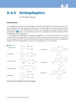
Jean-Martin Charcot was born in Paris, France, late in
1825 (Fig. 1.1). Although he was a 19th century scientist,
his influence carried on into the next century, especially
in the work of some of his well-known students, amongst
them Alfred Binet, Pierre Janet and Sigmund Freud
(Ekbom, 1992). He was a professor at the University of
Paris for 33 years, and in 1862 he began an association
with Paris’s Salpêtrière Hospital that lasted throughout
his life, ultimately becoming director of the hospital. In
1882, his focus turned to neurology, and he has been
called by some the founder of modern neurology. He
established a neurological clinic at the Salpêtrière that was
unique in Europe, and in so doing established the
bases for a neurological classification which have endured
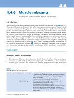
(see Fig. 1.2). He described multiple sclerosis [The
combination of nystagmus, intention tremor and
*Correspondence to: Andrew Eisen, Professor Emeritus, ALS Clinic, Vancouver General Hospital #322 Willow Pavilion, 805 West
12th Avenue, Vancouver, BC V5Z 1M9, Canada. E-mail: , Tel: +1(604)-875-4405, Fax: +1(604)-875-5867.
Fig. 1.1. Jean-Martin Charcot 1825–1893.
Ch01-N51894 9/8/06 10:26 AM Page 1
scanning or staccato speech, Charcot’s triad, is some-
times but not always associated with multiple sclerosis]
(Charcot, 1879). He attributed progressive and acute
muscular atrophy to lesions of the anterior horns of the
spinal cord and locomotor ataxia to the posterior horn
and spinal root. He gathered together the data leading to
the description of amyotrophic lateral sclerosis as is dis-
cussed below.
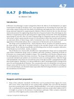
In 1873 he replaced Dr Alfred Vulpian (see Fig. 1.3)
as the Chair of Pathological Anatomy which he held for
a decade. He added histology to macroscopic anatomy
and undertook the exploration of the enormous
resources in pathology at Salpêtrière (Bonduelle, 1994,
1997). In the study of ‘Localizations of diseases of the
spinal cord (1873–74)’ he specified the anatomy and
physiology of the cord and subsequently cerebral locali-
zations of motor activities, both integral to his and
our understanding of amyotrophic lateral sclerosis
(known as Charcot’s disease before it popularly became
Lou Gehrig’s disease) (Bonduelle, 1994, 1997; Goetz,
1994, 2000).
An eminent scientist, Charcot was recognized as one
of the world’s most prominent professors of neurology.
In his time, he was both highly respected and chastized
as a third-rate show-off. His scientific career was a
continuous mixture of rigorous clinical neurology
(including detailed descriptions of amyotrophic lateral
sclerosis, Parkinson’s disease, brain anatomy, etc.) and
uncontrolled, controversial and sometimes even theatri-
cal experiments in the field of hysteria. Charcot’s fame
was as much the result of the unquestionable quality of
his scientific work as that of his theatrical presentations.
In the arts as in politics, he was more of a conservative
than an opportunist; authoritarian, shy, and brusque,
gloomy and taciturn, he nonetheless had a remarkable
power to attract.
Charcot’s understanding of ALS evolved over a
decade and was based on amazingly few patients (Goetz,
2000; Pearce, 2002). At the time of Charcot’s descrip-
tions of ALS, primarily 1850 to 1874, clinical diagnosis
was rudimentary and the distinction between upper and
lower motor neurons had not yet been made, and there
was no understanding of the role of the corticospinal
tract in connecting them (Goetz et al., 1995). The earli-
est description of ALS (1865) was that of a young
woman whose deficit was restricted to the upper motor
2
A. A. EISEN
Fig. 1.2. Saltpêtrière in 1882, the year that Charcot turned his thoughts to neurology.
Ch01-N51894 9/8/06 10:26 AM Page 2
neurons (in fact more likely primary lateral sclerosis).
She had been thought to be suffering from hysteria.
Autopsy showed ‘sclerotic changes limited to the lateral
columns of the spinal cord’ (Charcot, 1865). Four years
later (1869), in a series of papers written together with
Joffroy, Charcot reported cases of infantile and juvenile
spinal muscular atrophy in whom the lesions were
restricted to the anterior horn cells (Charcot and Joffroy,
1869a,b,c). Further clinical studies revealed a combina-
tion of upper and lower motor neuron signs which led
Charcot to coin the term ‘amyotrophic lateral sclerosis’
(Charcot, 1874, 1880). ‘We encountered several patients
with the following conditions: paralysis with spasms of
the arms and principally the legs (without any loss of
sensation), together with progressive amyotrophy, which
was confined mostly to the upper limbs and trunk’
(Charcot and Joffroy, 1869c).
He thought the anterior horn pathology followed
and was caused by disease of the lateral columns
and drew a parallel with anterior horn cell pathology
in multiple sclerosis, a concept not now in favor.
Gowers (1886) strongly contested Charcot’s notion that
ALS commenced in the descending motor tracts and
argued that the upper and lower motor neuron lesions
occurred independently of each other, which is the
general consensus. Eisen and Krieger (1998), however,
have adduced physiologic evidence that reinforced
Charcot’s ideas about the significance of upper and
lower motor neuron pathology.
His clinico-pathological observations led Charcot to
believe there was a two-part motor system organization.
Anterior horn cell disease resulted in weakness with
atrophy, and sclerosis of the lateral columns produced
spasticity with contractures (Charcot, 1880). Charcot
was not the first to describe cases of ALS, but did coin
the term amyotrophic lateral sclerosis (Rowland, 2001).
Charles Bell and others reported cases as early as 1824.
Having distinguished the motor functions of anterior
spinal nerve roots and the sensory functions of the
posterior roots, Bell was interested in finding patients
with purely motor disorders (Goldblatt, 1968). By mid-
century there were fiery debates among famous neurol-
ogists. Among the syndromes characterized by limb
weakness and muscle atrophy, they ultimately came to
separate neurogenic and myopathic diseases. It was not
clear whether some syndromes were variants of the
same condition or totally different disorders; this puzzle
included progressive muscular atrophy, progressive
bulbar palsy, primary lateral sclerosis and ALS.
Fortunately Charcot’s thoughts were also recorded
in English translations of the Tuesday Lectures at the
Hôpital de la Salpêtrière (references cited by Rowland
(2001)) and in translation by George Sigerson, who
included the essential concepts of Charcot’s ALS lec-
tures in English and Goetz has brought the translations
up-to-date (Goetz, 2000). The Tuesday Lectures also
exemplified Charcot’s zest for theatrical performance.
For example, during one lecture of 1888, Charcot said:
‘(To the patient): Give me your left arm. (Using a pin,
M. CHARCOT pricks at different points the arm and
the hand ).’ Charcot followed this performance with
another test, explaining to the audience as he did so
that: ‘You see that I am pulling the patient’s finger, even
a little brutally perhaps, without her suffering at all
[sans qu’elle éprouve rien] .’Turning to his subject, he
asked: ‘What am I doing to you?’ She replied: ‘I feel
nothing.’ The reality and authority of Charcot’s lecture
demonstrations was largely guaranteed by the fact
that they unfolded in real time before the audience
(Goetz, 1987).
Even though Charcot is credited as describing the
pathology of ALS, Cruveilhier (1853a,b) made an
essential contribution earlier, when he noted atrophy of
the anterior roots and suspected malfunction of the ante-
rior horn cells. Charcot knew of that work and compared
it with his own observations of anterior horn cell pathol-
ogy in infantile spinal muscular atrophy, poliomyelitis
and other disorders characterized by muscular atrophy.
HISTORICAL ASPECTS OF MOTOR NEURON DISEASES
3
Fig. 1.3. Alfred Vulpian who preceded Charcot as Professor
of Anatomy at Saltpêtrière.
Ch01-N51894 9/8/06 10:26 AM Page 3
The terminology of these cases was not clarified
for decades. Gowers (1886) is sometimes credited
for introducing the term ‘motor neuron disease’ in
1886–1888. However, that term must have come later
because Gowers used only the terms chronic spinal
muscular atrophy, ALS or chronic poliomyelitis. Brain
(1933) may have been the first to use ‘motor neuron dis-
ease’; in the first edition of his textbook, published in
1933. He gave ‘motor neuron disease’ as a synonym for
ALS (without mentioning why he used the new name).
It was 5 years after Charcot’s initial case report that
he first used the term ‘amyotrophic lateral sclerosis,’
which appeared in the title of the paper (http://clearx.
library.ubc.ca:2796/cgi/content/full/58/3/512-REF-
NHN7430-1; Charcot, 1874). In part IV of that series,
he recorded more observations that have become stan-
dard teachings: Amyotrophic paralysis starts in the
upper limbs as a cervical paraplegia. After 4, 5, 6
months or more, the emaciation spreads and there is
protopathic muscular atrophy, which advances for 2 or
3 years. After a delay of 6 or 9 months, the legs are
affected…but the muscles are conserved and contrast
singularly with the state of the upper limbs.
There is no paralysis of the bladder. The patient has
more difficulty walking and then cannot stand After
some time, the patient has noticed that, in bed or sitting,
the legs sometimes extend or flex until a position is pro-
duced involuntarily…and the legs come to resemble a
rigid bar. The rigidity is exaggerated when the patient is
held up by assistants who want to walk him. The feet
take on a posture of equinus varus. This rigidity, often
extreme, affects all joints by a spasmodic action of
the muscle. The tremor interferes with standing and
walking.
He summarized the features of amyotrophic lateral
sclerosis:
(1) Paralysis without loss of sensation of the upper
limbs, accompanied by rapid emaciation of the
muscles At a certain time, spasmodic rigidity
always takes over with the paralyzed and atrophic
muscles, resulting in permanent deformation by
contracture.
(2) The legs are affected in turn. Shortly, standing and
walking are impossible. Spasms of rigidity are first
intermittent, then permanent and complicated at
times by tonic spinal epilepsy. The muscles of the
paralyzed limb do not atrophy to the same degree
as the arms and hands. The bladder and rectum are
not affected. There is no tendency to the formation
of bedsores.
(3) In the third period, the preceding symptoms worsen
and bulbar symptoms appear. These three phases
happen in rapid succession – 6 months to 1 year
after the onset, all the symptoms have appeared and
become worse. Death follows in 2 or 3 years, on
average, from the onset of bulbar symptoms. This
is the rule but there are a few anomalies. Symptoms
may start in the legs or be limited to one side of the
body, a form of hemiplegia. In two cases, it started
with bulbar symptoms.
At present, the prognosis is grave. As far as I know,
there is no case in which all the symptoms occurred and
a cure followed. Is this an absolute block? Only the
future will tell. Charcot, therefore, gave a complete pic-
ture of ALS, emphasizing lower motor neuron signs in
the arms and upper motor neuron signs in the legs. His
description of the natural history, lamentably, has not
changed much in ensuing years. He described the
bulbar syndrome in detail. He described clonus and he
may have been the first to use the term ‘primary lateral
sclerosis.’
Charcot’s own assessment of ALS was clearly
stated:
‘
I do not think that elsewhere in medicine, in
pulmonary or cardiac pathology, greater precision can
be achieved. The diagnosis as well as the anatomy and
physiology of the condition “amyotrophic lateral scle-
rosis” is one of the most completely understood condi-
tions in the realm of clinical neurology’ (Charcot,
1874). Charcot died in 1893 in Morvan, France.
1.2. Notable names with ALS
Because ALS is rare (an incidence of < 2 per 100,000)
the list of famous or household names of people that
have or had the disease is rather short. Amongst these is
David Niven, the English actor, Dimitri Shostakovich,
the Russian composer and Mao Tse Tung, the revolu-
tionary leader of China. Nelson Butters was one of
America’s most distinguished neuropsychologists of
the last 25 years. He died from ALS in 1995 at age 58.
Like Stephen Hawking (see below), Dr Butters, toward
the end made use of computers to communicate and
work. This permitted him to edit a major journal in
neuropsychology, even when he could move only one
finger and then only one toe. With these small movements
he used Email to write to colleagues everywhere –
usually on professional matters, but also to transmit
amusing academic gossip. However, the two names that
have had the most impact are Lou Gehrig (Figs. 1.4 and
1.5) and Stephen Hawking (Fig. 1.6).
Of all the players in baseball history, none possessed
as much talent and humility as Lou (Henry Louis)
Gehrig. It seems that Lou Gehrig demonstrated the
characteristic ‘nice personality’ of so many patients
with ALS. His accomplishments on the field made him
an authentic American hero, and his tragic early death
4
A. A. EISEN
Ch01-N51894 9/8/06 10:26 AM Page 4
made him a legend. Gehrig’s later glory came from
humble beginnings. He was born on June 19, 1903 in
New York City. The son of German immigrants, Gehrig
was the only one of four children to survive. Is it there-
fore possible that Lou Gehrig had hereditary ALS, but
that his siblings never survived long enough to develop
the disease? His mother, Christina, worked tirelessly,
cooking, cleaning houses and taking in laundry to make
ends meet. His father, Heinrich, often had trouble find-
ing work and had poor health.
Gehrig’s consecutive game streak of 2,130 games
(a record that stood until Cal Ripken, Jr. broke it in
1995) did not come easily. He played well every day
despite a broken thumb, a broken toe and back spasms.
Later in his career Gehrig’s hands were X-rayed and
doctors were able to spot 17 different fractures that had
‘healed’ while Gehrig continued to play. Despite having
pain from lumbago one day, he was listed as the short-
stop and leadoff hitter. He singled and was promptly
replaced but kept the streak intact. His endurance and
strength earned him the nickname ‘Iron Horse.’ In 1938,
Gehrig fell below 0.300 average for the first time since
1925 and it was clear that something was wrong. He
lacked his usual strength. Teammate, Wes Ferrell noticed
that on the golf course, instead of wearing golf cleats,
Gehrig was wearing tennis shoes and sliding his feet
along the ground. Gehrig played the first eight games of
the 1939 season, but he managed only four hits. On a ball
hit back to pitcher Johnny Murphy, Gehrig had trouble
getting to first in time for the throw. On June 2, 1941, Lou
Gehrig succumbed to ALS and the country mourned.
Eleanor, his wife, received over 1,500 notes and telegrams
of condolence at their home in Riverdale, New York.
President Franklin Delano Roosevelt even sent her
flowers. Gehrig was cremated and his ashes were buried
at Kensico Cemetery in Valhalla, New York.
Stephen Hawking was born on the 300th anniversary
of Galileo’s death. He has come to be thought of as the
greatest mind in physics since Albert Einstein. With
similar interests – discovering the deepest workings of
HISTORICAL ASPECTS OF MOTOR NEURON DISEASES
5
Fig. 1.4. Lou Gehrig’s farewell speech.
Fig. 1.5. Lou Gehrig.
Fig. 1.6. Stephen Hawking.
Ch01-N51894 9/8/06 10:26 AM Page 5
the universe – he has been able to communicate arcane
matters not just to other physicists but to the general
public.
He grew up outside London in an intellectual family.
His father was a physician and specialist in tropical dis-
eases; his mother was active in the Liberal Party. He
was an awkward schoolboy, but knew from an early age
that he wanted to study science. He became increas-
ingly skilled in mathematics and in 1958 he and some
friends built a primitive computer that actually worked.
In 1959 he won a scholarship to Oxford University and
in 1962 he got his degree with honors and went to
Cambridge University to pursue a PhD in cosmology.
There he became intrigued with black holes (first pro-
posed by Robert Oppenheimer) and ‘space-time singu-
larities’ or events in which the laws of physics seem to
break down. After receiving his PhD, he stayed at
Cambridge, becoming known even in his 20s for his
pioneering ideas and use of Einstein’s formulae, as
well as his questioning of older, established physicists.
In 1968 he joined the staff of the Institute of Astronomy
in Cambridge and began to apply the laws of thermo-
dynamics to black holes by means of very complicated
mathematics.
At the remarkably young age of 32, he was named a
fellow of the Royal Society. He received the Albert
Einstein Award, the most prestigious in theoretical
physics. And in 1979, he was appointed Lucasian
Professor of Mathematics at Cambridge, the same post
held by Sir Isaac Newton 300 years earlier. In 1988
Hawking wrote A Brief History of Time: From the Big
Bang to Black Holes, explaining the evolution of his
thinking about the cosmos for a general audience. It
became a best-seller of long standing and established
his reputation as an accessible genius.
He remains extremely busy, his work hardly slowed
by amyotrophic lateral sclerosis. “My goal is simple. It
is complete understanding of the universe, why it is as
it is and why it exists at all.”
It is worthy and appropriate to mention one other
name, that of Professor Richard Olney, who, at the time
of writing, is in the terminal stages of ALS. It is impos-
sible to imagine the nightmare of a neurologist, dedi-
cated to ALS developing the disease that has occupied
his career. Richard, a personal friend of mine and many
of the contributors of this volume, started the ALS
Clinic at the University of California (San Francisco) in
1993. He was dedicated to the disease and care of
patients suffering from it. He contributed considerably
to the advancement of understanding ALS, especially
physiological aspects. He self-diagnosed the disease
about 2 years ago when on vacation he began stum-
bling. There is no other recorded precedent of an ALS
specialist developing the disease.
1.3. The first ALS gene
Charcot claimed that ALS was never hereditary. He
clearly overlooked Aran’s (1850) cases published 20
years earlier. As highlighted by Andersen (2003),
amongst Aran’s patients was a 43-year-old sea captain
presenting with cramps in the upper limb muscles and
subsequent wasting and weakness. He died within
2 years of onset of his disease and most likely had ALS.
Aran reports that one of the patient’s three sisters and
two maternal uncles had died of a similar disease. It
seems that this was the first hereditary case of ALS. It
took another 143 years before the superoxide dismutase
gene (SOD1) was discovered to be associated with famil-
ial ALS (Rosen et al., 1993). Eleven missence mutations
were found in 13 of 18 familial ALS (FALS) pedigrees.
1.4. Western Pacific ALS
It is not the role of this chapter to discuss similarities
and differences between Western Pacific ALS and the
disease elsewhere. However, the differences may be
more apparent than real. Evidence indicates that ALS
was prevalent on the island of Guam at least since 1815,
some 50 years before Charcot’s first descriptions
(Lavine et al., 1991). Had Charcot been able to visit
Guam one wonders what he would have made of the
disease. Although he described the pathology and
clinical picture so accurately it seems strange that there
was little reference if any as to its possible cause.
During the early years of American occupation of
Guam (1898–1920) death certificates were written in
Spanish and there were frequent deaths attributed to
“paralytico” or “lytico” terms the Chamorro used for
ALS. The term ‘rayput’ or ‘bodig’ (slowness or lazi-
ness) was used for Parkinsonism-dementia. The
Western Pacific form of ALS has been of interest for
over 50 years because its incidence, prevalence and
mortality rates were initially 50 to 100 times those of
ALS elsewhere. The male:female ratio approximated
2:1, the median age at onset was 44 years, familial
aggregation was recognized and ALS was associated
frequently with a Parkinsonism/dementia complex
(PDC) (Armon, 2003). Recently, the frequency of
Western Pacific ALS has declined, implying a tempo-
rary exposure to an environmental risk factor, possibly
in a genetically susceptible population. This has fueled
decades of research and speculation.
Marjorie Whiting, a nutritionist who lived with the
Chamorros in Guam, became convinced that the disease
resulted from ingestion of the cycad nut used to prepare
flour (Whiting, 1963). During the Japanese occupation
of Guam during World War II, many Chamorros fled
into the forests and may have eaten more cycad flour
6
A. A. EISEN
Ch01-N51894 9/8/06 10:26 AM Page 6
than usual. However, there is at least one well recorded
case of chronic cycad intake, without apparent harm, in
Sergeant Soichi Yokoi of the Japanese Imperial Army.
He was captured after 28 years as a fugitive in the jun-
gles of Guam and was wearing clothes that he had made
himself from fibers he had peeled from the bark of a
Pago tree. Such was the astonishing level of his self-
sufficiency that he was met with total disbelief until he
explained to his captors how he was able to survive for
over a quarter of a century by living off the natural
resources of the land. A principal part of his diet was
fadang. Remarkably, Sergeant Yokoi not only discov-
ered that fadang was edible but, astonishingly, devised a
way to prepare the nuts properly before cooking. He
lived to be 82, dying in 1997.
The cycad hypothesis was abandoned because two
similar clusters of neurodegenerative disease were
found in remote indigenous populations in Japan and
Papua New Guinea, neither of whom seemed to eat
cycad nuts. Also a good animal model never really
evolved (see Chapter 18). However, the cycad story
may have come back to life. It has now been suggested
that the answer may lie in the Chamorro’s favorite
entree: flying fox bats boiled in coconut cream (Cox
and Sacks, 2002). The bats have been especially desir-
able food items to the Chamorro, possibly because the
tradition is one of few retained from older times before
four centuries of upheaval and cultural oppression
which began with Spanish colonial rule in 1565. They
were served at weddings, fiestas, birthdays and the like.
The etiquette of bat-eating and preparation involves
rinsing off the outside of the animal like you would a
cucumber and tossing it in boiling water. The animals
are then served whole in coconut milk and are con-
sumed in their entirety. Meat, internal organs, fur, eyes
and wing membranes are all eaten.
So why the dramatic decrease in incidence of ALS on
Guam? Flying foxes are slow breeders, with females
needing to be 3 years old before they can successfully
give birth and rear babies. Then they rear only one young-
ster each year. Add this to the high death rate that is
common in any young wild animal. In fact, numbers of
flying foxes has dropped alarmingly towards extinction.
1.5. Spinal muscular atrophies
The clarity with which Charcot was able to describe
ALS was not matched by early descriptions of diseases
that appeared to be restricted to the lower motor
neuron, manifesting primarily by limb weakness.
This is hardly surprising when one considers that as
recently as 2003 classification of lower motor neuron
syndromes (including diffuse, proximal, distal and
monomelic) is still very much under discussion
(Van den Berg-Vos et al., 2003). The issue is further
complicated by early descriptions of primary muscle
disease, especially Duchenne muscular dystrophy,
which were being published about the same time as the
first descriptions of spinal muscular atrophy. Tyler
(2003) has recently reviewed the historical roots of
Duchenne muscular dystrophy in the 19th century,
citing early papers by Conte, Bell, Partridge and
Meryon through to the classic monographs of Duchenne
and Gowers. It is clear that a number of these cases
turned out to be anterior horn cell disease and not
primary muscle disease.
In 1850, Francois-Amilcar Aran described cases
using the name “progressive spinal muscular atrophy”
(Aran, 1850). However, there had not been an autopsy
study of these patients and there was no clinical distinc-
tion between neurogenic and myopathic diseases,
a notion that was yet to come. Aran was born in
Bordeaux, where he commenced his medical studies
but graduated in Paris. He published his first paper even
before he had become MD, for which honor he deliv-
ered an inaugural thesis entitled Des palpitations
du coeur, considérées principalement dans leur nature
et leur traitement (Aran, 1843).
He was active in the publishing of several journals,
among them Archives générales de médecine and the
Union médicale, to which he was one of the most pro-
lific contributors, publishing both his own papers as
well as analyses of English works. As professor agrégé,
he held private courses of therapy at the École pratique.
At the Hôtel-Dieu, as deputy to Léon Louis Rostand
(1790–1866), Aran’s clinical lectures were tremen-
dously successful. In the final years Aran preferably
concerned himself with studies of materia medica,
while still a prolific writer. One of his papers was on
acute rheumatism, from which he himself had suffered
repeatedly, and which caused his premature death on
February 22, 1861, at the age of only 44 years. He left
a large number of unfinished works, one of them a
Dictionnaire de thérapeutique, of which only the first
letters had been put on paper.
Duchenne (Fig. 1.7) claimed equal priority to
describing spinal muscular atrophy. He had studied all
of Aran’s patients with electrical stimulation (Duchenne
de Boulogne, 1851). However, it is not clear whether
Aran described Duchenne’s patients or vice versa.
Duchenne’s bid for priority was based on a notice of
50 words, not a scientific paper. The announcement
stated that, at a weekly meeting of the Academy (French
Academy of Science), he presented a collection of
papers, which he called ‘Recherches Electro-
Physiologiques’ and which he intended to be used as
evidence by future commission of authorities that never
left a record, if it ever existed. The ultimate compromise
HISTORICAL ASPECTS OF MOTOR NEURON DISEASES
7
Ch01-N51894 9/8/06 10:26 AM Page 7
was the eponym ‘Aran-Duchenne’ syndrome for what
we now regard as the broader categories of spinal mus-
cular atrophies.
Guillaume Benjamin Amand Duchenne descended
from a family of fishermen, traders and sea captains
who had resided in the harbor city Boulogne-sur-Mer in
Northern France since the first half of the 18th century.
He was predestined for a career at sea, as his father was
the commander Jean Duchenne who had been a ship’s
captain during the Napoleonic wars and expected his
son to follow in his keel waters.
Despite his father’s efforts to induce him to follow the
family seafaring tradition, his love of science prevailed.
Duchenne went to a local college at Douai, where he
received his baccalauréat at the age of 19. From 1827,
aged 21, he studied medicine under teachers like René-
Théophile-Hyacinthe Laënnec (1781–1826), Baron
Guillaume Dupuytren (1777–1835), François Magendie
(1783–1855) and Léon Cruveilhier (1791–1874). He
graduated in medicine in Paris in 1831 and, probably
influenced by Dupuytren, presented his Thèse de
médecine, a monograph on burns.
However, Duchenne’s early years in medicine were
undistinguished. His interest in “electropuncture,”
recently invented by Magendie and Jean-Baptiste
Sarlandière (1787–1838), enticed him back to Paris where
he was met with a rather cool reception, being ridiculed
for his provincial accent and his coarse manners.
Duchenne was never offered, and never applied for, an
appointment at a Paris teaching hospital or at the uni-
versity. He was known under the name of Duchenne de
Boulogne to avoid confusion with Édouard Adolphe
Duchesne (1894–1869), a fashionable society physician.
Nevertheless, Duchenne was a diligent investigator and
meticulous at recording clinical histories. When neces-
sary he would follow his patients from hospital to
hospital to complete his studies. In this way he achieved
an exceptionally rich and exquisite research material.
Toward the end of his life Duchenne became estab-
lished and popular, paradoxically Jean-Martin Charcot
was amongst his friends, and they held each other in
considerable esteem. His clinical ability was such that
the great Charcot dubbed him ‘The Master.’ At this
stage of his career he had become an international
celebrity. Every month he gave several dinner parties
for his colleagues (Charcot, Pierre Paul Broca, Auguste
Nélaton and Edmé Félix Alfred Vulpian). During
these get-togethers histological slides were projected
and discussed – mixed with funny pictures to please
Duchenne’s grandchild. These were the first attempts
at muscle pathology. Duchenne was probably the
first person to use biopsy procedure to obtain tissue
from a living patient for microscopic examination.
This aroused a deal of controversial discussion in
the lay press concerning the morality of examining
living tissues. In order to perform histopathological
diagnostics Duchenne constructed a biopsy needle, which
made possible percutaneous muscle biopsies without
anesthesia.
1.6. Spino-bulbar muscular atrophy
(SBMA) – Kennedy’s disease
[I am most grateful to my friend and colleague,
Professor William Kennedy for much of what is tran-
scribed verbatim from his records.]
In 1966 William Kennedy (Fig. 1.8) and co-workers
described an anterior horn cell disease characterized by
X-linked inheritance, onset in the 4th and 5th decades
and with slow progression with predominantly proxi-
mal spinal and bulbar muscle involvement and tongue
muscle furrowing. They commented on the associated
features of gynecomastia, diabetes and absence of long
tract signs (Kennedy et al., 1966).
8
A. A. EISEN
Fig. 1.7. Duchenne de Boulogne.
Ch01-N51894 9/8/06 10:26 AM Page 8
“In July 1964, George B., age 57, entered my
(Dr Kennedy’s) office.” He was of French-Indian descent
from a large family that lived on Grey Cloud island in the
Mississippi river. George complained of increasing gen-
eralized weakness and pain, mainly in the neck and
shoulders. As a youth he could run and work as well as
others. Since about age 35 he hadn’t felt strong. Muscle
cramps began in his chest, abdomen and calf and there
was twitching in his chin and shaking with his arms out-
stretched. Later he had definite weakness noticeable
when lifting objects over his head. Distal strength in his
hands remained good. When George was 37, a neuropsy-
chiatrist diagnosed primary muscular atrophy and com-
mented on the grooves in George’s tongue. Yet, from age
38 to 43 he worked in a slaughterhouse where he split
pigs down the back with a 16 lb cleaver. For about 20
years cold weather had hampered fine motions such as
buttoning his shirt. At about age 54 he began to aid chew-
ing by holding his chin up with his hands. At the same
time his voice changed pitch and began to be slurred. By
1964 walking required great effort.
At examination muscle weakness was generalized,
but more severe proximally. The gait was waddling. He
could not walk on his toes, hop, squat or rise. Reaching
overhead and heelwalking were moderately weak. The
biceps tendon reflex was depressed; all others were absent.
Large fasciculations were visible in the chin muscles. The
tongue was grooved and atrophic. Facial weakness was
marked and the lips protruded, but smiling was possible.
The voice was low pitched and gravelly. Word pro-
nunciation was poor. Sensation seemed normal.
Conduction hearing was decreased. There was bilateral
gynecomastia. The patient had been diagnosed with
diabetes at age 59.
Motor nerve conduction velocity was normal. EMG
showed scattered fibrillation potentials and giant motor
unit action potentials (MUAPs) in several muscles.
Muscle histology showed groups of atrophic muscle
fibers with small prominent groups of very large hyper-
trophic fibers with central nuclei and some basophilia.
He died of pneumonia at home at age 60. There was no
autopsy.
George’s father, Victor B, died at age 57. He had a
marked tremor at age 30. He was a farmhand until age
40 when he became too weak. He could ride a tractor
but could not mount or dismount alone. He needed a
railing to climb stairs. Rising from a chair required use
of his arms. George thought his father had had fascicu-
lations and a shrunken tongue. He was never hospital-
ized. There are no medical records. His possible
involvement initially caused us much confusion.
On August 7, 1964, Robert G, age 68, of German
and Swiss descent, was referred for anterior horn cell
disease. Robert had generalized weakness, areflexia
and the now familiar facies with fasciculations. NCV
was normal. EMG showed very large MUAPs. Median
nerve sensory responses were absent. The patient and
brother Alfred had been previously diagnosed by sev-
eral neurologists with primary muscular atrophy in
1951 and again in 1957. Alfred died in a university hos-
pital without autopsy. Brother William, with the same
disability, died of pneumonia in 1957. Robert died in
1967. Robert’s wife requested that autopsy material be
sent to me. There was marked reduction of anterior
horn cells at all levels, but the cells of Clarke’s column
and of the posterior horn were preserved. The anterior
spinal nerve roots contained fewer myelinated nerve
fibers than expected as compared with the posterior
spinal nerve roots. Muscle biopsy was identical to that
of George B. Similar cases had been described earlier,
but not fully appreciated (Kurland, 1957; Gross, 1966).
It was not until the 1980s that the association of
depressed or absent reflexes and small or absent sen-
sory potentials was described (Barkhaus et al., 1892;
Harding et al., 1982). In 1991 that genetic cause of
SBMA was identified by Albert La Spada and Kenneth
Fischbeck as the expansion of a polymorphic CAG
repeat sequence in the first exon of the gene encoding
the androgen receptor (La Spada et al., 1991).
Dr Kennedy was unaware that the disease he had
discovered was given his name until it appeared as such
in a paper by Schoenen et al. (1979).
HISTORICAL ASPECTS OF MOTOR NEURON DISEASES
9
Fig. 1.8. Dr William Kennedy the year he described spinob-
ulbar muscular atrophy.
Ch01-N51894 9/8/06 10:26 AM Page 9
1.7. Upper neuron syndromes
About a decade following Charcot’s (1865) original
description of amyotrophic lateral sclerosis (ALS), Erb
(1875) described a disorder characterized by exclusive
involvement of the corticospinal tract which he named
“spastic spinal paralysis.” Several cases given the name
of ‘lateral sclerosis’ were described even earlier, and
four of these were familial. In retrospect they most
likely represented some form of hereditary spastic para-
paresis, or one of the recently described infantile ALS
syndromes (Lerman-Sagie et al., 1996; Devon et al.,
2003). It appears that Charcot’s first case of ALS was in
fact a case of PLS.
Konzo was first identified in 1936 by Tessitore
(Trolli, 1938), a district medical officer in the Kahemba
District in the south-eastern part of the Bandundu
Province of the Democratic Republic of Congo (DRC).
There, konzo is known to be endemic with a prevalence
as high as 5% in certain villages. However, amongst 146
identified cases there were reports of some cases being
affected 30 to 40 years prior to 1937 when Tessitore
identified 140 new cases of konzo in the same area.
Konzo was brought to scientific attention by two epi-
demic outbreaks, each numbering more than 1,000 cases.
The first was in Bandundu Region in present-day Zaire
in 1936–37 and the second in Nampula Province of
Northern Mozambique in 1981. Smaller outbreaks in rural
areas have subsequently been reported from Zaire,
Mozambique, Tanzania and the Central African Republic.
Sporadic cases of konzo also occur in affected areas,
years after an extensive outbreak.
References
Alzheimer A (1991). A contribution concerning the patholog-
ical anatomy of mental disturbances in old age, 1899.
Alzheimer Dis Assoc Disord 5: 69–70.
Alzheimer A, Forstl H, Levy R (1991). On certain peculiar dis-
eases of old age. Hist Psychiatry 2: 71–101.
Alzheimer A, Stelzmann RA, Schnitzlein HN, Murtagh FR
(1995). An English translation of Alzheimer’s 1907 paper,
‘Uber eine eigenartige Erkankung der Hirnrinde’. Clin
Anat 8: 429–431.
Andersen PM (2003). Genetic aspects of amyotrophic lateral
sclerosis/motor neuron disease. In: Shaw PJ, Strong MJ
(Eds.) Motor Neuron Disorders. pp. 207–235.
Aran FA (1843). Des palpitations du coeur, considerees
prinicipalement dans leurs causes, leur nature et leur
traitement. Doctoral thesis. 31 pages. Paris, 1843, No. 98.
Aran FA (1850). Recherches sur une maladie non encore
decrite du systeme musculaire (atrophie musculaire pro-
gressive). Archs Gen Med 3: 5–35.
Armon C (2003). Western Pacific ALS/PDC and flying foxes:
what’s next. Neurology 61: 291–292.
Barkhaus PE, Kennedy WR, Stern LZ, Harrington RB (1892).
Hereditary proximal spinal and bulbar motor neuron dis-
ease of late onset. Arch Neurol 39: 112–116.
Bonduelle M (1994). [Charcot, anatomo-pathologist]. Arch
Anat Cytol Pathol 42: 171–180.
Bonduelle M (1997). [The Salpêtrière from Mazarin to
Charcot]. Hist Sci Med 31: 163–170.
Brain WR (1933). Diseases of the Nervous System. Oxford
University Press, London, UK.
Charcot J-M (1865). Sclerose des cordons lateraux de la
moelle epiniere chez une femme hysterique atteinnte de
contracture permanente des quatre membres. Bull Soc
Med (Paris): 24–35.
Charcot J-M (1874). Sclerose laterale amyotrophique. Oeuvres
Completes. Bureaux du Progres Medical 2: 249–266.
Charcot J-M (1879). Diagnostic des formes frustes de la scle-
rose en plaque. Progres Medical (Paris) 7: 97–99.
Charcot J-M (1880). Troubles trophiques consecutifs aux
lesions des nerfs. Oeuvres Completes. Bureaux du Progres
Medical 1: 1–32.
Charcot J-M, Joffroy A (1869a). Deux cas d’atrophie muscu-
laire progressive avec lesions de la substance grise et de
faisceaux anterolateraux de la moelle epiniere. Arch
Physiol Norm Pathol 1: 354–367.
Charcot J-M, Joffroy A (1869b). Deux cas d’atrophie muscu-
laire progressive avec lesions de la substance grise et de
faisceaux anterolateraux de la moelle epiniere. Arch
Physiol Norm Pathol 2: 628–649.
Charcot J-M, Joffroy A (1869c). Deux cas d’atrophie muscu-
laire progressive avec lesions de la substance grise et de
faisceaux anterolateraux de la moelle epiniere. Arch
Physiol Norm Pathol 3: 744–757.
Cox PA, Sacks OW (2002). Cycad neurotoxins, consumption
of flying foxes, and ALS-PDC disease in Guam.
Neurology 58: 956–959.
Cruveilhier J (1853a). Sur le paralysie musuclaire, progressive,
atrophique. Bull Acad Med 18: 490–501.
Cruveilhier J (1853b). Sur le paralysie musculaire, progressive,
atrophique. Bull Acad Med 18: 546–583.
Devon RS, Helm JR, Rouleau GA, Leitner Y, Lerman-Sagie
T, Lev D, Hayden MR (2003). The first nonsense mutation
in alsin results in a homogeneous phenotype of infantile-
onset ascending spastic paralysis with bulbar involvement
in two siblings. Clin Genet 64: 210–215.
Duchenne de Boulogne GB (1851). Recherches electro-phys-
iologiques et therapeutiques. Comp Rend Seances Acad
Sci 32: 506.
Eisen A, Krieger C (1998). Amyotrophic Lateral Sclerosis. A
Synthesis of Research and Clinical Practice. Cambridge
University Press, Cambridge, UK.
Ekbom K (1992). The man behind the syndrome: Jean-Martin
Charcot. J Hist Neurosci 1: 39–45.
Erb W (1875). Uber einen wenig bekannten Spinalen
Symptomencomplex. Berl Klin Wschr 12: 357–359.
Garcia-Ruiz PJ (2000). [Abnormal movements. Historical
notes]. Rev Neurol 31: 59–60.
Goetz CG (1987). Charcot, the Clinician: The Tuesday
Lessons. Raven Press, New York.
10
A. A. EISEN
Ch01-N51894 9/8/06 10:26 AM Page 10
Goetz C (1994). [Charcot: a scientific Janus]. Rev Neurol
(Paris) 150: 485–489.
Goetz CG (2000). Amyotrophic lateral sclerosis: early contribu-
tions of Jean-Martin Charcot. Muscle Nerve 23: 336–343.
Goetz CG, Bonduelle M, Gelfand T (1995). Charcot:
Constructing Neurology. Oxford University Press,
Oxford, UK.
Goldblatt D (1968). Motor neuron disease: historical intro-
duction. In: Norris FH, Kurland LT (Eds.) Motor Neuron
Disease. Grune & Stratton, New York, pp. 3–11.
Gowers WR (1886). Manual of Diseases of the Nervous
System. Churchill, London, UK.
Graeber MB, Kosel S, Egensperger R, Banati RB, Muller U,
Bise K, Hoff P, Moller HJ, Fujisawa K, Mehraein P (1997).
Rediscovery of the case described by Alois Alzheimer in
1911: historical, histological and molecular genetic analysis.
Neurogenetics 1: 73–80.
Gross M (1966). Proximal spinal muscular atrophy. J Neurol
Neurosurg Psychiatry 29: 20–34.
Harding AE, Thomas PK, Baraitser M, Bradbury PG, Morgan
Hughes JA, Ponsford JR (1982). X-linked recessive bul-
bospinal neuronopathy: a report of ten cases. J Neurol
Neurosurg Psychiatry 45: 1012–1019.
Hornykiewicz O (1977). Historical aspects and frontiers of
Parkinson’s disease research. Adv Exp Med Biol 90: 1–20.
Kennedy WR, Alter M, Foreman RT (1966). Hereditary prox-
imal spinal muscular atrophy of late onset. Neurology 16:
671–680.
Keppel Hesselink JM (1983). Some historical aspects of
Parkinson’s disease. The progression of insights in the
period 1817–1868. Janus 70: 263–279.
Kurland LT (1957). Epidemiologic investigation of amy-
otrophic lateral sclerosis. III A genetic interpretation of
incidence and geographic distribution. Proc Staff Meet
Mayo Clinic 32: 449–462.
La Spada AR, Wilson EM, Lubahn DB, Harding AE,
Fishbeck KH (1991). Androgen receptor gene mutations
in X-linked spinal and bulbar muscular atrophy. Nature
352: 77–79.
Lavine L, Steele JC, Wolfe N, Calne DB, O’Brien PC,
Williams DB, Kurland LT, Sxhoenberg BS (1991).
Amyotrophic lateral sclerosis/parkinson-dementia com-
plex in southern Guam: is it disappearing. In: Rowland LP
(Ed.) Advances in Neurology, Vol. 56: Amyotrophic Lateral
Sclerosis and Other Motor Neuron Diseases. Raven Press,
New York, pp. 271–285.
Lerman-Sagie T, Filiano J, Smith DW, Korson M (1996).
Infantile onset of hereditary ascending spastic paralysis
with bulbar involvement. J Child Neurol 11: 54–57.
Pearce JM (2002). Amyotrophic lateral sclerosis. J Neurol
Neurosurg Psychiatry 73: 671.
Rosen DR, Siddique T, Patterson D, Figlewicz DA, Sapp P,
Hentati A, Donaldson D, Goto J, O’Regan JP, Deng HX
(1993). Mutations in Cu/Zn superoxide dismutase gene are
associated with familial amyotrophic lateral sclerosis.
Nature 362: 59–62.
Rowland LP (2001). How amyotrophic lateral sclerosis got its
name: the clinical-pathologic genius of Jean-Martin
Charcot. Arch Neurol 58: 512–515.
Schoenen J, Delwaide PJ, Legros JJ, Franchimont P (1979).
Hereditary motor neuron disease: the proximal, adult,
sex-linked for Kennedy disease. Clinical and neuroen-
docrinologic observations. J Neurol Sci 41: 343–357.
Schwartz MF, Stark JA. (1992). The distinction between
Alzheimer’s disease and senile dementia: historical consid-
erations. J Hist Neurosci 1: 169–187.
Trolli G (1938). Paraplégie spastique épidémique, ‘Konzo’
des indigènes du Kwango. In: Trolli G (Ed.) Résumé des
observations réunies, au Kwango, au sujet de deux affec-
tions d’origine indéterminée. Fonds Riene Elizabeth,
Brussels, pp. 1–36.
Tyler KL (2003). Origins and early descriptions of ‘Duchenne
muscular dystrophy’. Muscle Nerve 28: 402–422.
Van den Berg-Vos RM, Visser J, Franssen H, de Visser M,
de Jong JMBV, Kakmijn S, Wokke JHJ, Van den Berg LH
(2003). Sporadic lower motor neuron disease with
adult onset: classification of subtypes. Brain 126:
1036–1047.
Whiting MG (1963). Toxicity of cycads. Econ Bot 17:
271–302.
HISTORICAL ASPECTS OF MOTOR NEURON DISEASES
11
Ch01-N51894 9/8/06 10:26 AM Page 11
Handbook of Clinical Neurology, Vol. 82 (3rd series)
Motor Neuron Disorders and Related Diseases
A.A. Eisen, P.J. Shaw, Editors
© 2007 Elsevier B.V. All rights reserved
Chapter 2
Comparative anatomy and physiology of the
corticospinal system
MARC H. SCHIEBER*
Departments of Neurology and Neurobiology & Anatomy and the Brain Injury Rehabilitation Program at
St. Mary’s Hospital, University of Rochester School of Medicine and Dentistry, Rochester, New York, USA
2.1. Introduction
In 1905, Campbell compiled his histologic studies of
the cerebral cortex in normal apes and humans, in a
number of human amputees and in two patients with
amyotrophic lateral sclerosis (ALS). He noted that the
giant Betz cells of the precentral gyrus: (1) occupied
the cortical territory where Grunbaum and Sherrington
had elicited contralateral movement with electrical
stimulation in the same individual apes, (2) underwent
retrograde transneuronal degeneration after amputa-
tion, and (3) degenerated in ALS (Campbell, 1905).
Synthesizing these observations, Campbell recognized
that the cortex containing Betz cells provided the most
direct connections from the cerebral cortex to spinal
motoneurons, a corticospinal projection.
Since Campbell’s time, anatomical and physiological
studies in both humans and animals have revealed that
the corticospinal system is more complex than a single
pathway directly connecting Betz cells in one hemi-
sphere to motoneurons in the contralateral spinal cord.
Much of what we know about the corticospinal system
in man, however, is based on extrapolation from phylo-
genetic trends identified in the more precise and detailed
studies that can be performed in experimental animals.
Care must be taken in extrapolating this information to
humans, as species differences clearly exist. For example,
the number of axons in the pyramidal tract increases
along the phylogenetic scale as follows: rat 73,000; cat
186,000; monkey 554,000; chimpanzee 800,000; human
1,100,000 (Lassek and Rasmussen, 1939; Lassek, 1941;
Lassek and Wheatley, 1945). In addition to the greatest
number of descending axons, humans probably have
more direct corticomotoneuronal connections than any
other species, and humans therefore are more dependent
on their corticospinal tract for normal movement.
Nevertheless, a comparative approach offers the most
detailed understanding possible of the corticospinal
tract, which has been studied in numerous mammalian
species (Heffner and Masterton, 1975; Armand, 1982).
For comparison with humans, we will focus here on the
most intensively studied non-human species, the domes-
tic cat (Felix domesiticus), and old world, macaque
monkeys (Macaca species). Other species – rodents,
new world monkeys, baboons, apes, etc. – will be men-
tioned only to develop specific points. For detailed and
extensive information, the interested reader is referred to
a number of comprehensive monographs (Lassek, 1954;
Phillips and Porter, 1977; Porter and Lemon, 1993).
2.2. The corticospinal tract
The general course of the corticospinal tract is well-known,
descending from the motor cortex to the medullary pyra-
mid and then decussating to the dorsolateral funiculus of
the contralateral spinal cord. Weakness, therefore, is con-
tralateral to lesions of this pathway in the brain. A more
detailed consideration of the corticospinal tract reveals
additional complexities, however, that may account for a
wider variety of phenomena observed clinically.
In all species, the corticospinal tract arises by and
large from Brodmann’s area 4, which is considered to
be the primary motor cortex (M1). In cats, area M1 lies
within the lateral aspect of the cruciate sulcus and
extends on to the surrounding hemispheric surface. In
macaque monkeys, M1 lies in the anterior bank of the
*Correspondence to: Marc H. Schieber, MD, PhD, University of Rochester Medical Center, Department of Neurology, 601 Elmwood
Avenue, Box 673, Rochester, NY 14642, USA. E-mail: , Tel: +1(585)-275-3369, Fax: +1(585)-244-2529.
Ch02-N51894 9/8/06 10:27 AM Page 15
central sulcus and extends onto the posterior half of the
surface of the precentral gyrus. In humans, M1 lies
largely within the anterior bank of the central sulcus,
extending on to the surface of the precentral gyrus
primarily in the medial, leg representation (see
Fig. 2.8(A,B)) (Campbell, 1905; Zilles et al., 1995).
The pyramidal somata of corticospinal tract neurons
are located in the deepest part of cortical layer V.
Although the giant Betz cells often are assumed to be
the only neurons from which the corticospinal projec-
tion originates, many other large and moderate sized
pyramid-shaped somata in layer V contribute axons to
the corticospinal tract. This can be illustrated by com-
paring the number of Betz cells in the human M1,
34,000, to the number of axons in human pyramid,
1,100,000 (Lassek and Wheatley, 1945). Only 3% of
the tract arises from Betz cells, while the remaining
97% arises from smaller neurons.
As axons descend from layer V toward the white
matter, some give off collaterals that travel horizontally
within the cortical gray up to several millimeters, pro-
viding interconnections within the major body part rep-
resentations of M1 (Ghosh and Porter, 1988; Huntley
and Jones, 1991). Descending through the centrum
semiovale, corticospinal axons from M1 converge in
the middle third of the posterior limb of the internal
capsule. Descending to the level of the midbrain, corti-
cospinal fibers lie in the middle third of the cerebral
peduncle, with corticobulbar fibers from more anterior
regions of the frontal lobe in the medial third and those
from the parietal and temporal lobes in the lateral third
of the peduncle. As they enter the base of the pons, the
descending fibers of the cerebral peduncle become
intermingled with the somata and crossing axons of the
neurons of the pontine nuclei. The bulk of corticofugal
fibers that enter the pons from the cerebral peduncle
terminate here in the pontine nuclei. Fibers destined for
the spinal cord emerge on the ventral aspect of the
medulla as the pyramid.
In the pyramid of macaque monkeys, axons that
have descended from the face representation tend to lie
dorsally, while those that have descended from the
upper extremity and lower extremity representations
are intermingled throughout the cross-sectional area
(Coxe and Landau, 1970). As they approach the cervi-
comedullary junction, fibers from the face representa-
tion turn dorsally to enter the medullary tegmentum, the
majority decussating to the opposite side to innervate
the pontomedullary reticular formation and bulbar
nuclei. In monkeys, cortical innervation of the facial
nucleus is directed primarily to the lateral motoneurons
that innervate lower facial muscles, whereas the dorsal
motoneurons that innervate upper facial muscles
receive relatively little cortical innervation (Jenny and
Saper, 1987). In humans, this difference may account in
part for the relative sparing of upper facial strength
after unilateral cortical lesions.
As fibers from the upper and lower extremity repre-
sentations reach the cervicomedullary junction, the
majority likewise leave their position on the ventral
aspect of the neuraxis, turn dorsally and decussate,
entering the lateral column of the spinal cord, where
they concentrate in the dorsolateral funiculus (Fig. 2.1).
(In rodents, however, the crossed corticospinal fibers
descend in the ventral-most base of the dorsal column
(Brown, 1971; Wise and Donoghue, 1986).) A minority
(~10%) of corticospinal fibers remain in their ventral
location, uncrossed, in the anterior column of the cord
adjacent to the anterior fissure. In approximately 75%
of human cases, the lateral and anterior corticospinal
tracts are asymmetric, with the right side larger than the
left (Nathan et al., 1990). Such asymmetry has not been
reported in monkeys and may be one anatomical feature
related to human handedness.
16
M. H. SCHIEBER
Fig. 2.1. The human corticospinal tracts. Sections of the
medulla and spinal cord stained with the Marchi method for
degenerating fibers are shown from a 69-year-old man who
sustained an extensive infarct in the territory of the right
middle cerebral artery 17 days before death. The right
medullary pyramid and left dorsolateral funiculus show
numerous degenerating fibers. In the C3 section, the anterior
corticospinal tract can be seen as a crescent of degenerating
fibers in the right anterior column adjacent to the central fis-
sure. Calibration bars represent 1 mm. Modified from Nathan
et al. (1990).
Ch02-N51894 9/8/06 10:27 AM Page 16
The lateral and anterior corticospinal tracts typically
are assumed to be crossed versus uncrossed, respec-
tively. In monkeys, however, a small number of
uncrossed axons can be observed in the dorsolateral
funiculus (Liu and Chambers, 1964). These uncrossed
axons in the lateral column when stimulated are suffi-
cient to excite motoneurons ipsilateral to the hemisphere
of origin (Bernhard et al., 1953). Other corticospinal
axons that have decussated at the cervicomedullary
junction and descend in the dorsolateral funiculus cross
back through the gray matter commissure of the spinal
cord, terminating ipsilateral to the hemisphere of origin
(Chambers and Liu, 1957; Liu and Chambers, 1964;
Galea and Darian-Smith, 1997a). Similarly, a small
number of corticospinal axons that have decussated at
the cervicomedullary junction descend close to the
medial aspect of the ventral horn. Some of these decus-
sated fibers eventually cross back in the anterior white
matter commissure, terminating ipsilateral to the hemi-
sphere of origin (Chambers and Liu, 1957; Liu and
Chambers, 1964; Nathan et al., 1990). Both uncrossed
and doubly decussating fibers may contribute to obser-
vations in human patients that, although only weakness
contralateral to a lesion above the pyramidal decussation
may be appreciated clinically, ipsilateral weakness can
be measured objectively as well (Colebatch and
Gandevia, 1989; Adams et al., 1990).
Nevertheless, the bulk of the lateral corticospinal
tract is crossed and descends in a position close to the
dorsolateral aspect of the ventral horn, where the motor
nuclei of distal limb musculature are located (Kuypers,
1982; Dum and Strick, 1996). In contrast, the bulk of
the anterior corticospinal tract is uncrossed and
descends in a position close to the anteromedial aspect
of the ventral horn, where the motor nuclei of proximal
limb musculature are located. In general the lateral cor-
ticospinal tract exerts greater control over distal limb
than axial musculature, whereas the anterior corti-
cospinal tract exerts more control over axial and proxi-
mal limb than distal limb musculature.
Fibers descending from the hand and foot repre-
sentations in the motor cortex are intermingled in the lat-
eral and ventral tracts. The majority of fibers originating
in the upper extremity representation terminate in the
gray matter of the cervical enlargement, while the major-
ity of fibers from the leg representation terminate in the
lumbosacral enlargement. Some fibers from the motor
cortex forelimb representation, however, send collaterals
to terminate at cervical levels, and then continue to
descend in the dorsolateral funiculus down to lum-
bosacral levels, where they terminate in the intermediate
zone of the spinal gray (Kuypers, 1960; Liu and
Chambers, 1964; Shinoda et al., 1976). Some fibers from
the hindlimb representation conversely send collaterals
to terminate in the cervical enlargement as they descend
to end at lumbosacral levels. These corticospinal axons
that terminate at somatotopically inappropriate spinal
levels may play a role in coordinating posture and move-
ment of the upper and lower extremities.
2.3. Terminations in the spinal gray matter
As they reach the appropriate spinal levels, descending
corticospinal axons enter the spinal gray matter.
Ascending the phylogenetic scale from rat to cat
through monkey and chimpanzee to the human, cortico-
spinal terminations shift progressively more ventrally
in the spinal gray (Fig. 2.2). While maintaining some
contact with the dorsal horn, the corticospinal termina-
tions overall achieve progressively closer interneuronal
access to motor output and ultimately increasingly
numerous, direct synaptic contacts to the spinal
motoneurons themselves (Kuypers, 1958, 1960, 1982).
A parallel trend from sensory to increasingly direct
motor innervation is found comparing the corticobulbar
projection in different species.
COMPARATIVE ANATOMY AND PHYSIOLOGY OF THE CORTICOSPINAL SYSTEM
17
Fig. 2.2. Comparative trends in the corticospinal tract and its
terminations. The position of descending corticospinal fibers
and their terminations in the gray matter of the brachial
enlargement is illustrated schematically for four species:
opossum, cat, Rhesus (macaque) monkey and chimpanzee. In
rodents and marsupials the corticospinal tract descends pri-
marily in the contralateral dorsal column and terminates
largely in the dorsal horn. In higher mammals the tract
descends largely in the contralateral dorsolateral funiculus,
although some uncrossed fibers are found in the ipsilateral
dorsolateral column and others in the anterior column, partic-
ularly in primates. Terminations in these species are largely in
the intermediate zone, though in monkeys and chimpanzees
increasingly numerous terminations are found among the
motoneuron cell columns (lamina IX). Reproduced from
Kuypers (1982) with permission from Elsevier.
Ch02-N51894 9/8/06 10:27 AM Page 17
Within the gray matter, corticospinal axons ramify
and synapse extensively on interneurons. In all species
that have been studied, the greatest number of cortico-
spinal terminations are found in Rexed’s laminae V and
VI (Heffner and Masterton, 1975). In cats, the bulk of
terminations are found in the base of the dorsal horn and
intermediate zone (Chambers and Liu, 1957). Some ter-
minations extend as well into lamina VII, but do not
reach lamina IX (Futami et al., 1979; Shinoda et al.,
1986). Although motoneuron dendrites extend into the
intermediate zone of the spinal gray matter and some
light microscopy studies have visualized corticospinal
boutons on motoneurons (Liang et al., 1991), in rats and
cats corticospinal axons do not make physiologically
evident synaptic contact with motoneurons (Lloyd,
1941; Hern et al., 1962; Alstermark et al., 2004).
Instead, feline corticospinal axons synapse on
interneurons in the intermediate zone. Many of these
interneurons have excitatory effects on motoneurons.
Through excitatory interneurons, trains of pyramidal
tract stimulation can facilitate the mono-synaptic reflex
as well as oligo-synaptic reflexes (Lloyd, 1941) and
evoke movement (Adrian et al., 1939; Landau, 1952).
Pyramidal volleys can facilitate cutaneous reflexes as
well (Sasaki et al., 1996). Especially well studied in
cats are certain classes of inhibitory interneurons. Ia
inhibitory interneurons receive synaptic inputs from
primary muscle spindle afferents and deliver synaptic
inhibition to heteronymous motoneurons. Ib inhibitory
interneurons receive synaptic input from Golgi tendon
organs and deliver synaptic inhibition to homonymous
motoneurons. These Ia and Ib inhibitory interneurons
receive additional synaptic inputs from numerous other
segmental and descending sources, including cortico-
spinal neurons (Lundberg, 1979). Via these synaptic
connections to spinal interneurons, the corticospinal
system can influence basic reflexes. In monkeys as
well, corticospinal neurons inhibit motoneurons via
spinal inhibitory interneurons (Preston and Whitlock,
1960, 1961). Though studied less directly in humans,
the organization of these reflex interneurons and their
control by the corticospinal system appear to be gener-
ally similar to that in the cat (Jankowska and Hammar,
2002; Petersen et al., 2003).
The loss of corticospinal control may account in part
for reflex changes associated with corticospinal lesions.
Reduced corticospinal input to Ia and Ib inhibitory
interneurons may contribute to hyperreflexia. Also fol-
lowing corticospinal lesions, tendon jerks that normally
elicit reflex contraction only in the stretched muscle may
elicit contraction in additional muscles. Stretch of the
flexor digitorum profundus tendons, for example, may
elicit an abnormal reflex contraction of the flexor polli-
cis longus (Hoffmann’s sign). In cats, muscle afferents
normally facilitate many heteronymous motoneurons in
addition to homonymous motoneurons (Fritz et al., 1989;
Wilmink and Nichols, 2003). Transmission through
these heteronymous reflex pathways normally may be
checked by corticospinal influence on inhibitory
interneurons in the spinal cord. Loss of this influence
then may lead to the abnormal spread of reflexes
observed after corticospinal lesions in humans.
In addition to modulating spinal reflex pathways, the
corticospinal system has influence over neuronal cir-
cuits in the spinal cord that constitute the central pattern
generators (CPGs) for cyclical motor behaviors such as
walking. In rats, repetitive discharge of a single motor
cortex neuron (driven by intracellular depolarization)
can be sufficient to activate the CPG that drives whisk-
ing movements of the vibrissae (Brecht et al., 2004). In
cats, although the corticospinal tract is not essential for
initiation of locomotion or for ambulation on a flat sur-
face (Drew et al., 2002), pyramidal tract neurons dis-
charge intensely in lifting the foot over an obstacle or
during complex locomotion on the rungs of a horizon-
tal ladder (Beloozerova and Sirota, 1993a; Drew, 1993;
Widajewicz et al., 1994). The primary role of the corti-
cospinal system in feline locomotion thus may lie in
modifying the basic rhythmic pattern to adapt to com-
plex circumstances.
Corticospinal neurons also contact a special class of
long propriospinal neurons at cervical levels just above
the brachial enlargement, C3–C4. In cats, these pro-
priospinal neurons help mediate visually guided reaching.
Collaterals of descending corticospinal, rubrospinal and
tectospinal inputs converge on these C3–C4 pro-
priospinal neurons, which in turn send their axons in the
ventrolateral funiculus down to forelimb motoneurons
in the lower cervical segments (Illert et al., 1978;
Alstermark et al., 1991). Stimulation of the pyramidal
tract produces disynaptic EPSPs in forelimb motoneu-
rons, which persist after a lesion of the lateral corti-
cospinal tract at C5, but are abolished by an additional
lesion of the ventrolateral funiculus at C5. These obser-
vations indicate that the lateral corticospinal tract
excites the C3–C4 propriospinal neurons, which in turn
excite distal forelimb motoneurons in the C6–T1 spinal
segments. Lesions of the corticospinal tract at C2, or of
the ventrolateral funiculus at C5, result in inaccurate
reaching, indicating that the information transmitted by
the descending corticospinal and rubrospinal systems to
the C3–C4 propriospinal neurons and thence to distal
forelimb motoneurons, plays an important role in visu-
ally guided reaching (Alstermark et al., 1981).
In primates, the disynaptic EPSPs in forelimb
motoneurons characteristic of the C3–C4 propriospinal
neurons are weaker and less common than in cats
(Maier et al., 1998). Administration of strychnine,
18
M. H. SCHIEBER
Ch02-N51894 9/8/06 10:27 AM Page 18
however, reveals disynaptic EPSPs that are abolished
by a lesion in the dorsolateral funiculus at C2, but not
by a similar lesion at C5 (Alstermark et al., 1999).
These observations suggest that C3–C4 propriospinal
neurons in primates receive more glycinergic inhibition
than do the homologous neurons in cats. Alternatively,
the strength of C3–C4 propriospinal input to forelimb
motoneurons may decrease from cats through different
species of primates to humans, as the strength of direct
corticomotoneuronal projections increase (Nakajima
et al., 2000). Nevertheless, in macaque monkeys the
C3–C4 propriospinal system still appears to contribute
to accurate control of dexterous finger movements
(Sasaki et al., 2004).
In humans, the presence of similar propriospinal neu-
rons is indicated by the facilitation of H-reflexes result-
ing from stimulation of cutaneous or mixed nerve
afferents. The central latency of such facilitation (typi-
cally 3–6 milliseconds) is too long to be attributed to
segmental interneurons, but too short to be mediated by
supraspinal loops, suggesting that the afferent impulses
act via neurons a few spinal segments away from the
motoneurons probed by the H-reflex (Burke et al., 1992;
Gracies et al., 1994; Mazevet et al., 1996). Additional
facilitation appears during weak voluntary contraction
of the muscle, suggesting that descending and afferent
inputs converge on human propriospinal interneurons.
2.4. Direct cortico-motoneuronal connections
Although the bulk of corticospinal terminations still
are found in the intermediate zone of the spinal gray,
in macaque monkeys many corticospinal axons
extend ventrally into lamina IX of the spinal gray
matter (Figs 2.2 and 2.9(A)) (Liu and Chambers, 1964;
Kuypers, 1982; Dum and Strick, 1996). Here they
ramify and make direct synaptic contact with motoneu-
rons (Hoff and Hoff, 1934; Lawrence et al., 1985). The
ramifications and terminations of corticospinal axons in
lamina IX are denser still in chimpanzees (Kuypers,
1982) and humans (Schoen, 1969). These corticospinal
connections with motoneurons may be particularly
associated with relatively fine, independent digit move-
ments, which are more highly developed as one pro-
gresses from monkeys to apes to humans. Comparing
two species of new world monkeys, for example,
revealed that the more dexterous cebus monkey has
more corticospinal terminations in lamina IX than the
less dexterous squirrel monkey (Bortoff and Strick,
1993). Certain dexterous carnivores, including the rac-
coon and the kinkajou, also have corticospinal termina-
tions in lamina IX (Heffner and Masterton, 1975).
Experimental demonstration of physiologically
active synapses made directly on motoneurons by
corticospinal axons has been obtained in monkeys and
baboons. The delay from arrival of an electrically
evoked descending corticospinal volley at a given
spinal segment to the appearance of an evoked volley in
the ventral roots (Bernhard et al., 1953), to facilitation
of monosynaptic reflexes (Preston and Whitlock, 1960)
or to the onset of EPSPs in motoneurons (Preston and
Whitlock, 1961; Clough et al., 1968; Jankowska et al.,
1975), all are consistent with monosynaptic transmis-
sion. Based on the phylogenetic trend from monkeys to
apes to humans, these direct cortico-motoneuronal
(CM) synaptic connections generally are inferred to be
even more important for normal function in humans
than in monkeys.
Individually, these corticomotoneuronal connections
are not necessarily the strongest synaptic inputs
received by motoneurons. In macaque monkeys,
a single corticospinal axon makes only 1–2 synaptic
boutons on the proximal dendrites of a given cervical
motoneuron (Fig. 2.3(A,B)) (Lawrence et al., 1985).
Minimal cortically evoked monosynaptic EPSPs in
lumbar motoneurons are smaller than minimal Ia
EPSPs (Porter and Hore, 1969). The time constants of
corticomotoneuronal EPSPs also are longer than those
of Ia EPSPs, indicating that the CM synapses are situ-
ated more peripherally on the motoneuron dendrites.
Nevertheless, the maximal CM EPSPs in baboon cervi-
cal motoneurons evoked by stimulation of the cortical
surface are larger than the maximal homonymous Ia
EPSPs evoked by stimulation of the muscle nerve, sug-
gesting a greater total input to the motoneurons from
CM cells than from Ia afferents (Clough et al., 1968). In
humans, CM synaptic boutons may be located in part
on the motoneuron somata (Schoen, 1969), which
suggests a stronger synaptic effect than in monkeys.
The effectiveness of primate CM synapses is
enhanced further by facilitation at higher frequencies
(Fig. 2.3(C)). When the cortex is stimulated with short,
high frequency bursts (e.g. above 50 Hz), the sequential
CM EPSPs within a burst become progressively larger,
beyond simple temporal summation (Landgren et al.,
1962b) and this facilitation becomes more prominent as
stimulation frequency increases (Muir and Porter,
1973). The corticospinal volley recorded in the lateral
column does not facilitate during such bursts, and facil-
itation also is seen when the pyramidal tract is stimu-
lated, suggesting that this facilitation involves a
mechanism within the spinal cord (Phillips and Porter,
1964). Such facilitation is not seen when stimulation of
the peripheral nerve produces Ia EPSP volleys in the
same temporal pattern, and thus the facilitating EPSPs
appear to be a property specific to CM synapses.
Facilitation at higher frequencies also has been shown
for the projection of single CM cells on a motoneuron
COMPARATIVE ANATOMY AND PHYSIOLOGY OF THE CORTICOSPINAL SYSTEM
19
Ch02-N51894 9/8/06 10:27 AM Page 19
pool in awake behaving monkeys (Lemon and Mantel,
1989). Thought to result from a mechanism in the
presynaptic terminals, this facilitation makes CM
EPSPs more effective at discharging motoneurons
when CM cells discharge high frequency bursts. After
lesions of the corticospinal system in humans, loss of
this potent excitation of motoneurons may result in a
reduced ability to recruit motoneurons and to drive
them at high frequencies, with consequent weakness
and slowness of voluntary movement.
Thus, as one ascends the scale from cats to monkeys
to chimpanzees, a trend becomes apparent for increased
numbers of direct corticomotoneuronal connections.
Although histologic evidence of such CM synapses in
humans is limited (Schoen, 1969), the trend generally is
used to project that in humans direct CM connections
are more numerous than in any other species.
Physiologically, the presence of CM connections in
humans has been inferred from the features of post-
stimulus time histogram peaks in the discharge times of
single motor units aligned on the delivery of transcra-
nial magnetic stimulation pulses to the motor cortex
(Palmer and Ashby, 1992; Petersen et al., 2003). The
extent of direct corticomotoneuronal connections in
humans, however, has yet to be fully explored.
2.5. Divergence and convergence in the cortico-
motoneuronal projection
The common clinical appellation, ‘upper motor
neuron,’ has fostered the assumption that individual
corticospinal neurons contact only spinal motoneurons
innervating a single muscle. However, evidence accu-
mulated over the last three decades shows that this is
not the case. Single pyramidal tract neurons initially
were shown to be antidromically activated by electrical
microstimulation in the motor nuclei of more than one
hindlimb muscle in the monkey lumbar enlargement
(Asanuma et al., 1979) and at more than one segmental
level in the cervical enlargement (Shinoda et al., 1979).
Filling with HRP then revealed that single corticospinal
axons indeed give off multiple collaterals that enter the
spinal gray at different segmental levels and ramify in
the motor nuclei of multiple muscles (Fig. 2.4(A))
(Shinoda et al., 1981).
That such terminal ramifications indeed provide
physiological innervation of multiple motoneuron
pools from single CM cells has been shown by spike-
triggered averaging of EMG activity in awake behaving
monkeys. Averages of rectified EMG triggered from the
spikes discharged by a single neuron in the primary
motor cortex sometimes show post-spike facilitatory
peaks. Such peaks indicate that excitatory input arrived
in the motoneuron pool at a fixed latency consistent
with a monosynaptic connection from the recorded M1
neuron to spinal motoneurons contributing to the EMG.
Such post-spike facilitation has been observed in the
EMG from multiple muscles recorded simultaneously
with the spike discharge of a single monkey M1 neuron
(Fig. 2.4(B)). Shown first in forearm muscles acting on
the wrist and fingers (Fetz and Cheney, 1980), multiple
intrinsic muscles of the hand also may receive input
from a single M1 neuron (Buys et al., 1986). Post-spike
facilitation from CM cells is more prevalent in intrinsic
20
M. H. SCHIEBER
Fig. 2.3. The corticomotoneuronal synapse. (A) Camera
lucida drawing of a corticospinal axon ramifying in lamina IX
and contacting a proximal dendrite of a motoneuron with a
single bouton (arrow). (B) Light micrograph of the same
synapse indicated by the arrow in A. Calibration bar represents
10 µ. (C) Facilitation of corticomotoneuronal EPSPs. Each
trace shows the intracellular voltage recorded from a motoneu-
ron averaged across 256 repetitions of the same stimulus. In
the top trace, a single cortical shock produced an EPSP. In the
next trace, double cortical shocks produced two temporally
summated EPSPs, with the second (V
1
) larger than the first
(V
0
) or the single EPSP. The difference trace (double minus
single) emphasizes the larger amplitude of the second EPSP. In
the bottom trace, triple cortical shocks evoked progressively
larger EPSPs, V
0
, V
1
and V
2
. Calibrations apply to all traces.
A and B are reproduced with permission from Lawrence et al.
(1985), C from Muir and Porter (1973).
Ch02-N51894 9/8/06 10:27 AM Page 20
hand muscles than in forearm muscles, including the
extrinsic finger muscles (Fig. 2.6). Nevertheless, single
M1 neurons also have been observed to produce post-
spike effects in muscles at multiple proximodistal
levels, acting on the fingers, wrist, elbow and shoulder
(McKiernan et al., 1998). Both anatomical and physio-
logical studies in monkeys thus have shown that single
CM cells may have projections that diverge to innervate
multiple motoneuron pools.
Although spike-triggered averaging of EMG has not
been applied to human M1 neurons, evidence of diver-
gent projections from human CM cells has been
obtained through studies of short-term synchronization
between motor units. Cross-correlation histograms of
the spike trains discharged by pairs of motor units
recorded simultaneously in the same or in different mus-
cles sometimes reveal a tendency (beyond what can be
attributed to chance alone) for action potentials to be
discharged synchronously (within a few milliseconds)
by the two motor units (Datta and Stephens, 1990;
Bremner et al., 1991). Such short-term synchronization
implies that the two motor units both receive synaptic
input from branches of the same axon. Furthermore,
short-term synchronization is reduced or abolished in
humans with corticospinal lesions (Datta et al., 1991;
Farmer et al., 1993). Hence, the observation that short-
term synchronization can be seen in motor units
recorded from different muscles indicates that in
humans, as in monkeys, CM cell axons diverge to inner-
vate multiple motoneuron pools. However, because such
studies typically have been performed with only two
simultaneous motor unit recordings, the extent of such
divergence in humans has yet to be assessed fully.
Divergence of the output from single CM cells to
multiple motoneuron pools indicates that different mus-
cles acting on closely related parts of a limb are not rep-
resented in spatially separate regions of the primary
motor cortex (M1). Conversely, the cortical territory
that provides corticospinal input to a given motoneuron
pool has a considerable spatial extent in M1, and over-
laps extensively with the territory providing input to
other nearby muscles. Although somatotopic segrega-
tion of within-limb representation appears to have
increased along the phylogenetic scale, even in humans
considerable evidence indicates overlapping territo-
ries controlling different movements and muscles
(Schieber, 2001).
Some of the earliest evidence of this overlap came
from studies that mapped the movements evoked by
electrical stimulation at different points in M1.
Electrical stimulation of the cortical surface in mon-
keys (Woolsey et al., 1952), apes (Leyton and
Sherrington, 1917) and humans (Penfield and Boldrey,
1937) demonstrated distinct M1 representations of the
face, upper extremity and lower extremity. Within any
of these major representations, however, overlap of the
representations of nearby body parts was found.
Movement of a single finger, for example, rarely was
evoked by stimulation at any point in the upper extrem-
ity representation. Rather, multiple fingers typically
were moved. Movement of a given finger was evoked
by stimulation at several different loci, and the territory
from which movement of a given finger was evoked
overlapped with the territory from which movement of
any other finger, or the wrist, was evoked.
More recent studies using more focal, intracortical
microstimulation (ICMS) have produced similar obser-
vations in new world monkeys (with a comparatively
lissencephalic cortex) (Gould et al., 1986), in old world
COMPARATIVE ANATOMY AND PHYSIOLOGY OF THE CORTICOSPINAL SYSTEM
21
Fig. 2.4. Divergent projections of single corticospinal neu-
rons. (A) A corticospinal axon reconstructed in the transverse
plane of the ventral horn of the monkey spinal cord entered
the spinal gray matter from the lateral column and then
branched to supply terminal ramifications in the outlined
motoneuron pools of four different muscles. (Reproduced
from Shinoda et al. (1981).) (B) Averages of rectified EMG
from six muscles – extensor digitorum secundi et tertii
(ED23), extensor carpi ulnaris (ECU), extensor digitorum
quarti et quinti (ED45), extensor digitorum communis (EDC),
extensor carpi radialis longus (ECRL) and extensor carpi
radialis brevis (ECRB) – that act on the wrist and/or fingers
in macaque monkeys, each was triggered from several thou-
sand spikes discharged by the simultaneously recorded M1
neuron whose averaged action potential is shown in the top
trace. The brief (~10 ms) peaks that begin shortly after the
neuron spike in each of the top four EMG averages indicate
that motoneurons innervating these four muscles received
synaptic excitation at a short and fixed latency following the
spikes of the M1 neuron. Modified from Fetz and Cheney
(1980); composite Figure reproduced from Schieber (2001).
Ch02-N51894 9/8/06 10:27 AM Page 21
macaques (Kwan et al., 1978) and in baboons (Waters
et al., 1990). Even with ICMS, movements of a given
joint or body part typically can be evoked by threshold
stimulation at many foci within a major body part
representation. Although some of these foci are con-
tiguous, others are scattered through the major repre-
sentation. This gives the impression of a complex
mosaic of within limb representation, with intermin-
gling of the cortex controlling different parts of the
limb. When stimulating current is increased beyond
the threshold for the slightest movement, not only does
the initially observed movement become more intense,
but movements of additional body parts in the same
major representation are evoked.
Other studies have examined the activation of a
number of muscles simultaneously – either by recording
evoked EMG or by measuring tendon tensions – while
stimulating different foci in M1 (Chang et al., 1947;
Donoghue et al., 1992; Park et al., 2001). Like studies
showing that movements of different body parts can be
evoked by stimulation at a given location, these studies
show that multiple muscles are activated during stimula-
tion of any given point. In baboons, the cortical territories
from which outputs converge on a single upper extremity
motoneuron pool can be on the order of 20 mm
2
(Landgren et al., 1962a) – a large fraction of the ~50 mm
2
upper extremity representation (Waters et al., 1990). Even
when motor units were recorded simultaneously from the
thenar eminence, the first dorsal interosseous and the
extensor digitorum communis of a baboon, the three ter-
ritories from which ICMS evoked responses of motor
units in the three different muscles all overlapped
(Fig. 2.5) (Andersen et al., 1975). Similarly in the hindlimb
representation of macaques, which covers approximately
22
M. H. SCHIEBER
Fig. 2.5. For full color figure, see plate section. Cortical territories from which inputs converge on motor units in three different
muscles. Maps are shown of points stimulated using intracortical microstimulation of up to 80 µA in 12 microelectrode penetra-
tions (denoted A through N) down the anterior wall of a baboon’s central sulcus. Single motor units were recorded simultaneously
from three different muscles that acted on different digits and were served by different peripheral nerves: extensor digitorum
communis (EDC, which extends all four fingers, radial nerve innervation); the thenar eminence (Thenar, which act only on the
thumb, median nerve innervation); and the first dorsal interosseous (FDI, which acts on the index finger, ulnar nerve innervation).
Black dots indicate locations where stimulation was ineffective for evoking motor unit discharges, whereas numbers indicate
threshold current (µA). Lateral is to the viewer’s left and medial to the right. With currents up to 20 µA (red), multiple small zones
are revealed from which the motor units in each muscle could be discharged. Though largely intermingled, on close inspection
these small zones also overlapped to some extent. At higher currents (up to 40 (orange) or 80 µA (yellow)) the zones for each
motor unit expanded and coalesced into large cortical territories, with increased mutual overlap. Current spread could not account
for these observations. Modified from Andersen et al. (1975); colorized figure reproduced from Schieber (2001).
Ch02-N51894 9/8/06 10:27 AM Page 22
30 mm
2
, single spinal motoneurons may receive EPSPs
from cortical territories of 1–3 mm
2
, and the total territory
from which a single motoneuron pool receives EPSPs
may cover 20 mm
2
(Jankowska et al., 1975). TMS map-
ping in humans is consistent with extensive overlap of
different upper extremity muscle representations as well
(Wassermann et al., 1992).
Because of the divergence and convergence in the
corticospinal projection, corticospinal lesions affect
functionally related muscles in parallel. Whereas a
radial nerve lesion will paralyse the brachioradialis
muscle while leaving the biceps brachii strong, a corti-
cospinal lesion will weaken the elbow flexors all to a
similar degree. Likewise, even small lesions affect many
muscles, multiple body parts and several joints concur-
rently (Schieber, 1999).
Paradoxically perhaps, the same divergence and
convergence may underlie the normal ability to make
fine, relatively independent movements, such as the
finger movements used in buttoning or typing. Because
of the complex mechanics of the musculoskeletal
system, controlling such movements requires not only
activity in particular muscles to produce the intended
movement, but also activity in other muscles to check
unintended motion (Beevor, 1903; Schieber, 1995). In
flexing the index finger, for example, the contractions
of the flexor digitorum superficialis and profundus
would flex the wrist too, if the wrist were not stabilized
by concurrent activity in extensor muscles.
When this aspect of normal corticospinal function is
lost, one body part cannot be moved without an abnor-
mal degree of motion in adjacent body parts. While
particularly evident in the impairment of individuated
finger movements (Lang and Schieber, 2003), the same
phenomenon is present in movements of the entire
upper extremity (Zackowski et al., 2004) and can affect
the face and lower extremity as well. This loss of indi-
viduation reflects not only a loss of stabilizing contrac-
tions, but also contraction of inappropriate muscles.
When a patient with pure motor hemiplegia attempts to
move a given finger, for example, contraction occurs in
intrinsic muscles of the hand that normally would
remain inactive (Lang and Schieber, 2004). Remaining
movements of the arm tend to be limited to a few
stereotyped patterns of synergistic contraction in multi-
ple muscles, which presumably are mediated via non-
corticospinal descending pathways (Brunstrom, 1970;
Dewald et al., 1995; Beer et al., 2004). Beyond weak-
ness, corticospinal lesions impair the ability to generate
stabilizing muscle contractions and volitional effort
activates additional, inappropriate muscle contractions.
The result is an inability to generate the fine, relatively
independent motion of discrete body parts that nor-
mally characterizes human movement.
Normal corticospinal output is not distributed evenly
to all motoneuron pools. The compound monosynaptic
EPSPs evoked in single motoneurons by stimulating the
baboon cortex is stronger in the motoneurons of distal
muscles than in those of proximal muscles (Phillips and
Porter, 1964) and stronger still for intrinsic muscles of
the hand and the extrinsic extensor digitorum commu-
nis than for other forearm muscles (Clough et al.,
1968). Cortically evoked EPSPs are also more common
and larger in the motoneurons of distal than proximal
macaque hindlimb muscles (Jankowska et al., 1975).
Similar findings have been obtained using spike-
triggered averaging of EMG activity in macaque mon-
keys (Fig. 2.6). Post-spike effects are more common in
wrist and digit muscles than in shoulder and elbow
muscles (McKiernan et al., 1998) and more common in
intrinsic than extrinsic finger muscles (Buys et al.,
1986). In humans, TMS indicates greater distal than
proximal representation in the corticospinal output to
the upper extremity, although some exceptions may be
found in the lower extremity (Petersen et al., 2003).
These observations on the distribution of corticospinal
output correlate with the distribution of weakness typi-
cally observed following corticospinal lesions in humans,
COMPARATIVE ANATOMY AND PHYSIOLOGY OF THE CORTICOSPINAL SYSTEM
23
Effects per Recorded Muscle Average Effect Magnitude
Suppressions Facilitations
SIIL
−16
−14
−12
−10
−8
−6
−4
−2
0
−16
−14
−12
−10
−8
−6
−4
−2
0
−14
−12
−10
−8
−6
−4
−2
0
ELB WRS DIG INT
SHL ELB WRS DIG INT
SHL ELB WRS DIG
SHL
Extensors
Flexors
ELB WRS DIG
DB
A
C
0
2
4
6
8
10
12
14
16
18
20
Fig. 2.6. Distribution of corticomotoneuronal input to upper
extremity muscles as quantified with spike-triggered averag-
ing in the macaque monkey. In the left column, the average
number of facilitatory (A) and suppressive (B) post-spike
effects is shown separately for flexor (filled) and extensor
(open) muscles acting about the different parts of the Rhesus
monkey upper extremity: shoulder (SHL), elbow (ELB), wrist
(WRS) and digits (DIG). In the right column the average peak
percent increase of facilitatory (C) or peak percent decrease
of suppressive (D) post-spike effects is shown, including
effects in the intrinsic muscles of the hand (INT). Overall,
corticomotoneuronal inputs are more frequent in wrist and
digit muscles than in shoulder and elbow muscles and are
slightly stronger in the more distal muscles as well. Modified
with permission from McKiernan et al. (1998).
Ch02-N51894 9/8/06 10:27 AM Page 23