Báo cáo khoa học: Binding of ATP at the active site of human pancreatic glucokinase – nucleotide-induced conformational changes with possible implications for its kinetic cooperativity doc
Bạn đang xem bản rút gọn của tài liệu. Xem và tải ngay bản đầy đủ của tài liệu tại đây (1.07 MB, 15 trang )
Binding of ATP at the active site of human pancreatic
glucokinase – nucleotide-induced conformational changes
with possible implications for its kinetic cooperativity
Janne Molnes
1,2,3
, Knut Teigen
3
, Ingvild Aukrust
1,2,3
, Lise Bjørkhaug
2,4
, Oddmund Søvik
2
, Torgeir
Flatmark
3
and Pa
˚
l Rasmus Njølstad
1,2
1 Department of Pediatrics, Haukeland University Hospital, Bergen, Norway
2 Department of Clinical Medicine, University of Bergen, Norway
3 Department of Biomedicine, University of Bergen, Norway
4 Center for Medical Genetics and Molecular Medicine, Haukeland University Hospital, Norway
Introduction
Glucokinase (GK) or hexokinase IV (EC 2.7.1.1) catal-
yses the phosphorylation of a-d-glucose (Glc) to form
glucose 6-phosphate, the entry point of Glc into gly-
colysis, using MgATP
2)
as the phosphoryl donor.
Human GK (hGK) is expressed in the liver [1], pan-
creas [2], brain, and endocrine cells of the gut [3,4]. It
is a key regulatory enzyme in the human pancreatic
b-cell (isoform 1), playing a crucial role in the regulation
Keywords
ATP binding; catalytic mechanism; GCK
maturity onset diabetes of the young (GCK-
MODY); glucokinase; kinetic cooperativity
Correspondence
T. Flatmark, Department of Biomedicine,
University of Bergen, N-5009 Bergen,
Norway
Fax: +47 55586360
Tel: +47 55586428
E-mail: torgeir.fl
Note
The atomic coordinates of the molecular
dynamics simulated structural models are
available from
(Received 7 April 2011, revised 20 April
2011, accepted 4 May 2011)
doi:10.1111/j.1742-4658.2011.08160.x
Glucokinase (GK) is the central player in glucose-stimulated insulin release
from pancreatic b-cells, and catalytic activation by a-
D-glucose binding has
a key regulatory function. Whereas the mechanism of this activation is well
understood, on the basis of crystal structures of human GK, there are no
similar structural data on ATP binding to the ligand-free enzyme and how
it affects its conformation. Here, we report on a conformational change
induced by the binding of adenine nucleotides to human pancreatic GK, as
determined by intrinsic tryptophan fluorescence, using the catalytically
inactive mutant form T228M to correct for the inner filter effect. Adeno-
sine-5¢-(b,c-imido)triphosphate and ATP bind to the wild-type enzyme with
apparent [L]
0.5
(ligand concentration at half-maximal effect) values of
0.27 ± 0.02 m
M and 0.78 ± 0.14 mM, respectively. The change in protein
conformation was further supported by ATP inhibition of the binding of
the fluorescent probe 8-anilino-1-naphthalenesulfonate and limited proteol-
ysis by trypsin, and by molecular dynamic simulations. The simulations
provide a first insight into the dynamics of the binary complex with ATP,
including motion of the flexible surface ⁄ active site loop and partial closure
of the active site cleft. In the complex, the adenosine moiety is packed
between two a-helices and stabilized by hydrogen bonds (with Thr228,
Thr332, and Ser336) and hydrophobic interactions (with Val412 and
Leu415). Combined with enzyme kinetic analyses, our data indicate that
the ATP-induced changes in protein conformation may have implications
for the kinetic cooperativity of the enzyme.
Abbreviations
AdN, adenine nucleotide; AMP-PNP, adenosine-5¢-(b,c-imido)triphosphate; ANS, 8-anilinonaphthalene-1-sulfonate; ATPcS, adenosine-5¢ -O-(3-
thiotriphosphate); GCK-MODY, GCK maturity-onset diabetes of the young; GK, glucokinase; GKA, glucokinase activator; Glc, a-
D-glucose;
GST, glutathione-S-transferase; hGK, human glucokinase; ITF, intrinsic tryptophan fluorescence; MD, molecular dynamic; n
H
, Hill coefficient;
PDB, Protein Data Bank; WT, wild-type.
2372 FEBS Journal 278 (2011) 2372–2386 ª 2011 The Authors Journal compilation ª 2011 FEBS
of insulin secretion, and is therefore termed th e p ancre-
atic b-cell glucose sensor [5]. In humans, more than
600 different mutations in the glucokinase gene (GCK)
have been detected in patients suffering from familial,
mild fasting hyperglycaemia [GCK maturity onset dia-
betes of the young (GCK-MODY), GCK permanent
neonatal diabetes mellitus, and GCK congenital hyper-
insulinism of infancy [6–11]. Some of the mutations
greatly reduce the binding affinity of MgATP
2)
[11,12], which is compatible with a direct interaction of
these residues with the nucleotide at the active site.
The catalytic mechanism of GK has been the subject
of several detailed analyses, and is still a partly unre-
solved issue. Although some theoretical evidence has
been presented in support of a random order mecha-
nism, in which the enzyme interacts with the substrate
and cosubstrate in a random fashion [13], enzyme
kinetic studies support an ordered mechanism in which
Glc binds to the enzyme before the cosubstrate [14–
16]. The discussion is reminiscent of that related to the
catalytic mechanism of yeast hexokinase [17]. For both
enzymes, part of the discussion has been related to the
question of whether ATP binds to the Glc-free enzyme
and the possibility of a nucleotide-triggered change in
protein conformation.
In this work, we have studied the interaction of
ATP and analogues with the human pancreatic enzyme
with the aims of: (a) presenting experimental evidence
for equilibrium binding to the ligand-free super-open
conformation; (b) demonstrating possible conforma-
tional changes associated with ATP binding; (c)
obtaining insights into the active site contact residues
involved in ATP binding; and (d) relating this informa-
tion to steady-state enzyme kinetic data. To achieve
these aims, we used a combined experimental approach
including intrinsic tryptophan fluorescence (ITF),
extrinsic 8-anilino-1-naphthalenesulfonate (ANS) fluo-
rescence, limited proteolysis, and molecular dynamic
(MD) simulations. Additionally, enzyme kinetic analy-
ses were performed to evaluate the functional implica-
tions of the structural data. The different approaches
provide new insights into the interaction of ATP with
hGK, with possible implications for the positive kinetic
cooperativity with respect to Glc.
Results
Recombinant proteins
The average yields of soluble recombinant pancreatic
glutathione-S-transferase (GST)–hGK fusion proteins
were 4.0 mg L
)1
(wild type and T228M) and
2.0 mg L
)1
(L146R). As the recombinant wild-type
(WT) hGK and WT GST–hGK enzymes demonstrate
similar steady-state kinetic parameters and the same
apparent K
d
for Glc in the ITF equilibrium binding
assay [18], the fusion proteins were used in kinetic
studies and ITF equilibrium binding analyses with Glc.
In the adenine nucleotide (AdN) equilibrium binding
studies, we compared nontagged and GST-tagged GK.
In all other experiments, only the nontagged proteins
were used.
Characterization of the T228M mutant reference
enzyme
The T228M mutant form, causing GCK-MODY in the
heterozygous state and GCK permanent neonatal dia-
betes mellitus in the homozygous state [9,19], was
selected as a non-ATP-binding reference enzyme on
the basis of its previously described kinetic properties
[9,20,21]. Here, equilibrium binding of Glc, as deter-
mined by ITF, demonstrated an increased affinity
(K
d
= 3.1 ± 0.1 mm) in comparison with WT GST–
hGK (K
d
= 4.3 ± 0.1 mm), and a fluorescence
enhancement signal response [(DF
eq
⁄ F
0
)
max
· 100] simi-
lar to that of the wild type (Table 1). Steady-state
kinetic analyses demonstrated a 9000-fold reduced
catalytic activity (k
cat
7 · 10
)3
s
)1
) (Table 1).
Thr228 is a highly conserved residue at the active site
of the hexokinase family of enzymes, positioned in the
phosphate-binding loop and part of a classical ATP-
binding motif (phosphate 2 site) in hexokinases and
homologous proteins [22]. In the crystal structures of
Table 1. The steady-state kinetics and ITF properties of WT GST–
hGK and two GCK-MODY mutant forms. NM, not measurable.
WT T228M
a,b
L146R
k
cat
(s
)1
)
c
67.6 ± 1.3 7 · 10
)3
0.77 ± 0.03
k
cat
(s
)1
)
d
68.4 ± 0.9 NM 0.61 ± 0.03
Relative catalytic
activity (%)
100 0.01 1.0
[S]
0.5
Glc (mM) 8.23 ± 0.26 NM 352 ± 25
K
m
MgATP
2)
(mM) 0.16 ± 0.01 NM 0.24 ± 0.04
Hill coefficient (n
H
)
c
1.95 ± 0.19 NM 1.29 ± 0.04
Hill coefficient (n
H
)
d
1.15 ± 0.04 NM 0.73 ± 0.04
Glc response (%)
[(DF
eq
⁄ F
o
)
max
· 100]
28.7 ± 1.5 29.2 ± 0.1 5.3 ± 0.5
K
d
Glc (mM)
e
4.3 ± 0.1 3.1 ± 0.1 19.3 ± 3.8
a
The n
H
, [S]
0.5
and K
d
values were not measured, because of low
catalytic activity.
b
The ITF responses to 200 mM Glc were 33.2
and 36.0 arbitrary fluorescence units for the fusion protein and the
isolated T228M hGK mutant, respectively.
c
Assay with Glc as the
variable substrate.
d
Assay with ATP as the variable substrate.
e
Obtained from equilibrium binding measurements by intrinsic Trp
fluorescence spectroscopy.
J. Molnes et al. ATP binding at active site of human glucokinase
FEBS Journal 278 (2011) 2372–2386 ª 2011 The Authors Journal compilation ª 2011 FEBS 2373
human and yeast hexokinases, the hydroxyl group of
this conserved Thr interacts with the a-phosphate of
ATP [21,23,24], and a Thr fi Met substitution in hGK
is inferred to eliminate this important contact (see the
in silico studies below). According to the coordinates
of the closed (Glc-bound) conformation of WT hGK
[Protein Data Bank (PDB) ID 1v4s], the T228M
mutation is predicted to be destabilizing, as measured
by the free energy of thermal unfolding (DDG =
)4.07 kcalÆmol
)1
) and the free energy of folding
(DDG = 0.85 kcalÆmol
)1
). However, the far-UV CD
spectrum was very similar, if not identical, to that of
WT hGK (Fig. S1), and no significant differences in
the apparent T
m
values (on thermal unfolding) of WT
hGK (Fig. 1) and the mutant protein (data not shown)
were observed. Thus, the Thr fi Met substitution has
little impact on the protein fold.
Equilibrium binding of adenosine-5¢-(b,c-imido)
triphosphate (AMP-PNP), ATP and MgATP to the
ligand-free enzyme
To study binding of AdNs to the ligand-free nontagged
enzyme, we first measured the change in ITF
[(DF
eq
⁄ F
0
) · 100] at 25 °C as a function of the AdN
concentration. In contrast to the enhancement of the
ITF signal observed with Glc [18,25], the ATP analogue
AMP-PNP resulted in quenching of the fluorescence
(Fig. 2), consistent with a previous report [26]. However,
the inner filter effect resulting from nucleotide absor-
bance at the excitation wavelength (295 nm), which was
not considered in that report, made a significant contri-
bution to the quenching. To correct for this effect, a sim-
ilar titration was performed with the non-ATP-binding
mutant T228M and with free Trp (Fig. 2A,C). Of the
two reference titrations, the T228M mutant gave the
preferred correction (Fig. 2A), as the mutant also dem-
onstrated quenching of the ITF at low concentrations
(£ 0.1 mm). From the fluorescence difference data
(Fig. 2B), an apparent [L]
0.5
(ligand concentration at
half-maximal effect) value of 0.27 ± 0.02 mm (25 °C)
was estimated by nonlinear regression analysis. The net
(specific) fluorescence quenching observed for AMP-PNP
was modest, but significant [ D(DF
eq
⁄ F
0
)
max
· 100 =
)2.6% ± 0.2%], suggesting that one or more of the
three Trp residues (Trp99, Trp167, and Trp257) undergo
small changes in quantum yield, but without any signifi-
cant spectral shift. A similar result was obtained with
the respective GST–hGK fusion proteins (Fig. 2C,D),
with an [L]
0.5
value of 0.16 ± 0.04 mm and D(DF
eq
⁄ F
0
)-
max
· 100 = )2.2% ± 0.2%. In the ITF titrations of
the wild type and the T228M mutant (control) with
increasing concentrations of ATP (Fig. 2E), a net
decrease in fluorescence intensity similar to the AMP-
PNP response was observed. The differential binding
data (Fig. 2E) were fitted to a hyperbolic binding iso-
therm by nonlinear regression (r
2
> 0.97), giving a half-
maximal effect ([L]
0.5
) at 0.78± 0.14 mm and
D(DF
eq
⁄ F
0
)
max
· 100 = )1.5% ± 0.1%. Similar
titrations with MgATP gave comparable maximal
quenching of ITF of D(DF
eq
⁄ F
0
)
max
· 100=
)2.2% ± 0.3%.
A
B
Fig. 1. Thermal refolding–unfolding and aggregation of WT hGK.
The experiments were performed as described in Experimental pro-
cedures. (A) The thermal refolding–unfolding profile of WT hGK
(23 l
M) in the absence of Glc was determined by following the
change in ellipticity at 222 nm at a constant heating rate of
40 °CÆh
)1
. An apparent transition temperature (T
m
) of 42.4 ± 0.2 °C
was determined from the first derivative of the smoothed denatur-
ation curve. No significant difference in the profile was observed in
the presence of Glc (data not shown). The observed optical activity
is expressed as the mean residue molar ellipticity ([h]
MR
). (B) The
pseudo-absorbance data were obtained at the same time as the
CD data in (A), reporting on the biphasic heat-induced increase in
absorbance. The regression lines, based on data points in the tem-
perature interval 24–79 °C, indicate an inflection point at 42 °C
and increasing aggregation of the protein above this temperature;
above 80 °C, the absorbance decreased, probably owing to pre-
cipitation of the protein.
ATP binding at active site of human glucokinase J. Molnes et al.
2374 FEBS Journal 278 (2011) 2372–2386 ª 2011 The Authors Journal compilation ª 2011 FEBS
Thermal refolding and unfolding
As previously demonstrated by ITF, ligand-free WT
hGK senses temperature shifts from 4 to 39 °C directly
by a slow (seconds to minutes) conformational change
(hysteresis), with a biphasic time course in temperature
jump (4–39 °C) experiments [18]. The far-UV CD spec-
troscopy at 222 nm confirmed this conformational
change by an apparent change in the secondary struc-
ture in the same temperature range (Fig. 1A). At
higher temperatures, the enzyme demonstrated rela-
tively low global thermodynamic stability, with an
apparent T
m
of 42.4 ± 0.2 °C and increasing aggrega-
tion at temperatures ‡ 42 °C, as measured from the
associated high-voltage (pseudo-absorbance) curve
obtained at the same time (Fig. 1B). Similarly, the iso-
thermal (25 °C) chemical unfolding caused by guani-
dine chloride also resulted in aggregation of the
protein (data not shown). This instability of the pro-
tein precluded an estimate of equilibrium thermody-
namic parameters, and thus also measurement of the
effect of ligands on such conformational equilibria.
Effect of ATP and Glc on extrinsic ANS
fluorescence and limited proteolysis
ANS is an extrinsic fluorophore with affinity for hydro-
phobic clusters in proteins that are not tightly packed
in a fully folded structure or become exposed in par-
tially unfolded structures [27]. The weak fluorescence of
− (ΔF
eq
/F
o
) x 100
[AMP-PNP] (mM)
WT hGK
T228M hGK
Tryptophan
hGK hGK
A
0.2 0.4 0.6 0.8
0.0
0.5
1.0
1.5
2.0
2.5
3.0
− [Δ(ΔF
eq
/F
o
)
]
x 100
[AMP-PNP] (mM)
B
0.1 0.2 0.3 0.4 0.5
1.0
2.0
3.0
4.0
5.0
− (ΔF
eq
/F
o
) x 100
[AMP-PNP] (mM)
WT GST-hGK
T228M GST-hGK
Tryptophan
GST–hGK GST–hGK
GST–hGK
C
[AMP-PNP] (mM)
− [Δ(ΔF
eq
/F
o
)
]
x 100
0.1 0.2 0.3 0.4 0.5
0.2
0.4
0.6
0.8
1.0
1.2
1.4
1.6
1.8
D
− [Δ(ΔF
eq
/F
o
)
]
x 100
123456
0.2
0.4
0.6
0.8
1.0
1.2
1.4
1.6
[ATP] (mM)
E
Fig. 2. Equilibrium binding of AMP-PNP (A–D) and ATP (E) in the absence of Glc. (A) The change in fluorescence intensity [(DF
eq
⁄ F
0
) · 100]
was measured at 25 °C upon subsequent additions of ligand. (A) AMP-PNP titration curves of WT hGK (d), the non-ATP-binding mutant
T228M hGK (s), and free Trp (at a concentration giving the same F
0
value as the enzyme) (.). The data were fitted to binding isotherms by
nonlinear regression analysis, with r
2
> 0.99 for both WT hGK and T228M hGK. Data points and error bars represent the mean ± SD of three
independent titrations. (B) The net fluorescence quenching [D(DF
eq
⁄ F
0
)
max
· 100] of WT hGK as a function of [AMP-PNP], with a calculated
[L]
0.5
value of 0.27 ± 0.02 mM. The data points and the solid line represent the difference between the WT and T228M hGK titrations. (C)
The same experiment as in (A), but performed on the GST fusion proteins. The titration curves of WT GST-hGK (d), the non-ATP-binding
mutant T228M GST–hGK (s), and free Trp (at a concentration giving the same F
0
value as the enzyme) (.). The data were fitted to binding
isotherms by nonlinear regression analysis, with r
2
> 0.99 for both WT GST–hGK and T228M GST–hGK. (D) The net fluorescence quenching
[D(DF
eq
⁄ F
0
)
max
· 100] of WT GST–hGK as a function of [AMP-PNP], with a calculated [L]
0.5
value of 0.16 ± 0.04 mM. The data points and
the solid line represent the difference between the WT and T228M GST–hGK titrations. (E) Equilibrium binding of ATP to WT GST–hGK in
the absence of Glc. The figure shows the net decrease in ITF [D(DF
eq
⁄ F
0
)
max
· 100] with increasing concentrations of ATP (25 °C), calculated
in a similar manner as in (B) and (D), representing the difference between the WT GST–hGK and T228M GST–hGK titrations. The data were
fitted to a hyperbolic binding isotherm by nonlinear regression analysis (r
2
> 0.97), and an [L]
0.5
value for ATP of 0.78 ± 0.14 mM was calcu-
lated. The data points (d) represent the means of duplicate titration experiments.
J. Molnes et al. ATP binding at active site of human glucokinase
FEBS Journal 278 (2011) 2372–2386 ª 2011 The Authors Journal compilation ª 2011 FEBS 2375
ANS was greatly enhanced upon binding to ligand-free
WT hGK (Fig. 3A), with a maximum at 480 nm
(blue shift), indicative of ANS binding to exposed
hydrophobic clusters. As seen from Fig. 3B, both ATP
and Glc significantly reduced the ANS fluorescence sig-
nal [Glc (P = 0.00004) > ATP (P = 0.004)], compati-
ble with a decrease in accessible hydrophobic clusters
as compared with the ligand-free enzyme.
In our studies on mutant forms of hGK, their sus-
ceptibilities to limited proteolysis by trypsin have
proved to be a valuable conformational probe (unpub-
lished data). Here, it was demonstrated (Fig. 3C) that
the ligand-free WT hGK (at 25 °C) is partly stabilized
by its association with ATP and Glc (Glc > ATP).
Effect of nonhydrolysable ATP analogues on the
equilibrium binding of Glc
The equilibrium binding of Glc to the ligand-free WT
hGK and its binary AdN complexes was determined
by its enhancement of the ITF signal (Table 2). In the
absence of AdNs, a hyperbolic binding isotherm for
Glc was observed, with a K
d
value of 4.2 ± 0.1 mm at
25 °C. Titration with Glc in the presence of Mg-adeno-
sine-5¢-O-(3-thiotriphosphate) (ATPcS) and MgAMP-
PNP also gave hyperbolic binding isotherms; however,
the apparent affinity for Glc increased (Table 2), i.e.
about two-fold with 5 mm MgAMP-PNP (P = 0.002).
A similar effect was observed for the GCK-MODY
L146R mutant in the presence of 2.5 mm ATPcS; that
is, the apparent K
d
decreased from 19.3 ± 3.8 mm to
14.0 ± 1.4 mm (Fig. 4), and there was a 25% incre-
ase in the fluorescence signal response [(DF
eq
⁄ F
0
)
max
·
100]. The mutant demonstrated a 100-fold reduction
in k
cat
and a 40-fold increase in the [S]
0.5
(substrate
concentration at half-maximal activity) value for Glc
(Table 1). The positive kinetic cooperativity with
respect to Glc was partly lost in the mutant
(n
H
= 1.29 ± 0.04), and in contrast to previous find-
ings [28], the K
m
for ATP (0.24 ± 0.04 mm) was only
slightly increased.
In silico dynamic and conformational effects of
ATP binding
In the MD simulations, the starting crystal structure
(PDB ID 1v4t) of the ligand-free super-open confor-
mation was modified to include the 23 missing residues
(Glu157–Asn179) in a surface loop structure (see
Experimental procedures). The C
a
rmsd value for the
modelled structure and the crystal structure was
2.3 A
˚
when the Glu157–Asn179 loop residues were
not included. From the computed B-factor values (Figs
A
B
C
Fig. 3. ANS fluorescence measurements and limited proteolysis.
(A) Emission fluorescence spectra (k
ex
= 385 nm) of free ANS in
buffer and ANS in the presence of 0.75 l
M WT hGK. A final ANS
concentration of 60 l
M was used. (B) The effect of ATP and Glc on
ANS binding to WT hGK. The ANS binding experiments were per-
formed at a temperature of 38 °C, as described in Experimental
procedures, with 60 l
M ANS and a protein concentration of
0.75 l
M. The concentrations of Glc and ATP were 30 mM and
2m
M, respectively. Each column represents the mean ± SD of
three independent experiments. Statistical significance was deter-
mined with Student’s t-test: **P < 0.01 and ***P < 0.0001. (C)
Time-course for the limited proteolysis of WT hGK by trypsin. WT
GST–hGK (0.5 mgÆmL
)1
) was cleaved with factor Xa for 2 h at 4 °C,
and subsequently subjected to limited proteolysis by trypsin at
25 °C (trypsin ⁄ hGK ratio of 1 : 400 by mass) in the absence of
ligand (d), or in the presence of either 40 m
M Glc ( )or2mM
ATP ⁄ 4mM MgAc (s). Data points and error bars represent the
mean ± SD of three independent experiments.
ATP binding at active site of human glucokinase J. Molnes et al.
2376 FEBS Journal 278 (2011) 2372–2386 ª 2011 The Authors Journal compilation ª 2011 FEBS
5A and S2B), the region that fluctuates the most is
Glu157–Asn179, consistent with the observed disorder
in the crystal structure. MD simulations of the mod-
elled binary GK–ATP complex revealed that the global
rmsd of the structure converged at the end of the 2-ns
simulation period (Fig. S2A). The dynamic changes in
the active site cleft opening over the 2-ns equilibration
period (Fig. 5C), as defined by the residues Lys169–
Gly223 (‘hinge’)–Gly229, suggest partial closure of the
interdomain cleft ( 15°). These defining residues were
previously used to monitor the opening of the cleft
Table 2. The effect of ATP analogues on the equilibrium binding
affinity of Glc as determined by ITF fluorescence titrations on WT
GST–hGK.
Concentration (m
M) K
d
(mM)
No ligand 4.2 ± 0.1
a
MgAMP-PNP
1 2.6 ± 0.1
b
5 2.1 ± 0.1
c
MgATPcS
1 4.0 ± 0.1
b
3 2.8 ± 0.1
b
a
Mean ± SD of five independent titration experiments.
b
Based on
nonlinear regression analysis of single binding isotherms (r
2
> 0.99)
(n = 12 data points).
c
Mean ± SD of three independent titration
experiments.
Fig. 4. The Glc binding isotherm for the mutant L146R GST–hGK.
The enhancement of ITF was measured at 25 °C with increasing
concentrations of Glc in the absence (d) and presence (s)of
2.5 m
M ATPcS. The solid lines represent the fit of the data to two
hyperbolas as obtained by nonlinear regression analyses, giving K
d
values of 19.3 ± 3.8 m M (r
2
> 0.98) and 14.0 ± 1.4 mM (r
2
> 0.99)
in the absence and presence of ATPcS, respectively, and a fluores-
cence signal response [(DF
eq
⁄ F
0
)
max
· 100] of 5%. For compari-
son, the (DF
eq
⁄ F
0
)
max
· 100 was 30% for WT GST–hGK. Data
points and error bars represent the mean ± SD of three indepen-
dent experiments.
asl
aslasl
*
70
30
40
50
60
Model 1
Model 2
Model 3
Time (ps)
Angle (°)
0 500 1000 1500 2000
10
20
Model 4
A
B
C
Fig. 5. (A, B) Computed B-factor values and changes in the interdo-
main cleft angle. The computed B-factor values for the MD simu-
lated model structures of the apoenzyme and the hGK–ATP binary
complex. The values are colour-coded onto the 3D ribbon structure
of (A) the apoenzyme and (B) the hGK–ATP binary complex, with
red corresponding to the most mobile region (B-factor ‡ 400 A
˚
2
),
blue corresponding to the most stable region (B-factor £ 40 A
˚
2
),
and green corresponding to B-factor values in the range 40–400 A
˚
2
.
Note also the change in secondary structure of the flexible active
site loop (asl), comprising residues Ser151–Cys181, on binding of
ATP (*). The B-factor values versus residue numbers are shown in
Fig. S2B. (C) The changes in the interdomain cleft angle during the
2-ns MD simulations at 300 K. The change in the cleft angle was
defined by the residues Lys169–Gly223 (‘hinge’)–Gly229, compati-
ble with a partial closure of 15°. Model 1: hGK super-open con-
formation (including coordinates for the Glu157–Asn179 loop).
Model 2: hGK super-open conformation with inserted Glc. Model 3:
hGK super-open conformation with inserted ATP. Model 4: hGK ter-
nary complex with Glc and ATP.
J. Molnes et al. ATP binding at active site of human glucokinase
FEBS Journal 278 (2011) 2372–2386 ª 2011 The Authors Journal compilation ª 2011 FEBS 2377
( 50°) on MD simulations of Glc dissociation from
the binary hGK–Glc complex [29]. A molecular
motion was further indicated by the dyndom algo-
rithm [30], with the coordinates obtained for the
ligand-free form and the hGK–ATP complex at the
end of the simulations (Figs 6B,C and S3; Table S2),
also indicating partial closure of the cleft ( 33°) and
an apparent domain motion, which were less dramatic
than for the Glc-induced conformational transition
(Table S2). In the final structure of the binary complex
(Fig. 6A; Table S1), the adenosine moiety is packed
between helices 12 and 15 in the L-domain [29] and
stabilized by hydrogen bonds (with Thr332 and Ser336
in helix 12) and hydrophobic interactions (with Val412
and Leu415 in helix 15).
A conformational change was also indicated by the
MD simulations of the modelled ternary hGK–Glc–
ATP complex. In the final structure of the simulations,
the interactions of the adenosine moiety were similar
to those observed in the binary ATP complex, with the
a-phosphate and b-phosphate oxygen atoms forming
hydrogen bonds with Thr228 and Ser411 in the
L-domain (data not shown).
For comparison, when the MD simulations were
performed with Glc in the super-open conformation
(Fig. 5C), no significant change in the interdomain
cleft was observed. The substrate was found to be
positioned at the active site, as expected [18], including
the interactions with the primary contact residues
Asn204 and Asn231 (data not shown). However, no
interactions with Thr168 and Lys169 were seen, as the
Ser151–Val181 surface loop was not displaced in the
direction of Glc, and there was no measurable closure
of the active site cleft during the 2-ns MD simulations
(Fig. 5C), as observed in the crystal structures of the
binary GK–Glc complex [18,31]. Thus, in this case, the
simulation time (2 ns) was too short to demonstrate
the large global conformational transition observed by
ITF upon Glc binding, which has a millisecond to
minute time scale [18,25,32], characteristic of this hys-
teretic enzyme, and thus out of reach of nanosecond-
scale MD simulations.
Steady-state kinetics
The steady-state kinetic properties of WT GST-hGK
were determined with Glc as the variable substrate at
high or low concentrations of MgATP (Table 3). Posi-
tive cooperativity was observed with respect to Glc at
5mm (saturating) MgATP (n
H
= 1.95 ± 0.10)
(Fig. 7A) with an [S]
0.5
value of 8.2 ± 0.3 mm. How-
ever, at 0.05 mm MgATP, the cooperativity was reduced
to n
H
= 1.07 ± 0.07 (Fig. 7B), and the [S]
0.5
value was
A
**
B
C
D205
R447
K169
K296
T332
S336
L415
V412
S411
T228
ATP
[helix 12]
[helix 15]
α
α
β
γ
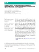
Fig. 6. The ATP-binding site in the MD simulated model structure
of the binary hGK–ATP complex and the domain motion induced by
ATP binding to the hGK apoenzyme. (A) Close-up view of the ATP-
binding site in the MD simulated model structure of the binary
hGK-ATP complex, showing the main contact residues with ATP;
for a presentation of all contact residues, see Table S1. For helix
nomenclature, see [47]. (B, C) The domain motion induced by ATP
binding to the apoenzyme with partial closure of the active site
cleft and a rotation angle of 33°. The coordinates were those
obtained for (B) the modelled super-open conformation, including
the Glu157–Asn179 loop, and (C) the modelled open conformation
with inserted ATP (GK–ATP). The C
a
rmsd values were 4.01 A
˚
for
the whole protein, 2.09 A
˚
for the fixed domain (349 residues), and
3.91 A
˚
for the moving domain (87 residues). The dynamic domains
were identified with the
DYNDOM program [30]. The C
a
backbone
structures, shown in line presentation, were colour-coded as fol-
lows: blue, fixed domain; red, moving domain; and green, connect-
ing residues. For comparison, the corresponding data for the
domain motion induced by Glc binding to the apoenzyme are
shown in Table S2. **ATP.
ATP binding at active site of human glucokinase J. Molnes et al.
2378 FEBS Journal 278 (2011) 2372–2386 ª 2011 The Authors Journal compilation ª 2011 FEBS
increased to 14.3 ± 1.7 mm. The fact that the kinetic
cooperativity is dependent on the MgATP concentration
is consistent with previous data reported for the rat liver
isoform [33,34]. With MgATP as the variable substrate,
a hyperbolic curve was obtained at a high Glc concen-
tration (60 mm), with a K
m
of 0.16 ± 0.01 mm (Table 3;
Fig. 7C). However, at a low Glc concentration
(0.5 mm), negative cooperativity was observed with
respect to MgATP (n
H
= 0.87 ± 0.06) (Fig. 7D), con-
sistent with a previous report on the rat liver isoform
[34], and the K
m
was reduced to 0.04 ± 0.003 mm.
Interestingly, the L146R mutant, with a severely
reduced affinity for Glc (Table 1), demonstrated similar
negative kinetic cooperativity with respect to MgATP as
the variable substrate (n
H
= 0.73 ± 0.04).
Discussion
The bisubstrate reaction catalysed by monomeric GK
is mechanistically characterized by diffusion-controlled
binding of Glc to thermodynamically favoured ligand-
free conformations of the enzyme (Scheme 1), followed
by global hysteretic isomerization of the enzyme to a
closed conformation [29,31].
From crystallographic, biophysical and kinetic stud-
ies on GK, it is known that both substrate binding
and catalysis require substantial conformational
changes in the enzyme. Ligand-free hGK is structur-
ally dominated by a super-open conformation [31],
which, in the crystal structure, is locked in an inactive
state by electrostatic and hydrophobic interactions
between the C-terminal helix (helix 17) and helix 6
[18]. Three residues (Asn204, Asn231, and Glu256) in
the large domain [31] function as primary contact res-
idues in the binding of Glc [18,31]. Pre-steady-state
analyses of Glc binding to WT hGK [26,32,35] have
provided evidence that the ligand-free enzyme in solu-
tion is in a pre-existing equilibrium between at least
two conformers (marked as GK
à
and GK
„
in
Scheme 1), i.e. the super-open conformation ( 80–
95%) and an alternative (presumably less open) con-
formation ( 5–20%) with a higher affinity for Glc
[26,35], which adds to the kinetic complexity of this
reaction. Recent high-resolution NMR analyses and
pre-steady-state Glc binding experiments also suggest
that GK is capable of sampling multiple conforma-
tional states, both in the absence and the presence of
Glc [32,36]. The global conformational changes trig-
gered by Glc binding have been defined crystallo-
graphically [31]. In the closed conformation (marked
as GK* in Scheme 1), precise alignment of additional
substrate contact residues (notably Thr168 and
Lys169 in the flexible surface ⁄ active site loop) [18,29]
and the subsequent higher affinity for Glc efficiently
accelerate the chemical reaction (k
3
) on binding of
the cosubstrate MgATP. The overall binding constant
K
1
for Glc and the values for the forward ( k
2
) and
reverse (k
)2
) rates of the conformational transition,
which probably includes intermediates [29,31,35,36],
have been estimated by stopped-flow fluorescence
spectroscopy [25]. In that study, the GK–
Glc M GK*–Glc interconversion was found to
be slow, with k
2
= 0.45 s
)1
and k
)2
= 0.28 s
)1
(K
2
= 1.6), favouring the forward rate and isomeriza-
tion, whereas the isomerization was unfavourable with
2-deoxyglucose as the substrate (K
2
= 0.8). Here, we
present experimental evidence that ATP binds to the
ligand-free form, and that this also results in changes
in the protein conformation.
ATP binds to the ligand-free open conformation
of hGK
Previous attempts to demonstrate direct binding
of ATP to the ligand-free form of rat GK by ITF
Table 3. The kinetic constants for WT GST–hGK at high and low concentrations of the fixed substrate. The catalytic activity was measured
at 37 °C, as described in Experimental procedures. Kinetic parameters were calculated by nonlinear regression analyses with the Hill and
Michaelis–Menten equations.
Conditions Hill coefficient [S]
0.5
Glc (mM) K
m
MgATP
2)
(mM) k
cat
(s
)1
)
Glc as variable substrate
5m
M ATP 1.95 ± 0.10 8.23 ± 0.26 – 67.6 ± 1.3
0.05 m
M ATP 1.07 ± 0.07 14.3 ± 1.7 – 27.8 ± 1.4
ATP as variable substrate
60 m
M Glc 1.15 ± 0.04 – 0.16 ± 0.01 68.4 ± 0.9
0.5 m
M Glc 0.87 ± 0.06 – 0.04 ± 0.002 0.70 ± 0.01
GK
‡
+ Glc
GK
‡ /
≠
·Glc GK*·Glc GK*·Glc6P GK + Glc6P
K
1
‡
k
-2
k
2
k
3
k
4
k
-3
GK
≠
+ Glc
K
1
≠
ADP
MgATP
Scheme 1. Reaction scheme for mammalian glucokinase.
J. Molnes et al. ATP binding at active site of human glucokinase
FEBS Journal 278 (2011) 2372–2386 ª 2011 The Authors Journal compilation ª 2011 FEBS 2379
spectroscopy [37] and hGK by differential scanning
calorimetry [25] were reported to be unsuccessful. The
topic was more recently readdressed [26] with a non-
hydrolysable ATP analogue (AMP-PNP) and ITF,
and relatively large quenching of the fluorescence sig-
nal was demonstrated, interpreted as a nucleotide-
induced conformational change. However, as no cor-
rections were made for a large inner filter effect,
owing to the significant absorbance of the nucleotide
at the excitation wavelength (285 nm), we have cor-
rected for this effect here (at k
ex
= 295 nm) as well
as for any effect of nonspecific binding to the enzyme
(i.e. not in the active site), with the non-ATP-binding
mutant form T228M (Table 1) as a reference enzyme.
Our analyses revealed that AMP-PNP and ATP do
indeed bind to hGK (Fig. 2) in the ligand-free open
conformation, and the MD simulations (Fig. 6) fur-
ther support this conclusion and also show the resi-
dues (including the mutated residue Thr228) directly
contacting ATP at the active site of WT hGK.
The partial quenching effect of AMP-PNP on the
T228M reference enzyme with disrupted ATP binding
at the active site (Fig. 2A,C) suggests a contribution
of nonspecific binding of that nucleotide (i.e. not in
the active site) in addition to its inner filter effect,
as observed for free Trp. The idea that AMP, in
contrast to MgADP
)
⁄ MgATP
2)
, can bind to more
than one site has been suggested for the rat liver
isoform [16].
Binding of ATP to ligand-free hGK results in a
conformational change
High-resolution NMR analyses [36] have revealed that
GK is an intrinsically mobile enzyme whose structure
and dynamics are modulated by temperature and
ligand binding. Here, we provide the first experimental
evidence of ATP-dependent structural changes in WT
hGK. Specifically, our ITF quenching (Fig. 2) and
MD simulations (Figs 5 and 6) indicate a significant
conformational change upon ATP binding, including
motion of the flexible surface ⁄ active site loop and par-
tial closure of the active site cleft (Figs 5C, 6B,C and
S3). A change in conformation is further supported
by the significant protective effect of ATP on binding
of the extrinsic fluorescence probe ANS (Fig. 3A,B)
and on the limited proteolysis by trypsin (Fig. 3C). In
both assay systems, Glc showed more potent inhibi-
tion than ATP, which may be related to the larger
conformational change and more effective closure of
A
C
B
D
Fig. 7. Steady-state kinetic properties of WT GST–hGK with Glc and MgATP as the variable substrates. (A) At 5 mM MgATP, positive coo-
perativity with respect to Glc was observed (n
H
= 1.95 ± 0.10). (B) At a low (0.05 mM) concentration of MgATP, the cooperativity with
respect to Glc was reduced (n
H
= 1.07 ± 0.07). (C) At 60 mM Glc, the binding curve for MgATP was hyperbolic (n
H
= 1.15 ± 0.04). (D) At a
low (0.5 m
M) concentration of Glc, negative cooperativity with respect to MgATP binding was observed (n
H
= 0.87 ± 0.06). For all nonlinear
regressions, the correlation coefficient (r
2
) was > 0.99. The steady-state kinetic constants are summarized in Table 3.
ATP binding at active site of human glucokinase J. Molnes et al.
2380 FEBS Journal 278 (2011) 2372–2386 ª 2011 The Authors Journal compilation ª 2011 FEBS
the active site cleft induced by Glc binding (Fig. S3;
Table S2).
Kinetic cooperativity with respect to Glc
In general, the mechanism for cooperativity observed
in enzyme kinetic studies represents an experimental
challenge. For monomeric GK, several models have
been considered to explain the positive kinetic cooper-
ativity with respect to Glc, including: (a) a random
order mechanism of substrate (Glc and MgATP
2)
)
addition [13,38]; and (b) a sequential order mechanism
[15,39,40], in which the binding of Glc as the first sub-
strate induces a slow, concentration-dependent confor-
mational transition [34,40] characteristic of a hysteretic
enzyme [41,42] (Scheme 1). The Glc-induced multipha-
sic ITF enhancement (millisecond to minute time scale)
of WT and mutant forms of GK [18,25,26,35,37,43]
strongly favours the second mechanism, and support
the idea that the cooperativity can be explained by an
equilibrium between conformational states with differ-
ent affinities for Glc [18,25,26,35,37,43]. However, little
experimental effort has been made to include or
exclude any contribution of (Mg)ATP binding to the
kinetic cooperativity.
The ligand-free and the binary GK–Glc complex are
dynamic entities [32,36], and binding of (Mg)ATP may
shift the equilibrium between different enzyme confor-
mations, as shown for Glc [18,25,26,35,37,43]. In this
study, the binding of ATP to the ligand-free enzyme
was found, by four independent criteria, to trigger con-
formational changes, including partial closure of the
active site cleft (Figs 5B, 6B,C and S3). Moreover, pre-
vious [34] and present (Table 1) steady-state kinetic
analyses are also compatible with conformational con-
trol of GK catalytic activity by the binding of
(Mg)ATP, with possible implications for kinetic coo-
perativity with respect to Glc. Thus, the cooperativity
is largely reduced (n
H
= 1.07 ± 0.07) at low concen-
trations of MgATP (Fig. 7B; Table 1) [34] and when
MgATP is replaced by MgITP, a poor phosphoryl
donor ATP analogue [44].
In most previous steady-state kinetic analyses, rat GK
(liver) was observed to be noncooperative with respect
to (Mg)ATP [33,45,46]. However, Neet et al. [34]
reported negative kinetic cooperativity (n
H
= 0.84)
when measurements were made in the presence of 30%
glycerol at a low Glc concentration (0.5 mm), and this
was also observed here for the recombinant pancreatic
hGK in the absence of glycerol (n
H
= 0.87 ± 0.06).
However, when hGK activity was measured at high glu-
cose concentrations, the Hill coefficient for (Mg)ATP
approached 1.0 (Fig. 7C), as expected from studies on
rat liver GK [33,45,46]. Negative cooperativity
(n
H
= 0.73 ± 0.04) was also observed for the GCK-
MODY mutant L146R, which has severely reduced
affinity for Glc (Table 1). Moreover, our studies on this
mutant revealed that the analogue ATPcS (at 2.5 mm)
increases the mutant’s low equilibrium binding affinity
for Glc (K
d
decreases from 19.3 ± 3.8 mm to
14.0 ± 1.4 mm), as well as the Glc-induced fluorescence
enhancement (by 25%) (Fig. 4). These effects may be
related to partial catalytic activation following
(Mg)ATP binding at physiological concentrations of
Glc. Similar or possibly larger effects of ATP in promot-
ing a catalytically competent state may occur in other
mutations associated with GCK-MODY.
Conclusions
Using biochemical and biophysical methods, we have
obtained experimental evidence in support of binding
of ATP to the ligand-free hGK, resulting in a change
of protein conformation. The MD simulations indicate
that the binding triggers molecular motion of the flexi-
ble surface ⁄ active site loop and partial closure of the
interdomain active site cleft. The modelled structure of
the hGK–ATP binary complex shows the residue con-
tacts involved in ATP binding at the active site. Our
findings further support conformational regulation of
GK by ATP binding, with possible implications for
kinetic cooperativity with respect to Glc. Further
mutational studies, notably of GCK-MODY-associated
mutations, may contribute to a better understanding
of the mechanistic and functional implications of the
multiple conformational equilibria and the conforma-
tional transitions induced by both Glc and (Mg)ATP,
with possible future clinical implications.
Experimental procedures
Materials
The oligonucleotide primers used for site-directed mutagen-
esis were from Invitrogen (Carlsbad, CA, USA). The
QuickChange XL Site-directed Mutagenesis Kit was from
Stratagene (La Jolla, CA, USA). Glutathione Sepharose 4B
was from Amersham Biosciences (GE Healthcare Europe
GMBH, Oslo, Norway). Glc was from Calbiochem (San
Diego, CA, USA). Magnesium chloride, magnesium ace-
tate, guanidine hydrochloride, trypsin (bovine pancreas),
trypsin inhibitor (soybean), pyruvate kinase (rabbit muscle),
phospho(enol)pyruvate, ATP, ANS and AMP-PNP were
from Sigma-Aldrich (St Louis, MO, USA). ATPcS was
obtained from Roche Diagnostics Corporation (Indianapo-
lis, IN, USA). All chemicals and buffers used for fluores-
cence measurements were of the highest analytical grade.
J. Molnes et al. ATP binding at active site of human glucokinase
FEBS Journal 278 (2011) 2372–2386 ª 2011 The Authors Journal compilation ª 2011 FEBS 2381
Site-directed mutagenesis
The mutations T228M and L146R were introduced into the
cDNA of the pancreatic (isoform 1) WT hGK with the
QuikChange XL Site-directed Mutagenesis Kit. The pGEX-
3X vector (kindly provided by F. M. Matschinsky, Univer-
sity of Pennsylvania, PA, USA), containing a protease fac-
tor Xa cleavage site, was used as the template. The
following specific oligonucleotide primers were used for
mutagenesis (mutated nucleotides are underlined): T228M
forward, 5¢-GC ATG ATC GTG GGC A
TG GGC TG
C AAT GCC TGC 3¢; T228 reverse, 5¢-GCA GGC ATT
GCA GCC C
AT GCC CAC GAT CAT GC-3¢; L146R for-
ward, 5¢-CAC AAG AAG CTG CCC C
GG GGC TTC AC
C TTC TCC-3¢; and L146R reverse, 5 ¢-GGA GAA GGT
GAA GCC C
CG GGG CAG CTT CTT GTG-3¢. Mutations
were confirmed by DNA sequencing.
Expression and purification of hGK
The WT and mutant recombinant proteins were generated
and expressed in the form of GST fusion proteins in Esc-
herichia coli BL21 cells, as previously described [47]. For
the CD and guanidine hydrochloride unfolding experi-
ments, the WT hGK was isolated by removing the GST
fusion protein as previously described [18]. Purified protein
was stored in liquid nitrogen in the absence of glucose
(10 mm glutathione, 50 mm Tris ⁄ HCl, pH 8.0). The protein
concentration was determined with the following absorp-
tion coefficients: A
280 nm
(1 mgÆmL
)1
Æcm
)1
) = 1.05 (fusion
protein); and A
280 nm
= 0.65 (isolated protein) [18].
Steady-state kinetics
The catalytic activity of GST–hGK was measured spectro-
photometrically (A
340 nm
)at37°CbyanNAD
+
-coupled
assay with glucose-6-phosphate dehydrogenase, as previ-
ously described [18]. Kinetic studies were performed with
10–12 dilutions of Glc or ATP. The protocol with Glc as
the variable substrate was carried out with 5 mm ATP
(2.5 mm excess of Mg
2+
), whereas in the assay with
MgATP as variable substrate (2.5 mm excess of Mg
2+
), sat-
urating amounts of Glc were used. In the case of the
severely inactivating mutation T228M [9,20], an ATP con-
centration of 10 mm was used. For determination of Hill
coefficients of WT hGK, assays were performed with 12
different Glc concentrations (range: 0–60 mm) or with 11
or 12 concentrations of MgATP (range: 0–3 mm). In the
assays with a low ATP concentration, an ATP-regenerating
system was used, including 2 mm phospho(enol)pyruvate
and 10 U of pyruvate kinase, to ensure a constant ATP
level. Steady-state kinetic parameters were calculated by
nonlinear regression analyses with the Hill and Michaelis–
Menten equations. The reaction rates were measured from
the linear part of the initial time-course.
GST–hGK fluorescence measurements
ITF measurements were performed on a Perkin-Elmer LS-
50B spectrometer (1-cm pathlength) at 25 °C in a buffer
containing 20 mm Hepes, 100 mm NaCl, and 1 mm dith-
iothreitol (pH 7.0), and a protein concentration of
0.03 mgÆmL
)1
(a concentration of 0.04 mgÆmL
)1
was used
for the mutant L146R GST–hGK). ATP titration experi-
ments were performed in the same buffer containing 1 mm
EDTA to complex trace amounts of Mg
2+
. The excitation
and emission wavelengths used were 295 nm and 340 nm,
respectively. Steady-state emission spectra were recorded
from 305 nm to 500 nm, and slit widths for excitation and
emission were set at 3 nm and 7 nm, respectively. All spec-
tra represent an average of four scans. The binding iso-
therms and the apparent equilibrium dissociation constants
(K
d
for Glc and [L]
0.5
for ATP) were determined by titra-
tion experiments; small aliquots of concentrated ligand
(Glc ⁄ (Mg)ATP ⁄ AMP-PNP) were successively added to the
enzyme solution every time that equilibrium was achieved
after the previous addition (150–200 s). The change in
intrinsic fluorescence intensity [(DF
eq
⁄ F
0
) · 100] at k
max
(340 nm) was recorded as a function of the concentration
of added ligand, with slit widths for excitation and emission
set at 4 nm and 7 nm, respectively. The concentrations used
were 0–60 mm Glc, 0–5 mm (Mg)ATP, and 0–450 lm
AMP-PNP. To correct for the inner filter effect, caused by
nucleotide absorbance at the excitation wavelength, the
mutant T228M hGK, which is unable to bind ATP at
the active site [9,20,21,24], was used as a reference enzyme.
The specific WT hGK fluorescence response to added nucleo-
tide was determined by subtracting the mutant T228M fluo-
rescence response (inner filter effect) to added nucleotide.
Titrations in the presence of free Trp (at a concentration
giving the same F
0
value as the enzyme) were performed as
an additional control. The fluorescence intensity was
adjusted for dilutions of protein, and baseline corrections
were obtained with buffer without protein. Linear and non-
linear regression analyses of the data were performed as
described previously [18], with sigmaplot technical graph-
ing software (Systat Software, San Jose, CA, USA).
ANS fluorescence measurements
Binding of ANS was performed at 38 °C on a Perkin-
Elmer LS50B spectrometer (1-cm pathlength), with an exci-
tation wavelength of 385 nm and excitation ⁄ emission slit
widths of 6 nm. The cuvette, containing 60 lm ANS in
20 mm Hepes, 100 mm NaCl, and 1 mm dithiothreitol
(pH 7.0), was pre-equilibrated to 38 °C before the reaction
was started by the addition of hGK (0.75 lm). Five minutes
later, the stable fluorescence emission spectra were recorded
(400–600 nm). All spectra represent an average of four
scans. When the effect of Glc (30 mm) or ATP (2 mm)on
ANS binding was investigated, the ligand was added to the
ATP binding at active site of human glucokinase J. Molnes et al.
2382 FEBS Journal 278 (2011) 2372–2386 ª 2011 The Authors Journal compilation ª 2011 FEBS
buffer in the cuvette and pre-equilibrated at the appropriate
temperature before addition of enzyme. Prior to addition,
the enzyme was preincubated for 7 min in the presence of
the same concentration of ligand.
CD spectroscopy
Far-UV CD spectra (185–260 nm, light path 1 mm) were
recorded at 20 °C on a Jasco J-810 spectropolarimeter. The
isolated WT and T228M proteins were diluted in 20 m m
sodium phosphate buffer (pH 7.2) with 0.7 mm dithiothrei-
tol to a final concentration of 23 lm. The proteins were
analysed in the absence and presence of 40 mm Glc. The
spectra obtained represent an average of four scans (scan
rate of 50 nmÆmin
)1
), all background-corrected and
smoothed. Secondary structure analyses were performed
with the CD Neural Network algorithm [48]. Thermal
unfolding (20–90 °C) was determined by following the
change in ellipticity at 222 nm at a constant heating rate of
40 °CÆh
)1
. The midpoint of the transition (T
m
) was deter-
mined from the first derivative of the smoothed denatur-
ation curve. The associated high-voltage (pseudo-
absorbance) increase was recorded at the same time as the
CD data; this was found to be important for monitoring
the quality and validity of the data (Fig. 1).
Limited proteolysis with trypsin
WT GST–hGK was cleaved with factor Xa for 2 h at 4 °C,
and the rate of limited proteolysis by trypsin was subse-
quently measured at 25 °C in a 100-lL reaction mixture
containing 20 mm Hepes, 50 mm NaCl, and 2 mm dith-
iothreitol (pH 7.0), at a final GST–hGK concentration of
0.5 lgÆlL
)1
and a GST–hGK ⁄ trypsin ratio of 400 : 1 (by
mass). The effect of preincubation with the substrates Glc
(40 mm) or MgATP (2 mm ATP ⁄ 4mm MgAc) on the
extent of proteolysis of WT hGK was studied. At timed
intervals (0, 5, 10, 20 and 30 min), aliquots of 15 lL were
taken from the proteolytic reaction, quenched with 14 lL
of SDS sample buffer containing soybean trypsin inhibitor
[protease ⁄ inhibitor ratio of 1 : 1.5 (by mass)], heated for
15 min at 56 °C, and subjected to 10% SDS ⁄ PAGE analy-
ses. Bands were visualized by Coomassie Blue staining, and
the band corresponding to full-length hGK was quantified
with quantity one 1-d analysis software from Bio-Rad
(Hercules, CA, USA). The data were fitted by nonlinear
regression analysis with sigmaplot technical graphing
software.
Modelling of the hGK apoenzyme in the
super-open conformation
The initial coordinates of the hGK apoenzyme were taken
from the X-ray crystal structure solved at 3.4 A
˚
resolution
(PDB ID 1v4t) [31]. As the coordinates of a highly flexible
loop structure, comprising residues Glu157–Asn179, were
not fully resolved, we refined this crystal structure computa-
tionally. Initial coordinates for the 23 missing amino acids
were taken from the structure of the binary glucose–hGK
complex (PDB ID 1v4s) [including a GK activator (GKA)
(compound A)] [31]. The dihedral angle of the peptide bond
of Gly170 was adjusted so that the terminal amino acids
were in an optimal position for filling the gap in the apoen-
zyme. Insertion of the missing residues was followed by
energy minimization and 3-ns MD simulation (allowing
only the inserted residues to move). The first 2 ns of simu-
lation were performed in an implicit water environment
(generalized Born model) to allow fast relaxation of the
inserted peptide (avoiding friction with explicit water mole-
cules). The resulting model was then solvated in explicit
water in a periodic box, and simulated for another nanosec-
ond to further relax the model.
Structure modelling and MD simulations
The binary hGK–Glc complex (PDB ID 1v4s) [including a
GKA (compound A)] was structurally aligned with the
modelled structure of the complete hGK apoenzyme by
applying the combinatorial extension method as imple-
mented in the ce software [49]. Coordinates for glucose
were extracted from the binary complex, and saved together
with the hGK apoenzyme coordinates to construct an ini-
tial model of glucose bound to the super-open conforma-
tion. Furthermore, the coordinates of human hexokinase
type I in complex with AMP-PNP (PDB ID 1qha) were
aligned with our model to build the initial model for ATP-
bound super-open hGK, and with the coordinates of the
binary complex with Glc (PDB ID 1v4s) to generate start-
ing coordinates for the ternary complex. These procedures
provided us with the following four structural models:
model 1, hGK super-open conformation (including coordi-
nates for the Glu157–Asn179 loop); model 2, hGK super-
open conformation with inserted Glc; model 3, hGK super-
open conformation with inserted ATP; and model 4, hGK
ternary complex with Glc and ATP.
Coordinates for all four models were relaxed by MD sim-
ulation in explicit water. The changes in rmsd values during
the 2-ns MD simulations, relative to the starting structures,
are given in Fig. S2A. Mg was not included in the initial
simulation of the ternary complex.
Software and hardware
MD simulations were performed with amber10 software
[50]. The four models were all simulated in a truncated
octahedral periodic box with TIP3P water, keeping bonds
involving hydrogen atoms fixed, allowing a 2-fs time step
between calculations of forces acting on the atoms. The
temperature was increased from 0 K to 300 K under
constant volume during the first 20 ps of the simulation,
J. Molnes et al. ATP binding at active site of human glucokinase
FEBS Journal 278 (2011) 2372–2386 ª 2011 The Authors Journal compilation ª 2011 FEBS 2383
keeping the protein coordinates fixed with weak restraints.
The systems were further equilibrated at 300 K for 200 ps
before restraints were removed, and the models were
allowed to relax for 2 ns under constant pressure and tem-
perature. Coordinates were saved every 10th picosecond
from the 2-ns simulations, and trajectories were analysed
with the ambertools suite of programs (http://amber-
md.org/#AmberTools). All simulations were performed on
an HP BL 460c Linux cluster equipped with Xeon 2.66-
GHz quad-core processors. Each job was distributed over
32 CPUs, and the average computation time was
11.5 hÆns
)1
.
By use of the crystal structure of the super-open (PDB
ID 1v4t) and the closed (Glc and GKA-bound) (PDB
ID 1v4s) forms, two structure-based methods were used for
the estimation of the free energy of thermal unfolding [51]
and the folding free energy [52] of hGK mutants.
The dyndom program [30] was used to determine
dynamic domains and connecting residues involved in hinge
bending motions on binding of Glc and ATP to the super-
open conformation of hGK. Structural images were gener-
ated with pymol, version 1.0 [53]. icm-pro [54] was used to
analyse the resulting complexes after MD simulations and
to calculate the contact area of residues interacting with
Glc and ATP presented in Table S1.
Acknowledgements
A M. Nordbø and A. Sepulveda Muno
˜
z are thanked
for expert technical assistance and French press prepa-
ration of recombinant enzymes, respectively. This work
was supported by Helse Vest, Haukeland University
Hospital, the Research Council of Norway, the Novo
Nordisk Foundation, the University of Bergen, Inno-
vest AS, the Norwegian Diabetes Association, the Aar-
skog Foundation and the Meltzer Foundation.
References
1 Tanizawa Y, Koranyi LI, Welling CM & Permutt MA
(1991) Human liver glucokinase gene: cloning and
sequence determination of two alternatively spliced
cDNAs. Proc Natl Acad Sci USA 88, 7294–7297.
2 Nishi S, Stoffel M, Xiang K, Shows TB, Bell GI &
Takeda J (1992) Human pancreatic beta-cell glucoki-
nase: cDNA sequence and localization of the polymor-
phic gene to chromosome 7, band p 13. Diabetologia
35, 743–747.
3 Hughes SD, Quaade C, Milburn JL, Cassidy L &
Newgard CB (1991) Expression of normal and novel
glucokinase mRNAs in anterior pituitary and islet cells.
J Biol Chem 266, 4521–4530.
4 Jetton TL, Liang Y, Pettepher CC, Zimmerman EC,
Cox FG, Horvath K, Matschinsky FM & Magnuson
MA (1994) Analysis of upstream glucokinase promoter
activity in transgenic mice and identification of glucoki-
nase in rare neuroendocrine cells in the brain and gut.
J Biol Chem 269, 3641–3654.
5 Matschinsky FM (2002) Regulation of pancreatic beta-
cell glucokinase: from basics to therapeutics. Diabetes
51, S394–S404.
6 Froguel P, Vaxillaire M, Sun F, Velho G, Zouali H,
Butel MO, Lesage S, Vionnet N, Clement K, Fou-
gerousse F et al. (1992) Close linkage of glucokinase
locus on chromosome 7p to early-onset non-insulin-
dependent diabetes mellitus. Nature 356, 162–164.
7 Vionnet N, Stoffel M, Takeda J, Yasuda K, Bell GI,
Zouali H, Lesage S, Velho G, Iris F, Passa P et al.
(1992) Nonsense mutation in the glucokinase gene
causes early-onset non-insulin-dependent diabetes mell-
itus. Nature 356, 721–722.
8 Glaser B, Kesavan P, Heyman M, Davis E, Cuesta A,
Buchs A, Stanley CA, Thornton PS, Permutt MA,
Matschinsky FM et al. (1998) Familial hyperinsulinism
caused by an activating glucokinase mutation. N Engl J
Med 338, 226–230.
9 Njølstad PR, Søvik O, Cuesta-Mun
˜
oz A, Bjørkhaug L,
Massa O, Barbetti F, Undlien DE, Shiota C, Magnuson
MA, Molven A et al. (2001) Neonatal diabetes mellitus
due to complete glucokinase deficiency. N Engl J Med
344, 1588–1592.
10 Njølstad PR, Sagen JV, Bjørkhaug L, Odili S, Shehadeh
N, Bakry D, Sarici SU, Alpay F, Molnes J, Molven A
et al. (2003) Permanent neonatal diabetes caused by glu-
cokinase deficiency: inborn error of the glucose–insulin
signaling pathway. Diabetes 52, 2854–2860.
11 Osbak KK, Colclough K, Saint-Martin C, Beer NL,
Bellanne
´
-Chantelot C, Ellard S & Gloyn AL (2009)
Update on mutations in glucokinase (GCK), which
cause maturity-onset diabetes of the young, permanent
neonatal diabetes, and hyperinsulinemic hypoglycemia.
Hum Mutat 30, 1512–1526.
12 Gloyn AL, Odili S, Buettger C, Njølstad PR, Shiota C,
Magnuson MA & Matschinsky FM (2004) Glucokinase
and the regulation of blood sugar. a mathematical
model predicts the threshold for glucose stimulated
insulin release for GCK gene mutations that cause
hyper- and hypoglycemia. In Glucokinase and Glycemic
Disease: From Basics to Novel Therapeutics (Matschin-
sky FM & Magnuson MA eds), pp. 92–109. Karger,
Basel.
13 Pettersson G (1986) Mechanistic origin of the sigmoidal
rate behaviour of glucokinase. Biochem J 233, 347–350.
14 Storer AC & Cornish-Bowden A (1977) Kinetic evi-
dence for a ‘mnemonical’ mechanism for rat liver gluco-
kinase. Biochem J 165, 61–69.
15 Cornish-Bowden A & Storer AC (1986) Mechanistic
origin of the sigmoidal rate behaviour of rat liver hexo-
kinase D (‘glucokinase’). Biochem J
240, 293–296.
ATP binding at active site of human glucokinase J. Molnes et al.
2384 FEBS Journal 278 (2011) 2372–2386 ª 2011 The Authors Journal compilation ª 2011 FEBS
16 Monasterio O & Ca
´
rdenas ML (2003) Kinetic studies of
rat liver hexokinase D (‘glucokinase’) in non-co-opera-
tive conditions show an ordered mechanism with
MgADP as the last product to be released. Biochem J
371, 29–38.
17 Wilson JE & Schwab DA (1996) Functional interaction
of hexokinase with ATP requires participation by both
small and large lobes of the enzyme: implications for
other proteins using the actin fold as a nucleotide bind-
ing motif. FASEB J 10, 799–801.
18 Molnes J, Bjørkhaug L, Søvik O, Njølstad PR & Flat-
mark T (2008) Catalytic activation of human glucoki-
nase by substrate binding: residue contacts involved in
the binding of D-glucose to the super-open form and
conformational transitions. FEBS J 275, 2467–2481.
19 Stoffel M, Froguel P, Takeda J, Zouali H, Vionnet N,
Nishi S, Weber IT, Harrison RW, Pilkis SJ, Lesage S
et al. (1992) Human glucokinase gene: isolation, charac-
terization, and identification of two missense mutations
linked to early-onset non-insulin-dependent (type 2)
diabetes mellitus. Proc Natl Acad Sci USA 89, 7698–
7702.
20 Davis EA, Cuesta-Mun
˜
oz A, Raoul M, Buettger C,
Sweet I, Moates M, Magnuson MA & Matschinsky
FM (1999) Mutants of glucokinase cause hypoglyca-
emia- and hyperglycaemia syndromes and their analysis
illuminates fundamental quantitative concepts of glu-
cose homeostasis. Diabetologia 42, 1175–1186.
21 Mahalingam B, Cuesta-Mun
˜
oz A, Davis EA, Matschin-
sky FM, Harrison RW & Weber IT (1999) Structural
model of human glucokinase in complex with glucose
and ATP: implications for the mutants that cause hypo-
and hyperglycemia. Diabetes 48, 1698–1705.
22 Bork P, Sander C & Valencia A (1992) An ATPase
domain common to prokaryotic cell cycle proteins,
sugar kinases, actin, and hsp70 heat shock proteins.
Proc Natl Acad Sci USA 89, 7290–7294.
23 Harrison RW & Weber IT (2004) Molecular models of
human glucokinase and the implications for glycemic
diseases. In Glucokinase and Glycemic Disease: From
Basics to Novel Therapeutics (Matschinsky FM &
Magnuson MA eds), pp. 135–144. Karger, Basel.
24 Marotta DE, Anand GR, Anderson TA, Miller SP,
Okar DA, Levitt DG & Lange AJ (2005) Identification
and characterization of the ATP-binding site in human
pancreatic glucokinase. Arch Biochem Biophys 436,
23–31.
25 Heredia VV, Thomson J, Nettleton D & Sun S (2006)
Glucose-induced conformational changes in glucokinase
mediate allosteric regulation: transient kinetic analysis.
Biochemistry 45, 7553–7562.
26 Kim YB, Kalinowski SS & Marcinkeviciene J (2007) A
pre-steady state analysis of ligand binding to human
glucokinase: evidence for a preexisting equilibrium.
Biochemistry 46, 1423–1431.
27 Semisotnov GV, Rodionova NA, Razgulyaev OI, Uver-
sky VN, Gripas AF & Gilmanshin RI (1991) Study of
the ‘molten globule’ intermediate state in protein fold-
ing by a hydrophobic fluorescent probe. Biopolymers
31, 119–128.
28 Sagen JV, Odili S, Bjørkhaug L, Zelent D, Buettger C,
Kwagh J, Stanley C, Dahl-Jørgensen K, de Beaufort C,
Bell GI et al. (2006) From clinicogenetic studies of
maturity-onset diabetes of the young to unraveling com-
plex mechanisms of glucokinase regulation. Diabetes 55,
1713–1722.
29 Zhang J, Li C, Chen K, Zhu W, Shen X & Jiang H
(2006) Conformational transition pathway in the allo-
steric process of human glucokinase. Proc Natl Acad
Sci USA 103, 13368–13373.
30 Hayward S & Berendsen HJ (1998) Systematic analysis
of domain motions in proteins from conformational
change: new results on citrate synthase and T4 lyso-
zyme. Proteins 30, 144–154.
31 Kamata K, Mitsuya M, Nishimura T, Eiki J-i & Nagat-
a Y (2004) Structural basis for allosteric regulation of
the monomeric allosteric enzyme human glucokinase.
Structure 12, 429–438.
32 Larion M & Miller BG (2010) Global fit analysis of glu-
cose binding curves reveals a minimal model for kinetic
cooperativity in human glucokinase. Biochemistry 49,
8902–8911.
33 Storer AC & Cornish-Bowden A (1976) Kinetics of rat
liver glucokinase. Co-operative interactions with glucose
at physiologically significant concentrations. Biochem J
159, 7–14.
34 Neet KE, Keenan RP & Tippett PS (1990) Observation
of a kinetic slow transition in monomeric glucokinase.
Biochemistry 29, 770–777.
35 Antoine M, Boutin JA & Ferry G (2009) Binding
kinetics of glucose and allosteric activators to human
glucokinase reveal multiple conformational states.
Biochemistry 48, 5466–5482.
36 Larion M, Salinas RK, Bruschweiler-Li L, Bruschweiler
R & Miller BG (2010) Direct evidence of conforma-
tional heterogeneity in human pancreatic glucokinase
from high-resolution nuclear magnetic resonance.
Biochemistry 49, 7969–7971.
37 Lin S-X & Neet KE (1990) Demonstration of a slow
conformational change in liver glucokinase by fluores-
cence spectroscopy. J Biol Chem 265, 9670–9675.
38 Ca
´
rdenas ML, Rabajille E & Niemeyer H (1979)
Kinetic cooperativity of glucokinase with glucose. Arch
Biol Med Exp (Santiago) 12, 571–580.
39 Gregoriou M, Trayer IP & Cornish-Bowden A (1981)
Isotope-exchange evidence for an ordered mechanism
for rat-liver glucokinase, a monomeric cooperative
enzyme. Biochemistry 20, 499–506.
40 Ca
´
rdenas ML, Rabajille E & Niemeyer H (1984) Sup-
pression of kinetic cooperativity of hexokinase D (glu-
J. Molnes et al. ATP binding at active site of human glucokinase
FEBS Journal 278 (2011) 2372–2386 ª 2011 The Authors Journal compilation ª 2011 FEBS 2385
cokinase) by competitive inhibitors. A slow transition
model. Eur J Biochem 145, 163–171.
41 Frieden C (1970) Kinetic aspects of regulation of meta-
bolic processes. The hysteretic enzyme concept. J Biol
Chem 245, 5788–5799.
42 Neet KE & Ainslie GR Jr (1980) Hysteretic enzymes.
Methods Enzymol 64, 192–226.
43 Heredia VV, Carlson TJ, Garcia E & Sun S (2006)
Biochemical basis of glucokinase activation and the
regulation by glucokinase regulatory protein in natu-
rally occuring mutations. J Biol Chem 281, 40201–
40207.
44 Pollard-Knight D & Cornish-Bowden A (1987) Kinetics
of hexokinase D (‘glucokinase’) with inosine triphos-
phate as phosphate donor. Loss of kinetic
co-operativity with respect to glucose. Biochem J 245,
625–629.
45 Parry MJ & Walker DG (1967) Further properties and
possible mechanism of action of adenosine 5¢-triphos-
phate-D-glucose 6-phosphotransferase from rat liver.
Biochem J 105, 473–482.
46 Cornish-Bowden A & Ca
´
rdenas ML (2004) Glucoki-
nase: a monomeric enzyme with positive cooperativity.
In Glucokinase and Glycemic Disease: From Basics to
Novel Therapeutics (Matschinsky FM & Magnuson MA
eds), pp. 125–134. Karger, Basel.
47 Bjørkhaug L, Molnes J, Søvik O, Njølstad PR & Flat-
mark T (2007) Allosteric activation of human glucoki-
nase by free polyubiquitin chains and its ubiquitin-
dependent cotranslational proteasomal degradation.
J Biol Chem 282, 22757–22764.
48 Bo
¨
hm G, Muhr R & Jaenicke R (1992) Quantitative
analysis of protein far UV circular dichroism spectra by
neural networks. Protein Eng 5, 191–195.
49 Shindyalov IN & Bourne PE (1998) Protein structure
alignment by incremental combinatorial extension (CE)
of the optimal path. Protein Eng 11, 739–747.
50 Case DA, Darden TA, Cheatham TE, Simmerling CL,
Wang J, Duke RE, Luo R, Crowley M, Walker RC,
Zhang W et al. (2008) AMBER 10. University of
California, San Francisco, CA.
51 Parthiban V, Gromiha MM & Schomburg D (2006)
CUPSAT: prediction of protein stability upon point
mutations. Nucleic Acids Res 34, W239–W242.
52 Benedix A, Becker CM, de Groot BL, Caflisch A &
Bo
¨
ckmann RA (2009) Predicting free energy changes
using structural ensembles. Nat Methods 6, 3–4.
53 DeLano Scientific LLC (2007) The PyMOL Molecular
Graphics System. South San Francisco, CA, USA.
54 Molsoft LLC (2008) ICM version 3.6. La Jolla, CA,
USA.
Supporting information
The following supplementary material is available:
Fig. S1. Far-UV CD spectra of WT and T228M hGK.
Fig. S2. The atom-positional backbone rmsd of the
MD trajectory structures during the 2-ns MD simula-
tions, relative to the starting structures, and calculated
B-factor values based on fluctuations of C
a
carbons
during the 2-ns MD simulations of the four modelled
structures.
Fig. S3. Surface presentations of the ligand-free (open),
ATP (partly closed) and Glc + ATP (closed) modelled
structures of hGK.
Table S1. Contact area in percentage of exposed area
for residues interacting with Glc and ATP in the bin-
ary complex.
Table S2. Domain motions in the conformational
transitions induced by Glc and ATP binding to the
apoenzyme.
This supplementary material can be found in the
online version of this article.
Please note: As a service to our authors and readers,
this journal provides supporting information supplied
by the authors. Such materials are peer-reviewed and
may be re-organized for online delivery, but are not
copy-edited or typeset. Technical support issues arising
from supporting information (other than missing files)
should be addressed to the authors.
ATP binding at active site of human glucokinase J. Molnes et al.
2386 FEBS Journal 278 (2011) 2372–2386 ª 2011 The Authors Journal compilation ª 2011 FEBS