Báo cáo khoa học: The catalytic mechanism of dye-decolorizing peroxidase DyP may require the swinging movement of an aspartic acid residue potx
Bạn đang xem bản rút gọn của tài liệu. Xem và tải ngay bản đầy đủ của tài liệu tại đây (528.8 KB, 8 trang )
The catalytic mechanism of dye-decolorizing peroxidase
DyP may require the swinging movement of an aspartic
acid residue
Toru Yoshida
1
, Hideaki Tsuge
2
, Hiroki Konno
1
, Toru Hisabori
1
and Yasushi Sugano
1
1 R1-7 Chemical Resources Laboratory, Tokyo Institute of Technology, Japan
2 Department of Bioresources and Environmental Sciences, Kyoto Sangyo University, Japan
Introduction
Heme peroxidase, one of the best-studied and most
ubiquitous enzymes, oxidizes a variety of substrates,
including phenolic and azo compounds. To date,
many heme peroxidases have been isolated from vari-
ous organisms, and the details of these can be found
in the high-quality PeroxiBase (http://peroxibase.
toulouse.inra.fr/index.php) database [1]. Heme perox-
idases are classified into six families: animal peroxid-
ases, nonanimal peroxidases, catalases, diheme
cytochrome c peroxidases, dye-decolorizing peroxidase
(DyP)-type peroxidases, and haloperoxidases. The cat-
alytic cycle of peroxidases has been well studied [2],
and proceeds as follows:
Resting state þ H
2
O
2
! compound I þ H
2
O
Compound I þ AH
2
! compound II þ AH
Compound II þ AH
2
! resting state þ AHþH
2
O
Combined with the above:
2AH
2
þ H
2
O
2
! 2H
2
O þ 2AH
AH
2
and AH• mean a substrate and a radical pro-
duct, respectively. In essence, enzymes that catalyze the
above reaction are defined as peroxidases, even though
details of the catalytic cycles or the nature of the
Keywords
catalytic mechanism; DyP; DyP-type
peroxidase; heme protein; swing
mechanism
Correspondence
Y. Sugano, R1-7 Chemical Resources
Laboratory, Tokyo Institute of Technology,
4259 Nagatsuta, Midori-ku Yokohama
226-8503, Japan
Fax: +81 45 924 5268
Tel: +81 45 924 5235
E-mail:
(Received 23 February 2011, revised 1 April
2011, accepted 4 May 2011)
doi:10.1111/j.1742-4658.2011.08161.x
The dye-decolorizing peroxidase (DyP)-type peroxidase family is a unique
heme peroxidase family. The primary and tertiary structures of this family
are obviously different from those of other heme peroxidases. However,
the details of the structure–function relationships of this family remain
poorly understood. We show four high-resolution structures of DyP
(
EC 1.11.1.19), which is representative of this family: the native DyP
(1.40 A
˚
), the D171N mutant DyP (1.42 A
˚
), the native DyP complexed with
cyanide (1.45 A
˚
), and the D171N mutant DyP associated with cyanide
(1.40 A
˚
). These structures contain four amino acids forming the binding
pocket for hydrogen peroxide, and they are remarkably conserved in this
family. Moreover, these structures show that OD2 of Asp171 accepts a
proton from hydrogen peroxide in compound I formation, and that OD2
can swing to the appropriate position in response to the ligand for heme
iron. On the basis of these results, we propose a swing mechanism in com-
pound I formation. When DyP reacts with hydrogen peroxide, OD2 swings
towards an optimal position to accept the proton from hydrogen peroxide
bound to the heme iron.
Abbreviations
DyP, dye-decolorizing peroxidase; PDB, Protein Data Bank.
FEBS Journal 278 (2011) 2387–2394 ª 2011 The Authors Journal compilation ª 2011 FEBS 2387
intermediates obtained may differ from those of stan-
dard ubiquitous heme peroxidases. Compound I repre-
sents an intermediate with an Fe
4+
oxoferryl center [3]
and a porphyrin cationic radical [4,5], whereas com-
pound II is obtained when one electron has been
removed from compound I. The resulting AH• (an AH
radical) is further converted to various products via non-
enzymatic reactions (e.g. radical coupling steps).
A great deal of information is available on the first
step of the cycle, i.e. the generation of compound I
using H
2
O
2
. In the most popular model, a distal histi-
dine is essential, serving as an acid–base catalyst, and
an equally essential arginine supports compound I for-
mation [6]. In other words, most heme peroxidases
have both a distal histidine and an essential arginine
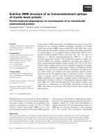
[7]. A simplified scheme of compound I formation,
according to many studies [8–10], is shown in Fig.1.
The reaction starts with deprotonation of hydrogen
peroxide by histidine (an acid–base catalytic residue).
Although the proposed peroxidase–H
2
O
2
complex
existing just before the formation of compound I (the
boxed state in Fig. 1) has not been experimentally
demonstrated, it is believed that H
2
O
2
must interact
with heme iron prior to compound I formation [8].
Deprotonation of H
2
O
2
is extremely improbable,
because the pK
a
of this molecule is 11.6 [11]. However,
H
2
O
2
bound to heme iron (Fe
3+
) is estimated to have
apKa of 3.2–4.0 [8]. If this is indeed the case, deproto-
nation of H
2
O
2
appears to be reasonable, and a classi-
cal reaction path (termed the Poulos–Kraut
mechanism) has been proposed [6].
DyP (
EC 1.11.1.19) from the fungus Bjerkandera ad-
usta Dec 1 (formerly called Thanatephorus cucumeris
Dec 1) is a type of heme peroxidase, but also mediates
hydrolysis of anthraquinone rings, indicating that the
enzyme is bifunctional [12,13]. DyP from B. adusta
Dec 1 is a member of the DyP-type peroxidase family
[14], which is further subdivided into subfamilies A, B,
C, and D, according to PeroxiBase. Research on
DyP-type peroxidase family enzymes commenced only
15 years ago, much later than work on the more com-
mon peroxidases. However, in recent times, several
major studies of DyP-type peroxidases have appeared
[15–17]. The DyP-type peroxidase AnaPX from the
cyanobacterium Anabaena sp. PCC 7120 (type D)
shares some characteristics with DyP from B. adusta
Dec 1 [18]. However, two other types of DyP-type per-
oxidase, YcdB (type A) [19] and YfeX (type B) from
Escherichia coli, have been reported to cooperatively
capture iron [20]. Surprisingly, the functions of YcdB
and YfeX thus appear to have little to do with peroxi-
dase activity. These studies suggest that DyP-type per-
oxidases represent a novel form of heme enzyme,
expressing activities that are not confined to peroxidase
action. Moreover, such enzymes are widely distributed
from bacteria to metazoa, indicating that the DyP-type
peroxidase family is not exceptionally small, but is
rather a sizeable grouping of proteins sharing type-
unique characteristics. Thus, a key cluster of proteins
has evolved to play various roles in a variety of organ-
isms [14]. It is thus important to understand the vari-
ous characteristics of DyPs. In the present study, we
focused on elucidation of the catalytic mechanism,
employing tertiary structural analysis.
Three X-ray crystal structures of members of the
DyP-type peroxidase family, bound to heme or proto-
porphyrin IX, have been deposited in the Protein Data
Bank (PDB). YcdB (PDB ID 2WX7, 2.3 A
˚
resolution)
is a type A enzyme; TyrA (PDB ID 2iiz, 2.3 A
˚
resolu-
tion) is a type B enzyme [21]; and DyP (PDB
ID 2D3Q, 2.96 A
˚
resolution) is a type D enzyme [12].
Surprisingly, the levels of primary structural identity
between DyP (2D3Q) and the other enzymes are very
low (< 5%), although the overall tertiary structures
are very similar [14]. One of the most interesting char-
acteristics of DyP-type peroxidases is that the catalytic
residue is not histidine but rather aspartic acid
(Asp171 in DyP) [12]. However, the details of struc-
ture–function relationships in the DyP family remain
poorly understood. Here, we obtained four structures
of a DyP (type D) enzyme at 1.40–1.45 A
˚
resolution:
native DyP (native); the D171N mutant DyP (D171N),
native DyP complexed with cyanide (CN) (native-CN),
and the D171N mutant DyP associated with CN
(D171N-CN), and identified the precise positions of
Asp171 and associated residues. These structures show
that OD2 accepts a proton from H
2
O
2
bound to the
heme iron. Nevertheless, in the native structure, OD2
is too far away to accept the proton from the H
2
O
2
.
On the other hand, the four structures obviously show
that OD2 is able to swing to the appropriate position
Fe
3+
Fe
3+
Fe
4+
+
H
O
Fe
3+
:
:
:
OH
O
OH
H
2
O
Compound IResting state
N
NH
NH+
NH
N
NH
N
NH
–
O
Fig. 1. Schematic diagram of the most popular mechanism
advanced to explain compound I formation by peroxidases. Struc-
tures of the resting state, two deduced intermediates and com-
pound I are shown. The black bars enclosing iron atoms represent
porphyrin rings. The acid–base catalytic residue is indicated as the
imidazole group of histidine. In compound I, the plus sign and
the black dot indicate when the porphyrin ring is a cationic radical.
The box indicates the region that is the focus of the present study.
Catalytic residue Asp171 swings in view T. Yoshida et al.
2388 FEBS Journal 278 (2011) 2387–2394 ª 2011 The Authors Journal compilation ª 2011 FEBS
in response to ligand for heme iron. On the basis of
these analyses, we propose a new mechanism for the
formation of compound I by DyP. When H
2
O
2
binds
to the heme iron of DyP, OD2 of the catalytic residue
Asp171 swings to a position that is optimal for interac-
tion with the H
2
O
2
.
Results
Approach pathway and binding site of H
2
O
2
In all heme peroxidases, H
2
O
2
is believed to bind to the
heme-distal side. This means that an H
2
O
2
approach
pathway from the enzyme molecular surface to the
heme-distal region must exist. To date, no details of any
such pathway have been reported for DyP-type peroxid-
ases. In the present study, we obtained a native structure
at 1.40-A
˚
resolution (PDB ID 3AFV). This high-resolu-
tion structure showed much more precise details of
enzyme conformation and molecular surface arrange-
ment than were previously available [12]. Heme was not
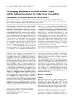
exposed to the molecular surface. However, a large cav-
ity was evident towards the heme-distal side (Fig. 2A),
and formed a small cylindrical pocket about 3 A
˚
in both
diameter and height. H
2
O
2
appeared to fit into this
pocket. In contrast, bulky substrates, such as anthraqui-
none, did not appear to bind into this pocket. Further-
more, this region was surrounded by side chains of four
amino acids: Asp171 (the catalytic residue), Arg329,
Leu354, and Phe356. Overall, the results suggest that
H
2
O
2
may reach the heme-distal side by passing through
the cavity, and then bind and react (with heme) within
the pocket (Fig. 2B).
Conservation of residues forming the binding
pocket for H
2
O
2
Primary sequence identities among different types of
DyP-type peroxidases are, at most, 15%. Indeed, this
value falls to < 5% when types A and D are
compared. Nevertheless, YcdB, TyrA, and DyP,
of types A, B, and D, respectively, have very similar
b-barrel folds and a-helical structural regions. As
described above, we confirmed that the side chains of
Asp171, Arg329, Leu354 and Phe356 form a binding
pocket for H
2
O
2
. On the basis of X-ray crystal struc-
tures and multiple sequence alignments among all
DyP-type peroxidase family members (types A, B, C,
and D), we confirmed conservation of the four residues
that form binding pockets for H
2
O
2
. In YcdB
(type A), the relevant residues are Asp200, Arg312,
Leu331, and Phe333. In TyrA (type B), the residues
are Asp151, Arg242, Leu255, and Phe257. Although
BtDyP (type B) does not bind heme or protoporphy-
rin IX (PDB ID 2gvk), the relevant residues in this
protein seem to be Asp157, Arg245, Thr260, and
Phe262. No X-ray crystal structure of a type C enzyme
has been obtained. However, multiple sequence align-
ment of all DyP-type peroxidases in PeroxiBase
showed that the four residues discussed above were
remarkably conserved as compared with other residues
(Fig. S1). These results show that the b-barrel fold, the
a-helical structural regions and the binding pocket
for H
2
O
2
are conserved in members of the DyP-type
peroxidase family.
D171N structure
We previously reported that the enzyme activity of the
D171N was 1 ⁄ 3000th that of native [12]. We obtained
a D171N structure at 1.42-A
˚
resolution (PDB
ID 3MM1) (Fig. 3), and compared this with the native
structure at 1.40 A
˚
resolution. Both structures were
superimposed on the heme plane and several rmsd val-
ues were calculated (Table 1). The overall structure of
the two enzymes was very similar. The structures at
the heme-distal side were also similar. In both struc-
tures, two water molecules were positioned in the
H
2
O
2
D171
R329
L354
L354
F356
75°
AB
R329
D171
F356
Heme plane
H
2
O
2
3 Å
Large cavity
3 Å
3 Å
Fig. 2. Approach pathway and binding site of H
2
O
2
in DyP. (A) The cutaway view indicates the molecular surface of the entire structure.
The black square indicates the heme plane. The broken arrow in the large cavity shows the pathway taken by H
2
O
2
when it approaches the
heme-distal side. (B) Close-up views of an end of the cavity [circled in (A)]. This region of the cavity forms a binding pocket for H
2
O
2
. The
broken arrow shows the approach pathway of H
2
O
2
. The pocket is delineated by double-headed arrows. Four residues forming the pocket
are shown in stick format.
T. Yoshida et al. Catalytic residue Asp171 swings in view
FEBS Journal 278 (2011) 2387–2394 ª 2011 The Authors Journal compilation ª 2011 FEBS 2389
binding pocket of H
2
O
2
, and the positional relation-
ships of the four residues forming the binding pocket
were very similar. OD1 of Asp171 (Asn171) formed
hydrogen bonds with the amide nitrogen of Gly172
and NH1 of Arg329, but did not form a hydrogen
bond with a water molecule in the binding pocket.
OD2 (ND2) did not form a hydrogen bond with the
peptide chain, but formed a bond with a water mole-
cule in the binding pocket. No polar atoms that could
form hydrogen bonds with OD2 (ND2) were noted in
the peptide chain within 5.0 A
˚
of OD2 (ND2). Excep-
tionally, the positions of Asp171 and Asn171 seemed
to differ between the two structures. In fact, the rmsd
for Asp171(Asn171) was notably larger than that for
other residues. It is of particular interest that the rmsd
for CA of Asp171(Asn171) was very small, but the
rmsd for OD2 (ND2) was clearly large. This probably
results in alternation of proton acceptor and donor
between the native and the D171N. In the native pro-
tein, OD2 is the proton acceptor and W1227 the pro-
ton donor. In contrast, in the D171N mutant, ND2 is
the proton donor and W1200 the proton acceptor.
These results strongly support the previous suggestion
that Asp171 functions as a catalytic residue [12].
Coordination of CN
CN coordination was examined by spectroscopy and
X-ray crystallography. Because the speed of reaction
between peroxidase and H
2
O
2
is very high, the binding
mode has not been experimentally demonstrated. On
the other hand, CN binds stably to the heme iron.
Actually, complexes of peroxidase with CN have been
believed to mimic peroxidase–H
2
O
2
complexes [22].
Although the binding mode of the heme iron differs
between H
2
O
2
and CN, the position of the carbon
atom of CN bound to the heme iron mimics the posi-
tion of the proximal oxygen of H
2
O
2
bound to the
heme iron during compound I formation. Therefore,
the carbon atom position of the CN should provide
basic information for understanding the interaction
between OD2 of Asp171 and the proximal oxygen
of H
2
O
2
bound to the heme iron in compound I
formation.
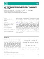
The addition of CN to native led to a shift in the
Soret band from 406 nm to 421 nm, and created an
additional absorption maximum at 535 nm with a
shoulder at 565 nm (Fig. 4). This change was similar
to that observed in horseradish peroxidase and Arthro-
myces ramosus peroxidase upon binding of CN [2,23].
The data suggest that CN bound to the heme iron of
native, and that the electron state of the iron then
changed from high-spin to low-spin. We obtained a
Native
Native-CN
D171N
D171N-CN
D171
D171
N171
N171
G172
G172
G172
G172
R329
R329
R329
R329
L354
L354
L354
L354
F356
F356
F356
F356
CN
CN
W1200
W1227
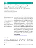
Fig. 3. Structures at the heme-distal side of the native, D171N,
native-CN and D171N-CN enzymes. Heme molecules are shown as
white sticks. Blue spheres represent water molecules and W
means oxygen of water. Broken lines between two atoms indicate
that the distance between these atoms is < 3.4 A
˚
. The 2F
o
) F
c
electron density map at 1r is shown in pink for water molecules
and cyanide ions. Brown circles represent the OD1 atom of
Asp171 or Asn171, and black circles represent the OD2 atom of
Asp171 or the ND2 atom of Asn171.
Table 1. Rmsd values between two structures.
Native and
D171N
Native and
native-CN
Native and
D171N-CN
All CA 0.08 0.38 0.57
Heme plane
a
0.02 0.04 0.04
Asp171 (Asn171)
All atoms 0.29 0.53 0.46
Main chain 0.20 0.40 0.34
Side chain 0.35 0.64 0.55
CG, OD1, OD2 (ND2)
atoms
0.39 0.70 0.59
OD2 (ND2) atom 0.61 1.03 0.78
Arg329
All atoms 0.08 0.24 0.36
Main chain 0.07 0.24 0.34
Side chain 0.09 0.25 0.36
Leu354
All atoms 0.04 0.29 0.22
Main chain 0.04 0.31 0.25
Side chain 0.04 0.28 0.18
Phe356
All atoms 0.16 0.22 0.27
Main chain 0.05 0.26 0.30
Side chain 0.20 0.18 0.24
a
The heme plane represents the 24 atoms of porphyrin.
Catalytic residue Asp171 swings in view T. Yoshida et al.
2390 FEBS Journal 278 (2011) 2387–2394 ª 2011 The Authors Journal compilation ª 2011 FEBS
native-CN enzyme structure at 1.45 A
˚
resolution (PDB
ID 3MM2) and one of D171N-CN at 1.40 A
˚
resolu-
tion (PDB ID 3MM3) (Fig. 3). In both structures, CN
bound almost vertically to the heme plane, and the
nitrogen atom of CN formed a hydrogen bond with an
adjacent water molecule. When the native-CN struc-
ture was compared with that of the native, by superim-
position in the heme plane, the rmsd for Asp171,
especially OD2, was apparently large (Table 1). When
D171N-CN was compared with the native by superim-
position on the heme plane, the rmsd for Asn171,
especially ND2, was again rather large. Interestingly,
OD2 of Asp171 and ND2 of Asn171 formed hydrogen
bonds with different molecules. In native CN, OD2
formed a bond with the water molecule adjacent to
CN. In contrast, ND2 formed a hydrogen bond with
CN of D171N-CN.
In the four structures obtained in the present study,
OD1 could not always form a hydrogen bond with a
molecule in the binding pocket of H
2
O
2
, but always
participated in formation of two hydrogen bonds with
the peptide chain. In contrast, OD2 (ND2) could
always form a hydrogen bond with a molecule in the
binding pocket of H
2
O
2
, but could not always engage
in hydrogen bonding with the peptide chain. These
results strongly suggest that OD2, rather than OD1,
accepts a proton from H
2
O
2
in the binding pocket.
Discussion
Swinging of Asp171
It is important to note that OD2 of Asp171 in native-
CN and ND2 of Asn171 in D171N-CN formed hydro-
gen bonds with different molecules. As a result, the
positions of OD2 and ND2 were very different. This
appears to have been induced by variation in the pro-
tonation state of OD2 and ND2. In native-CN, OD2
is a proton acceptor at pH 6.0. The crystallization con-
dition was also at pH 6.0. Therefore, OD2 cannot
form a hydrogen bond with CN, which is a proton
acceptor, but can form such a bond with a water mole-
cule adjacent to CN, which serves as a proton donor.
Thus, OD2 is shifted in position, in a direction away
from the heme plane, as compared with the native. On
the other hand, in D171N-CN, ND2 is a proton donor
at pH 6.0. Therefore, ND2 can form a hydrogen bond
with CN. As a result, ND2 is shifted in position in a
direction towards the heme plane, as compared with
the native. This suggests that OD2 can change position
in response to ligand status in the binding pocket. This
flexibility of OD2 seems to be associated with the fact
that OD2 does not possess a polar atom that can form
a hydrogen bond. Moreover, such flexibility was
strongly supported by superimposition of the struc-
tures of the native, D171N, native-CN and D171N-
CN enzymes in the heme plane (Fig. 5). Because of
differences in the chosen hydrogen bond partners,
OD2 (ND2) seems to swing around OD1, by over 37°.
These results suggest that OD2 can swing in response
to ligand status.
The catalytic residue Asp171 swings to form the
compound I intermediate
At which position does OD2 accept a proton from
H
2
O
2
? Because an X-ray crystal structure at 1.4 A
˚
reso-
lution cannot show hydrogen atoms, we discuss this
issue from the viewpoint of the distance between OD2
and the proximal oxygen. We replace the carbon atom
position of CN with a position of the proximal oxygen
Native
Native-CN
5
Fig. 4. UV–visible spectra of native and native-CN enzymes. Spec-
tra from 450 nm to 700 nm are shown at five-fold magnification.
Solution conditions were 4 l
M DyP in 25 mM citrate buffer (pH 5.5)
containing 0.5
M NaCl, with or without 100 mM KCN.
CN
2.05 A
37.0
OD1
OD2
(ND2)
D171
(N171)
D171
(N171)
OD1
OD2
(ND2)
60
Fig. 5. Comparison of Asp171 (Asn171) locations between native
(green), D171N (blue), native-CN (yellow) and D171N-CN (red)
enzymes. These four structures are superimposed on the heme
planes. Gray broken lines show the shortest distances between
OD2 (ND2) and the carbon atom of CN for each structure. The dis-
tance between the CN carbon atom and the iron atom of heme is
2.05 A
˚
. On the right, the relevant four residues are rotated by 60°
to assist in an understanding of differences in residue positions.
T. Yoshida et al. Catalytic residue Asp171 swings in view
FEBS Journal 278 (2011) 2387–2394 ª 2011 The Authors Journal compilation ª 2011 FEBS 2391
of H
2
O
2
as described in Results. The distances between
OD2 (ND2) and the proximal oxygen are shown in
Fig. 5. In the native and D171N-CN structures, the dis-
tances are 4.06 A
˚
and 3.46 A
˚
, respectively. We think
that this indicates a significant difference. That is
because this difference is caused by the difference
between the positions of hydrogen bond partners of
OD2 in the native and ND2 in D171N-CN. In the
native, the distance, 4.06 A
˚
, is too long to permit reac-
tion with H
2
O
2
. This shows that this position of OD2 in
the native is not appropriate for involvement in the reac-
tion. In contrast, in D171N-CN, the distance, 3.46 A
˚
,is
less than in the native. The position of OD2 in D171N-
CN is appropriate for involvement in the reaction. Our
argument is illustrated in Fig. 6A. Thus, OD2 never
accepts a proton when in the position occupied by OD2
in the native, but does so when in the position occupied
by OD2 in D171N-CN (Fig. 6A).
On the basis of the dual conclusions that OD2
accepts a proton when in the position occupied by
OD2 in D171N-CN, and that OD2 can swing from the
position occupied in the native to the position seen in
D171N-CN, we propose a swing mechanism for the
formation of compound I by DyP (Fig. 6B). To accept
a proton, OD2 of Asp171 swings towards the position
occupied by OD2 in D171N-CN. After compound I
formation, OD2 of Asp171 returns to the position
characteristic of the native. The side chain of the cata-
lytic residue of DyP is not located just above the heme
iron, unlike in other heme peroxidases. Moreover, the
side chain of DyP is arranged in parallel rather than
vertically to the heme plane. This novel location and
arrangement seems to produce the swing mechanism.
However, this swing mechanism may be needed in
type C and D DyP-type peroxidases. Nevertheless, we
believe that this structural study paves the way to
understanding the structure–function relationships of
the DyP-type peroxidase family.
Experimental procedures
Crystallization of native and D171N proteins
Native and D171N proteins were purified with a modifica-
tion of published procedures [12,24,25]. Purified samples
were deglycosylated by endoglycosidase H (Roche Diagnos-
tics, Tokyo, Japan). Samples were loaded onto Superdex 75
columns (GE Healthcare Japan, Tokyo, Japan), and the de-
glycosylated fractions were concentrated to 20 mgÆmL
)1
by
ultrafiltration. Crystallization was achieved with the hang-
Fig. 6. The swinging mechanism of Asp171. (A) Interpretation of distances between Asp171 and a virtual proximal oxygen of H
2
O
2
in the
native and D171N-CN. The native and D171N-CN structures are superimposed on the heme planes. Note that Asn171 of D171N-CN is
shown as Asp171. To avoid misunderstanding, only the heme of the native is shown in white. The position of the CN carbon mimics the
position of the proximal oxygen of H
2
O
2
. Double-headed arrows show whether the distance between Asp171 and the virtual proximal oxy-
gen is appropriate to permit reaction. (B) Schematic diagram of the proposed mechanism of compound I formation by DyP. Structures of the
resting state, two deduced intermediates, and compound I are shown. The two black bars enclosing an iron atom indicate the porphyrin ring.
Broken lines and arrows indicate hydrogen bonds and the swinging direction of the OD2 atom of Asp171, respectively. In compound I, a
plus sign and a black dot indicate when the porphyrin ring is a cationic radical.
Catalytic residue Asp171 swings in view T. Yoshida et al.
2392 FEBS Journal 278 (2011) 2387–2394 ª 2011 The Authors Journal compilation ª 2011 FEBS
ing drop vapor diffusion method. Drops containing 1 lLof
a20mgÆmL
)1
protein solution (0.1 m Mes at pH 6.0; 0.5 m
NaCl) and 1 lL of mother solution [0.1 m Mes at pH 6.0;
48% (w ⁄ v) poly(ethylene glycol) 8000] were equilibrated
against 500 lL of reservoir solution [0.1 m Mes at pH 6.0;
0.25 m NaCl; 28% (w ⁄ v) poly(ethylene glycol) 8000] at
278 K. Hexagonal crystals appeared after 2–3 weeks. Prior
to data collection, crystals were soaked briefly in a cryopro-
tective solution containing 0.1 m Mes at pH 6.0, 0.25 m
NaCl, 30% (w ⁄ v) poly(ethylene glycol) 8000, and
25% (v ⁄ v) glycerol, and flash frozen in liquid nitrogen.
CN-complexed crystals of native and D171N
proteins
CN-complexed crystals were prepared by soaking native and
D171N proteins in a cryoprotective solution containing
0.1 m Mes at pH 6.0, 0.25 m NaCl, 30% (w ⁄ v) poly(ethylene
glycol) 8000, 25% (w ⁄ v) glycerol, and 120 mm KCN. For
both crystal types, binding of CN appeared to be complete
within a few seconds, as assessed by visual monitoring of the
crystal color change from brown to red. The crystals were
flash frozen in liquid nitrogen.
X-ray data collection and structural refinement
Data collection from native, the D171N, native-CN and
D171N-CN was performed with a wavelength of 1.0 A
˚
on
a beamline PF-AR NE3A or PF-AR NW12A instrument at
the Photon Factory (Tsukuba, Japan). Subsequent proce-
dures, including processing, scaling, and refinement, were
identical for all crystals. Datasets were processed and scaled
with the hkl2000 program [26]. Structures were solved with
the molecular replacement software of the ccp4 program
suite [27] (molrep), employing a 2.96-A
˚
-resolution structure
of DyP (PDB ID 2D3Q) as the starting point. Iterative
refinement and model-building were subsequently per-
formed with refmac5 [28] and coot [29]. Data collection
and refinement statistics are summarized in Table S1. For
native DyP, three datasets were collected, refined, and
superimposed on the heme plane. The rmsd values for
Asp171 were very small in all three datasets (Fig. S2;
Table S2). Two datasets were collected and refined for the
D171N, and superimposed on the heme plane. The rmsd
values of Asn171 were rather large in both datasets. This
was because the hydrogen bond between the protonated
ND2 of Asn171 and a water molecule in the binding pocket
was weak.
UV–visible spectrophotometry (solution studies)
All spectra were obtained with a Shimadzu UV-2400 PC
spectrophotometer (Shimadzu Co., Kyoto, Japan) at 30 °C,
with a spectral bandwidth of 1.0 nm, employing cuvettes of
light path 1 cm. Solution conditions were 4 lm DyP in
25 mm citrate buffer (pH 5.5) containing 0.5 m NaCl, with
or without 100 mm KCN.
Acknowledgements
This research was undertaken with the assistance of
the Photon Factory in KEK (proposal numbers
2010G011 and 2008G063) and was partly supported
by a Grant-in-Aid for Scientific Research
(no. 22570136) from the Japan Society for the Promo-
tion of Sciences.
References
1 Koua D, Cerutti L, Falquet L, Sigrist CJ, Theiler G,
Hulo N & Dunand C (2009) PeroxiBase: a database
with new tools for peroxidase family classification.
Nucleic Acids Res 37, D261–266.
2 Dunford HB (1999) Heme Peroxidases. Wiley,
New York.
3 Lang G, Spartalin K & Yonetani T (1976) Mo
¨
ssbauer
spectroscopic study of compound ES of cytochrome c
peroxidase. Biochim Biophys Acta 451, 250–258.
4 Browlett WR & Stillman MJ (1981) Evidence for heme
pi cation radical species in compound I of horseradish
peroxidase and catalase. Biochim Biophys Acta 660, 1–7.
5 Kaneko Y, Tamura M & Yamazaki I (1980) Formation
of porphyrin pi cation radical in zinc-substituted horse-
radish peroxidase. Biochemistry 19, 5795–5799.
6 Poulos TL & Kraut J (1980) The stereochemistry of
peroxidase catalysis. J Biol Chem 255, 8199–8205.
7 Welinder KG (1992) Superfamily of plant, fungal and
bacterial peroxidases. Curr Opin Struct Biol 2, 388–393.
8 Jones P & Dunford HB (2005) The mechanism of com-
pound I formation revisited. J Inorg Biochem 99, 2292–
2298.
9 Raven EL (2003) Understanding functional diversity
and substrate specificity in haem peroxidases: what can
we learn from ascorbate peroxidase? Nat Prod Rep 20,
367–381.
10 Derat E, Shaik S, Rovira C, Vidosich P & Alfonso-Pri-
eto M (2007) The effect of a water molecule on the
mechanism of formation of compound 0 in horseradish
peroxidase. J Am Chem Soc 129, 6346–6347.
11 Uri N & Evans MG (1949) The dissociation constant of
hydrogen peroxide and the electron affinity of the HO
2
radical. Trans Faraday Soc 45, 224–230.
12 Sugano Y, Muramatsu R, Ichiyanagi A, Sato T &
Shoda M (2007) DyP, a unique dye-decolorizing peroxi-
dase, represents a novel heme peroxidase family:
Asp171 replaces the distal histidine of classical peroxid-
ases. J Biol Chem 282, 36652–36658.
T. Yoshida et al. Catalytic residue Asp171 swings in view
FEBS Journal 278 (2011) 2387–2394 ª 2011 The Authors Journal compilation ª 2011 FEBS 2393
13 Sugano Y, Matsushima Y, Tsuchiya K, Aoki H, Hirai
M & Shoda M (2009) Degradation pathway of an
anthraquinone dye catalyzed by a unique peroxidase
DyP from Thanatephorus cucumeris Dec 1.
Biodegradation 20, 433–440.
14 Sugano Y (2009) DyP-type peroxidases comprise a
novel heme peroxidase family. Cell Mol Life Sci 66,
1387–1403.
15 Ogola HJ, Hashimoto N, Miyabe S, Ashida H,
Ishikawa T, Shibata H & Sawa Y (2010) Enhancement
of hydrogen peroxide stability of a novel Anabaena sp.
DyP-type peroxidase by site-directed mutagenesis of
methionine residues. Appl Microbiol Biotechnol 87,
1727–1736.
16 Hofrichter M, Ullrich R, Pevyna MJ, Liers C & Lundell
T (2010) New and classic families of secreted fungal
heme peroxidases. Appl Microbiol Biotechnol 87,
871–897.
17 Liers C, Bobeth C, Pecyna M, Ullich R & Hofrichter
M (2010) DyP-like peroxidases of the jelly fungus
Auricularia auricular-judae oxidize nonphenolic lignin
model compounds and high-redox potential dyes. Appl
Microbiol Biotechnol 85, 1869–1879.
18 Ogola HJ, Kamiike T, Hashimoto N, Ashida H, Ishika-
wa T, Shibata H & Sawa Y (2009) Molecular character-
ization of a novel peroxidase from the cyanobacterium
Anabaena sp. strain PCC 7120. Appl Environ Microbiol
75, 7509–7518.
19 Sturm A, Schierhorn A, Lindenstrauss U, Lilie H &
Bru
¨
ser T (2006) YcdB from Escherichia coli reveals a
novel class of Tat-dependently translocated hemopro-
teins. J Biol Chem 281, 13972–13978.
20 Le
´
toffe
´
S, Heuck G, Delepelaire P, Lange N &
Wandersman C (2009) Bacteria capture iron from heme
by keeping tetrapyrrol skeleton intact. Proc Natl Sci
Acad USA 106, 11719–11724.
21 Zubieta C, Joseph R, Krishna SS, McMullan D,
Kapoor M, Axelrod HL, Miller MD, Abdubek P,
Acosta C, Astakhova T et al. (2007) Identification and
structural characterization of heme binding in a novel
dye-decolorizing peroxidase, TyrA. Proteins 69, 234–
243.
22 Edwards SL & Poulos TL (1990) Ligand binding and
structural perturbations in cytochrome c peroxidase.
J Biol Chem 265, 2588–2595.
23 Fukuyama K, Kunishima N, Amada F, Kubota T &
Matsubara H (1995) Crystal structures of cyanide- and
triiodide-bound forms of Arthromyces ramosus peroxi-
dase at different pH values. Perturbations of active site
residues and their implication in enzyme catalysis.
J Biol Chem 270, 21884–21892.
24 Sugano Y, Nakano R, Sasaki K & Shoda M (2000)
Efficient heterologous expression in Aspergillus oryzae
of a unique dye-decolorizing peroxidase, DyP, of Geo-
trichum candidum Dec 1. Appl Environ Microbiol 66,
1754–1758.
25 Saijo S, Sato T, Tanaka N, Ichiyanagi A, Sugano Y &
Shoda M (2005) Precipitation diagram and optimization
of crystallization conditions at low ionic strength
for deglycosylated dye-decolorizing peroxidase from a
basidiomycete. Acta Crystallogr F 61, 729–732.
26 Otwinowski Z & Minor W (1997) Processing of x-ray
diffraction data collected in oscillation mode. Methods
Enzymol 276, 307–326.
27 Collaborative Computational Project Number 4 (1994)
The CCP4 suite: programs for protein crystallography.
Acta Crystallogr D 50, 760–763.
28 Vagin A & Teplyakov A (2000) An approach to multi-
copy search in molecular replacement. Acta Crystallogr
D 56, 1622–1624.
29 Emsley P & Cowtan K (2004) Coot: model-building
tools for molecular graphics. Acta Crystallogr D 60,
2126–2132.
Supporting information
The following supplementary material is available:
Fig. S1. Structure-based sequence alignments of YcdB,
BtDyP, TyrA, and DyP.
Fig. S2. Comparison of the locations of Asp171
(Asn171) side chains among structures obtained with
different datasets.
Table S1. Data collection and refinement statistics.
Table S2. Rmsd values between two structures.
This supplementary material can be found in the
online version of this article.
Please note: As a service to our authors and readers,
this journal provides supporting information supplied
by the authors. Such materials are peer-reviewed and
may be re-organized for online delivery, but are not
copy-edited or typeset. Technical support issues arising
from supporting information (other than missing files)
should be addressed to the authors.
Catalytic residue Asp171 swings in view T. Yoshida et al.
2394 FEBS Journal 278 (2011) 2387–2394 ª 2011 The Authors Journal compilation ª 2011 FEBS