Báo cáo khoa học: Binding of cGMP to the transducin-activated cGMP phosphodiesterase, PDE6, initiates a large conformational change involved in its deactivation ppt
Bạn đang xem bản rút gọn của tài liệu. Xem và tải ngay bản đầy đủ của tài liệu tại đây (934.67 KB, 19 trang )
Binding of cGMP to the transducin-activated cGMP
phosphodiesterase, PDE6, initiates a large conformational
change involved in its deactivation
Akio Yamazaki
1,2,3
, Fumio Hayashi
4
, Isao Matsuura
5
and Vladimir A. Bondarenko
6
1 Kresge Eye Institute, Wayne State University, Detroit, MI, USA
2 Department of Ophthalmology, Wayne State University, Detroit, MI, USA
3 Department of Pharmacology, Wayne State University, Detroit, MI, USA
4 Department of Biology, Kobe University, Japan
5 Division of Molecular and Genomic Medicine, National Health Research Institutes, Zhunan Town, Taiwan
6 College of Osteopathic Medicine, Touro University, Henderson, NV, USA
Keywords
cGMP binding; cGMP-binding-dependent
protein conformational change; GAF
domains; G-protein-mediated signal
transduction; PDE
Correspondence
V. A. Bondarenko, College of Osteopathic
Medicine, Touro University, Henderson,
NV 89014, USA
Fax: +1 702 777 1799
Tel: +1 702 777 1806
E-mail:
(Received 30 January 2011, revised 17
March 2011, accepted 22 March 2011)
doi:10.1111/j.1742-4658.2011.08104.x
Retinal photoreceptor phosphodiesterase (PDE6), a key enzyme for photo-
transduction, consists of a catalytic subunit complex (Pab) and two inhibi-
tory subunits (Pcs). Pab has two noncatalytic cGMP-binding sites. Here,
using bovine PDE preparations, we show the role of these cGMP-binding
sites in PDE regulation. Pabcc and its transducin-activated form, Pabc,
contain two and one cGMP, respectively. Only Pabc shows [
3
H]cGMP
binding with a K
d
50 nM and Pc inhibits the [
3
H]cGMP binding. Binding
of cGMP to Pabc is suppressed during its formation, implying that cGMP
binding is not involved in Pabcc activation. Once bound to Pabc,
[
3
H]cGMP is not dissociated even in the presence of a 1000-fold excess of
unlabeled cGMP, binding of cGMP changes the apparent Stokes’ radius of
Pabc, and the amount of [
3
H]cGMP-bound Pabc trapped by a filter is
spontaneously increased during its incubation. These results suggest that
Pabc slowly changes its conformation after cGMP binding, i.e. after for-
mation of Pabc containing two cGMPs. Binding of Pc greatly shortens the
time to detect the increase in the filter-trapped level of [
3
H]cGMP-bound
Pabc, but alters neither the level nor its Stokes’ radius. These results sug-
gest that Pc accelerates the conformational change, but does not add
another change. These observations are consistent with the view that Pabc
changes its conformation during its deactivation and that the binding of
cGMP and Pc is crucial for this change. These observations also imply that
Pabcc changes its conformation during its activation and that release of Pc
and cGMP is essential for this change.
Structured digital abstract
l
PDE6 alpha, PDE6 beta and PDE6 gamma physically interact by molecular sieving (View
interaction)
Abbreviations
GAF, a domain derived from cGMP-regulated cyclic nucleotide phosphodiesterases, certain adenylyl cyclases, the bacterial transcription
factor FhlA; GTPcS, guanosine 5¢-O-(3-thiotriphosphate); IBMX, 1-methyl-3-isobutylxanthine; OS, outer segments of retinal photoreceptors;
PDE, cGMP phosphodiesterase; PMSF, phenylmethylsulfonyl fluoride; Pa and Pb, rod PDE catalytic subunits; Pa¢, cone PDE catalytic
subunit; Pab ⁄ Pc,Pab complexes having an unknown number of Pc;Pd, a prenyl-binding protein; Pc, rod PDE inhibitory subunit; Pc¢, cone
PDE inhibitory subunit; T, transducin.
1854 FEBS Journal 278 (2011) 1854–1872 ª 2011 The Authors Journal compilation ª 2011 FEBS
Introduction
Cyclic GMP phosphodiesterase (EC 3.1.4.17), classified
as PDE6 in the PDE family, is one of the key enzymes
for phototransduction in the outer segments (OS) of
retinal photoreceptors. Its activation is G-protein-med-
iated: illuminated rhodopsin stimulates GTP ⁄ GDP
exchange on transducin (T)a, followed by dissociation
of GTP–Ta from Tbc. The GTP–Ta activates PDE,
resulting in a decrease in the cytoplasmic [cGMP], clo-
sure of cGMP-gated channels and hyperpolarization of
plasma membranes [1–3].
The inactive form of rod PDE is composed of a cat-
alytic subunit complex, Pab, and two inhibitory subun-
its, Pcs, i.e. Pabcc [4–10]. A study using electron
microscopy and image analysis of single particles [11]
shows that bovine Pabcc, 150 · 108 · 60 A
˚
, has the
shape of a flattened bell with a handle-like protrusion
( 30 A
˚
) and that the structure is divided into three
distinct substructures by two holes. Except for the pro-
trusion, the structure also appears to consist of two
homologous structures arranged side by side. These
characteristics are consistent with a model in which
Pabcc’s structure is determined by a dimer of homolo-
gous catalytic subunits consisting of two GAF (a
domain derived from cGMP-regulated cyclic nucleo-
tide phosphodiesterases, certain adenylyl cyclases, the
bacterial transcription factor FhlA) regions and one
catalytic region. Indeed, bovine Pabcc contains two
cGMPs and these bind tightly to substructures formed
by GAF regions [12]. These two substructures, called
the noncatalytic cGMP-binding sites, are similar, but
not identical, in shape and size [11]. This implies that
the manner of cGMP binding to each site and ⁄ or the
role of cGMP binding to each site in PDE regulation,
if present, may be different.
The current predominant model for PDE regulation
is simple [13]. For activation, GTP–Ta interacts with
Pc in Pabcc, and the GTP–TaÆPabcc complex, with-
out altering the firm interaction between Pab and Pc,
expresses a high cGMP hydrolytic activity. For deacti-
vation, GTP in the GTP–TaÆPabcc complex is hydro-
lyzed with the help of RGS9 and accessory proteins,
i.e. the GTP is hydrolyzed after formation of a huge
complex, and Pabcc is recovered after dissociation of
various proteins, including GDP-bound Ta (GDP–
Ta). This model conveniently explains the rapid acti-
vation and deactivation of PDE; however, there is no
clear evidence to show a firm and continuous interac-
tion between GTP–Ta and Pabcc during Pabcc acti-
vation, as would be shown by the isolation of a
complex of Pabcc with Ta containing a hydrolysis-
resistant GTP analogue such as guanosine 5¢
-O-(3-
thiotriphosphate) (GTPcS). In addition, there is no
definitive evidence to prove the formation of a GTP–
TaÆPabcc complex containing RGS9 and accessory
proteins and its decomposition during deactivation of
GTP–Ta-activated PDE.
Binding of cGMP to the noncatalytic site in Pab is
believed to be involved in PDE regulation. Two mod-
els, the cGMP-regulated Pab-Pc interaction model
[14–18] and the cGMP-binding direct regulation model
[19], have been proposed to explain the role of cGMP-
binding sites in PDE regulation. In the former model,
the interaction between Pab and Pc is dependent upon
the presence of cGMP at the noncatalytic site. When
the noncatalytic sites of Pabcc are saturated with
cGMP, GTP–Ta activates Pabcc without changing the
tight interaction between Pab and Pc, i.e. a GTP–TaÆ-
Pabcc complex is formed and the complex expresses a
high PDE activity. However, when the noncatalytic
sites are not saturated, GTP–Ta activates Pabcc
through dissociation of Pc complexed with GTP–Ta,
i.e. a Pc-depleted PDE(s) is produced. Pc in the GTP–
Ta complex enhances the GTPase activity of Ta; the
resulting GDP–Ta instantly releases Pc, and the
released Pc deactivates the GTP–Ta-activated PDE. In
the latter model, binding of cGMP to the noncatalytic
sites directly regulates PDE catalytic activity. These
two models appear to explain some observations of
cGMP binding to noncatalytic sites. However, as dis-
cussed later, these models have many ambiguous and
controversial points. Thus, it is difficult to integrate
these concepts smoothly into a coherent model for
PDE regulation.
We have recently challenged the dominant model for
PDE regulation by proposing a new and comprehen-
sive model [11,13,20] in which GTP–Ta activates
Pabcc by forming a complex with a Pc, thereby disso-
ciating the PcÆGTP–Ta complex. This occurs on mem-
branes and is independent of the cytoplasmic [cGMP].
A significant portion of the PcÆGTP–Ta complex is
then released into the soluble fraction. Thus, Pabc is
the GTP–Ta-activated PDE. After hydrolysis of GTP,
both soluble and membranous PcÆGDP–Ta complexes
deactivate Pabc without liberating Pc. These PcÆGDP–
Ta complexes appear to have a preferential order in
deactivating Pabc. This new model is based on the fol-
lowing observations: (a) Pabc, but not Pab, is isolated
only when OS homogenates are incubated with
GTPcS; (b) the ratio of Pc ⁄ Pab in Pabcc and Pabc is
2 : 1; (c) the enzymatic activity of Pabc is 12 times
higher than that of Pabcc and is inhibited by 30 nm
Pc; (d) the basic structure of these PDE species is not
A. Yamazaki et al. Roles of cGMP binding in PDE6 regulation
FEBS Journal 278 (2011) 1854–1872 ª 2011 The Authors Journal compilation ª 2011 FEBS 1855
changed when Pabcc is shifted to Pabc; (e)
PcÆGTPcS–Ta is isolated from membranous and solu-
ble fractions; (f) both membranous and soluble
PcÆGDP–Ta complexes deactivate Pabc without liber-
ating Pc; (g) the membranous PcÆGDP–Ta complex
appears to be consumed earlier than the soluble
PcÆGDP–Ta complex; and (h) PDE regulatory mecha-
nisms similar to this model are also found in mamma-
lian and amphibian photoreceptors, as well as in rods
and cones. During these studies, we have also shown
that: (a) the interaction between Pabcc and GTPcS–
Ta is short-lived, indicating that GTP–TaÆPabcc is an
intermediate, but not GTP–Ta-activated PDE; (b) free
Pc is not detected in any preparations, implying that
Pc always forms complexes with other proteins; (c)
Pabccd and Pabcdd are formed when Pabcc and Pabc
are solubilized with Pd, a prenyl-binding protein; (d)
the stoichiometry of Pabccd suggests that only one
lipid moiety may be involved in the interaction of
Pabcc with membranes; and (e) the stoichiometry of
Pabcdd suggests that a lipid moiety in Pab is also
affected by Pc dissociation.
In this study, we extend our model by integrating
the role of cGMP binding to the noncatalytic site. We
demonstrate that Pabcc and Pabc contain two and
one cGMP, respectively, that only Pabc expresses
[
3
H]cGMP-binding activity and that Pc inhibits
[
3
H]cGMP binding to Pabc. We also show that the
cGMP binding to Pabc is suppressed during Pabcc
activation, i.e. cGMP binding is not involved in Pabcc
activation. We also suggest that cGMP binding to
Pabc slowly changes its conformation and that binding
of Pc accelerates the conformational change. Based on
these studies, we propose that binding of cGMP to
Pabc is the first step in PDE deactivation.
Results
Binding of [
3
H]cGMP to OS membranes
Bovine OS membranes contain a [
3
H]cGMP-binding
site(s) ( Fig. 1A). Both GTPcS-treated and nontreated
membranes showed [
3
H]cGMP-binding activities; how-
ever, the activity in GTPcS-treated membranes was
much higher than in GTPcS-nontreated membranes,
indicating that GTPcS–Ta somehow enhances the
[
3
H]cGMP-binding activity. By contrast, the soluble
fraction, whether obtained from GTPcS-treated or
nontreated OS homogenates, showed only negligible
[
3
H]cGMP-binding activity (data not shown). This sug-
gests that no protein in the soluble fraction contains the
[
3
H]cGMP-binding site and ⁄ or expresses [
3
H]cGMP-
binding activity under our experimental conditions.
Solubilization and isolation of membranous proteins
showed that a [
3
H]cGMP-binding activity (Fig. 1B)
was detected only in the fraction containing a protein-
doublet (m 88 kDa) (Fig. 1C) and that the activity
appeared to be proportional to the level of the pro-
tein-doublet. These fractions also contained a PDE
activity that was proportional to the level of the pro-
tein-doublet (data not shown). The protein-doublet has
been identified as Pab and 70–80% of Pab is extracted
from membranes under these conditions [13,20]. These
results suggest that the [
3
H]cGMP-binding activity in
membranes is due to a Pab complex(s). This implies
that cone PDEs, Pa¢a¢⁄Pc¢ complexes, are also present
and that a Pa¢a¢⁄Pc¢ complex(s) expresses [
3
H]cGMP-
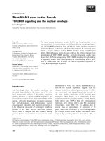
Fig. 1. Binding of [
3
H]cGMP to membranous PDE. (A) Levels of
[
3
H]cGMP binding to OS membranes treated with or without
GTPcS. OS homogenates (27.5 mg protein) were suspended in
18.4 mL of buffer A and divided into two portions. After incubation
of a portion with 50 l
M GTPcS overnight on ice, its membranes
were washed twice with 5 mL buffer A supplemented with 50 l
M
GTPcS, twice with 5 mL buffer A and suspended in 5 mL buffer A.
The other portion was treated in the same way but without GTPcS.
Binding of [
3
H]cGMP to these suspensions (10 lL) was assayed
using 1 l
M [
3
H]cGMP. (B,C) [
3
H]cGMP binding to proteins extracted
from OS membranes treated with or without GTPcS. OS homogen-
ates (27.7 mg protein) were suspended in 18 mL of buffer A,
divided into two portions and treated with or without GTPcS. Pro-
teins were extracted from membranes with 3 mL buffer B (·7),
concentrated to 0.5 mL and applied to Bio-Gel A 0.5-m column.
[
3
H]cGMP-binding activity (B) and PDE activity (not shown) were
assayed using 60 and 5 lL of the fraction, respectively. Protein pro-
files in the fraction (90 lL) were analyzed by SDS ⁄ PAGE and stain-
ing with Coomassie Brilliant Blue (C). The left end lane shows the
molecular mass of standard proteins, 94, 67 and 43 kDa.
Roles of cGMP binding in PDE6 regulation A. Yamazaki et al.
1856 FEBS Journal 278 (2011) 1854–1872 ª 2011 The Authors Journal compilation ª 2011 FEBS
binding activity. However, neither Pa¢ nor its
[
3
H]cGMP-binding activity could be identified. These
failures, we believe, are because of its small abundance
in OS. The soluble fraction also contained a Pab ⁄ Pc
complex (peak b in [13]); however, the complex showed
only negligible [
3
H]cGMP-binding activity (data not
shown). This is consistent with the above-mentioned
conclusion that [
3
H]cGMP-binding activity was not
detected in the soluble fraction.
Interestingly, the [
3
H]cGMP-binding activity in
GTPcS-treated PDE was higher than in GTPcS-non-
treated PDE (Fig. 1B). When OS homogenates are
incubated with GTPcS, the Pab content in membranes
is increased 20–30% by binding of the Pab ⁄ Pc com-
plex existing in the soluble fraction [13]. Therefore,
binding of the Pab ⁄ Pc complex to membranes and the
resulting expression of a [
3
H]cGMP-binding activity
could increase the activity in membranes. However,
the increase in the activity by GTPcS was much
higher, 2.4 times (Fig. 1B). In addition, Pab in the
Pab ⁄ Pc complex has two cGMP-binding sites at most
[12]. Therefore, we conclude that even if the Pab ⁄ Pc
complex could express [
3
H]cGMP-binding activity, the
greater part of the increase is due to an increase in the
activity of a Pab ⁄ Pc complex(s) located on mem-
branes. This is unexpected because previous studies
using frog PDE ⁄ membranes [21,22] showed that their
[
3
H]cGMP-binding activity in GTP-nontreated PDE
was much higher than that in GTP-treated PDE. We
also note that this result, with the observation shown
in Fig. 1A, implies that [
3
H]cGMP binding to solubi-
lized PDE species is similar to binding to membranous
PDE species, i.e. the properties of cGMP binding to
membranous PDE species may be estimated by study-
ing cGMP binding to solubilized PDE species.
Identification of PDE species expressing
[
3
H]cGMP-binding activity
GTPcS-nontreated membranes contain Pabcc, and
GTPcS-treated membranes have Pabcc and Pabc as
major species and a Pab ⁄ Pc complex as a minor species
[20]. These PDE species were extracted using a hypo-
tonic buffer (Fig. 2A) or Pd in an isotonic buffer
(Fig. 2C) and their [
3
H]cGMP-binding activities were
measured after isolation. The use of Pd in an isotonic
buffer may exclude a possible artifact(s) caused by the
hypotonic extraction. OS homogenates were also treated
with GTPcS in the presence of cGMP (GTPcS+
cGMP), and after isolation of Pab ⁄ Pc complexes, their
[
3
H]cGMP-binding activities were measured (Fig. 2B).
The result is compared with the results in Fig. 2A, as
shown later.
Pabcc extracted by a hypotonic buffer
Pabcc was obtained from GTPcS-nontreated mem-
branes (Fig. 2A, upper) and GTPcS-treated membranes
(Fig. 2A, lower). In the former preparation, the
[
3
H]cGMP-binding activity appeared to be proportional
to the level of Pab, implying that Pabcc may express
[
3
H]cGMP-binding activity. However, the molecular
ratio of [
3
H]cGMP to Pab was < 0.01, indicating that
only a negligible portion of the Pabcc expresses this
activity. In the latter preparation, a small [
3
H] radio-
activity was detected in the fraction close to the Pabcc
peak. However, the level of [
3
H] radioactivity was not
proportional to that of Pab in the Pabcc fraction, indi-
cating that the [
3
H] radioactivity is not attributable to
[
3
H]cGMP bound to the Pabcc, i.e. the Pabcc does not
show [
3
H]cGMP-binding activity and⁄or the Pabcc,
when it exists with GTP–Ta, appears to lose a portion
that may express [
3
H]cGMP-binding activity (Fig. 2A,
upper).
Pabcc extracted with Pd in an isotonic buffer
The Pabccd preparation was obtained from GTPcS-
nontreated membranes (data not shown) and GTPcS-
treated membranes (Fig. 2C). In the former prepara-
tion, the [
3
H]cGMP-binding activity appeared to be
proportional to the level of Pab; however, the molecu-
lar ratio of [
3
H]cGMP to Pab in the Pabccd was
< 0.01. These observations are identical to those for
Pabcc extracted with a hypotonic buffer (Fig. 2A,
upper). In the latter preparation, Pabccd appeared to
show a small [
3
H]cGMP-binding activity (Fig. 2C,
upper). However, the amount of binding was not
exactly proportional to the Pab level in the fraction,
indicating that the [
3
H] radioactivity was not due to
[
3
H]cGMP bound to the Pabccd.
As shown later (Fig. 7), Pabcc can be trapped by a
Millipore filter with a high efficiency, implying that the
lack of [
3
H]cGMP-binding activity and ⁄ or the negligi-
ble level of [
3
H]cGMP-binding activity in Pabcc prepa-
rations are not due to the failure to trap [
3
H]cGMP-
bound Pabcc. Taken together, our results strongly
suggest that Pabcc does not express [
3
H]cGMP-bind-
ing activity and that negligible activities occasionally
detected in fractions containing Pabcc may be artifacts
caused by experimental procedures. The level of [
3
H]
radioactivity was not proportional to the level of Ta
(Fig. 2C). This confirms that Ta has no cGMP-binding
site [23]. The amino acid sequence of Ta also supports
this notion. This is specifically noted here because we
use this information in a later discussion.
A. Yamazaki et al. Roles of cGMP binding in PDE6 regulation
FEBS Journal 278 (2011) 1854–1872 ª 2011 The Authors Journal compilation ª 2011 FEBS 1857
Pabc and Pab ⁄ Pc
Whether extracted with the hypotonic buffer (Fig. 2A,
lower) or with Pd in the isotonic buffer (Fig. 2C), frac-
tions containing these PDE species clearly showed
[
3
H]cGMP-binding activities. In addition, the level of
Pab was proportional to that of [
3
H]cGMP-binding
activity in these fractions. These results indicate that
both Pabc and Pab ⁄ Pc express [
3
H]cGMP-binding
activity.
We emphasize that [
3
H]cGMP-binding activity in the
fraction containing Pabcdd (Fig. 2C, upper) was similar
to that in the fraction containing Pabc (Fig. 2A, lower),
although these activities were apparently different due
to the use of different amounts of OS homogenates and
different volumes of the fraction in the assay. We con-
firmed this observation by comparing the [
3
H]cGMP-
binding activity of Pabc with that of Pabcdd (data not
shown). These results indicate that Pd binding to the
lipid moiety of Pab does not affect the level of
[
3
H]cGMP-binding activity in Pabc, implying that mem-
brane binding of Pabc may not affect its cGMP-binding
activity. This implication also supports our above-men-
tioned view that properties of cGMP binding to mem-
branous PDE species may be estimated by studying the
cGMP binding to solubilized PDE species. We also note
that the NaCl gradient in the study (shown in Fig. 2C)
was modified to collect both rod and cone PDEs with
fraction numbers similar to those for rod PDEs
(Fig. 2A). Therefore, their elution profile was slightly
different from that shown in Fig. 2A. We have already
shown that the elution profile of PDE species containing
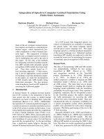
Fig. 2. Binding of [
3
H]cGMP to PDE species extracted from OS membranes. (A,B) PDE species extracted with a hypotonic buffer. Details of
the procedure are given in Experimental procedures. OS homogenates (50.4 mg protein) were suspended in 20 mL buffer A and divided into
three portions. After incubation with cGMP (A, upper), GTPcS (A, lower) or cGMP + GTPcS (B), proteins were extracted with buffer B (a
hypotonic buffer), applied to a TSK–DEAE 5PW column and eluted. Fractions containing PDE species were determined by SDS ⁄ PAGE and
assaying PDE activity. Elution profiles of the 88-kDa protein, Pab, are shown in each panel. The elution profile of other proteins is detailed
elsewhere [20]. PDE species were identified as described previously [20]. Binding of [
3
H]cGMP to the fraction (60 lL) was measured with
0.5 l
M [
3
H]cGMP. (C) PDE species extracted with Pd in an isotonic buffer. OS homogenates (12.4 mg) were suspended in 13 mL of buf-
fer A and divided into two portions. After incubation of a portion with GTPcS (50 l
M) for 1 h on ice, membranes were washed with 2 mL of
buffer A containing GTPcS (50 l
M) and 2 mL of buffer A. The other portion was treated in the same way but without GTPcS. These mem-
branes were suspended in 2.5 mL of buffer D, incubated with Pd (final 3 l
M) overnight on ice, and washed twice with 2 mL of buffer D. All
supernatants were collected and applied to a TSK–DEAE 5PW column. Rod and cone PDE species and their stoichiometry and transducin
subunits were identified as described previously [20]. Binding of [
3
H]cGMP to the fraction (50 lL) was measured with 0.5 lM [
3
H]cGMP
(upper). Protein profiles in fractions (40 lL) were analyzed by SDS ⁄ PAGE and staining with Coomassie Brilliant Blue (lower). Owing to the
limited space, only results from GTPcS-treated membranes are shown. Profiles of PDE species from GTPcS-nontreated membranes are
given in Yamazaki et al. [20].
Roles of cGMP binding in PDE6 regulation A. Yamazaki et al.
1858 FEBS Journal 278 (2011) 1854–1872 ª 2011 The Authors Journal compilation ª 2011 FEBS
Pd is identical to that of PDE species without Pd when
the same NaCl gradient was used [20]. Comparison of
the [
3
H]cGMP-binding activity of cone PDE with that
of Pabc is discussed later.
Contents of cGMP in Pabcc and Pabc
Pabcc and Pabc were purified from GTPcS-treated OS
homogenates (Fig. 3A). These PDE species were clearly
separated and characterization of these species includ-
ing their specific activity and Pc-sensitivity verified the
clear separation [20]. We also note that the level of pro-
tein staining with Coomassie Brilliant Blue is propor-
tional to the molecular mass calculated based on its
amino acid sequence under our staining conditions, i.e.
the Pc ⁄ Pab ratios also showed the clear separation [20].
Molecular sieve chromatography of these PDE species
also showed that the Pc ⁄ Pab ratio in these PDE species
was not changed during their storage.
We found that 3.0 pmol of the Pabcc contained
6.5 pmol of cGMP (Fig. 3B). This indicates that
Pabcc contains two cGMPs. Pabcc isolated from
GTPcS-nontreated OS homogenates also contained two
cGMPs (data not shown). These results indicate that
noncatalytic sites of Pabcc, whether located with or
without GTP–Ta, are saturated by cGMP. These results
also suggest that saturation is a reason for the lack of
[
3
H]cGMP-binding activity in Pabcc. These Pabcc
preparations had been exposed to cGMP-free conditions
for at least 1 week. This suggests that these cGMPs bind
tightly to Pabcc, confirming previous observations [12].
Pabc, 6.0 pmol, contained 6.1 pmol of cGMP
(Fig. 3B). This indicates that Pabc contains one
cGMP, i.e. one of the noncatalytic sites in Pabc is
empty. The possibility that cGMP existing in Pabc can
be exchanged by [
3
H]cGMP during the assay of
[
3
H]cGMP binding is quite low, as discussed later.
Therefore, we conclude that the [
3
H]cGMP-binding
activity in Pabc we observed is due to the binding of
[
3
H]cGMP to the empty site, i.e. [
3
H]cGMP-bound
Pabc contains one original cGMP and one [
3
H]cGMP.
These results also indicate that GTP–Ta dissociates
not only a single Pc, but also one cGMP from Pabcc
during its activation. In other words, PDE activation
is the mechanism by which Pabcc having two cGMPs
changes to Pabc having one cGMP, and PDE deacti-
vation is the mechanism by which Pabc having one
cGMP shifts to P abcc having two cGMPs. Pab ⁄ Pc
(Fig. 2A lower and C upper) is a minor species that is
difficult to purify [20]. Therefore, the content of cGMP
in Pab ⁄ Pc could not be measured.
Pabc was exposed to cGMP-free conditions for
> 3 days. Under these conditions, the molecular ratio
of cGMP to Pab in Pabc is always 1.0 (Fig. 3B).
This observation suggests that the affinity for cGMP is
clearly different in Pabcc’s two noncatalytic sites and
that GTPcS–Ta (GTP–Ta) releases cGMP only from
the same one site in Pabcc during its activation. This
also implies that GTP–Ta dissociates Pc from the
same site in Pabcc during its activation.
Characterization of [
3
H]cGMP binding to Pabc
Purified Pabc showed a [
3
H]cGMP-binding activity
(Fig. 4A). The level of [
3
H]cGMP binding reached a
plateau as the [
3
H]cGMP concentration increased.
Scatchard plotting of this saturable [
3
H]cGMP binding
(Fig. 4A, insert) indicates that Pabc has one type of
cGMP-binding site with K
d
50 nm. This is consistent
with the above-mentioned view that [
3
H]cGMP binds
to the same site in Pabc. The level of bound
[
3
H]cGMP reached a plateau in < 2 min under these
conditions (Fig. 4B). Unlabeled cGMP, but not
cAMP, competitively inhibited [
3
H]cGMP binding
(Fig. 4C). This indicates that the [
3
H]cGMP-binding
site in Pabc is cGMP-specific.
Trapping of [
3
H]cGMP-bound Pabc to a Millipore
filter
After incubation with [
3
H]cGMP, Pabc was applied to a
molecular sieve column and the amount of [
3
H]cGMP
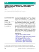
Fig. 3. Levels of cGMP contained in Pabcc and Pabc.Pabcc
(6.50 lgÆ20 lL
)1
) and Pabc (4.75 lgÆ50 lL
)1
) were purified from
GTPcS-treated OS homogenates. (A) Purity of these PDE prepara-
tions. Preparations of Pabcc (10 lL) and Pabc (25 lL) were applied
to SDS ⁄ PAGE followed by staining with Coomassie Brilliant Blue.
(B) Levels of cGMP contained in these PDE species. Contents of
cGMP were measured using a cGMP immunoassay kit.
A. Yamazaki et al. Roles of cGMP binding in PDE6 regulation
FEBS Journal 278 (2011) 1854–1872 ª 2011 The Authors Journal compilation ª 2011 FEBS 1859
bound to Pabc was calculated based on the [
3
H] radio-
activity in the Pabc fraction (Fig. 4D). We found that
70 lL of fraction 15, the peak fraction, contained 3.2 lg
Pabc (15.5 pmol) and 13.1 pmol of [
3
H]cGMP, i.e.
83% of Pabc in the fraction was occupied by
[
3
H]cGMP. The average level of the occupation was
86% in three experiments. These results indicate that
100% of Pabc binds [
3
H]cGMP under these condi-
tions. The result is also confirmed later (Fig. 5). How-
ever, only 17% of the activity was detected when
70 lL of the fraction was applied to the filter and the
[
3
H]cGMP-binding activity was obtained based on the
[
3
H] radioactivity trapped by the filter (Fig. 4D). This
shows that the Millipore filter traps 17% of the
[
3
H]cGMP-bound Pabc existing in the assay mixture.
We could not get a result showing that 100% of Pabc
expressed [
3
H]cGMP-binding activity. We believe that
this is resulted from an artifact caused by our experi-
mental procedures, because the Pabc preparation we
obtained appears to contain one type of Pabc [20], and
[
3
H]cGMP, once bound to Pabc, is not dissociated even
in the presence of a 1000-fold excess of unlabeled cGMP
(Fig. 5). In Fig. 4D, fraction 15 apparently shows that
100% of Pabc binds [
3
H]cGMP. This is due to our
intention to show the ratio of [
3
H]cGMP-binding activ-
ity measured by the filter. It should be noted that
18.2% of the [
3
H]cGMP-bound Pabc in the assay
mixture was trapped by the filter in the studies shown in
Fig. 4A, however this low rate does not affect the prop-
erties shown in Fig. 4A–C, because these properties are
not affected by the low efficiency of the filter to trap
[
3
H]cGMP-bound Pabc.
Fig. 4. Binding of [
3
H]cGMP to Pabc. (A) Concentration of [
3
H]cGMP. [
3
H]cGMP binding to Pabc (1.92 lg) was measured with the indicated
concentrations of [
3
H]cGMP. The [
3
H]cGMP-binding activity was analyzed by Scatchard plotting (insert). (B) Time-course. Pabc (17.3 lg) was
incubated in 55 m
M Tris ⁄ HCl, (pH 7.5) containing 4.4 mM EDTA and 1.1 mM IBMX (final volume, 720 lL) on ice for 10 min. The [
3
H]cGMP
binding was initiated by adding 80 lLof10l
M [
3
H]cGMP. After incubation for the indicated periods, an aliquot (80 lL) was taken and applied
to a Millipore filter. (C) The cyclic nucleotide specificity. After incubation of Pabc (1.92 lg) with the indicated concentration of unlabeled
cGMP (
•
) or cAMP (s) on ice for 10 min, [
3
H]cGMP binding was measured with 1 lM [
3
H]cGMP. The 100% activity indicates that
1.46 pmol [
3
H]cGMP bound to Pabc in tubes. (D) Levels of [
3
H]cGMP-bound Pabc trapped by the filter. OS homogenates (18.9 mg protein)
were suspended in 9.7 mL of buffer A. After isolation by the TSK–DEAE 5PW column chromatography and concentration to 0.3 mL, the
Pabc preparation ( 80 lg) was incubated with 1 l
M [
3
H]cGMP for 30 min on ice and applied to a TSK 250 column that had been equili-
brated with buffer D. The level of [
3
H]cGMP bound to Pabc was calculated based on the [
3
H] radioactivity in 70 lL of the fraction (
•
). The
fraction (70 lL) was also applied to a Millipore filter and the [
3
H] radioactivity on the filter was measured (h). Only fractions containing Pabc
are shown. (Insert) The rate of [
3
H] radioactivity on the filter per the level of [
3
H] radioactivity in the fraction. The 100% radioactivity indicates
the [
3
H] radioactivity detected in fraction 15. Fraction 15 (70 lL) contained 3.2 lgPabc (15.5 pmol) and 13.1 pmol of [
3
H]cGMP.
Roles of cGMP binding in PDE6 regulation A. Yamazaki et al.
1860 FEBS Journal 278 (2011) 1854–1872 ª 2011 The Authors Journal compilation ª 2011 FEBS
GTPcS–Ta-activated and Pd-extracted cone PDE,
Pa¢a¢c¢dd [20], expressed [
3
H]cGMP-binding activity
(Fig. 2C, upper). We note that all Pa¢a¢c¢c¢ complexes
present were activated to Pa¢a¢c¢ under our conditions
[20]. Interestingly, the level of [
3
H]cGMP-binding
activity in fraction 13 was approximatley five times
higher than that of fraction 27 (Fig. 2C, upper). A
similar observation was also obtained when these
PDEs were extracted with a hypotonic buffer (data not
shown). These results indicate that 85% of
[
3
H]cGMP-bound Pa¢a¢c¢ was trapped by the Millipore
filter under the following assumptions: (a) the content
of Pab and Pa¢a¢ in these fractions are similar, (b)
[
3
H]cGMP binds to all Pa¢a¢c¢ complexes, (c) Pa¢a¢c¢
has one cGMP binding site, (d) Pd binding does not
affect the level of [
3
H]cGMP-binding activity in
Pa¢a¢c¢, and (e) 17% of [
3
H]cGMP-bound Pabc is
trapped by the Millipore filter. We note that the level
of protein staining with Coomassie Brilliant Blue is
proportional to the molcular mass calculated based on
its amino acid sequence under our staining conditions
[20]. Thus, amounts of Pab and Pa¢a¢ can be compared
by comparing their staining levels in the same gel. We
found that the stained level of Pab was similar to that
of Pa¢a¢ (Fig. 2C, lower). This indicates that levels of
Pab and Pa¢a¢ are similar, i.e. assumption (a) was pro-
ven. As described, we found that the [
3
H]cGMP-bind-
ing activity of Pa¢a¢c¢dd was similar to that of Pa¢a¢c¢,
i.e. assumption (d) was proven. Assumption (e) was
also proven, as described above. Assumptions (b) and
(c) are not yet proven; however, these assumptions are
reasonable if characteristics of the [
3
H]cGMP binding
to Pabc are taken into consideration. Therefore, we
conclude that the low trapping rate is specific to
[
3
H]cGMP-bound Pabc.
Conformational change of Pabc by cGMP binding
After incubation with [
3
H]cGMP for 30 min (i.e. after
binding of [
3
H]cGMP to 100% of Pabc), dissocia-
tion of [
3
H]cGMP bound to Pabc was followed with
or without 1 mm unlabeled cGMP (Fig. 5A). We
found that the level of [
3
H]cGMP binding to Pabc was
not changed even in the presence of 1 mm unlabeled
cGMP, at least for the first 5 min. Under similar con-
ditions, [
3
H]cGMP binding to Pabc reached a maxi-
mum in < 2 min (Fig. 4B), indicating that the 5-min
incubation was enough to chase [
3
H]cGMP bound to
Pabc, if indeed [
3
H]cGMP could be chased. Therefore,
this observation indicates that [
3
H]cGMP, once bound
Fig. 5. Change of Pabc’s characteristics by cGMP binding. (A) Dissociation of [
3
H]cGMP bound to Pabc. Purified Pabc (16.0 lg) suspended
in 640 lL of 55.5 m
M Tris ⁄ HCl (pH 7.5) containing 4.44 mM EDTA and 1.11 mM IBMX, and [
3
H]cGMP binding was initiated by adding 80 lL
of 9 l
M [
3
H]cGMP. After incubation for 30 min on ice, an aliquot (72 lL) was withdrawn, applied to a Millipore filter, and its radioactivity was
designated as the level at time 0. Simultaneously, 72 lLof10m
M unlabeled cGMP (
•
) or water (s) was added to the assay mixture. After
incubation for 0.25, 0.5, 0.75, 1, 2, 5, 10 and 20 min, an aliquot (80 lL) was withdrawn, applied to a Millipore filter, and its [
3
H] radioactivity
was measured. The arrow indicates the addition of cGMP or water. The 100% activity indicates that 1.32 pmol of [
3
H]cGMP was detected
in 1.6 lgofPabc (7.72 pmol). (B) Elution profile of Pabc from a gel-filtration column. Purified Pabc (70 lg) was incubated with (black) or
without (red) unlabeled cGMP (0.5 m
M) in 0.5 mL of 25 mM Tris ⁄ HCl, pH 7.5, 0.1 mM EDTA and 1 mM IBMX for 30 min on ice and applied
to a Superdex 200 HR column that had been equilibrated with buffer E. Detailed conditions for this elution are in the Experimental proce-
dures. PDE activity was assayed using 5 lL of the fraction (
•
). The 100% PDE activity indicates that 12.5 nmol cGMP was hydrolyzed per
min per tube. [
3
H]cGMP binding activity was measured using 50 lL of the fraction (h). The 100% activity indicates that 1.50 pmol of
[
3
H]cGMP was detected in the assay mixture.
A. Yamazaki et al. Roles of cGMP binding in PDE6 regulation
FEBS Journal 278 (2011) 1854–1872 ª 2011 The Authors Journal compilation ª 2011 FEBS 1861
to Pabc, cannot be dissociated. This strongly suggests
that Pabc, after binding of [
3
H]cGMP, changes its
conformation, particularly that of its noncatalytic site
and ⁄ or a region(s) near the noncatalytic site, and that
Pabc, after changing its conformation, firmly holds the
[
3
H]cGMP. A large conformational change initiated by
cGMP binding has been reported in one of GAF
domains in cone PDE [24]. A similar conformational
change may occur when [
3
H]cGMP binds to Pabc, i.e.
when Pabc having one cGMP is shifted to Pabc hav-
ing two cGMPs.
To further prove that binding of cGMP changes the
conformation of Pabc , we directly compared the rela-
tive compactness (Stokes’ radius) of cGMP-treated
Pabc with that of cGMP-nontreated Pabc (Fig. 5B).
This method has been used to show a conformational
change by cGMP binding in PDE5 [25–27]. After incu-
bation of Pabc with or without cGMP for 30 min on
ice, these Pabcs were applied to a gel-filtration column
and PDE activity was measured to identify the fraction
containing Pabc. As expected, the cGMP-nontreated
Pabc was eluted as a single peak with the peak activity
in fraction 38. [
3
H]cGMP-binding activity was also
observed in these fractions. However, cGMP-treated
Pabc was eluted as two peaks, the major peak in frac-
tion 34 and the minor peak in fraction 38, and only
Pabc in fraction 38 showed [
3
H]cGMP-binding activ-
ity. These observations indicate that the apparent
Stokes’ radius of cGMP-treated Pabc was 4–7 A
˚
larger
than that of cGMP-nontreated Pabc, i.e. the Stokes’
radius of Pabc appears to be increased when Pabc
having one cGMP is shifted to Pabc having two
cGMPs. We note that the difference in the Stokes’
radius was observed in Tris buffer; however, the differ-
ence was less clear in a phosphate buffer (data not
shown). This may be because of a tendency of Pabc to
change its structure in Tris buffer [11]. We also note
that 50 lL of the peak fraction of the cGMP-nontreat-
ed Pabc contained 2.4 lgPabc (11.6 pmol of Pabc)
and bound 9.90 pmol [
3
H]cGMP. This indicates that
85% of the Pabc expressed [
3
H]cGMP-binding
activity, confirming that almost all Pabc complexes
show [
3
H]cGMP-binding activity (Fig. 4D). We also
note that the major peak of the cGMP-treated Pabc
showed no ability to bind [
3
H]cGMP, confirming that
cGMP, once bound to Pabc, is not dissociated
(Fig. 5A).
Rate of the conformational change in Pabc
The level of [
3
H]cGMP-binding increased abruptly
after a 10-min incubation (Fig. 5A). The level was
increased approximately three times the level at time 0
after 20 min (Fig. 5A) and approximately four times
after 40 min (data not shown). Because 100% of
Pabc present bound [
3
H]cGMP during preincubation,
these observations indicate that the amount of
[
3
H]cGMP-bound Pabc trapped by the filter increased
abruptly during incubation.
Incubation of [
3
H]cGMP-bound Pabc was initiated
by the addition of unlabeled cGMP or water (Fig. 5A).
An increase in the trapped level of [
3
H]cGMP-bound
Pabc was observed after addition of 1 mm unlabeled
cGMP, indicating that the increase is not due to new
binding of [
3
H]cGMP to Pabc. The increase was also
detected after addition of water, implying that the
unlabeled cGMP is not involved in this increase. Addi-
tion of unlabeled cGMP or water slightly diluted the
mixture, by 10%; however, it is unlikely that such a
small dilution could cause this increase. Modification
of the Pabc during incubation could also be ignored
because the Pabc was pure (Fig. 3A) and the incu-
bation was carried out on ice. Taken together,
these observations deny the possibility that the increase
is attributed to a reaction that occurred during
incubation.
During preincubation, [
3
H]cGMP bound to Pabc.
As another important change during preincubation,
the buffer in the Pabc preparation, a phosphate buffer
containing Mg
2+
, was changed to a Tris buffer con-
taining 1-methyl-3-isobutylxanthine (IBMX), but not
Mg
2+
.Pabc appears to have a tendency to change its
structure in a Tris buffer, but not in a phosphate buf-
fer [11], and Mg
2+
binds to Pab [28,29]. IBMX may
also increase the cGMP affinity of noncatalytic sites,
as discussed later. Therefore, these changes might
affect the properties of Pabc and this change might
increase the level of [
3
H]cGMP-bound Pabc trapped
by the filter. However, this increase was observed with
either Pabc stored in the original buffer or in the
preincubation buffer, a Tris buffer without Mg
2+
(data not shown). The increase was also detected with
or without IBMX (data not shown). Therefore, these
explanations may be disregarded. Modification of
Pabc during preincubation could also be ignored, as
described above. Taken together, these observations
strongly suggest that [
3
H]cGMP binding to Pabc dur-
ing preincubation is the sole reason for the increase in
the filter-trapping level of [
3
H]cGMP-bound Pabc, i.e.
the increase appears to be caused by a conformational
change in Pabc upon binding of [
3
H]cGMP.
This increase in the filter-trapping level of
[
3
H]cGMP-bound Pabc was observed only after 10-
min incubation, i.e. 40 min appeared to be required
to detect the increase (Fig. 5A). Why is the increase
detected after such a long incubation if it is due to
Roles of cGMP binding in PDE6 regulation A. Yamazaki et al.
1862 FEBS Journal 278 (2011) 1854–1872 ª 2011 The Authors Journal compilation ª 2011 FEBS
[
3
H]cGMP binding? We believe that the conforma-
tional change caused by [
3
H]cGMP binding progresses
consistently, but slowly, and that the increase is
detected only after the Pabc with an altered conforma-
tion accumulates to a certain level. In other words,
there is a threshold to trap the [
3
H]cGMP-bound
Pabc. We emphasize that a mechanism to accelerate
the conformational change should be present if
this conformational change is indeed involved in PDE
regulation.
Suppression of cGMP binding during activation
of Pabcc
Two possible stages for cGMP binding to Pabc are
expected in PDE regulation: during Pabcc activation
to Pabc and during Pabc deactivation to Pabcc.
First, we investigate whether cGMP binds to Pabc
during the activation of Pabcc to Pabc. After incuba-
tion of OS homogenates with GTPcS in the presence
(Fig. 2B) or absence (Fig. 2A, lower) of cGMP, PDE
species were extracted with buffer B and applied to a
TSK–DEAE column, and their [
3
H]cGMP-binding
activities were measured. Both OS homogenates were
incubated in the presence of IBMX and > 20% of
added cGMP remained in the cGMP-added homoge-
nate when membranes were isolated. We found that
the [
3
H]cGMP binding activity of cGMP-treated Pabc
appeared to be slightly higher than that of cGMP-
nontreated Pabc. However, the difference was not
clear in another two studies. Therefore, we conclude
that cGMP-incubated P abc has the ability to bind
[
3
H]cGMP similar to that seen in Pabc obtained with-
out cGMP. The same result was obtained when Pabc
was extracted with Pd in a isotonic buffer (data not
shown). Pabc, once it binds cGMP, holds the cGMP
and cannot accept [
3
H]cGMP (Fig. 5B). Therefore,
the [
3
H]cGMP-binding activity we observed (Fig. 2B)
indicates that Pabc cannot bind cGMP during activa-
tion of Pabcc to Pabc.
Pab ⁄ Pc, the minor GTPcS–Ta-activated PDE
(Fig. 2), lost its [
3
H]cGMP-binding activity when the
fraction containing Pab ⁄ Pc was pretreated with cGMP
(data not shown). However, Pab ⁄ Pc obtained from
cGMP-treated OS homogenates showed a [
3
H]cGMP-
binding activity (Fig. 2B) similar to that of Pab ⁄ Pc
obtained from cGMP-nontreated homogenates
(Fig. 2A, lower). This suggests that binding of cGMP
to Pab ⁄ Pc is suppressed during its formation.
Together, our observations indicate that the cGMP-
binding activity of GTP–Ta-activated PDE species is
suppressed during its formation. This, we believe, is a
critical finding to identify the function of cGMP bind-
ing in PDE regulation. We note that the Pab ⁄ Pc was
eluted slightly earlier when OS homogenates were incu-
bated with cGMP, as previously shown [20]. The pres-
ence of cGMP may be crucial for the early elution;
however, the real reason is unknown.
Binding of cGMP during deactivation of Pabc
Next, we investigated whether cGMP binds to Pabc
during deactivation of Pabc to Pabcc. Binding of
cGMP may be involved in Pabc deactivation in two
ways: after interaction with Pc and before interaction
with Pc. First, we studied whether cGMP binds to
Pabc after Pc binding to Pabc. We assayed
[
3
H]cGMP-binding activity of Pabc after incubation of
Pabc with Pc or its mutants (Fig. 6). Here, these P abc
complexes are termed PabcÆPc or PabcÆPc-mutant to
emphasize that [
3
H]cGMP-binding activity is assayed
after formation of these complexes. PcÆGDP–Ta,
instead of Pc, should be used, because PcÆGDP–Ta,
but not free Pc, is the endogenous inhibitor of Pabc
[13,20]. However, it is not known whether the Pc
mutants we used form a complex with GDP–Ta.
Therefore, free Pc was used in this study.
[
3
H]cGMP binding to Pabc (control)
The level of [
3
H]cGMP binding to Pabc reached a pla-
teau in < 2 min and was not changed during the incu-
bation period of at least 40 min (Figs 4B and 6B).
After reaching the plateau, 100% of Pabc bound
[
3
H]cGMP in the mixture. However, the plateau indi-
cates the level of [
3
H]cGMP-bound Pabc trapped by
the filter. In this case, the filter trapped 16% of the
Pabc existing in the mixture.
[
3
H]cGMP binding to PabcÆPc
The level of bound [
3
H]cGMP was reduced when
PabcÆPc was formed (Fig. 6A). A reason for the reduc-
tion is that binding of [
3
H]cGMP to PabcÆPc was slow
and, even after 30 min incubation, did not reach the
level that Pabc could reach in 2 min (Fig. 6B). The K
d
for cGMP in Pabc ⁄ Pc is 0.33 lm (Fig. 6C), indicat-
ing that the binding of Pc to Pabc reduces its affinity
for cGMP by 6.5 times. This reduction may be a rea-
son for the slow binding of [
3
H]cGMP to PabcÆPc. She
efficiency of a Millipore filter for trapping [
3
H]cGMP-
bound Pabc is increased when Pc binds to [
3
H]cGMP-
bound Pabc, as shown below (Fig. 7A). Therefore, the
reduction in the level of [
3
H]cGMP binding to PabcÆPc
is not due to a reduction in the Millipore filter’s ability
to trap the [
3
H]cGMP-bound PabcÆPc.
A. Yamazaki et al. Roles of cGMP binding in PDE6 regulation
FEBS Journal 278 (2011) 1854–1872 ª 2011 The Authors Journal compilation ª 2011 FEBS 1863
[
3
H]cGMP binding to PabcÆC18del and PabcÆC10del
Both C18Sub and C10del drastically reduced the level
of [
3
H]cGMP bound (Fig. 6A,B). A simple explanation
for the reduction is that Pc mutants lacking the C-ter-
minus interrupt the entry of [
3
H]cGMP into a noncata-
lytic site in Pabc. This explanation appears to be
correct because Pc’s domains other than the C-termi-
nus, in particular its N-terminus, are located in the
vicinity of GAF domains [30–32]. However, the reduc-
tion was much larger than that caused by Pc (Fig. 6A),
suggesting that the method of binding of these Pc
mutants to Pabc is slightly different from that of Pc
and that the inhibitory effect is neutralized, although
partially, by its C-terminus. Alternatively, all of the
PabcÆC-terminal mutant complexes would bind
[
3
H]cGMP; however, only the filter might trap a negli-
gible amount of these complexes. However, the level
trapped by the filter was increased when C10del was
added to [
3
H]cGMP-bound Pabc (Fig. 7A). Therefore,
this scenario is unlikely.
[
3
H]cGMP binding to PabcÆN18del and PabcÆN22del
The apparent level of [
3
H]cGMP binding was increased
when Pabc formed a complex with N18del and N22del
(Fig. 6). There are two possible explanations for this
observation: (a) the level of [
3
H]cGMP binding to
these PabcÆN-terminal mutant complexes was really
increased, and (b) the level trapped by the filter of
[
3
H]cGMP-bound Pabc was increased. We compared
[
3
H]cGMP binding to Pabc with that to PabcÆN22del
(and PabcÆN18del). We found that: (a) both Pabc
(Figs 4A and 6C) and PabcÆN22del (Fig. 6C) had one
type of [
3
H]cGMP-binding site, (b) the rate of
[
3
H]cGMP binding to PabcÆN22del appeared to be
similar or slightly faster than that to Pabc (Fig. 6B),
and (c) the affinity of PabcÆN22del for [
3
H]cGMP was
higher than that of Pabc (Fig. 6C). These findings
indicate that PabcÆN22del binds [
3
H]cGMP more effec-
tively than does Pabc. As described, 100% of Pabc
rapidly binds [
3
H]cGMP under these conditions.
Therefore, we conclude that 100% of PabcÆN22del
also rapidly binds [
3
H]cGMP and that the apparent
increase in the level of [
3
H]cGMP-bound PabcÆN22del
is due to the effective trapping of [
3
H]cGMP-bound
PabcÆN22del by the filter. In other words, explanation
(b), but not explanation (a), is appropriate. The small
difference in levels of [
3
H]cGMP binding to PabcÆN22-
del and PabcÆN18del (Fig. 7A) may also be caused by
the difference in levels trapped by the filter of
[
3
H]cGMP-bound PabcÆN22del and PabcÆN18del.
Fig. 6. Effects of Pc and its mutants on [
3
H]cGMP binding to Pabc. (A) Effect on the level of [
3
H]cGMP binding. After incubation of Pabc
(1.92 lg) with various concentrations of Pc or its mutants, the [
3
H]cGMP-binding activity was measured. The 100% activity indicates that
1.46 pmol [
3
H]cGMP bound to Pabc in tubes. Following Pc and its mutants were used: (
•
) wild-type Pc,(h) N18del, (4) N22del, ( )
C18Sub, and (.) C10del. (B) The effect on the time-course of [
3
H]cGMP-binding. Pabc (17.3 lg) was incubated with 1.11 lM Pc or its
mutants in 55 m
M Tris ⁄ HCl, (pH 7.5) containing 4.4 mM EDTA and 1.1 mM IBMX (final volume, 720 lL) on ice for 30 min. The [
3
H]cGMP
binding was initiated by adding 80 lLof10l
M [
3
H]cGMP. After incubation for the indicated periods on ice, an aliquot (80 lL) was taken and
applied to a Millipore filter. The following Pc and its mutants were used: (s) control, (
•
) wild-type Pc,(4) N22del, and ( ) C18Sub. (C) The
effect on the Scatchard plot. Pabc (1.92 lg) was incubated with 1 m
M of wild-type Pc (
•
) or N22del (4). As a control, Pabc alone was incu-
bated (s). Then, [
3
H]cGMP binding was initiated by adding indicated concentrations of [
3
H]cGMP (C-1). The [
3
H]cGMP binding in C-1 is ana-
lyzed by Scatchard plotting (C-2).
Roles of cGMP binding in PDE6 regulation A. Yamazaki et al.
1864 FEBS Journal 278 (2011) 1854–1872 ª 2011 The Authors Journal compilation ª 2011 FEBS
In conclusion, Pc appears to suppress [
3
H]cGMP
binding to Pabc by interrupting the entry of [
3
H]cGMP
into a noncatalytic site in Pabc. This indicates that the
scheme in which Pc binds to Pabc first and then cGMP
binds to the Pabcc is unlikely. This also implies that Pc
in Pabcc interferes with cGMP release. This may be a
reason for the observations that cGMPs are tightly
bound to Pabcc (Fig. 3) [12] and that release of cGMP
from Pabcc is coupled with Pc dissociation (Fig. 2).
Effect of Pc on cGMP-bound Pabc
Next, we studied how Pc affects [
3
H]cGMP-bound
Pabc, i.e. Pabc having two cGMPs. Because 100%
of Pabc binds [
3
H]cGMP and the Pabc firmly folds
[
3
H]cGMP, we investigate the effect of Pc on the filter-
trapping level of [
3
H]cGMP-bound Pabc (Fig. 7A).
The study is based on the results shown in Fig. 5. The
increase in the filter-trapping level of [
3
H]cGMP-bound
Pabc is an indicator of Pabc’s conformational change
on cGMP binding. PcÆGDP–Ta, instead of Pc, should
be used in this study because PcÆGDP–Ta, but not free
Pc, is the inhibitor of Pabc [13,20]. However, as
described above, it is not known whether Pc mutants
we used form a complex with GDP–Ta. Therefore,
free Pc was used in this study.
When Pc was added to [
3
H]cGMP-bound Pabc in
exactly the same way as in the study shown in Fig. 5A,
the level of [
3
H]cGMP-bound Pabc trapped by the fil-
ter increased in < 15 s, but the level of [
3
H]cGMP-
bound Pabc trapped was not changed (Fig. 7A). A
simple explanation for these phenomena is that Pc
shortens the time required to detect the slow increase,
i.e. Pc accelerates the conformational change in
[
3
H]cGMP-bound Pabc.
To strengthen the above-mentioned conclusion, the
relative compactness, the Stokes’ radius, of cGMP-pre-
treated Pabc,Pabc having two cGMPs, was also com-
pared with or without P c (Fig. 7B). Purified Pabcc
was used as the Pabcc having two cGMPs because
Pabcc has two cGMPs (Fig. 3). As expected, the
cGMP-pretreated Pabc eluted as two peaks, the major
peak of Pabc eluted in fraction 34, which showed no
[
3
H]cGMP-binding activity. The important point is
that Pabcc was also eluted in fraction 34 (Fig. 7B).
This strongly suggests that Pc does not change the rel-
ative compactness of P abc having two cGMPs, i.e.
binding of Pc does not change the conformation of
Pabc having two cGMPs. Together with the observa-
tion that cGMP-binding to Pabc slowly changes its
conformation (Fig. 5) and Pc shortens the time
required to detect the slow increase (Fig. 7A), the
Fig. 7. Pc effect on the conformational change of Pabc by cGMP binding. (A) The time to detect the increase in the level of [
3
H]cGMP-
bound Pabc trapped by the filter. The experiment was carried out as a part of the study depicted in Fig. 5A. Pabc (16.0 lg) was suspended
in 640 lL of 55.5 m
M Tris ⁄ HCl (pH 7.5) containing 4.44 mM EDTA and 1.11 mM IBMX, and [
3
H]cGMP binding to the Pabc was initiated by
adding 80 lLof9l
M [
3
H]cGMP. After incubation for 30 min, an aliquot (72 lL) was withdrawn and applied to a Millipore filter, and its radio-
activity was designated as the level at time 0. Simultaneously, a mixture (72 lL) of Pc or its mutant (10 l
M) with or without cGMP (10 mM)
was added to the assay mixture: (
•
), with cGMP; and (s), without cGMP. After incubation for 0.25, 0.5, 0.75, 1, 2, 5, 10 and 20 min, an ali-
quot (80 lL) was withdrawn, applied to a Millipore filter, and its radioactivity was measured. The arrow (›) indicates the addition of Pc (or Pc
mutant) with or without cGMP. The arrow (‹) indicates levels of [
3
H]cGMP-bound Pabc with ( ) or without (4) N18del. The 100% activity
indicates that 1.32 pmol of [
3
H]cGMP was detected in 1.6 lgofPabc (7.72 pmol). Data shown in Fig. 5A was used as a control for this
study. (B) Elution profile of PDE species from a gel-filtration column. Purified Pabc (70 lg) was incubated with unlabeled cGMP (0.5 m
M)in
0.5 mL of 25 m
M Tris ⁄ HCl, pH 7.5, 0.1 mM EDTA and 1 mM IBMX for 30 min on ice and applied to a Superdex 200 HR column that had
been equilibrated with buffer E (black). The chromatography conditions are given in the Experimental procedures. PDE activity (
•
) was
assayed using 5 lL of the fraction. [
3
H]cGMP-binding activity (h) was measured using 50 lL of the fraction. Pabcc (90 lg) was also applied
to the column and eluted in the same manner (red). PDE activity was assayed using 20 lL of the fraction (
•
). The 100% PDE activity indi-
cates that 12.1 nmol cGMP was hydrolyzed per min per tube. [
3
H]cGMP-binding activity was measured using 50 lL of the fraction (h). The
100% [
3
H]cGMP-binding activity indicates that 2.4 pmol of [
3
H]cGMP was detected in the assay mixture.
A. Yamazaki et al. Roles of cGMP binding in PDE6 regulation
FEBS Journal 278 (2011) 1854–1872 ª 2011 The Authors Journal compilation ª 2011 FEBS 1865
result strongly suggests that binding of Pc accelerates
cGMP-dependent conformational change in Pabc.
The rapid increase by Pc in the level of [
3
H]cGMP-
bound Pabc trapped by the filter (Fig. 7A) might be
due to a change by Pc in the surface of [
3
H]cGMP-
bound Pabc and ⁄ or in the total charge of Pabc.In
such cases, a slow increase (Fig. 5A) could also be
detected, however, in this case, the slow increase was
missing. Therefore, this possibility is unlikely. We also
note that the result (Fig. 7B) does not eliminate the
possibility that Pc binding causes a small and ⁄ or local-
ized conformational change in [
3
H]cGMP-bound Pabc.
However, the clear change in its Stokes’ radius
(Fig. 5B) suggests that this type of conformational
change is not involved.
When C10del was added, the level of [
3
H]cGMP-
bound Pabc trapped by the filter was also rapidly
increased and the increase on longer incubation
(Fig. 5A) disappeared (Fig. 7A). Similar observations
were also made when N22del was added (Fig. 7A).
These results suggest that binding of C10del or
N22del, similar to Pc binding, accelerates the confor-
mational change in [
3
H]cGMP-bound Pabc and that a
Pc domain(s) located between N22 and C10 is involved
in this acceleration. We note that the level of
[
3
H]cGMP-bound Pabc increased by N22del was smal-
ler than that increased by Pc or C10del; however, the
increase with N18del was similar to that with Pc or
C10del (Fig. 7A). The level of [
3
H]cGMP binding to
PabcÆN18del was also consistently higher than that
binding to PabcÆN22del (Fig. 6A). These results sug-
gest that all or some of the four amino acid residues
between amino acids 19–22 in the Pc sequence may be
crucial for the acceleration.
In conclusion, these results strongly suggest that Pc
binding accelerates the cGMP-binding-initiated confor-
mational change in Pabc. Together with the result
showing that Pc inhibits cGMP-binding to Pabc
(Fig. 6), these results imply that the scheme by which
cGMP binds to P abc first and Pc then binds to the
Pabc is appropriate for Pabc deactivation, the process
to shift Pabc having one cGMP to Pabcc having two
cGMPs (Fig. 8). These results also imply a large con-
formational change during Pabcc activation, the pro-
cess to shift Pabcc having two cGMPs to Pabc having
one cGMP, although its direction is opposite to that
for Pabc deactivation (Fig. 8).
Discussion
Using bovine PDE preparations, we have recently pro-
posed a new and comprehensive model for PDE regu-
lation [13,20]. In this study, we try to integrate the role
of noncatalytic and cGMP-specific binding sites in Pab
in this new model. We show that Pabcc, the inactive
form, and Pabc, the GTP–Ta-activated form, contain
two and one cGMP, respectively, and that only Pabc
shows [
3
H]cGMP binding. We also show that the abil-
ity of Pabc to bind cGMP is suppressed during forma-
tion of Pabc. We also strongly suggest that cGMP
binding slowly changes the conformation of Pabc and
that Pc binding accelerates this change. These findings
are consistent with the view that Pabc rapidly changes
its conformation during deactivation and that binding
of cGMP and Pc play crucial roles in this change
(Fig. 8). These findings also imply that Pabcc rapidly
changes its conformation during its activation and that
the release of Pc and cGMP play important roles in
this change (Fig. 8). To the best of our knowledge, this
is the first model in which the role of noncatalytic
binding sites is smoothly integrated in PDE regulation.
Pabc is the PDE species expressing
[
3
H]cGMP-binding activity
Identification of a PDE species expressing [
3
H]cGMP-
binding activity is the first step in the exploration of
the role of cGMP binding to the noncatalytic site in
PDE regulation. We have shown that GTP–Ta-acti-
vated PDE in membranes has a high [
3
H]cGMP-bind-
ing activity (Fig. 1), that Pabc contains one cGMP, i.e.
Fig. 8. Role of the noncatalytic cGMP-binding site in PDE regula-
tion. GTP–Ta activates Pabcc ⁄ 2cGMPs (Pabcc having two cGMPs)
to Pabc ⁄ cGMP (Pabc having one cGMP). At the initial stage, cGMP
is present in OS; however, binding of cGMP to the empty site on
Pabc ⁄ cGMP is suppressed. After hydrolysis of cGMP, retinal gua-
nylate cyclase initiates to produce cGMP from GTP. When the
[cGMP] in OS is increased to 50 n
M, the cGMP binds to
Pabc ⁄ cGMP and Pabc ⁄ 2cGMPs is formed. The PDE species then
slowly changes its conformation. Interaction with PcÆGDP–Ta accel-
erates the conformational change and swiftly establishes the inac-
tive form of PDE (Pabcc ⁄ 2cGMPs).
Roles of cGMP binding in PDE6 regulation A. Yamazaki et al.
1866 FEBS Journal 278 (2011) 1854–1872 ª 2011 The Authors Journal compilation ª 2011 FEBS
Pabc has one empty noncatalytic site (Fig. 3), and that
cGMP binds to Pabc (Figs 2–4) with K
d
of 50 nm
(Figs 4A and 6C). Three points related to these issues
need discussion. First, in the current dominant model,
Pabcc complexed with GTP–Ta is believed to be the
GTP–Ta-activated PDE. Some groups also suggest that
Pc-free Pab is a GTP–Ta-activated PDE. However,
neither the GTPcS–TaÆPabcc complex nor the Pc-free
Pab was detected in any OS homogenates [13,20].
Therefore, in their absence, we could not characterize
the [
3
H]cGMP-binding activities of these Pab com-
plexes. We believe that this failure may not be crucial
in exploring the role of cGMP binding in PDE regula-
tion. Second, when [
3
H]cGMP binding to Pabc was
assayed, cGMP present originally in Pabc could be
exchanged with [
3
H]cGMP. However, based on the
following two reasons, we conclude that the cGMP ⁄
[
3
H]cGMP exchange does not occur and that the [
3
H]
radioactivity we detected is only attributed to the
[
3
H]cGMP bound to the empty site in Pabc. (a) Pabc
had been exposed to cGMP-free conditions > 5 days
before the final purification step; however, purified
Pabc contains exactly one cGMP (Fig. 3). This sug-
gests that Pabc firmly holds the cGMP, i.e. the cGMP
is hardly exchanged with [
3
H]cGMP. This is true espe-
cially when the concentration of [
3
H]cGMP is low
(1 lm). (b) Fractions containing Pabc did not show
any [
3
H]cGMP-binding activity when the Pabc was
pretreated with unlabeled cGMP (Figs 5B and 7B).
This indicates that neither the originally bound cGMP
nor the newly bound cGMP on Pabc can be exchanged
with [
3
H]cGMP in the assay mixture. We emphasize
that the exchange did not occur even with 1 mm cGMP
(Figs 5A and 7A). Third, we added IBMX to suppress
the hydrolysis of [
3
H]cGMP when [
3
H]cGMP binding
to Pabc was assayed. We found that the affinity of a
PDE preparation for cGMP was increased when 1 mm
IBMX was present in the assay mixture (data not
shown). Under these conditions, a large part of the
[
3
H]cGMP was not yet hydrolyzed, even in the absence
of IBMX, suggesting that IBMX itself affects the affin-
ity of the PDE preparation for cGMP. IBMX also
enhances the cGMP affinity of frog PDE [23]. There-
fore, in OS, the affinity of Pabc for cGMP may be
lower than observed. We note that the assay mixture
for [
3
H]cGMP binding did not contain Mg
2+
to sup-
press the hydrolysis of [
3
H]cGMP. We found that both
the amount of [
3
H]cGMP bound to Pabc and the affin-
ity of Pabc for [
3
H]cGMP were not notably changed in
the presence or absence of 5 mm MgCl
2
(data not
shown). Thus, we believe that the lack of Mg
2+
in the
assay mixture does not affect the affinity of Pabc for
cGMP.
Binding of cGMP to Pabc is not involved PDE
activation
Binding of [
3
H]cGMP to Pabc implies that cGMP
binding may be involved in the activation of P abcc to
Pabc and ⁄ or in the deactivation of Pabc to Pabcc.
Here, we show that the ability of Pabc to bind cGMP
is suppressed during the formation of Pabc by
GTPcS–Ta in OS homogenates (Fig. 2). This indicates
that cGMP binding is not involved in the activation of
Pabcc to Pabc. It is not known now how this property
of Pabc is suppressed in OS homogenates containing
GTPcS–Ta. However, we emphasize that either mem-
brane-bound Pabc (Fig. 1A) or Pabc purified from
membranes (Fig. 4A) expresses a [
3
H]cGMP-binding
activity. This suggests that a soluble factor in the OS
homogenates is involved in the suppression of cGMP
binding. This factor may be GTPcS–Ta, because by
interacting with Pab [33,34], GTPcS–Ta may inhibit
the entry of cGMP into the noncatalytic site in Pabc.
Alternatively, the interaction with GTPcS–Ta may
compel Pabc to change its conformation and Pabc
with the altered conformation may not be able to bind
cGMP. We have shown that GTPcS–Ta releases not
only one Pc, but also one cGMP from Pabcc when
Pabcc is activated (Fig. 3). The GTPcS–Ta releases Pc
by forming a complex with Pc [13,20]; however, the
mechanism to release cGMP is not known because nei-
ther GTPcS–Ta nor Pc can bind [
3
H]cGMP (Fig. 2)
[23]. It is possible that this alternative mechanism may
also be involved in the release of cGMP.
Binding of cGMP to Pabc may be a mechanism
for light adaptation
Our results strongly suggest that binding of cGMP, i.e.
formation of Pabc having two cGMPs from Pabc hav-
ing one cGMP, changes the conformation of Pabc. The
important points are that this Pabc’s conformational
change is slow (Fig. 5A); however, binding of Pc to
Pabc having two cGMPs accelerates its conformational
change (Fig. 7). Overall, these findings indicate that
cGMP binds to Pabc first and then Pc binds to the
Pabc in Pabc deactivation (Fig. 8). In this scheme, the
residual [cGMP] is crucial for the rate of Pabc deacti-
vation. The residual [cGMP] in OS is dependent upon
the level of illumination: if illumination is low, the
residual [cGMP] is higher than the K
d
of Pabc and the
cGMP-binding-dependent conformational change may
be initiated on Pabc immediately after disappearance
of GTP–Ta. Thus, Pabc will be rapidly deactivated. If
illumination is high, the residual [cGMP] is lower than
the K
d
and the conformational change may be delayed.
A. Yamazaki et al. Roles of cGMP binding in PDE6 regulation
FEBS Journal 278 (2011) 1854–1872 ª 2011 The Authors Journal compilation ª 2011 FEBS 1867
Therefore, Pabc will be slowly deactivated. This may
be a mechanism for light adaptation. The important
point for this argument is whether the residual [cGMP]
in OS can be increased to the K
d
before deactivation of
Pabc. The K
d
of Pabc for cGMP we measured is
50 nm (Figs 4A and 6C), and the K
d
in OS may be
higher than 50 nm, as described above. However, the
K
d
in OS should be much lower than the dark level of
cGMP, 4–5 lm (2, 3). Therefore, a partial recovery of
the [cGMP] may be enough to initiate Pabc deactiva-
tion. The [cGMP] in OS is recovered by retinal guanyl-
ate cyclase. We have recently shown that retinal
guanylate cyclase is activated by a light-initiated, ATP-
stimulated and Ca
2+
-sensitive mechanism [35–37] and
that this activity is much higher than that expected by
the current model. Indeed, it has been reported that the
in vivo activation of retinal guanylate cyclase is much
higher than that shown based on the current model
[38–40]. Therefore, the [cGMP] in OS may be partially
recovered before complete shut down of Pabc and the
[cGMP], after partially recovered, may be enough to
initiate the cGMP-binding-dependent conformational
change on Pabc.
Using N18del and N22del, we suggest that all or some
of the four amino acids between residues 19 and 22 in
the Pc amino acid sequence may be involved in the
Pc-dependent acceleration of P abc’s conformational
change (Fig. 7A). This sequence includes part of the
Pro20–Xaa21–Thr22–Pro23–Arg24 sequence, which is
essential for phosphorylation of Thr22 by cyclin-
dependent protein kinase 5 [41–45]. It is not known
now whether Thr22 phosphorylation affects the Pc-
dependent acceleration of Pabc’s conformational
change; however, this study is very interesting. Compar-
ison with PDE5 [25–27] will also be of great interest for
future studies.
Previous models for the role of cGMP binding in
PDE regulation
It should be emphasized that all previous studies, includ-
ing ours, did not identify PDE species expressing
cGMP-binding activity. For frog PDE, species exiting in
OS membranes [14–16,21,22,33] or species treated with
trypsin [15] were used. For bovine PDE, species treated
with trypsin were used [17,18]. Therefore, the cGMP-
binding activity reported might be the activity ascribed
by the mixture of PDE species and ⁄ or affected by other
PDE species. Moreover, the cGMP-binding activity
obtained might be affected by different rates of filter
trapping. Therefore, conclusions in previous studies
may not be correct. For example, previous studies sug-
gested that Pc stimulated cGMP binding to GTP–Ta-
activated frog PDE ⁄ membranes [21,22]. We propose to
re-evaluate this conclusion. Trypsin-treated PDE species
have other problems. Trypsin digests one PDE species
in various ways, and the trypsin-treated PDE appears to
lose not only all P cs, but also all cGMPs [17,18]. The
presence of such PDE species in OS is doubtful [13,20].
Two previous models to explain the role of the non-
catalytic site in PDE regulation, the cGMP-regulated
Pab-Pc interaction model [14–18] and the cGMP-bind-
ing-direct regulation model [19], were also based on
unclear identification of the PDE species expressing
cGMP-binding activity. For example, the first implies
the presence of a GTP–TaÆPabcc complex containing
two cGMPs or the presence of a GTP–TaÆPabcc com-
plex containing one or no cGMP and cGMP binding
to the complex. However, neither the presence of these
complexes nor cGMP binding to the complex have
been verified. In addition, the mechanism by which
GTP–Ta activates Pabcc without changing the interac-
tion between Pab and Pc has not been shown. The first
model also implies that GTP–Ta releases Pc when the
[cGMP] in OS is low or absent, and that the released
Pc accelerates deactivation of GTP–Ta-activated PDE.
These implications are also problematical. First, Pc is
released from Pabcc to accelerate PDE deactivation.
This appears to be a self-contradiction. Second, cGMP
is released from Pabcc when [cGMP] is low or absent.
However, as shown previously [12] and in this study,
this is not the case in bovine PDE. Third, P c is
released from GDP–T
a. However, Pc forms a tight
complex with GDP–Ta [33,46,47], and the mechanism
to release Pc from the complex is not known (we have
shown that PcÆGDP–Ta, without Pc liberation, inhibits
Pabc [13]). Fourth, the first model indicates, to our
understanding, that the released Pc enhances GTPase
activity of the Ta that is already complexed with Pc in
Pabcc. However, this mechanism is unknown. In the
second model, there are no data showing which PDE
species binds cGMP, how cGMP binding changes the
PDE catalytic activity and how GTP–Ta is involved in
the mechanism. Therefore, it is difficult to integrate
these models in PDE regulation. We and others [48–
50] have also proposed a model in which the noncata-
lytic sites serve as a cytoplasmic buffer for cGMP. This
model appears not to be directly related to PDE regu-
lation, and thus we do not discuss here.
Experimental procedures
Materials
Dark-adapted frozen bovine retinas were obtained from JA
Lawson Co. (Lincoln, NE, USA). [
3
H]cGMP was from
Roles of cGMP binding in PDE6 regulation A. Yamazaki et al.
1868 FEBS Journal 278 (2011) 1854–1872 ª 2011 The Authors Journal compilation ª 2011 FEBS
PerkinElmer Life Sciences (Waltham, MA, USA). Other
chemical reagents and materials were purchased from vari-
ous sources described in previous articles [13,20].
Preparation of OS membranes and isolation of
PDE species
Membranes were prepared from OS homogenates as
described previously [13]. PDE species were solubilized with
a hypotonic buffer or Pd in an isotonic buffer and purified
by using DEAE and molecular sieve columns [13,20]. In
each study, the protein content in the OS homogenates and
the buffer volumes used to wash membranes or to extract
PDE species were slightly different. These differences have
been shown not to be critical [13,20].
Use of a hypotonic buffer
Details have been published previously [20]. Here, as an
example, the procedure for the experiment shown in Fig. 2
is described. OS homogenates ( 50 mg protein) were sus-
pended in 20 mL buffer A (20 mm Hepes, pH 7.5, 5 m m
dithiothreitol, 5 mm MgCl
2
,5lm leupeptin, 5 lm pepstatin
A, 0.1 mm phenylmethane sulfonyl fluoride (PMSF), 1 mm
benzamidine and 150 mm NaCl) and divided into three por-
tions. To each portion, cGMP (final 1 mm), GTPcS (final
50 lm), or cGMP + GTPcS was added. After overnight
incubation on ice, membranes were isolated and washed
twice with 7 mL buffer A supplemented with cGMP
(1 mm), GTPcS (50 lm) or cGMP + GTPcS. Finally, all
membranes were washed twice with 7 mL buffer A. PDE
species were extracted from these membranes with 7 mL
buffer B (5 mm Tris ⁄ HCl, pH 7.5, 5 mm dithiothreitol,
0.5 mm MgCl
2
,5lm leupeptin, 5 lm pepstatin A, 0.1 mm
PMSF and 1 mm benzamidine) (·7) and applied to a TSK–
DEAE 5PW column. After assaying PDE activity and iden-
tifying a 88-kDa protein-doublet by SDS ⁄ PAGE, fractions
containing PDE species were concentrated to 0.5 mL and
applied to a Superdex 200 HR column. The 88-kDa-dou-
blet has been identified as Pab [13,20]. When cGMP was
added, IBMX (final 1 mm) was also added to all portions.
Use of Pd in an isotonic buffer
Details of this procedure have been reported elsewhere [20].
After incubation of OS homogenates with or without
GTPcS, membranes were isolated and washed as described
above. These membranes were suspended in buffer C
(10 mm Na-phosphate, pH 6.8, 5 mm dithiothreitol, 5 mm
MgCl
2
,5lm leupeptin, 5 lm pepstatin A, 0.1 mm PMSF,
1mm benzamidine, 100 mm NaCl), incubated with Pd (final
3 lm) overnight on ice, and washed with twice buffer C
[13,20]. All supernatants were combined together and
applied to a TSK–DEAE 5PW column. Pabcc and Pabc
are isolated as Pabccd and Pabcdd, respectively [20]. We
also obtained a PDE preparation using a Bio-Gel A 0.5-m
column (9 · 600 mm) that had been equilibrated with buf-
fer D (10 mm Na-phosphate, pH 6.8, 1 mm dithiothreitol,
2mm MgCl
2
,5lm leupeptin, 5 lm pepstatin A, 0.1 mm
PMSF, 1 mm benzamidine, 150 mm NaCl and 15% glyc-
erol). Proteins were eluted with buffer D (Fig. 1B,C). The
chromatography conditions were: flow rate, 0.8 mLÆ13 min
)1
;
and fraction volume, 0.7 mL. The resin was chosen because
the resin does not have any charge and only a small
amount of proteins may stick to the resin.
Preparation of P c and its mutants
Bovine Pc and its mutants, N18del (a Pc mutant in which
18 amino acids in the N-terminus were deleted), N22del (a
Pc mutant in which 22 amino acids in the N-terminus were
deleted), C18Sub (a Pc mutant in which 18 amino acids in
the C-terminus were substituted with a frame-shift muta-
tion) and C10del (a Pc mutant in which 10 amino acids in
the C-terminus were deleted), were used. Oligonucleotides
[49] and procedures to express and purify Pc and these Pc
mutants [51] have been reported.
Contents of cGMP in Pabcc and Pabc
Contents of cGMP bound to purified Pabcc and Pabc were
measured using a cGMP immunoassay kit, the Correlate-
EIA Direct Cyclic GMP Enzyme Immunoassay Kit (Assay
Designs, Ann Arbor, MI, USA). After incubation of
100 lL of these preparations with 900 lL 0.1 m HCl for
2 h on ice, supernatants were obtained by centrifugation
(350 000 g, 20 min, 4 °C) and diluted with 0.1 m HCl. The
amount of cGMP in these mixtures (100 lL) was measured
as the instruction of the kit indicated. Apparent M
r
values
of P abcc (216,894) and Pabc (207,226) were used for the
calculation of cGMP concentrations.
Determination of [
3
H]cGMP-binding activity in
PDE
Two methods were used filter assay and gel filtration.
Filter assay
This assay was performed as described previously [23,30].
Typically, Pabc ( 15 lg before purification, and 2 lg,
after purification) was incubated with 0.5 or 1 lm
[
3
H]cGMP (1 mCiÆmL
)1
) in the assay medium (final vol-
ume, 100 lL) containing 25 mm Tris ⁄ HCl, pH 7.5, 0.1 mm
EDTA and 1 mm IBMX for 30 min on ice, and 80-lL
aliquots were applied to a Millipore filter (HA, pore size
0.45 lm) that had been wetted with 20 mm sodium phos-
phate buffer (pH 6.5). The filter was washed three times
with 3 mL NaCl ⁄ P
i
and dissolved in 4 mL of scintillation
cocktail. When 1 lm [
3
H]cGMP was used, the maximum
A. Yamazaki et al. Roles of cGMP binding in PDE6 regulation
FEBS Journal 278 (2011) 1854–1872 ª 2011 The Authors Journal compilation ª 2011 FEBS 1869
level ( 1.7% of the added counts) was reached in 2 min
and the binding was stable for at least 40 min (Fig. 4B).
Addition of a 1000-fold excess of unlabeled cGMP to
the [
3
H]cGMP inhibited [
3
H]cGMP binding by > 99%. A
linear relationship existed between the level trapped by the
filter of [
3
H]cGMP-bound Pabc and the protein level.
Gel filtration
This method was used to estimate the efficiency of the Milli-
pore filter to trap [
3
H]cGMP-bound Pabc. After incubation
with 1 lm [
3
H]cGMP in 0.5 mL of 25 mm Tris ⁄ HCl, pH 7.5,
containing 0.1 mm EDTA and 1 mm IBMX for 30 min on
ice, Pabc (40–90 lg) was applied to a Superdex 200 HR
column that had been equilibrated with buffer D. The chro-
matography conditions were: room temperature, 0.3–0.5
mLÆmin
)1
flow rate and 0.3–0.5 mL fraction volume. The
level of [
3
H]cGMP bound to Pabc was calculated based on
the [
3
H] radioactivity in 50–70 lL of the fraction. The frac-
tion (50–70 lL) was also applied to a Millipore filter and the
[
3
H] radioactivity on the filter was measured. The efficiency
of the filter’s ability to trap [
3
H]cGMP-bound Pabc was esti-
mated by comparison of these [
3
H] radioactivities.
Determination of Stokes# radii of Pabc treated
with or without cGMP
Purified Pabc (70 lg) was incubated with or without unla-
beled cGMP (0.5 mm) in 0.5 mL of 25 mm Tris ⁄ HCl,
pH 7.5, 0.1 mm EDTA and 1 mm IBMX for 30 min on ice.
After concentration to 0.25 mL, the mixture was applied
to a Superdex 200 HR column which had been equilibrated
with buffer E (10 mm Tris ⁄ HCl, pH 7.5, 1 mm dithiothrei-
tol, 2 mm MgCl
2
,5lm leupeptin, 5 lm pepstatin A,
0.1 mm PMSF, 1 mm benzamidine, 100 mm NaCl and 10%
glycerol). The chromatography conditions were: room tem-
perature, 0.25 mLÆmin
)1
flow rate and 0.25 mL fraction
volume. PDE activity was assayed using 5–20 lL of the
fraction. [
3
H]cGMP-binding activity was measured using
50 lL of the fraction. Neither IBMX nor cGMP was added
to the elution buffer, buffer E, because IBMX may change
the affinity of Pabc for cGMP, as discussed in this study,
and cGMP, once bound to Pabc, is not dissociated. The
apparent Stokes’ radii of these PDE species were calculated
as described previously [25–27]. The following proteins were
used to standardize the column: ovalbumin, 30.5 A
˚
; conalu-
bumin, 40.4 A
˚
; aldolase, 48.1 A
˚
; and thyroglobulin, 85.0 A
˚
.
Analytical procedures
PDE activity was assayed as described previously [30]. The
protein concentration was measured with BSA as the stan-
dard [52]. SDS ⁄ PAGE using 8–16% gradient gels was car-
ried out as described previously [53]. Protein staining with
Coomassie Brilliant Blue was also performed as described
previously [54]. The advantage of this staining method has
been described elsewhere [13,20]. The method was also used
to estimate roughly the content of Pab in gels. In our
results, individual points represent the average values of
duplicate assays. All experiments were carried out at least
three times and the results were similar. The data shown
are representative of these experiments.
Acknowledgements
We especially thank Dr Richard Needleman, Wayne
State University, for critical reading of the manuscript.
This work was supported in part by National Institute
of Health Grants EY07546 and EY09631, Jules and
Doris Stein Professorship and an unrestricted grant
from Research to Prevent Blindness, and an unre-
stricted grant from College of Osteopathic Medicine,
Touro University.
References
1 Hurley JB (1987) Molecular properties of the cGMP
cascade of vertebrate photoreceptors. Annu Rev Physiol
49, 793–812.
2 Yau KW & Baylor DA (1989) Cyclic GMP-activated
conductance of retinal photoreceptor cells. Annu Rev
Neurosci 12, 289–327.
3 Miller WH (1990) Dark mimic. Invest Ophthalmol Vis
Sci 31, 1659–1673.
4 Miki N, Baraban JM, Keirns JJ, Boyce JJ & Bitensky
MW (1975) Purification and properties of the light-acti-
vated cyclic nucleotide phosphodiesterase of rod outer
segments. J Biol Chem 250, 6320–6327.
5 Baehr W, Devlin MJ & Applebury ML (1979) Isolation
and characterization of cGMP phosphodiesterase from
bovine rod outer segments. J Biol Chem 254, 11669–
11677.
6 Hurley JB & Stryer L (1982) Purification and character-
ization of the gamma regulatory subunit of the cyclic
GMP phosphodiesterase from retinal rod outer
segments. J Biol Chem 257, 11094–11099.
7 Deterre P, Bigay J, Forquet F, Robert M & Chabre M
(1988) cGMP phosphodiesterase of retinal rods is regu-
lated by two inhibitory subunits. Proc Natl Acad Sci
USA 85, 2424–2428.
8 Fung BK, Young JH, Yamane HK & Griswold-Prenner
I (1990) Subunit stoichiometry of retinal rod cGMP
phosphodiesterase. Biochemistry 29, 2657–2664.
9 Ovchinnikov YA, Lipkin VM, Kumarev VP, Gubanov
VV, Khramtsov NV, Akhmedov NB, Zagranichny VE
& Muradov KG (1986) Cyclic GMP phosphodiesterase
from cattle retina. Amino acid sequence of the gamma-
subunit and nucleotide sequence of the corresponding
cDNA. FEBS Lett 204, 288–292.
Roles of cGMP binding in PDE6 regulation A. Yamazaki et al.
1870 FEBS Journal 278 (2011) 1854–1872 ª 2011 The Authors Journal compilation ª 2011 FEBS
10 Lipkin VM, Khramtsov NV, Vasilevskaya IA, Atabek-
ova NV, Muradov KG, Gubanov VV, Li T, Johnston
JP, Volpp KJ & Applebury ML (1990) Beta-subunit of
bovine rod photoreceptor cGMP phosphodiesterase.
Comparison with the phosphodiesterase family. J Biol
Chem 265, 12955–12959.
11 Kajimura N, Yamazaki M, Morikawa K, Yamazaki A
& Mayanagi K (2002) Three-dimensional structure of
non-activated cGMP phosphodiesterase 6 and compari-
son of its image with those of activated forms. J Struct
Biol 139, 27–38.
12 Gillespie PG & Beavo JA (1989) cGMP is tightly bound
to bovine retinal rod phosphodiesterase. Proc Natl Acad
Sci USA 86, 4311–4315.
13 Yamazaki A, Bondarenko VA, Matsuura I, Tatsumi M,
Kurono S, Komori N, Matsumoto H, Hayashi F,
Yamazaki RK & Usukura J (2010) Mechanism for the
regulation of mammalian cGMP phosphodiesterase 6.
1: identification of its inhibitory subunit complexes and
their roles. Mol Cell Biochem 339, 215–233.
14 Arshavsky VY, Dumke CL & Bownds MD (1992) Non-
catalytic cGMP-binding sites of amphibian rod cGMP
phosphodiesterase control interaction with its inhibitory
gamma-subunits. A putative regulatory mechanism of
the rod photoresponse. J Biol Chem 267, 24501–24507.
15 D’Amours MR & Cote RH (1999) Regulation of pho-
toreceptor phosphodiesterase catalysis by its non-cata-
lytic cGMP-binding sites. Biochem J 340, 863–869.
16 Norton AW, D’Amours MR, Grazio HJ, Hebert TL &
Cote RH (2000) Mechanism of transducin activation of
frog rod photoreceptor phosphodiesterase. Allosteric
interaction between the inhibitory gamma subunit and
the noncatalytic cGMP-binding sites. J Biol Chem 275,
38611–38619.
17 Mou H, Grazio HJ 3rd, Cook TA, Beavo JA & Cote
RH (1999) cGMP binding to noncatalytic sites on
mammalian rod photoreceptor phosphodiesterase is
regulated by binding of its gamma and delta subunits.
J Biol Chem 274, 18813–18820.
18 Mou H & Cote RH (2001) The catalytic and GAF
domains of the rod cGMP phosphodiesterase (PDE6)
heterodimer are regulated by distinct regions of its inhib-
itory gamma subunit. J Biol Chem 276, 27527–27534.
19 Zhang XJ, Cahill KB, Elfenbein A, Arshavsky VY &
Cote RH (2008) Direct allosteric regulation between the
GAF domain and catalytic domain of photoreceptor
phosphodiesterase PDE6. J Biol Chem 283, 29699–29705.
20 Yamazaki A, Tatsumi M, Bondarenko VA, Kurono S,
Komori N, Matsumoto H, Matsuura I, Hayashi F,
Yamazaki RK & Usukura J (2010) Mechanism for the
regulation of mammalian cGMP phosphodiesterase 6.
2: isolation and characterization of the transducin-
activated form. Mol Cell Biochem 339, 235–251.
21 Yamazaki A, Bartucca F, Ting A & Bitensky MW
(1982) Reciprocal effects of an inhibitory factor on
catalytic activity and noncatalytic cGMP binding sites
of rod phosphodiesterase. Proc Natl Acad Sci USA 79,
3702–3706.
22 Cote RH, Bownds MD & Arshavsky VY (1994) cGMP
binding sites on photoreceptor phosphodiesterase: role
in feedback regulation of visual transduction. Proc Natl
Acad Sci USA 91, 4845–4849.
23 Yamazaki A, Sen I, Bitensky MW, Casnellie JE &
Greengard P (1980) Cyclic GMP-specific, high affinity,
noncatalytic binding sites on light-activated phospho-
diesterase. J Biol Chem 255, 11619–11624.
24 Martinez SE, Heikaus CC, Klevit RE & Beavo JA
(2008) The structure of the GAF A domain from
phosphodiesterase 6C reveals determinants of cGMP
binding, a conserved binding surface, and a large
cGMP-dependent conformational change. J Biol Chem
283, 25913–25919.
25 Chu DM, Corbin JD, Grimes KA & Francis SH (1997)
Activation by cyclic GMP binding causes an apparent
conformational change in cGMP-dependent protein
kinase. J Biol Chem 272, 31922–31928.
26 Francis SH, Chu DM, Thomas MK, Beasley A, Grimes
K, Busch JL, Turko IV, Haik TL & Corbin JD (1998)
Ligand-induced conformational changes in cyclic nucle-
otide phosphodiesterases and cyclic nucleotide-depen-
dent protein kinases. Methods 14, 81–92.
27 Zoraghi R, Bessay EP, Corbin JD & Francis SH (2005)
Structural and functional features in human PDE5A1
regulatory domain that provide for allosteric cGMP
binding, dimerization, and regulation.
J Biol Chem 280,
12051–12063.
28 Srivastava D, Fox DA & Hurwitz RL (1995) Effects
of magnesium on cyclic GMP hydrolysis by the bovine
retinal rod cyclic GMP phosphodiesterase. Biochem
J 308, 653–658.
29 He F, Seryshev AB, Cowan CW & Wensel TG (2000)
Multiple zinc binding sites in retinal rod cGMP phos-
phodiesterase, PDE6alpha beta. J Biol Chem 275,
20572–20577.
30 Yamazaki M, Li N, Bondarenko VA, Yamazaki RK,
Baehr W & Yamazaki A (2002) Binding of cGMP to
GAF domains in amphibian rod photoreceptor cGMP
phosphodiesterase (PDE). Identification of GAF
domains in PDE alphabeta subunits and distinct
domains in the PDE gamma subunit involved in stimu-
lation of cGMP binding to GAF domains. J Biol Chem
277, 40675–40686.
31 Guo LW, Muradov H, Hajipour AR, Sievert MK,
Artemyev NO & Ruoho AE (2006) The inhibitory
gamma subunit of the rod cGMP phosphodiesterase
binds the catalytic subunits in an extended linear
structure. J Biol Chem 281, 15412–15422.
32 Song J, Guo LW, Muradov H, Artemyev NO, Ruoho
AE & Markley JL (2008) Intrinsically disordered
gamma-subunit of cGMP phosphodiesterase encodes
A. Yamazaki et al. Roles of cGMP binding in PDE6 regulation
FEBS Journal 278 (2011) 1854–1872 ª 2011 The Authors Journal compilation ª 2011 FEBS 1871
functionally relevant transient secondary and tertiary
structure. Proc Natl Acad Sci USA 105, 1505–1510.
33 Yamazaki A, Yamazaki M, Tsuboi S, Kishigami A,
Umbarger KO, Hutson LD, Madland WT & Hayashi F
(1993) Regulation of G protein function by an effector
in GTP-dependent signal transduction. An inhibitory
subunit of cGMP phosphodiesterase inhibits GTP
hydrolysis by transducin in vertebrate rod photorecep-
tors. J Biol Chem 268, 8899–8907.
34 Liu W, Clark WA, Sharma P & Northup JK (1998)
Mechanism of allosteric regulation of the rod cGMP
phosphodiesterase activity by the helical domain of
transducin alpha subunit. J Biol Chem 273 , 34284–
34292.
35 Yamazaki A, Yu H, Yamazaki M, Honkawa H, Matsu-
ura I, Usukura J & Yamazaki RK (2003) A critical role
for ATP in the stimulation of retinal guanylyl cyclase
by guanylyl cyclase-activating proteins. J Biol Chem
278, 33150–33160.
36 Yamazaki A, Yamazaki M, Yamazaki RK & Usukura
J (2006) Illuminated rhodopsin is required for strong
activation of retinal guanylate cyclase by guanylate
cyclase-activating proteins. Biochemistry 45, 1899–1909.
37 Bondarenko VA, Hayashi F, Usukura J & Yamazaki A
(2010) Involvement of rhodopsin and ATP in the acti-
vation of membranous guanylate cyclase in retinal pho-
toreceptor outer segments (ROS-GC) by GC-activating
proteins (GCAPs): a new model for ROS-GC activation
and its link to retinal diseases. Mol Cell Biochem 334,
125–139.
38 Cornwall MC & Fain GL (1994) Bleached pigment acti-
vates transduction in isolated rods of the salamander
retina. J Physiol 480(Pt 2), 261–279.
39 Koutalos Y, Nakatani K, Tamura T & Yau KW (1995)
Characterization of guanylate cyclase activity in single
retinal rod outer segments. J Gen Physiol 106, 863–890.
40 Detwiler P (2002) Open the loop: dissecting feedback
regulation of a second messenger transduction cascade.
Neuron 36, 3–4.
41 Tsuboi S, Matsumoto H & Yamazaki A (1994) Phos-
phorylation of an inhibitory subunit of cGMP phospho-
diesterase in Rana catesbeiana rod photoreceptors. II.
A possible mechanism for the turnoff of cGMP
phosphodiesterase without GTP hydrolysis. J Biol Chem
269, 15016–15023.
42 Tsuboi S, Matsumoto H, Jackson KW, Tsujimoto K,
Williams T & Yamazaki A (1994) Phosphorylation of
an inhibitory subunit of cGMP phosphodiesterase in
Rana catesbeiana rod photoreceptors. I. Characteriza-
tion of the phosphorylation. J Biol Chem 269, 15024–
15029.
43 Matsuura I, Bondarenko VA, Maeda T, Kachi S,
Yamazaki M, Usukura J, Hayashi F & Yamazaki A
(2000) Phosphorylation by cyclin-dependent protein
kinase 5 of the regulatory subunit of retinal cGMP
phosphodiesterase. I. Identification of the kinase and its
role in the turnoff of phosphodiesterase in vitro. J Biol
Chem 275, 32950–32957.
44 Hayashi F, Matsuura I, Kachi S, Maeda T, Yamamoto
M, Fujii Y, Liu H, Yamazaki M, Usukura J & Yama-
zaki A (2000) Phosphorylation by cyclin-dependent pro-
tein kinase 5 of the regulatory subunit of retinal cGMP
phosphodiesterase. II. Its role in the turnoff of phos-
phodiesterase in vivo. J Biol Chem 275, 32958–32965.
45 Yamazaki A, Moskvin O & Yamazaki RK (2002) Phos-
phorylation by cyclin-dependent protein kinase 5 of the
regulatory subunit (Pc) of retinal cGMP phosphodies-
terase (PDE6): its implications in phototransduction.
Adv Exp Med Biol 514, 131–153.
46 Yamazaki A, Hayashi F, Tatsumi M, Bitensky MW &
George JS (1990) Interactions between the subunits of
transducin and cyclic GMP phosphodiesterase in
Rana catesbeiana rod photoreceptors. J Biol Chem 265,
11539–11548.
47 Otto-Bruc A, Antonny B, Vuong TM, Chardin P &
Chabre M (1993) Interaction between the retinal cyclic
GMP phosphodiesterase inhibitor and transducin. Kinet-
ics and affinity studies. Biochemistry 32, 8636–8645.
48 Yamazaki A, Yamazaki M, Bondarenko VA &
Matsumoto H (1996) Discrimination of two functions
of photoreceptor cGMP phosphodiesterase gamma
subunit. Biochem Biophys Res Commun 222, 488–493.
49 Yamazaki A, Bondarenko VA, Dua S, Yamazaki M,
Usukura J & Hayashi F (1996) Possible stimulation
of retinal rod recovery to dark state by cGMP release
from a cGMP phosphodiesterase noncatalytic site.
J Biol Chem 271, 32495–32498.
50 Cote RH & Brunnock MA (1993) Intracellular cGMP
concentration in rod photoreceptors is regulated by
binding to high and moderate affinity cGMP binding
sites. J Biol Chem 268, 17190–17198.
51 Bondarenko VA, Desai M, Dua S, Yamazaki M, Amin
RH, Yousif KK, Kinumi T, Ohashi M, Komori N,
Matsumoto H et al. (1997) Residues within the polycat-
ionic region of cGMP phosphodiesterase gamma subunit
crucial for the interaction with transducin alpha subunit.
Identification by endogenous ADP-ribosylation and site-
directed mutagenesis. J Biol Chem 272, 15856–15864.
52 Bradford MM (1976) A rapid and sensitive method for
the quantitation of microgram quantities of protein
utilizing the principle of protein–dye binding. Anal
Biochem 72, 248–254.
53 Yamazaki A, Tatsumi M, Torney DC & Bitensky MW
(1987) The GTP-binding protein of rod outer segments.
I. Role of each subunit in the GTP hydrolytic cycle.
J Biol Chem 262, 9316–9323.
54 Fairbanks G, Steck TL & Wallach DF (1971) Electro-
phoretic analysis of the major polypeptides of the
human erythrocyte membrane. Biochemistry 10, 2606–
2617.
Roles of cGMP binding in PDE6 regulation A. Yamazaki et al.
1872 FEBS Journal 278 (2011) 1854–1872 ª 2011 The Authors Journal compilation ª 2011 FEBS