Báo cáo khoa học: Roles of the SH2 and SH3 domains in the regulation of neuronal Src kinase functions pptx
Bạn đang xem bản rút gọn của tài liệu. Xem và tải ngay bản đầy đủ của tài liệu tại đây (642.56 KB, 11 trang )
Roles of the SH2 and SH3 domains in the regulation of
neuronal Src kinase functions
Bradley R. Groveman
1
, Sheng Xue
2
, Vedrana Marin
1
, Jindong Xu
2
, Mohammad K. Ali
1
,
Ewa A. Bienkiewicz
1
and Xian-Min Yu
1,2
1 Department of Biomedical Sciences, College of Medicine, Florida State University, Tallahassee, USA
2 Faculty of Dentistry, University of Toronto, Ontario, Canada
Introduction
Src family kinases (SFKs) are critically involved in the
regulation of many biological functions mediated
through growth factors, G-protein-coupled receptors
or ligand-gated ion channels. As such, SFKs have
become important targets for therapeutic treatments
[1,2]. Based on crystallographic studies of inactive and
active Src, the SH2 and SH3 domains are believed to
form a ‘regulatory apparatus’. Binding of the phos-
phorylated C-terminus to the SH2 domain and ⁄ or
binding of the SH2-kinase linker to the SH3 domain
inactivates SFKs [3–6]. It has been shown that
mutating Tyr527 to phenylalanine (Y527F) in the
Keywords
NMDA receptor regulation; phosphorylation;
Src; the SH2 domain; the SH3 domain
Correspondence
X M. Yu, 1115 West Call Street,
Tallahassee, FL 32306-4300, USA
Fax: +1 850 644 5781
Tel: +1 850 645 2718
E-mail:
(Received 10 September 2010, revised
3 November 2010, accepted 6 December
2010)
doi:10.1111/j.1742-4658.2010.07985.x
Previous studies demonstrated that intra-domain interactions between
Src family kinases (SFKs), stabilized by binding of the phosphorylated
C-terminus to the SH2 domain and ⁄ or binding of the SH2 kinase linker to
the SH3 domain, lock the molecules in a closed conformation, disrupt the
kinase active site, and inactivate SFKs. Here we report that the up-regula-
tion of N-methyl-
D-aspartate receptors (NMDARs) induced by expression
of constitutively active neuronal Src (n-Src), in which the C-terminus tyro-
sine is mutated to phenylalanine (n-Src ⁄ Y535F), is significantly reduced by
dysfunctions of the SH2 and ⁄ or SH3 domains of the protein. Furthermore,
we found that dysfunctions of SH2 and ⁄ or SH3 domains reduce auto-
phosphorylation of the kinase activation loop, depress kinase activity, and
decrease NMDAR phosphorylation. The SH2 domain plays a greater regu-
latory role than the SH3 domain. Our data also show that n-Src binds
directly to the C-terminus of the NMDAR NR2A subunit in vitro, with a
K
D
of 108.2 ± 13.3 nM. This binding is not Src kinase activity-dependent,
and dysfunctions of the SH2 and ⁄ or SH3 domains do not significantly
affect the binding. These data indicate that the SH2 and SH3 domains may
function to promote the catalytic activity of active n-Src, which is impor-
tant in the regulation of NMDAR functions.
Structured digital abstract
l
MINT-8074560: NR2A (uniprotkb:Q00959) binds (MI:0407)ton-Src (uniprotkb:P05480)by
surface plasmon resonance (
MI:0107)
l
MINT-8074641, MINT-8074668, MINT-8074679, MINT-8074693, MINT-8074813: n-Src
(uniprotkb:
P05480) and n-Src (uniprotkb:P05480) phosphorylate (MI:0217)byprotein kinase
assay (
MI:0424)
l
MINT-8074576, MINT-8074726, MINT-8074741, MINT-8074777: n-Src (uniprotkb:P05480)
phosphorylates (
MI:0217) NR2A (uniprotkb:Q00959)byprotein kinase assay (MI:0424)
Abbreviations
c-Src, cellular Src; NMDAR, N-methyl-
D-aspartate receptor; n-Src, neuronal Src; SFK, Src family kinase; v-Src, viral Src.
FEBS Journal 278 (2011) 643–653 ª 2010 The Authors Journal compilation ª 2010 FEBS 643
C-terminus of chicken cellular Src (c-Src), dephospho-
rylating phosphorylated Y527, or disrupting the SH2
or SH3 domain interactions by dysfunction of either
of these domains may significantly enhance the enzyme
activity of c-Src [3–6].
It is known that N-methyl-d-aspartate receptors
(NMDARs) are regulated by receptor-associated SFKs
[7–12]. This regulation was found to be a key mecha-
nism underlying the activity-dependent neuroplasticity
associated with many physiological and pathological
processes [11–13]. The C-termini of NMDAR NR2A
and NR2B subunits are primary targets for phosphor-
ylation by SFKs, such as Src and Fyn kinases [14–16].
However, the mechanism by which NMDARs are reg-
ulated by SFKs is still not completely understood.
To determine how NMDARs are regulated by Src
kinase, we examined the regulation of NMDARs
NR1-1a ⁄ NR2A, which represent a dominant NMDAR
subunit combination in the adult central nervous
system, by Src both in cell culture and in vitro. Our
results revealed that SH2 and SH3 domain interactions
may act not only to constrain the activation of Src,
but also to promote the enzyme activity of activated
Src, which is important in the regulation of NMDARs
by Src.
Results and Discussion
NMDARs NR1-1a ⁄ NR2A were co-expressed in HEK-
293 cells expressing viral Src (v-Src), wild-type neuro-
nal Src (n-Src) or n-Src mutants. Whole-cell currents
were evoked using l-aspartate or N-methyl-d-aspartate
(250 lm) applied through a double-barrel pipette
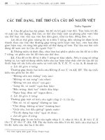
system. Figure 1A shows NMDAR-mediated current
traces before and after application of the SFK inhibi-
tor PP2 (10 lm). Co-transfection of constitutively
active Src, such as v-Src, significantly enhanced
NMDAR NR1-1a ⁄ NR2A-mediated current density
compared with that in cells without v-Src expression
(Fig. 1C). The mean peak amplitude of whole-cell cur-
rents recorded in HEK-293 cells expressing constitu-
tively active n-Src in which Tyr535 (corresponding to
Y527 in chicken c-Src) was mutated to phenylalanine
(Y535F) (see Table 1) was 760 ± 140 pA (n = 12,
mean ± SEM). Application of the SFK inhibitor PP2
significantly inhibited NR1-1a ⁄ NR2A receptor-medi-
ated whole-cell currents (Fig. 1A) without altering the
reversal potential of recorded currents (Fig. 1B). The
peak amplitudes of NMDAR-mediated currents were
reduced to 73 ± 7% (n = 7) of those observed prior
to PP2 application (Fig. 1D). In contrast, application
PP2AB
CD
0.5 nA
3 s
0
20
40
60
80
100
120
50
60
70
80
90
100
PP2 PP3
(7)(7)
(14)
(14) (14)
Percent control
##
Peak current density (pA/nF)
(6) (7)
##
v-Src: –
n-Src:
–
–60 20 40 60
0.1
0.2
0.3
–0.2
–0.3
V (mV)
I (nA)
PP2
Control
+
#
##
Fig. 1. Effects of inactivation of the SH3
and SH2 domains on the Src regulation of
NMDAR activity. (A) NR1-1a ⁄ NR2A recep-
tor-mediated whole-cell currents before and
during PP2 application recorded in HEK-293
cells co-transfected with cDNAs of
n-Src ⁄ Y535F. (B) Current–voltage
relationship recorded before (control) and
during PP2 application for a cell co-transfect-
ed with n-Src ⁄ Y535F. (C) Mean (± SEM)
NMDAR peak current density recorded in
HEK-293 cells transfected without ())or
with (+) v-Src. (D) Effects of PP2 application
on peak amplitudes of NMDAR currents,
normalized against those before PP2
application (100%, dashed line), recorded
from cells co-transfected or not with cDNAs
of n-Src mutants as indicated. #P < 0.05,
##P < 0.01 (independent group t test).
Values in parentheses indicate the number
of cells tested.
A novel function of Src SH2 and SH3 domains B. R. Groveman et al.
644 FEBS Journal 278 (2011) 643–653 ª 2010 The Authors Journal compilation ª 2010 FEBS
of PP3, the inactive form of PP2, had no such effect
(Fig. 1D). Consistent with results reported previously
[7,17], no significant change in NMDAR currents was
induced by PP2 application in cells without Src
co-transfection (Fig. 1D). No significant effect of PP2
on NMDAR currents was detected in cells co-express-
ing n-Src (K303R ⁄ Y535F), in which the lysine at resi-
due 303 in the kinase domain was mutated to arginine
(Table 1), thereby blocking the enzyme activity of Src
[3,18]. The peak amplitudes of NMDAR currents
during PP2 application were 96 ± 4% (n =7) of
those of controls before PP2 application (Fig. 1D).
Taken together, these data demonstrate that, by inhib-
iting the activity of Src, PP2 application decreases
NR1-1a ⁄ NR2A receptor activity.
Unexpectedly, however, the inhibition of NMDAR
currents induced by PP2 application was significantly
reduced in cells expressing n-Src ⁄ Y535F with the addi-
tional mutations D101N and R183K in the SH3 and
SH2 domains (Fig. 1D and Table 1). Previous studies
[3,18–21] have shown that D99 (corresponding to
D101 in n-Src) in the SH3 domain of c-Src forms a
salt bridge with an arginine located three residues
upstream of the conserved PXXP motif of the SH3
ligand. The D99N mutation prevents formation of this
salt bridge and disrupts the SH3 binding specificity.
R175 (corresponding to R183 in n-Src) in the SH2
domain of c-Src makes important connections with
phosphorylated tyrosine. Mutation of R175 to lysine
prevents this connection, and decreases SH2 interac-
tions with its ligand. D99N and R175K mutations
therefore inhibit interactions with ligands of the SH3
and SH2 domains, respectively, both intra- and inter-
molecularly, and thereby disrupt the overall functions
of Src kinase [3,18–21].
After PP2 application, peak amplitudes of NMDAR
currents were reduced to 89 ± 3% (n = 7) of those of
controls before PP2 application in cells co-expressing
active n-Src with dysfunctional SH3 and SH2 domains
(D101N ⁄ R183K ⁄ Y535F, Fig. 1D). The NMDAR
current reduction was significantly (P < 0.05, indepen-
dent group t test) smaller than that detected in cells
co-expressing constitutively active n-Src (Y535F,
Fig. 1D), raising the question: what roles do the SH3
and ⁄ or SH2 domains play in the regulation of
NMDARs by active Src?
To address this issue, we examined the activity of
n-Src expressed in HEK-293 cells. The gel shown in
Fig. 2A was loaded with lysates of HEK-293 cells
expressing wild-type n-Src or its mutants. Consistent
with previous findings [3,17], the Y535F mutation sig-
nificantly increased phosphorylation at Y424 (corre-
sponding to Y416 in chicken c-Src) compared with
that in wild-type n-Src (Fig. 2A). Dysfunction of the
kinase domain abolished phosphorylation of Y424 in
constitutively active n-Src (K303R ⁄ Y535F, Fig. 2A).
However, it was also noted that phosphorylation of
the activation loop, represented by phosphorylation
of Y424, in n-Src mutants with defective SH2 and ⁄ or
SH3 domains was reduced compared with that in
constitutively active n-Src (Y535F, Fig. 2A). These
findings suggest that dysfunction of the SH3 (D101N)
and ⁄ or SH2 (R183K) domains may down-regulate the
activity of active Src.
We then examined the enzyme activity in lysates of
HEK-293 cells expressing n-Src or its mutants by mea-
suring phosphorylation of the generic substrate poly-
Glu-Tyr. We found that the kinase activity in cells
expressing constitutively active n-Src was significantly
increased compared with that of cells expressing wild-
type n-Src (WT, Fig. 2B). Expression of inactive n-Src
(K303R ⁄ Y535F) did not produce detectable kinase
activity (Fig. 2B). Compared to cells expressing
constitutively active n-Src, the kinase activity was
significantly reduced by 27 ± 4% in cells expressing
active n-Src with a dysfunctional SH3 domain
Table 1. n-Src constructs listed by the residue(s) mutated and corresponding mutation(s) in chicken c-Src.
n-Src constructs
Corresponding
c-Src mutation Mutation location Phenotype
Wild-type None None Native
Y535F Y527F C-terminus Constitutively active
K303R ⁄ Y535F K297R ⁄ Y527F Kinase domain and C-terminus Kinase-dead
D101N ⁄ Y535F D99N ⁄ Y527F SH3 domain and C-terminus SH3 domain dysfunction
R183K ⁄ Y535F R175K ⁄ Y527F SH2 domain and C-terminus SH2 domain dysfunction
D101N ⁄ R183K ⁄ Y535F D99N ⁄ R175K ⁄ Y527F SH3, SH2 domain and C-terminus SH3 and SH2 domain dysfunction
Y535F
D1)258
Y527F
D1)250
N-terminal, SH3,
SH2 domain and C-terminus
Deletion of N-terminal, SH3 and
SH2 domain of active n-Src
K303R ⁄ Y535F
D1)258
K297R ⁄ Y527F
D1)250
N-terminal, SH3, SH2,
kinase domain and C-terminus
Deletion of N-terminal, SH3 and
SH2 domain of kinase-dead n-Src
B. R. Groveman et al. A novel function of Src SH2 and SH3 domains
FEBS Journal 278 (2011) 643–653 ª 2010 The Authors Journal compilation ª 2010 FEBS 645
(D101N ⁄ Y535F), by 96 ± 0.05% in cells expressing
active n-Src with a dysfunctional SH2 domain
(R183K ⁄ Y535F), and by 97 ± 0.04% in cells express-
ing active n-Src with dysfunctional SH3 and SH2
domains (D101N ⁄ R183K ⁄ Y535F, Fig. 2B). These data
not only suggest that dysfunction of the SH3 and⁄ or
SH2 domains significantly reduces the enzyme activity
of active Src expressed in HEK-293 cells, but also show
that the SH2 domain plays a greater role than the SH3
domain in regulation of n-Src activity. Consistent with
the finding that dysfunction of the SH3 and SH2
domains dramatically reduced n-Src activity (Fig. 2B),
we also found that, compared with constitutively active
n-Src (Y535F), neither auto-phosphorylation in the
activation loop nor kinase activity were present in the
n-Src mutant Y535F
D1)258
, from which the N-terminus
and both the SH3 and SH2 domains were deleted
(Fig. S1).
To confirm the effect of the SH3 and ⁄ or SH2
domain dysfunctions, n-Src and its mutants were
expressed in BL21(DE3) cells, purified as described
previously [22] and examined. Figure 3A shows these
purified proteins detected with antibodies as indicated.
Kinase activity on the generic substrate poly-Glu-Tyr
was measured 5–60 min after addition of n-Src or its
mutants (0.5 lm, Fig. 3B). Consistent with our previ-
ous findings [22], the enzyme activity of constitutively
active n-Src protein was significantly enhanced com-
pared to wild-type n-Src (Fig. 3B), but no enzyme
activity was detected in inactive n-Src protein
(Fig. 3B). Mutation of the SH3 or SH2 domain signifi-
cantly inhibited Src kinase activity, with a greater
effect resulting from dysfunction of the SH2 domain
(Fig. 3B), as was noted in HEK-293 cells.
Furthermore, we examined the auto-phosphorylation
of constitutively active n-Src, active n-Src with dys-
functional SH3 and SH2 domains, and inactive n-Src.
Each of these proteins (5 lg) was treated with a buffer
containing Lambda protein phosphatase (400 U) for
18 h at 30 °C. To initiate auto-phosphorylation,
a buffer containing 10 mm sodium orthovanadate,
50 mm sodium fluoride, 0.2 mm ATP and 10 mm
MgCl
2
was added to the samples to inactivate the
phosphatase for 0, 5, 10 or 20 min. The phosphoryla-
tion reaction was then stopped by addition of 6 · SDS
sample buffer supplemented with 50 mm EDTA. Y424
phosphorylation was subsequently analyzed by Wes-
tern blot (Fig. 3C). Ratios of band intensity detected
with anti-Src
pY416
IgG (rabbit) versus that detected
with anti-Src IgG (mouse) were calculated, and nor-
malized against the ratio obtained for n-Src ⁄ Y535F
protein that was not treated with Lambda protein
phosphatase (Fig. 3C). Decreased phosphorylation at
Y424 was observed in the active n-Src with dysfunc-
tional SH3 and ⁄ or SH2 domains compared with that
in constitutively active n-Src (Fig. 3C). However,
5 min after inactivation of Lambda protein phospha-
tase, Y424 phosphorylation of the active n-Src without
and with dysfunctional SH3 or SH2 domains or both
SH3 and SH2 domains reached similar levels
(75.4 ± 0.8%, 61.4 ± 9.8%, 75.0 ± 8.4% and
79.3 ± 3.4%, respectively) of their phosphorylation at
20 min. No such phosphorylation was observed in
inactive n-Src (Fig. 3C). Collectively, these data
Kinase activity (Abs
490 nm
)
#
#
#
0
0.5
1.0
1.5
2.5
93
50
93
A
B
50
93
50
Src
pY424
Src
Src
pY535
(8)
(8) (8) (8) (8) (8) (8)
2.0
#
##
Fig. 2. Effects of dysfunction of the SH3 and ⁄ or SH2 domains on
n-Src proteins expressed in HEK-293 cells. (A) Western blot
showing protein expression in lysates (20 lg) of HEK-293 cells.
The filters were sequentially immunoblotted with antibodies as
indicated: Src
pY535
(corresponding to Src
pY527
), probed with anti-
pY527 IgG (rabbit); Src
pY424
(corresponding to Src
pY416
), probed
with anti-pY416 IgG (rabbit); Src, probed with anti-Src IgG (mouse).
Values on the left indicate molecular mass (kDa). (B) Kinase activity
of n-Src proteins expressed in HEK-293 cells on a generic sub-
strate (poly-Glu-Tyr). Values in parentheses indicate the number of
experimental repeats. #P < 0.05 (independent group t test) in com-
parison with the kinase activity of constitutively active n-Src
(Y535F).
A novel function of Src SH2 and SH3 domains B. R. Groveman et al.
646 FEBS Journal 278 (2011) 643–653 ª 2010 The Authors Journal compilation ª 2010 FEBS
suggest that dysfunction of the SH3 or SH2 domains
does not alter the ability of active Src to phosphorylate
itself at Y424, but significantly reduces auto-phosphor-
ylation by modulating the kinase activity of the
enzyme.
To determine the roles of the SH3 and ⁄ or SH2
domains in Src regulation of NMDAR phosphoryla-
tion, the protein fragment corresponding to amino
acids K1096–V1464 in the NR2A C-tail was incubated
with wild-type n-Src or its mutants at a 1 : 1 concentra-
tion ratio for 1 h at 37 °C in the presence of 10 mm
MgCl
2
and 0.2 mm ATP. We found that the NR2A C-
tail protein was phosphorylated by wild-type n-Src, but
not by inactive n-Src (Fig. 4A). Incubation with active
Src
D101N/R183K/Y535F
93
50
93
50
Y535F
D101N/Y535F
R183K/Y535F
K303R/Y535F
Wt
Cms
A
C
B
WB
n-Src:
0
1.0
2.0
3.0
0
20
40 60
Time (min)
Kinase activity (a.u.)
Wt (3)
0 5 10 2015
Time (min)
0.00
0.05
0.10
0.15
0.20
Autophosphorylation (a.u.)
Y535F (3) R183K/Y535F (3)
D101N/Y535F (3) K303R/Y535F (3)
D101N/R183K/Y535F(3)
D101N/R183K/Y535F
C
Y535F
C
0 5 10 200 5 10 20 0 5 10 20 (min)
Src
pY424
Src
K303R/Y535F C
0 5 10 20 (min)0 5 10 20
Src
pY424
Src
R183K/Y535F CD101N/Y535F C
Y535F (5)
R183K/Y535F (6)
D101N/Y535F (6)
K303R/Y535F (4)
D101N/R183K/Y535F (5)
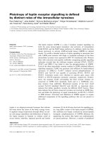
Fig. 3. Effects of dysfunction of the SH3 and ⁄ or SH2 domains on purified n-Src proteins in vitro. (A) Purified n-Src proteins expressed in
BL21(DE3) cells. Cms, Coomassie blue staining. WB, Western blot of purified n-Src proteins probed with anti-Src IgG. (B) Kinase activity of
purified n-Src proteins on a generic substrate (poly-Glu-Tyr). (C) Western blot showing n-Src auto-phosphorylation of Y424. The filters were
sequentially immunoblotted with antibodies against the proteins indicated. Lane C, untreated n-Src ⁄ Y535F protein. The graph shows the
results of densitometric analysis of Western blot data displayed as ratios of pY424 versus total Src (which were normalized against
untreated constitutively active n-Src (Y535F)). Values in parentheses indicate the number of experimental repeats.
B. R. Groveman et al. A novel function of Src SH2 and SH3 domains
FEBS Journal 278 (2011) 643–653 ª 2010 The Authors Journal compilation ª 2010 FEBS 647
n-Src resulted in an increased level of NR2A C-tail
phosphorylation compared with incubation with wild-
type n-Src. Active n-Src proteins with defective SH3
and ⁄ or SH2 domains resulted in a reduced level of
NR2A C-tail phosphorylation compared to constitu-
tively active Src (Fig. 4A). The time course of phos-
phorylation of the NR2A C-tail protein by wild-type
and mutant n-Src proteins is shown in Fig. 4B. The
highest tyrosine phosphorylation was produced by con-
stitutively active n-Src. At 10 min, phosphorylation of
NR2A C-tail by the constitutively active n-Src reached
a level similar to that produced by wild-type n-Src at
60 min (Fig. 4B). Dysfunction of the SH3 and ⁄ or SH2
domains affected the phosphorylation process of
NR2A C-tail proteins by active n-Src and reduced the
n-Src activity on NMDARs, with the greater effect pro-
duced by the dysfunction of the SH2 domain (Fig. 4).
To determine whether the reduced phosphorylation
and activity of NMDARs observed with dysfunction
of the SH3 and ⁄ or SH2 domains in Src may be due to
a change in interaction of Src with its substrate, bind-
ing of wild-type or mutant n-Src proteins with the
NR2A C-tail protein was examined using surface plas-
mon resonance (Fig . 5). We found that, in contrast to
bovine serum albumin, all of the n-Src proteins were
able to bind the NR2A C-tail with similar binding
affinities in the nanomolar range (Fig. 5). This indi-
cates that the ability of n-Src protein to bind to the
NR2A C-tail is independent of its kinase activity, and
that dysfunction of the SH3 and ⁄ or SH2 domains does
not affect this interaction.
The regulation of NMDARs by Src and other SFKs
[7–12] has been found to be a key mechanism underly-
ing activity-dependent neuroplasticity in the central
nervous system. SFKs are closely linked to NMDARs
in neurons [12] through binding to post-synaptic
density 95 (PSD-95) [23] or NADH dehydrogenase sub-
unit 2 (ND2) [24]. It is well known that the activity of
SFKs is tightly regulated by the reversible phosphoryla-
tion of Y527 in chicken c-Src in vivo. The phosphoryla-
tion of Y527 may decrease the activity of SFKs, with
dephosphorylation of phosphorylated Y527 having the
opposite effect [3–6]. Protein tyrosine phosphatise a
may selectively dephosphorylate phosphorylated Y527
[25,26], while C-terminal Src kinase specifically phos-
phorylates Y527 [3,27,28]. Protein tyrosine phospha-
tase a associates with NMDARs through binding to the
scaffold protein PSD-95, and constitutively up-regulates
NMDARs through endogenous SFKs [29]. C-terminal
Src kinase binds to phosphorylated NMDARs in
response to the actions of SFKs, depresses SFK activity
and thereby down-regulates NMDARs [17]. The close
proximity of C-terminal Src kinase, protein tyrosine
phosphatase a, SFKs and their substrate, NMDARs,
ensures that the complex forms a well-controlled molec-
ular network regulating receptor function and synaptic
plasticity [9,11,12,17,29].
Two types of Src, cellular Src (c-Src) and neuronal
Src (n-Src), are found in neurons. n-Src contains a six
amino acid insertion in the SH3 domain, and is only
expressed in neurons [3]. The SH3 and SH2 domains
in Src have been recognized to be involved in the nega-
tive regulation of Src. However, it has also been shown
that the SH2 domain may have positive effects on the
kinase activity and substrate interaction with the
kinase domain, for example in virus Fps (v-Fps) tyro-
sine kinase [30,31]. Recent detailed investigations
showed that, in active Fps kinase, the SH2 domain
tightly interacts with the kinase N-terminal lobe,
and positions the kinase aC helix in an active
configuration [32]. This structure is stabilized by ligand
binding to the SH2 domain [32]. Similarly, in active
NR2A:
+ + + + + + +
–
n-Src:
–
WT
Y535F
D101N/Y535F
R183K/Y535F
D101N/R183K/Y535F
Y535F
K303R/Y535F
93
50
Src
37
50
A
B
pY
37
50
NR2A
0204060
0
1.0
2.0
3.0
Time (min)
NR2A C-tail protein
phosphorylation (Abs
490 nm
)
Wt (3)
Y535F (3)
R183K/Y535F (3)
D101N/Y535F (3)
K303R/Y535F (3)
D101N/R183K/Y535F (3)
Fig. 4. Effects of dysfunction of the SH3 and ⁄ or SH2 domains on
phosphorylation of NMDAR NR2A C-tail protein by n-Src. (A) Wes-
tern blot showing phosphorylation of NR2A C-terminal fragment
(amino acids 1096-1464, 5 lg) incubated without ()) or with (+)
n-Src or its mutants as indicated. Duplicate filters were immunob-
lotted with antibodies as indicated: NR2A, probed with anti-NR2A
C-terminus IgG (rabbit); pY, probed with anti-phosphotyrosine IgG
(4G10, mouse); Src, probed with anti-Src IgG (mouse). (B) NR2A
C-terminus phosphorylation induced by n-Src proteins as indicated
and detected by color assay (see Experimental procedures). Values
in parentheses indicate the number of experimental repeats.
A novel function of Src SH2 and SH3 domains B. R. Groveman et al.
648 FEBS Journal 278 (2011) 643–653 ª 2010 The Authors Journal compilation ª 2010 FEBS
Response (RU)
0
6
12
18
24
30
Time (s)
BSA
0
10
50
100
200
400
Time (s)
Response (RU)
0
10
20
30
40
50
Wt
AB
CD
E
G
F
Normalized
response (a.u.)
0
0.2
0.4
0.6
0.8
1.0
[nM]
K
D
= 108.2 ± 13.3
Time (s)
Response (RU)
0
15
30
45
60
75
Y535F
K
D
= 96.0 ± 1.8
Normalized
response (a.u.)
0
0.2
0.4
0.6
0.8
1.0
[nM]
Time (s)
Response (RU)
0
10
20
30
40
50
R183K/Y535F
K
D
= 199.9 ± 31.1
Normalized
response (a.u.)
0
0.2
0.4
0.6
0.8
1.0
[nM]
Time (s)
Response (RU)
0
10
20
30
40
50
D101N/Y535F
K
D
= 227.3 ± 31.5
Normalized
response (a.u.)
0
0.2
0.4
0.6
0.8
1.0
[nM]
Time (s)
Response (RU)
0
6
12
18
24
30
D101N/R183K/Y535F
K
D
= 135.9 ± 26.1
Normalized
response (a.u.)
0
0.2
0.4
0.6
0.8
1.0
[nM]
Time (s)
0 100 200 300 400
0 100 200 300 400
0 100 200 300 400
0 100 200 300 400
0 100 200 300 400
0 100 200 300 400
0 100 200 300 400
Response (RU)
0
15
30
45
60
75
K303R/Y535F
K
D
= 151.0 ± 32.8
Normalized
response (a.u.)
0
0.2
0.4
0.6
0.8
1.0
0 100 200 300 400
0 100 200 300 400
0 100 200 300 400
0 100 200 300 400
0 100 200 300 400
0 100 200 300 400
[nM]
Fig. 5. Binding of n-Src and NR2A C-tail proteins. (A–F) Surface plasmon resonance showing binding of wild-type and mutant n-Src proteins
at concentrations of 0–400 n
M to NR2A C-tail protein immobilized on a CM5 chip to a surface density of 2000 response units (RU). Insets
show affinity curves fitted to a one-site binding model derived from surface plasmon resonance binding curves normalized to the response
at 400 n
M (mean ± SEM for each concentration of n-Src protein); K
D
, steady-state dissociation constant (mean ± SEM, n = 6). The sensor-
grams in (A) are displayed as overlaid triplicate experiments, while those in (B)–(G) are displayed as single representative experiments for
clarity. The degree of reproducibility of the triplicate runs in (B)–(G) was similar to that shown in (A). (G) Surface plasmon resonance sensor-
gram showing binding of bovine serum albumin at 400 n
M (negative control).
B. R. Groveman et al. A novel function of Src SH2 and SH3 domains
FEBS Journal 278 (2011) 643–653 ª 2010 The Authors Journal compilation ª 2010 FEBS 649
cellular Abl (c-Abl) tyrosine kinase, the SH2 and SH3
domains are redistributed from their auto-inhibitory
positions at the back site of the kinase domain, adopt-
ing an extended conformation and stimulating the cata-
lytic activity of the kinase [32]. Small-angle X-ray
scattering analysis showed that, in activated c-Abl, the
SH3, SH2, and kinase domains form an extended
arrangement [33]. This alternative conformation may
prolong the active state of the kinase by preventing it
from reverting to the auto-inhibitory state [33]. In Src
and Abl kinases, the SH2 domain can act in conjunction
with an additional SH2 or SH3 domain to maintain an
inactive state through intra-molecular interactions with
the catalytic domain, and is also critical for active
signaling [32]. Therefore, it is possible that the SH2
domain is bi-functional in regulation of kinase activity.
A previous study [14] reported that the tyrosine
phosphorylation of NMDAR NR2A and NR2B
subunits induced by incubation with recombinant Src
and Fyn may be significantly reduced by application
of SH2 domain binding peptides, which results in
blocking of the binding of the SH2 domain to the
substrate and thereby preventing interaction of
the substrate with the kinase domain. For active n-Src
in which the C-tail tyrosine was mutated to phenylala-
nine, dysfunctions of the SH2 and ⁄ or SH3 domains
reduced auto-phosphorylation of the kinase domain
activation loop, depressed kinase activity, and inhib-
ited Src-mediated NMDAR tyrosine phosphorylation
and channel activity regulation. Although the detailed
mechanisms underlying the actions of SH2 and SH3
domains in regulation of active n-Src remain to be
clarified, our study has revealed that SH2 and SH3
domain interactions may act not only to constrain the
activation of n-Src, but also to regulate the enzyme
activity of active n-Src, and that the SH2 domain
appears to play a greater role than the SH3 domain.
These findings may be important for understanding
the regulation of activity-dependent neuroplasticity in
the central nervous system.
Experimental procedures
HEK-293 cell culture and transfection
Cell culture and DNA transfection were performed as
described previously [17,29]. Briefly, HEK-293 cells were
grown in Dulbecco’s modified Eagle’s medium (Invitrogen,
Carlsbad, CA, USA) supplemented with 10% fetal bovine
serum (Invitrogen). These cells were then transfected using
Effecten (Qiagen, Valencia, CA, USA) or Lipofectamine
(Gibco-BRL, Carlsbad, CA, USA) according to the manu-
facturer’s instructions, with expression vectors (pcDNA3 or
pRcCMV) containing cDNAs encoding NR1-1a (0.4 lg),
NR2A (1.2 lg) and ⁄ or v-Src (0.2 lg), wild-type n-Src
(0.2 lg) or an n-Src mutant (0.2 lg): Y535F, D101N ⁄
Y535F, R183K ⁄ Y535F, K303R ⁄ Y535F, D101N ⁄ R183K ⁄
Y535F, Y535F
D1)258
or K303R ⁄ Y535F
D1)258
. D101, R183,
K303 and Y535 in mouse n-Src correspond to D99, R175,
K297 and Y527 in chicken c-Src, respectively (see Table 1).
For electrophysiological recordings, green fluorescence pro-
tein (GFP, 0.15 lg) was co-transfected. After 5–12 h, media
used for cDNA transfection were replaced with Dulbecco’s
modified Eagle’s medium supplemented with AP5 (500 lm)
for 48 h before recordings.
Whole-cell recordings in cultured cells
The methods used for whole-cell patch clamp recordings in
HEK-293 cells have been described previously [17,29]. In
brief, cells were bathed in a standard extracellular solution
containing NaCl (140 mm), CsCl (5 mm), CaCl
2
(1.2 mm),
HEPES (25 mm), glucose (32 mm), tetrodotoxin (TTX)
(0.001 mm), glycine (0.01 mm), pH 7.35 and osmolarity 310–
320 mOsm. Recording pipettes were pulled to a diameter of
1–2 lm at the tip, and filled with intracellular solution com-
prising 145 mm CsCl, 0.5 mm 1,2-bis(o-aminophenoxy)
ethane-N,N,N’,N’-tetraacetic acid (BAPTA), 10 mm
HEPES, 2 mm MgCl
2
,4mm potassium-adenosine-5’-tripho-
sphate (K-ATP), osmolarity 290–300 mOsm (DC resistance:
4–7 MX). Whole-cell currents were evoked by application of
l-aspartate or N-methyl-d-aspartate (250 lm) dissolved in
the extracellular solution for 3 s using a multi-barrel fast-
step perfusion system (SF-77B perfusion fast-step system,
Warner Instruments, Hamden, CT, USA). Recordings were
obtained under voltage-clamp conditions at a holding poten-
tial of )60 mV. Whole-cell currents were recorded using
Axopatch 200B amplifiers (Molecular Devices, Sunnydale,
CA, USA). Online data acquisition and off-line analysis were
performed using pClamp9 software (Molecular Devices).
Protein expression and purification
The techniques used for protein expression and purification
have been described previously [22]. In brief, cDNA encod-
ing full length wild-type n-Src, n-Src mutants (Y535F,
D101N ⁄ Y535F, R183K ⁄ Y535F, K303R ⁄ Y535F or D101N ⁄
R183K ⁄ Y535F) or amino acids K1096–V1464 of the NR2A
subunit was cloned into the pET15b vector and subsequently
transformed into Escherichia coli BL21(DE3) cells. The pro-
teins were expressed as N-terminal His
6
tag fusions in
Terrific Broth (VWR, Radnor, PA, USA) supplemented
with 100 lgÆmL
)1
ampicillin using a modified Autoinduc-
tionÔ protocol [34]. Cultures were grown at 37 °C for 3–4 h
and then cooled to 18 °C for protein expression for an
additional 18 h. Cells were then harvested by centrifugation
at 7500 g for 15 min at 4 °C. Pellets were resuspended in
buffer A (50 mm Tris ⁄ Cl, 0.5 m NaCl, 25 mm imidazole, pH
A novel function of Src SH2 and SH3 domains B. R. Groveman et al.
650 FEBS Journal 278 (2011) 643–653 ª 2010 The Authors Journal compilation ª 2010 FEBS
8.0) containing 1 mm phenylmethylsulfonyl fluoride, and
lysed using a sonicator. After centrifugation at 25 000 g at
4 °C, the supernatant was loaded onto a chelating Sepharose
column (Amersham Biosciences, Uppsala, Sweden). After
washing four times with 50 mL Buffer A, proteins were
eluted with 500 mm imidazole. The His tag was removed by
incubation with thrombin for 4 h at 37 °C. Protein purity
was assessed using SDS ⁄ PAGE and Western blotting
(Fig. 2B) and was at least 95%. Purified proteins were con-
centrated following extensive dialysis in buffer containing
30 mm sodium phosphate and 30 mm NaCl (pH 7.4), and
stored at 4 °C under reducing conditions (1 mm dithiothrei-
tol), and then analyzed using an electrospray ionization
(ESI) linear ion-trap mass spectrometer (LTQ MS) (Thermo
Finnigan, Waltham, MA, USA). The sequence coverage of
purified n-Src proteins was determined after analysis of tryp-
tic peptides using MS ⁄ MS [22]. Protein concentration was
determined spectrophotometrically in the presence of 6 m
urea at 280 nm using calculated extinction coefficients
().
Immunoblotting and in vitro kinase activity assay
Proteins purified from BL21(DE3) cells were subjected to
SDS ⁄ PAGE and Western blotting. Antibodies including
anti-Src IgG (Millipore, Billerica, MA, USA), anti-pY527
IgG (Cell Signaling, Danvers, MA, USA), anti-pY416 IgG
(Cell Signaling), anti-NR2A C-terminus IgG (Upstate,
Charlottesville, VA, USA) and anti-phosphotyrosine IgG
(4G10; Upstate) were used. To determine the kinase activity
of the n-Src proteins, a modified ELISA-based assay
(PTK101; Sigma, St Louis, MO, USA) was performed
using an exogenous tyrosine kinase-specific polymer sub-
strate, poly-Glu-Tyr (Sigma) or an NR2A protein fragment
corresponding to the C-tail amino acids K1096–V1464.
The phosphorylation reaction was initiated by addition of
n-Src proteins to tyrosine kinase reaction buffer containing
excess Mg
2+
(10 mm), Mn
2+
(10 mm) and ATP (0.2 mm)
in microtiter plates coated with poly-Glu-Tyr substrate or
NR2A C-tail. The phosphorylation reactions were stopped
by removing the reaction buffer and washing with NaCl ⁄
P
i
+Tween-20 at each time point as indicated. The
phosphorylated substrate was detected using horseradish
peroxidase-conjugated anti-phosphotyrosine IgG. A color
reaction was induced by adding the horseradish peroxidase
substrate o-phenylenediamine, and stopped using 0.25 m
sulfuric acid, followed by absorbance measurements at
490 nm using a spectrophotometer and a microplate ELISA
reader (Benchmark, Bio-Rad, Hercules, CA, USA). Steady-
state kinase activity assays for the proteins were performed
at room temperature for 60 min. All of the chemicals and
agents were purchased from Sigma except where indicated.
To examine the auto-phosphorylation of the proteins,
5 lg of n-Src ⁄ Y535F, n-Src ⁄ D101N ⁄ Y535F, n-Src ⁄ R183K ⁄
Y535F, n-Src ⁄ D101N ⁄ R183K ⁄ Y535F or n-Src ⁄ K303R ⁄
Y535F were dephosphorylated using 400 U of Lambda
protein phosphatase (New England BioLabs, Ipswich, MA,
USA) in the manufacturer-provided reaction buffer at
30 °C for 18 h. The phosphatase was inactivated by
addition of 10 mm sodium orthovanadate and 50 mm
sodium fluoride in a buffer containing 0.2 mm ATP and
10 mm MgCl
2
for 0, 5, 10, or 20 min. The reactions were
stopped by addition of 6 · SDS sample buffer supple-
mented with 50 mm EDTA. Auto-phosphorylation at
pY424 was analyzed by Western blot and quantified by
densitometric analysis using Image J (National Institutes of
Health, Bethesda, MD).
Surface plasmon resonance
The affinity interactions of Src mutants and NR2A C-tail
fragment were analyzed using a Biacore T-100 optical
biosensor (Biacore ⁄ GE Healthcare, Uppsala, Sweden). The
NR2A C-tail protein fragment was immobilized on a CM5
chip (Biacore ⁄ GE Healthcare) using amine coupling chemis-
try. This process consisted of surface chip activation using a
1 : 1 ratio of 0.4 m 1-ethyl-3-(3-dimethylaminopropyl)-car-
boimide and 0.1 m N-hydroxysuccinimide, followed by
NR2A C-tail protein immobilization to a level of
2000 response units (RU) using 10 lgÆmL
)1
protein in
10 mm sodium acetate immobilization buffer (pH 4.5), and
chip surface deactivation using 1 m ethanolamine ⁄ HCl (pH
8.5). All binding experiments were performed in a running
buffer containing 50 mm HEPES, 150 mm NaCl, 3 mm
EDTA, 0.05% p20 surfactant (Biacore ⁄ GE Healthcare), pH
7.4. Src at concentrations up to 400 lm was injected in
triplicate over the chip surface at a flow rate of 10 lLÆmin
)1
for 180 s. The surface was regenerated using 30 s bursts of
2 m NaCl followed by 0.05% SDS at a flow rate of 50 lLÆ-
min
)1
. All experiments were performed in triplicate on two
CM5 chips following the same protocol. The data were ana-
lyzed using BiaEvaluation 3.0 software (Biacore) and Sig-
maPlot (Systat Software Inc, Richmond, CA, USA) and
fitted to a 1 : 1 Langmuir binding model for calculation of
the equilibrium dissociation constants (K
D
).
Acknowledgements
This work was supported by a grant from the National
Institutes of Health (R01 NS053567) to X M.Y. Plas-
mids of v-Src, and n-Src and its mutants were kindly
provided by Dr T. Pawson (Department of Molecular
Genetics, University of Toronto, Canada) and Dr
S. Hanks (Department of Cell Biology, Vanderbilt
University, Nashville, TN), respectively. We gratefully
acknowledge the Biomedical Proteomics Laboratory at
the College of Medicine, Florida State University, for
the use of UV ⁄ Vis spectroscopy and surface plasmon
resonance instruments.
B. R. Groveman et al. A novel function of Src SH2 and SH3 domains
FEBS Journal 278 (2011) 643–653 ª 2010 The Authors Journal compilation ª 2010 FEBS 651
References
1 Cohen P (2002) Protein kinases – the major drug targets
of the twenty-first century? Nat Rev Drug Discov 1,
309–315.
2 Liu XJ, Gingrich JR, Vargas-Caballero M, Dong YN,
Sengar A, Beggs S, Wang SH, Ding HK, Frankland PW
& Salter MW (2008) Treatment of inflammatory and
neuropathic pain by uncoupling Src from the NMDA
receptor complex. Nat Med 14, 1325–1332.
3 Brown MT & Cooper JA (1996) Regulation, substrates
and functions of src. Biochim Biophys Acta 1287, 121–
149.
4 Xu W, Harrison SC & Eck MJ (1997) Three-dimen-
sional structure of the tyrosine kinase c-Src. Nature 385,
595–602.
5 Ingley E (2008) Src family kinases: regulation of their
activities, levels and identification of new pathways.
Biochim Biophys Acta 1784, 56–65.
6 Roskoski R Jr (2005) Src kinase regulation by phos-
phorylation and dephosphorylation. Biochem Biophys
Res Commun 331, 1–14.
7 Kohr G & Seeburg PH (1996) Subtype-specific regula-
tion of recombinant NMDA receptor-channels by pro-
tein tyrosine kinases of the src family. J Physiol 492,
445–452.
8 Miyakawa T, Yagi T, Kitazawa H, Yasuda M, Kawai N,
Tsuboi K & Niki H (1997) Fyn-kinase as a determinant
of ethanol sensitivity: relation to NMDA receptor func-
tion. Science 278, 698–701.
9 Yu X-M, Askalan R, Keil GJI & Salter MW (1997)
NMDA channel regulation by channel-associated
protein tyrosine kinase Src. Science 275, 674–678.
10 Hisatsune C, Umemori H, Mishina M & Yamamoto T
(1999) Phosphorylation-dependent interaction of the
N-methyl-d-aspartate receptor e2 subunit with
phosphatidylinositol 3-kinase. Genes Cells 4, 657–666.
11 Ali DW & Salter MW (2001) NMDA receptor regula-
tion by Src kinase signalling in excitatory synaptic
transmission and plasticity. Curr Opin Neurobiol 11,
336–342.
12 Salter MW & Kalia LV (2004) Src kinases: a hub for
NMDA receptor regulation. Nat Rev Neurosci 5, 317–
328.
13 MacDonald JF, Jackson MF & Beazely MA (2006)
Hippocampal long-term synaptic plasticity and signal
amplification of NMDA receptors. Crit Rev Neurobiol
18, 71–84.
14 Cheung HH & Gurd JW (2001) Tyrosine phosphoryla-
tion of the N-methyl-d-aspartate receptor by exogenous
and postsynaptic density-associated Src-family kinases.
J Neurochem 78, 524–534.
15 Nakazawa T, Komai S, Tezuka T, Hisatsune C,
Umemori H, Semba K, Mishina M, Manabe T &
Yamamoto T (2001) Characterization of Fyn-mediated
tyrosine phosphorylation sites on GluRe2 (NR2B)
subunit of the N-methyl-d-aspartate receptor.
J Biol
Chem 276, 693–699.
16 Yang M & Leonard JP (2001) Identification of mouse
NMDA receptor subunit NR2A C-terminal tyrosine
sites phosphorylated by coexpression with v-Src. J Neu-
rochem 77, 580–588.
17 Xu J, Weerapura M, Ali MK, Jackson MF, Li H,
Lei G, Xue S, Kwan CL, Manolson MF, Yang K
et al. (2008) Control of excitatory synaptic transmis-
sion by C-terminal Src kinase. J Biol Chem 283,
17503–17514.
18 Polte TR & Hanks SK (1997) Complexes of focal adhe-
sion kinase (FAK) and Crk-associated substrate
(p130(Cas)) are elevated in cytoskeleton-associated frac-
tions following adhesion and Src transformation.
Requirements for Src kinase activity and FAK proline-
rich motifs. J Biol Chem 272, 5501–5509.
19 Weng Z, Rickles RJ, Feng S, Richard S, Shaw AS,
Schreiber SL & Brugge JS (1995) Structure–function
analysis of SH3 domains: SH3 binding specificity
altered by single amino acid substitutions. Mol Cell Biol
15, 5627–5634.
20 Songyang Z & Cantley LC (1995) Recognition and
specificity in protein tyrosine kinase-mediated signalling.
Trends Biochem Sci 20, 470–475.
21 Verderame MF (1997) pp60v-src transformation of rat
cells but not chicken cells strongly correlates with low-
affinity phosphopeptide binding by the SH2 domain.
Mol Biol Cell 8, 843–854.
22 Marin V, Groveman BR, Qiao H, Xu J, Ali MK,
Fang X-Q, Lin S-X, Rizkallah R, Hurt MH,
Bienkiewicz EA et al. (2010) Characterization of
neuronal Src kinase purified from a bacterial expression
system. Protein Expr Purif 74, 289–297.
23 Tezuka T, Umemori H, Akiyama T, Nakanishi S &
Yamamoto T (1999) PSD-95 promotes Fyn-mediated
tyrosine phosphorylation of the N-methyl-d-aspartate
receptor subunit NR2A. Proc Natl Acad Sci USA 96,
435–440.
24 Gingrich JR, Pelkey KA, Fam SR, Huang Y,
Petralia RS, Wenthold RJ & Salter MW (2004) Unique
domain anchoring of Src to synaptic NMDA receptors
via the mitochondrial protein NADH dehydrogenase
subunit 2. Proc Natl Acad Sci USA 101, 6237–6242.
25 Ponniah S, Wang DZ, Lim KL & Pallen CJ (1999) Tar-
geted disruption of the tyrosine phosphatase PTPa leads
to constitutive downregulation of the kinases Src and
Fyn. Curr Biol 9, 535–538.
26 Su J, Muranjan M & Sap J (1999) Receptor protein
tyrosine phosphatase a activates Src-family kinases and
controls integrin-mediated responses in fibroblasts. Curr
Biol 9, 505–511.
27 Nada S, Okada M, MacAuley A, Cooper JA & Nakag-
awa H (1991) Cloning of a complementary DNA for a
A novel function of Src SH2 and SH3 domains B. R. Groveman et al.
652 FEBS Journal 278 (2011) 643–653 ª 2010 The Authors Journal compilation ª 2010 FEBS
protein-tyrosine kinase that specifically phosphorylates
a negative regulatory site of p60c-src. Nature 351, 69–
72.
28 Imamoto A & Soriano P (1993) Disruption of the csk
gene, encoding a negative regulator of Src family tyro-
sine kinases, leads to neural tube defects and embryonic
lethality in mice. Cell 73, 1117–1124.
29 Lei G, Xue S, Chery N, Liu Q, Xu J, Kwan CL, Fu Y,
Lu YM, Liu M, Harder KH et al. (2002) Gain control
of N-methyl-d-aspartate receptor activity by receptor-
like protein tyrosine phopshatase a. EMBO J 21, 2977–
2989.
30 Stone JC, Atkinson T, Smith M & Pawson T (1984)
Identification of functional regions in the transforming
protein of Fujinami sarcoma virus by in-phase insertion
mutagenesis. Cell 37, 549–558.
31 Sadowski I, Stone JC & Pawson T (1986) A noncatalyt-
ic domain conserved among cytoplasmic protein-tyro-
sine kinases modifies the kinase function and
transforming activity of Fujinami sarcoma virus
P130gag-fps. Mol Cell Biol 6, 4396–4408.
32 Filippakopoulos P, Kofler M, Hantschel O, Gish GD,
Grebien F, Salah E, Neudecker P, Kay LE, Turk BE,
Superti-Furga G et al. (2008) Structural coupling of
SH2-kinase domains links Fes and Abl substrate recog-
nition and kinase activation. Cell 134, 793–803.
33 Nagar B, Hantschel O, Seeliger M, Davies JM, Weis WI,
Superti-Furga G & Kuriyan J (2006) Organization of the
SH3–SH2 unit in active and inactive forms of the c-Abl
tyrosine kinase. Mol Cell 21, 787–798.
34 Grabski A, Mehler M & Drott D (2005) The Overnight
Express autoinduction system: high-density cell growth
and protein expression while you sleep. Nat Methods 2,
233–235.
Supporting information
The following supplementary material is available:
Fig. S1. Effects of deletion of the N-terminus and both
the SH3 and SH2 domains on the activity of n-Src
proteins expressed in HEK-293 cells.
This supplementary material can be found in the
online version of this article.
Please note: As a service to our authors and readers,
this journal provides supporting information supplied
by the authors. Such materials are peer-reviewed and
may be re-organized for online delivery, but are not
copy-edited or typeset. Technical support issues arising
from supporting information (other than missing files)
should be addressed to the authors.
B. R. Groveman et al. A novel function of Src SH2 and SH3 domains
FEBS Journal 278 (2011) 643–653 ª 2010 The Authors Journal compilation ª 2010 FEBS 653