Báo cáo khoa học: Neural retina leucine-zipper regulates the expression of Ppp2r5c, the regulatory subunit of protein phosphatase 2A, in photoreceptor development pdf
Bạn đang xem bản rút gọn của tài liệu. Xem và tải ngay bản đầy đủ của tài liệu tại đây (544.2 KB, 10 trang )
Neural retina leucine-zipper regulates the expression of
Ppp2r5c, the regulatory subunit of protein
phosphatase 2A, in photoreceptor development
Jung-Woong Kim, Sang-Min Jang, Chul-Hong Kim, Joo-Hee An, Eun-Jin Kang and
Kyung-Hee Choi
Department of Life Science (BK21 program), College of Natural Sciences, Chung-Ang University, Seoul, Korea
Introduction
Protein phosphatase 2A (PP2A) is a major cellular ser-
ine ⁄ threonine phosphatase that plays a critical role in
balancing phosphorylation signals that are important
for cellular proliferation and differentiation [1,2]. The
catalytic C-subunit of PP2A associates with the scaf-
folding A-subunit, and the A ⁄ C heterodimer also binds
to regulatory B-subunits to form a heterotrimeric holo-
enzyme [3]. B-subunits can be divided into four distinct
families on the basis of their homology, namely B
(B55 or PR55) [4–7], B¢ (B56 or PR61) [8–11], B¢¢
(PR48 ⁄ 59 ⁄ 72 ⁄ 130) [12,13] and B¢¢¢ (PR93 ⁄ 110) [14],
and the B56 family consists of at least five different
gene p roducts, a (PPP2R5A), b (PPP2R5B), c (PPP2R5C),
d (PPP2R5D), and e (PPP2R5E) [8]. The five B56 fam-
ily members have diverse functions, including a mitotic
checkpoint in Xenopus laevis and binding to APC pro-
tein, which acts as a scaffold for b-catenin, axin and
glycogen synthase kinase-b [15,16]. Moreover, B56e is
involved in Xenopus eye development through the insu-
lin-like growth factor–phosphoinositide 3-kinase–Akt
and hedgehog signaling pathways [17]. It is believed
that PP2A exercises regulatory flexibility and substrate
specificity through association of the core A ⁄ C hetero-
dimer with one of the regulatory B-subunits [1,18].
This characteristic of PP2A contributes to its ability
to regulate multiple cellular functions; however, the
Keywords
neural retina leucine-zipper; photoreceptor
development; PP2A regulatory subunit;
Ppp2r5c; target gene
Correspondence
K H. Choi, Department of Life Science
(BK21 program), College of Natural
Sciences, Chung-Ang University, 221
Heuksuk Dong, Dongjak Ku, Seoul 156-756,
South Korea
Fax: +82 2 824 7302
Tel: +82 2 820 5209
E-mail:
(Received 12 July 2010, revised 11
September 2010, accepted 11 October
2010)
doi:10.1111/j.1742-4658.2010.07910.x
Protein phosphatase 2A plays an important role in balancing phosphoryla-
tion signals that are critical for cell proliferation and differentiation. Here,
we report that Ppp2r5c (regulatory subunit of protein phosphatase 2A)
expression was regulated by the transcription factor neural retina leucine-
zipper (Nrl) through enhancement of its transcriptional activity on the
Ppp2r5c promoter. Using electrophoretic mobility shift assays and chroma-
tin immunoprecipitation, we also found that Nrl bound directly to the
Nrl-response element on the Ppp2r5c promoter. The affinity of binding of
Nrl to the Ppp2r5c promoter was tightly regulated during mouse photo-
receptor development. Overall, these results suggest that Ppp2r5c expres-
sion is regulated by Nrl during retinogenesis through direct binding to the
promoter region of Ppp2r5c.
Abbreviations
ChIP, chromatin immunoprecipitation; E, embryonic day; EMSA, electrophoretic mobility shift assay; GST, glutatione S-transferase;
NRE, neural retina leucine-zipper-response element; Nrl, neural retina leucine-zipper; NS, not significant; P, postnatal day; PP2A, protein
phosphatase 2A; siRNA, small interfering RNA; WT, wild type.
FEBS Journal 277 (2010) 5051–5060 ª 2010 The Authors Journal compilation ª 2010 FEBS 5051
precise molecular mechanisms underlying the transcrip-
tional control of PP2A genes and the effects of diverse
combinations of PP2A subunits have not yet been elu-
cidated.
Neural retina leucine-zipper (Nrl) belongs to the
basic motif leucine-zipper family of transcription
factors [19]. Nrl is conserved in vertebrates and is
specifically expressed in photoreceptors and the pineal
gland [19,20]. Nr1 is essential for rod differentia-
tion, and may act as a molecular switch in the deter-
mination of photoreceptor cell fate, as Nrl knockout
mice have a complete lack of rods but enhanced
S-cones [21]. In humans, missense mutations of NRL
are associated with autosomal dominant retinitis pig-
mentosa [22], and this disease may be a result of
altered transcriptional activity of the NRL [23]. Nrl
interacts with cone-rod homeobox [24], Flt-3-interact-
ing zinc-finger [25] and TATA box-binding protein
[26] to regulate the expression of rhodopsin [27],
NR2E3 [28], cGMP-phosphodiesterase-a, cGMP-phos-
phodiesterase-b [29,30] and rod-specific genes [21].
These observations have shown that Nrl plays a criti-
cal role in the differentiation of rod photoreceptors
that involves spatiotemporal regulation of its target
gene expression.
In this study, we identified Nrl as a novel transcrip-
tional factor that regulates Ppp2r5c gene expression in
photoreceptor development. Furthermore, an unbiased
motif search of Ppp2r5c promoter sequences revealed
that the Ppp2r5c promoter has putative Nrl-binding
sequences. Moreover, the functional roles of Nrl in
Ppp2r5c transcription were examined in vitro and
in vivo. We also present the association profiles of Nrl
on the Ppp2r5c promoter during mouse photoreceptor
development, with the goal of determining the critical
stage for Nrl-mediated Ppp2r5c expression.
Results
Conserved sequences of the Ppp2r5c promoter
contain Nrl-binding sites
In the search for conserved putative regulatory
elements, the sequence of the human Ppp2r5c pro-
moter from )300 to )1 relative to the transcriptional
start site was compared with corresponding regions of
mouse and cow sequences, with clustal w. Highly
conserved noncoding sequences were determined
among these Ppp2r5c promoters with a minimal
sequence similarity of 67% (Fig. 1A). To identify tran-
scription factors that might bind to the Ppp2r5c pro-
moter and regulate its expression, we used tfsearch
software (Searching transcription factor binding sites,
Version 1.3). As shown in Fig. 1B, the Nrl response
element (NRE) was found at )154 to )143 from the
transcription start site. Furthermore, the putative pro-
moter region of the Ppp2r5c gene contained binding
sites for MZF1, CREB, GATA1, Hsf1 ⁄ 2 and CdxA.
A
B
Fig. 1. A conserved region of the Ppp2r5c
promoter contains putative Nrl-binding
motifs. (A) The promoter sequences for
human, cow and mouse Ppp2r5c genes
were aligned using the multiple sequencing
alignment program
CLUSTAL W. Underlined
sequences represent putative NREs.
Asterisks are indicated within twenty
nucleotides. (B) The mouse Ppp2r5c
promoter (GeneID: 26931) was analyzed
with a motif searching program to identify
binding sites of transcription factors.
Consensus binding sites are underlined, the
Nrl-binding site is printed in bold, and the
transcription start site is shown as +1.
Regulation of Ppp2r5c expression by Nrl J W. Kim et al.
5052 FEBS Journal 277 (2010) 5051–5060 ª 2010 The Authors Journal compilation ª 2010 FEBS
Nrl increases the endogenous Ppp2r5c transcript
level and Ppp2r5c reporter gene activity
We first screened various cell lines to identify cells that
abundantly express Nrl mRNA and proteins (Figs S1B
and S2C). Mouse hippocampal HT22 cells showed
high-level expression of Nrl mRNA and protein. To
determine whether Nrl is truly a transcriptional regula-
tor of Ppp2r5c, HT22 cells were transiently transfected
with FLAG–CMV2–Nrl expression plasmids, and
Ppp2r5c mRNA expression was analyzed by quantita-
tive real-time PCR. As shown in Fig. 2A, overexpres-
sion of Nrl induced Ppp2r5c transcription (left panel),
and rhodopsin expression by Nrl was also confirmed as
a positive control (right panel). To further examine
whether the increased expression of Ppp2r5c transcripts
was specifically modulated by Nrl, we used Nrl small
interfering RNA (siRNA) transfectants in which the
expression of Nrl was approximately 60% abrogated
(Fig. S3A,B). As shown in Fig. 2B, transfection of Nrl
siRNA effectively decreased the levels of Ppp2r5c
(Fig. 2B, right panel) and rhodopsin (Fig. 2B, left
panel) mRNA. We then conducted luciferase reporter
assays with Ppp2r5c promoters to test the Nrl-mediated
transcriptional activity. The Ppp2r5c promoter frag-
ment from 800 to )1 was cloned into luciferase repor-
ter constructs that were transiently transfected with
the Nrl expression plasmids into HEK293 cells, which
do not express mRNA and protein of Nrl endoge-
nously (Figs S1B and S2C). In the presence of exoge-
nous Nrl, Ppp2r5c promoter activity was significantly
increased by Nrl in a dose-dependent manner (Fig. 2C,
left panel). Nrl also enhanced its known target
rhodopsin promoter activity (Fig. 2C, right panel). To
further investigate Nrl-mediated transcriptional activa-
tion through a putative NRE, we performed luciferase
reporter assays with various mutant forms of the
mouse Ppp2r5c promoter in HEK293 cells. To accom-
plish this, we cloned two different mutants, the NRE-
deleted mutant (DNRE) and the truncated mutant that
contained NRE (NRE) (Fig. 2D, upper panel). As
expected, the NRE included full length of wild-type
promoter. (WT) and the NRE mutant significantly
induced luciferase reporter activity in an Nrl concen-
tration-dependent manner (Fig. 2D, lanes 3, 4, 7 and
8). However, the NRE-deleted mutant promoter con-
struct (DNRE) abolished the luciferase activity under
conditions of Nrl overexpression (Fig. 2D, lanes 5 and
Ppp2r5c
6 6
Rhodopsin
***
2
3
4
5
2
3
4
5
***
Fold increase
Fold increase
1
0
1
0
Mock
Mock
Flag-Nrl
Flag-Nrl
Ppp2r5c
1.2
1.2
Rhodopsin
***
***
0.4
0.6
0.8
1.0
0.4
0.6
0.8
1.0
Fold increase
Fold increase
0.2
0
0.2
0
Mock
Mock
Ppp2r5c
10
12
Rhodopsin
***
4
6
8
10
**
***
***
Fold increase
2
0
Mock
Mock
Flag–Nrl Flag–Nrl
Fold increase
Ppp2r5c promoter
25
Luciferase
NRE
: WT
Luciferase
: ΔNRE
Luciferase
NRE
: NRE
–87
–800
–74
–260
**
***
5
10
15
20
25
***
***
0
5
Flag–Nrl
Flag–Nrl
Flag–Nrl
Flag–Nrl
Reporter :
Mock
WT
ΔNRE
NRE
NS NS
–1
A
B
C
D
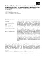
Fig. 2. Nrl increases endogenous Ppp2r5c
mRNA levels and Ppp2r5c reporter gene
activity. (A, B) HT22 cells were transfected
with plasmids expressing FLAG–Nrl and ⁄ or
Nrl siRNA vectors. RNA was extracted, and
quantitative real-time PCR analysis was con-
ducted using primers specific for rhodopsin,
Ppp2r5c and Gapdh. The Gapdh gene was
used as an internal control. (C, D) HEK293
cells were cotransfected with Ppp2r5c
promoter–Luc and pCMV–b-galactosidase
with increasing amounts (1 lg and 2 lg) of
plasmids encoding Nrl cDNA. Forty-eight
hours after transfection, luciferase activity
was measured. All data were normalized to
b-galactosidase activity. Data are expressed
as the fold increase over relative luciferase
units, normalized to the control. The statisti-
cal significant levels were considered
significant at P < 0.05 (*), very significant at
P < 0.01 (**), obviously significant at
P < 0.001 (***), or not significant (NS).
J W. Kim et al. Regulation of Ppp2r5c expression by Nrl
FEBS Journal 277 (2010) 5051–5060 ª 2010 The Authors Journal compilation ª 2010 FEBS 5053
6). These results suggest that the NRE at )87 to )74
is responsible for the Nrl-mediated transcriptional acti-
vation of mouse Ppp2r5c.
Nrl binds to the Ppp2r5c promoter in vitro
As the Ppp2r5c promoter contains a putative NRE
and its transcripts were increased by Nrl, we con-
ducted an electrophoretic mobility shift assay (EMSA)
with glutatione S-transferase (GST)-fused recombinant
Nrl (Fig. S4) to determine whether Nrl induces
Ppp2r5c transcription through direct binding to the
proximal promoter region of Ppp2r5c. An oligonucleo-
tide containing the consensus Nrl-binding site at )154
to )143 of the Ppp2r5c promoter was used as a hot
probe. As shown in the left panel of Fig. 3, incubation
of hot probes with Nrl produced slower-migrating
DNAÆprotein complexes in a dose-dependent manner
(lanes 4 and 5), whereas the control GST protein alone
did not form DNAÆprotein complexes (lanes 2 and 3).
The presence of Nrl in the proteinÆDNA complex was
verified with antibody against Nrl, which supershifted
a portion of the Nrl–probe complex (lane 6), whereas
the IgG negative control did not alter the binding pat-
tern (lane 7). The Ppp2r5c probe–Nrl protein–Nrl anti-
body triple complex disappeared when cold Ppp2r5c
probe was added as a competitor (lane 8). The rhodop-
sin promoter was used as a positive control (Fig. 3,
right panel). These results show that Nrl binds directly
to its response element ()154 to )143 from the tran-
scriptional start site) located in the promoter region of
Ppp2r5c in vitro.
Nrl was recruited to the Ppp2r5c promoter during
photoreceptor development
We next conducted a chromatin immunoprecipitation
(ChIP) assay with HT22 cells, to further examine the
binding of Nrl to the Ppp2r5c promoter in vivo. Nrl
binding to the Ppp2r5c and rhodopsin promoters was
examined by quantitative real-time of ChIP samples
with appropriate primers. As shown in Fig. 4A, Nrl
antibody specifically immunoprecipitated regions of
the Ppp2r5c and rhodopsin promoter containing the
NRE, whereas normal rabbit serum, used as a negative
control, did not precipitate the Ppp2r5c and rhodopsin
promoters in the HT22 cell lines. To confirm the asso-
ciation of Nrl with the Ppp2r5c promoter in mouse ret-
ina, we used postnatal day (P)10 mouse retina for the
quantitative ChIP assay, because Nrl expression was
highly upregulated after the P4 stage (data not shown).
The Ppp2r5c and rhodopsin promoters were specifically
precipitated with antibody against Nrl, but not with
rabbit control serum, in the mouse retina (Fig. 4B).
It was previously reported that rod–cone differentia-
tion is regulated by increases in the expression levels of
Nrl to modulate its specific target gene expression in
photoreceptor precursor cells [20,31]. To determine the
developmental stage-specific recruitment of Nrl to the
Ppp2r5c promoter, quantitative ChIP assays were con-
ducted with developing mouse retinas of various
stages, from embryonic day (E)15 to P42. Nrl binding
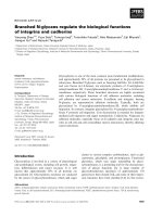
to the Ppp2r5c promoter increased approximately five-
fold from P10 to P14, and thereafter decreased to
basal levels until P21 (Fig. 4C). This sharp increase in
Fig. 3. Nrl directly binds to the Ppp2r5c pro-
moter consensus element in vitro. EMSA
showing the binding of Nrl to NRE sites in
the rhodopsin and Ppp2r5c promoters.
Lanes are as indicated below the autoradio-
graph. Two or four micrograms of purified
proteins was used for EMSA. For the
competition experiment, lane 8 included a
10-fold molar excess of unlabeled NRE
oligonucleotide. Lane 6 contains 0.1 lLof
antibody against Nrl, and lane 7 contains the
same quantity of rabbit serum as a negative
control for Nrl antibody. Arrowheads repre-
sent the specific shifted band (a, unbound
probes; b and c, bands shifted by Nrl;
d, supershifted band; e, nonspecific bands).
These experiments were conducted at least
three times, and similar results were
obtained.
Regulation of Ppp2r5c expression by Nrl J W. Kim et al.
5054 FEBS Journal 277 (2010) 5051–5060 ª 2010 The Authors Journal compilation ª 2010 FEBS
the Nrl binding affinity for the Ppp2r5c promoter was
tightly regulated during photoreceptor development
when compared with the patterns of binding of Nrl to
the rhodopsin promoter (Fig. 4C,D). The mRNA tran-
script levels of Ppp2r5c and rhodopsin in mouse retina
corresponded to the observed increase in Nrl binding
affinity for their promoter regions (Fig. 4E,F). Taken
together, these results clearly indicate that Nrl regu-
lated the expression of Ppp2r5c transcripts through
direct binding to the Ppp2r5c promoter during mouse
photoreceptor development.
Discussion
In this study, we found that Ppp2r5c or B56c (PP2A
regulatory subunit) expression was regulated by Nrl.
Ectopic expression or knockdown of Nrl modulated
the Ppp2r5c mRNA expression level and Nrl-mediated
luciferase activity on the Ppp2r5c and rhodopsin pro-
moters (Fig. 2). We also demonstrated that Nrl bound
to NRE on the Ppp2r5c promoter both in vitro and
in vivo (Figs 3 and 4). Furthermore, the binding affin-
ity of Nrl for the Ppp2r5c promoter was tightly regu-
lated during mouse photoreceptor development, and
was enhanced between P10 and P14 (Fig. 4C).
It has recently been reported that PP2A may play
important roles in developing eyes, and the functions of
PP2A appear to be highly regulated by various regula-
tory subunits [17]. The mRNA isoforms of the PP2A A-
subunit and B-subunit (PP2A-Aa ⁄ b and PP2A-Ba ⁄ b ⁄ c)
have also been shown to be highly expressed in the
mouse retina [32]. In this study, we first attempted to
Rho promoter
Ppp2r5c promoter
Mouse retina
***
***
6
8
10
Input (%)
12
***
2
4
0
IP:
Serum Nrl
Serum Nrl
Ppp2r5c promoter
Rho promoter
HT22 cells
***
Input (%)
3
4
5
6
**
1
2
0
IP:
Serum Nrl
Serum Nrl
Ppp2r5c
2
3
***
1.5
2.5
1
0
Fold increase
0.5
Rhodopsin
20
40
Fold increase
60
***
30
50
0
10
BA
E
F
Input (%)
0.4
0.6
0.8
1.0
1.2
ChIP: Ppp2r5c promoter
0.0
0.2
E15 E20 P0 P2 P4 P8 P10 P14 P21 P42
Input (%)
0.6
ChIP: Rho promoter
0.2
0.3
0.4
0.5
0.0
0.1
E15 E20 P0 P2 P4 P8 P10 P14 P21 P42
D
C
IP with Nrl antibody
IP with IgG
IP with Nrl antibody
IP with IgG
Fig. 4. Occupation of Nrl on its target gene
promoters in vivo. (A, B) ChIP analysis was
conducted on HT22 cells (A) and mouse
retina (B). Primers specific for the promoter
regions of the target genes were used to
detect the presence of putative promoter
regions in the immunoprecipitates (IP) by
quantitative real-time PCR. Target genes
examined included rhodopsin and Ppp2r5c.
(C, D) An antibody against Nrl was used to
immunoprecipitate the bound chromatin
fragments from developmental mouse retina
from mouse E15 to P42 to determine when
each of the various proteins binds relative to
transcription initiation. Primers specific to
the promoter regions of the target genes
[Ppp2r5c (C) and rhodopsin (D)] were used
to detect the presence of the putative pro-
moter regions by quantitative real-time PCR.
Total fragmented sequences were detected
by the specific target gene primer using
quantitative real-time PCR as an input
control. (E, F) E15 and P14 mouse retinal
RNA was extracted, and quantitative
real-time PCR analysis was performed using
primers specific for rhodopsin, Ppp2r5c and
Gapdh. The Gapdh gene was used as an
internal control. The statistical significant
levels were considered significant at
P < 0.05 (*), very significant at P < 0.01
(**), obviously significant at P < 0.001 (***),
or not significant (NS).
J W. Kim et al. Regulation of Ppp2r5c expression by Nrl
FEBS Journal 277 (2010) 5051–5060 ª 2010 The Authors Journal compilation ª 2010 FEBS 5055
identify transcription factors that regulate PP2A gene
expression in retinogenesis. To accomplish this, we used
a motif searching program, and found several putative
transcription factors that can bind to the highly con-
served PP2A gene promoters. Among the various PP2A
genes, we found that the Ppp2r5c (PP2A-B56c), Ppp2r2b
(PP2A-B55b; data not shown) and Ppp3cc (protein
phosphatase 3, catalytic subunit c; data not shown)
genes have an NRE on their promoter region. These
findings suggest that Nrl plays an important role during
eye development through regulation of the expression of
PP2A genes, including Ppp2r5c. In a previous study,
Yoshida et al. evaluated the gene expression patterns of
developing and mature Nrl
) ⁄ )
mouse retina by using
microarray experiments, and found that Ppp2r5c was
downregulated 2.12-fold in Nrl
) ⁄ )
mouse retina when
compared to Nrl
+ ⁄ +
mouse retina [33]. The current
results of our in silico-based biochemical approaches
revealed that the in vivo observations reported by Yosh-
ida et al. in Nrl knockout mouse might have been
caused by direct binding of Nrl to the Ppp2r5c promoter
as its direct downstream target gene.
By the use of quantitative ChIP and quantitative
real-time PCR assays in developing mouse retinas, we
showed that Nrl was significantly associated with
Ppp2r5c promoters in vivo (Fig. 4C). As in a study
conducted by Peng [31], in which the association of
Nrl with the rhodopsin promoter was shown, we also
detected increased patterns of Nrl recruitment on the
Ppp2r5c promoter from P10 to P14 in the mouse retina
(Fig. 4C), as well as the induction of Ppp2r5c tran-
scripts that corresponded to the transcription factor
association (Fig. 4E). However, prior to the associa-
tion of Nrl with the Ppp2r5c promoter in the early
stage of development (between E15 and P8), we also
detected the Ppp2r5c transcripts in the mouse retinas
(data not shown). These findings raise the possibility
that other important transcription factors in eye devel-
opment, such as GATA1, CREB and Hsf1 [34–36],
might regulate the expression of Ppp2r5c mRNA prior
to the appearance of Nrl in the retina. Like the
increase in Nrl expression during development,
Ppp2r5c mRNA expression was precisely controlled by
Nrl through direct binding to its target gene promoter.
Despite intensive studies, the mechanisms of PP2A
in photoreceptor cell physiology and development have
yet to be fully elucidated. In this respect, we have dem-
onstrated that Nrl, an important transcription factor
in photoreceptor development, directly regulates the
expression of Ppp2r5c, which is a regulatory subunit of
PP2A. These results support the potential benefits of
association between Nrl and Ppp2r5c as its target gene
during retinogenesis. Further investigations are needed
to define the crucial target substrate proteins of
Ppp2r5c and its molecular mechanisms in photorecep-
tor differentiation.
Experimental procedures
Cell culture and transfection
Mouse hippocampal HT22 cells were obtained from the
ATCC (Manassas, VA, USA). HT22 cells were maintained
in DMEM supplemented with 10% fetal bovine serum
(Invitrogen, Carlsbad, CA, USA) and penicillin–streptomy-
cin (50 units per mL). Transient transfection was conducted
using Lipofectamine 2000 (Invitrogen) according to the
manufacturer’s instructions.
Animal use
ICR strain mice (SAM IBRS#301) were originally purchased
from Samtaco (Osan, Korea), and bred and maintained at
the barrier facilities of Chun-Ang University (School of
Medicine) under a 12 h light ⁄ dark cycle. Mice were killed by
cervical dislocation, and the retinas were then excised rapidly
(with removal of the lens) on an ice plate, after which they
were stored at )70 °C. The Chung-Ang University Institu-
tional Review Board approved (approval No. 40) all proce-
dures involving mice and rabbits used in this study.
Plasmid constructs
The Nrl full-length coding region was amplified from E18
mouse eye cDNA generated by reverse transcriptase
(iNtRON Biotechnology, Sung-Nam, Korea), with the fol-
lowing primers: forward, 5¢-ATG GCT TTC CCT CCC
AGT CCC-3¢; reverse, 5¢-TCA GAG GAA GAG GTG
TGT GTG-3¢. Amplified Nrl cDNA was introduced into
the pCRII–TOPO vector (Invitrogen), and the Nrl clone
was verified by DNA sequencing. Nrl-full length cDNA
was subcloned into the pFLAG–CMV2 vector (Sigma-
Aldrich, St. Louis, MO, USA) and the pGEX4T1 vector
(Amersham Pharmacia Biotech, Uppsala, Sweden), and
then verified by DNA sequencing. Luciferase reporter con-
structs were generated by PCR amplification of the mouse
rhodopsin and Ppp2r5c promoter sequences ()800 to )1)
using mouse genomic DNA (rhodopsin promoter, forward,
5¢-ATG GTC ATC CCT CCC TGG-3¢; rhodopsin promoter,
reverse, 5¢-CCA CGC CTG TGA CGT TGG-3¢; Ppp2r5c pro-
moter (WT), forward, 5¢-TAG CAC TTC CTG ACT ATT-
3¢; Ppp2r5c promoter (WT), reverse, 5¢-AAA AAA AAG
ACA AAC TGA-3¢; Ppp2r5c promoter (DNRE), forward,
5¢-TAG CAC TTC CTG ACT ATT-3¢; Ppp2r5c promoter
(DNRE), reverse, 5¢-GAA GCT GCA ACT TAA AAT-3¢;
Ppp2r5c promoter (NRE), forward, 5¢-AGC AGG TAC
GGA TCA CTG-3¢; Ppp2r5c promoter (NRE), reverse,
Regulation of Ppp2r5c expression by Nrl J W. Kim et al.
5056 FEBS Journal 277 (2010) 5051–5060 ª 2010 The Authors Journal compilation ª 2010 FEBS
5¢-AAA AAA AAG ACA AAC TGA-3¢). The PCR product
was HindIII-digested, introduced into the pGL4.12 basic
vector (Promega, Madison, WI, USA), and verified by DNA
sequencing.
RNA preparation and quantitative real-time PCR
Total RNA was isolated from 100 mg of mouse retina and
various cell lines with TRIzol solution (Invitrogen), accord-
ing to the manufacturer’s specifications. Contaminated
genomic DNA was removed from 5 lg of total RNA by
incubation with 10 Units of RNase-free DNase I (New
England Biolabs, Ipswich, MA, USA) and 2 Units of
RNase inhibitor (New England Biolabs) in diethylpyrocar-
bonate-treated water. The reaction mixture was incubated
for 1 h at 37 °C and then for 10 min at 60 °C. RNA con-
centrations were determined by spectrophotometric analy-
sis. All RNA isolates had an A
260 nm
⁄ A
280 nm
between 1.8
and 2.0, indicating that the isolated RNA was suitable for
subsequent analyses. Oligo-dT (Intron Biotechnology) was
used as the primer in the first step of cDNA synthesis.
Total RNA (1 lg) was combined with 0.5 lg of oligo-dT,
200 lm dNTPs and H
2
O, and then preheated at 75 °C for
5 min to denature the secondary structures. The mixture
was then cooled rapidly to 20 °C, after which 4 lLof5·
RT buffer, 10 mm dithiothreitol and 200 U of avian myelo-
blastosis virus reverse transcriptase (Intron Biotechnology)
were added to give a total volume of 20 lL. The RT mix
was incubated at 42 °C for 60 min, after which the reaction
was stopped by heating at 95 °C for 5 min. The expression
levels of mouse rhodopsin and Ppp2r5c mRNA were mea-
sured by quantitative real-time PCR with the following spe-
cific primers: rhodopsin, forward, 5¢-TCA AGC CGG AGG
TCA ACA AC-3¢; rhodopsin, reverse, 5¢-TCT TGG ACA
CGG TAG CAG AG-3¢; Ppp2r5c, forward, 5¢-AGT ACC
TGG GGA TTG GC-3¢; Ppp2r5c, reverse, 5¢-CAT GGC
TTG ATA TAC AAC GC-3¢; Gapdh, forward, 5¢-GGG
CAC TTA CGG GTG TTA GA-3¢; Gapdh, reverse, 5¢-CCC
TGT CTG GTT TCC A CA GT-3¢. The primers were designed
with primer 3, and cross-checked by a blast search of the
NCBI da tabase. The specificity of each of the amplified prod ucts
generated was confirmed by melting curve analysis. The iQ
SYBR Green PCR Supermix (Bio-Rad, Hercules, CA, USA)
and the CFX96 Real-Time PCR Detection System (Bio-Rad)
were used to detect the real-time quantitative PCR products of
reverse-transcribed cDNA samples, according to the manufac-
turer’s instructions. T he Gapdh gene was used for normalization.
The relative mRNA e xpression was calculated by the 2
)(DDCt)
method, as previously described [37]. PCR was conducted in
duplicate for each experimental condition tested.
Luciferase assay
HEK293 cells were cultured in 60 mm dishes and transfect-
ed using Lipofectamine 2000, with the luciferase reporter
constructs (0.1 lg), pCMV–b-galactosidase and FLAG–Nrl.
The cells were lysed in reporter lysis buffer 48 h after trans-
fection (Promega). Cell extracts were analyzed with the
luciferase reporter assay system, using a glomax luminome-
ter (Promega). Luciferase activities were normalized on the
basis of the b-galactosidase activity of the cotransfected
vector. All transfection experiments were repeated indepen-
dently at least three times.
EMSA
Oligonucleotide labeling and EMSA were conducted as
described by Hellman et al. [38]. The synthesized upper
oligonucleotides (1 lg) were incubated with [
32
P]ATP[cP]
(Perkin Elmer, Covina, CA, USA) and T4 polynucleotide
kinase (New England Biolabs, Ipswich, MA, USA) for 1 h at
37 °C for radiolabeling. To stop the kinase reaction, 10 mm
Tris (pH 7.5), 1 mm EDTA and 100 mm NaCl were added to
the tubes. Complementary strands were denatured at 100 °C
for 5 min and annealed at room temperature. The dsDNA
(oligonucleotides for the rho promoter, 5¢-ATC TCG CGG
ATG CTG AAT CAG CCT CTG GC-3¢ and 5¢-GCC
AGA GGC TGA TTC AGC ATC CGC GAG AT-3¢; oli-
gonucleotides for the Ppp2r5c promoter, 5¢-CCC TGA
AGC CAG GAT GAG CCG CAG GGA AAG-3¢ and
5¢-TGG AGC TC G CTG ATT GGC CAG AAG CTG CAA-
3¢) was used for the following EMSA assay. The DNAÆpro-
tein binding reaction was conducted in a mixture including
10· binding buffer [100 mm Tris ⁄ Cl (pH 7.5), 10 mm EDTA,
1 m KCl, 1 mm dithiothreitol, 50% (v ⁄ v) glycerol,
0.1 mgÆmL
)1
BSA), 4000 c.p.m. of
32
P-labeled oligonucleo-
tides and affinity purified GST–Nrl for 30 min at 30 °C. In
some cases, double-stranded cold oligomers were added as a
cold competitor. This mixture was incubated on ice for
10 min without antibody or for 20 min with antibody in the
absence of the radiolabeled probe, and then for 30 min at
30 °C in the presence of the radiolabeled probe, after which
it was resolved on a 10% acrylamide gel that had been prerun
at 100 V for 1 h with 400 mm Tris ⁄ acetic acid ⁄ EDTA buffer.
The loaded gel was run at 200 V for 90 min, dried and then
placed on Kodak X-ray film (Eastman Kodak, Rochester,
NY, USA) to generate an autoradiogram. The film was
developed after overnight exposure at )20 °C.
ChIP
A ChIP assay was conducted following the protocol
provided by Upstate Biotechnology (Lake Placid, NY,
USA). Briefly, the indicated mouse retinal tissues were cut
into small pieces (1–3 mm
3
), and the tissues were cross-
linked with 1% paraformaldehyde (Sigma-Aldrich, St.
Louis, MO, USA) in NaCl ⁄ P
i
for 15 min at 37 °C. HT22
cells were also treated with 1% paraformaldehyde. The cells
were then washed with ice-cold NaCl ⁄ P
i
and resuspended
in 200 lL of SDS sample buffer containing a protease
J W. Kim et al. Regulation of Ppp2r5c expression by Nrl
FEBS Journal 277 (2010) 5051–5060 ª 2010 The Authors Journal compilation ª 2010 FEBS 5057
inhibitor mixture. The suspension was sonicated three times
for 10 s with a 1 min cooling period on ice, after which it was
precleared with 20 lL of protein A–agarose beads blocked
with sonicated salmon sperm DNA for 30 min at 4 ° C. The
beads were then removed, after which the chromatin solution
of each experimental group was immunoprecipitated over-
night with antibodies against Nrl at 4 °C; this was followed
by incubation with 40 lL of protein A–agarose beads (Milli-
pore, Bedford, MA, USA) for an additional 1 h at 4 °C. The
immune complexes were eluted with 100 lL of elution buffer
(1% SDS and 0.1 m NaHCO
3
), and formaldehyde cross-links
were reversed by heating at 65 °C for 4 h. Proteinase K was
added to the reaction mixtures, which were then incubated at
45 °C for 1 h. DNA of the immunoprecipitates and control
input DNA were purified with the PCR purification kit (Qia-
gen, Valencia, CA, USA), and then analyzed by quantitative
real-time PCR with the rhodopsin and Ppp2r5c promoter-spe-
cific primers (rhodopsin, forward, 5¢-ATG AGA CAC CCT
TTC CTT TCT-3¢; rhodopsin, reverse, 5¢-GTA GAC AGA
GAC CAA GGC TGC-3¢; Ppp2r5c, forward, 5¢-CCC TCT
AAG AGC TGG GAT TCT-3¢; Ppp2r5c, reverse, 5¢-CAA
ACT GAA GCT CTC TGC AGC-3¢).
Statistical analysis
Statistical analysis of variances between two different exper-
imental groups was conducted with Tukey’s post hoc com-
parison test, using spss (Version 12). All experiments were
repeated at least three times. The levels were considered sig-
nificant at P < 0.05 (*), very significant at P < 0.01 (**),
obviously significant at P < 0.001 (***), or not significant
(NS).
Antibody production
Details are given in Doc. S1.
Western blotting
Details are given in Doc. S2.
Acknowledgements
This work was supported by the Mid-career Researcher
Program through a National Research Foundation of
Korea (NRF) grant funded by the Korean government
(MEST) (grant nos. 2009-0079913 and 2010-0000409).
This work was supported by the Seoul R&BD program
(grant No. 10543) and the BK21 program.
References
1 Janssens V & Goris J (2001) Protein phosphatase 2A: a
highly regulated family of serine ⁄ threonine phosphata-
ses implicated in cell growth and signalling. Biochem J
353, 417–439.
2 Mumby M (2007) PP2A: unveiling a reluctant tumor
suppressor. Cell 130, 21–24.
3 Mayer-Jaekel RE & Hemmings BA (1994) Protein
phosphatase 2A – a ‘menage a trois’. Trends Cell Biol 4,
287–291.
4 Healy AM, Zolnierowicz S, Stapleton AE, Goebl M,
DePaoli-Roach AA & Pringle JR (1991) CDC55, a
Saccharomyces cerevisiae gene involved in cellular mor-
phogenesis: identification, characterization, and homol-
ogy to the B subunit of mammalian type 2A protein
phosphatase. Mol Cell Biol 11, 5767–5780.
5 Mayer RE, Hendrix P, Cron P, Matthies R, Stone SR,
Goris J, Merlevede W, Hofsteenge J & Hemmings BA
(1991) Structure of the 55-kDa regulatory subunit of
protein phosphatase 2A: evidence for a neuronal-spe-
cific isoform. Biochemistry 30, 3589–3597.
6 Strack S, Chang D, Zaucha JA, Colbran RJ & Wadzin-
ski BE (1999) Cloning and characterization of B delta,
a novel regulatory subunit of protein phosphatase 2A.
FEBS Lett 460, 462–466.
7 Zolnierowicz S, Csortos C, Bondor J, Verin A, Mumby
MC & DePaoli-Roach AA (1994) Diversity in the regu-
latory B-subunits of protein phosphatase 2A: identifica-
tion of a novel isoform highly expressed in brain.
Biochemistry 33, 11858–11867.
8 Csortos C, Zolnierowicz S, Bako E, Durbin SD &
DePaoli-Roach AA (1996) High complexity in the
expression of the B¢ subunit of protein phospha-
tase 2A0. Evidence for the existence of at least seven
novel isoforms. J Biol Chem 271, 2578–2588.
9 McCright B, Rivers AM, Audlin S & Virshup DM
(1996) The B56 family of protein phosphatase 2A
(PP2A) regulatory subunits encodes differentiation-
induced phosphoproteins that target PP2A to both
nucleus and cytoplasm. J Biol Chem 271, 22081–22089.
10 McCright B & Virshup DM (1995) Identification of
a new family of protein phosphatase 2A regulatory
subunits. J Biol Chem 270, 26123–26128.
11 Tehrani MA, Mumby MC & Kamibayashi C (1996)
Identification of a novel protein phosphatase 2A regula-
tory subunit highly expressed in muscle. J Biol Chem
271, 5164–5170.
12 Hendrix P, Mayer-Jackel RE, Cron P, Goris J,
Hofsteenge J, Merlevede W & Hemmings BA (1993)
Structure and expression of a 72-kDa regulatory sub-
unit of protein phosphatase 2A. Evidence for different
size forms produced by alternative splicing. J Biol Chem
268, 15267–15276.
13 Tanabe O, Nagase T, Murakami T, Nozaki H, Usui H,
Nishito Y, Hayashi H, Kagamiyama H & Takeda M
(1996) Molecular cloning of a 74-kDa regulatory sub-
unit (B¢¢ or delta) of human protein phosphatase 2A.
FEBS Lett 379, 107–111.
Regulation of Ppp2r5c expression by Nrl J W. Kim et al.
5058 FEBS Journal 277 (2010) 5051–5060 ª 2010 The Authors Journal compilation ª 2010 FEBS
14 Moreno CS, Park S, Nelson K, Ashby D, Hubalek F,
Lane WS & Pallas DC (2000) WD40 repeat proteins
striatin and S ⁄ G(2) nuclear autoantigen are members of
a novel family of calmodulin-binding proteins that asso-
ciate with protein phosphatase 2A. J Biol Chem 275,
5257–5263.
15 Seeling JM, Miller JR, Gil R, Moon RT, White R &
Virshup DM (1999) Regulation of beta-catenin signal-
ing by the B56 subunit of protein phosphatase 2A.
Science 283, 2089–2091.
16 Li X, Yost HJ, Virshup DM & Seeling JM (2001)
Protein phosphatase 2A and its B56 regulatory subunit
inhibit Wnt signaling in Xenopus. EMBO J 20, 4122–
4131.
17 Rorick AM, Mei W, Liette NL, Phiel C, El-Hodiri HM
& Yang J (2007) PP2A:B56epsilon is required for eye
induction and eye field separation. Dev Biol 302, 477–
493.
18 Sontag E (2001) Protein phosphatase 2A: the Trojan
Horse of cellular signaling. Cell Signal 13, 7–16.
19 Swaroop A, Xu JZ, Pawar H, Jackson A, Skolnick C &
Agarwal N (1992) A conserved retina-specific gene
encodes a basic motif ⁄ leucine zipper domain. Proc Natl
Acad Sci USA 89, 266–270.
20 Akimoto M, Cheng H, Zhu D, Brzezinski JA,
Khanna R, Filippova E, Oh EC, Jing Y, Linares JL,
Brooks M et al. (2006) Targeting of GFP to newborn
rods by Nrl promoter and temporal expression profiling
of flow-sorted photoreceptors. Proc Natl Acad Sci USA
103, 3890–3895.
21 Mears AJ, Kondo M, Swain PK, Takada Y, Bush RA,
Saunders TL, Sieving PA & Swaroop A (2001) Nrl is
required for rod photoreceptor development. Nat Genet
29, 447–452.
22 DeAngelis MM, Grimsby JL, Sandberg MA,
Berson EL & Dryja TP (2002) Novel mutations in the
NRL gene and associated clinical findings in patients
with dominant retinitis pigmentosa. Arch Ophthalmol
120, 369–375.
23 Kanda A, Friedman JS, Nishiguchi KM & Swaroop A
(2007) Retinopathy mutations in the bZIP protein NRL
alter phosphorylation and transcriptional activity. Hum
Mutat 28, 589–598.
24 Mitton KP, Swain PK, Chen S, Xu S, Zack DJ &
Swaroop A (2000) The leucine zipper of NRL interacts
with the CRX homeodomain. A possible mechanism of
transcriptional synergy in rhodopsin regulation. J Biol
Chem 275, 29794–29799.
25 Mitton KP, Swain PK, Khanna H, Dowd M, Apel IJ &
Swaroop A (2003) Interaction of retinal bZIP transcrip-
tion factor NRL with Flt3-interacting zinc-finger pro-
tein Fiz1: possible role of Fiz1 as a transcriptional
repressor. Hum Mol Genet 12, 365–373.
26 Friedman JS, Khanna H, Swain PK, Denicola R,
Cheng H, Mitton KP, Weber CH, Hicks D &
Swaroop A (2004) The minimal transactivation domain
of the basic motif-leucine zipper transcription factor
NRL interacts with TATA-binding protein. J Biol
Chem 279, 47233–47241.
27 Kumar R, Chen S, Scheurer D, Wang QL, Duh E,
Sung CH, Rehemtulla A, Swaroop A, Adler R &
Zack DJ (1996) The bZIP transcription factor Nrl stim-
ulates rhodopsin promoter activity in primary retinal
cell cultures. J Biol Chem 271, 29612–29618.
28 Oh EC, Cheng H, Hao H, Jia L, Khan NW &
Swaroop A (2008) Rod differentiation factor NRL
activates the expression of nuclear receptor NR2E3 to
suppress the development of cone photoreceptors.
Brain Res 1236, 16–29.
29 Pittler SJ, Zhang Y, Chen S, Mears AJ, Zack DJ,
Ren Z, Swain PK, Yao S, Swaroop A & White JB
(2004) Functional analysis of the rod photoreceptor
cGMP phosphodiesterase alpha-subunit gene promoter:
Nrl and Crx are required for full transcriptional activ-
ity. J Biol Chem 279
, 19800–19807.
30 Lerner LE, Gribanova YE, Ji M, Knox BE &
Farber DB (2001) Nrl and Sp nuclear proteins mediate
transcription of rod-specific cGMP-phosphodiesterase
beta-subunit gene: involvement of multiple response
elements. J Biol Chem 276, 34999–35007.
31 Peng GH & Chen S (2007) Crx activates opsin tran-
scription by recruiting HAT-containing co-activators
and promoting histone acetylation. Hum Mol Genet 16,
2433–2452.
32 Liu WB, Li Y, Zhang L, Chen HG, Sun S, Liu JP,
Liu Y & Li DW (2008) Differential expression of the
catalytic subunits for PP-1 and PP-2A and the
regulatory subunits for PP-2A in mouse eye. Mol Vis
14, 762–773.
33 Yoshida S, Mears AJ, Friedman JS, Carter T, He S,
Oh E, Jing Y, Farjo R, Fleury G, Barlow C et al.
(2004) Expression profiling of the developing and
mature Nrl) ⁄ ) mouse retina: identification of retinal
disease candidates and transcriptional regulatory targets
of Nrl. Hum Mol Genet 13, 1487–1503.
34 Ramirez M & Lamas M (2009) NMDA receptor
mediates proliferation and CREB phosphorylation in
postnatal Muller glia-derived retinal progenitors.
Mol Vis 15, 713–721.
35 Crawford SE, Qi C, Misra P, Stellmach V, Rao MS,
Engel JD, Zhu Y & Reddy JK (2002) Defects of the
heart, eye, and megakaryocytes in peroxisome prolifera-
tor activator receptor-binding protein (PBP) null
embryos implicate GATA family of transcription fac-
tors. J Biol Chem 277, 3585–3592.
36 Evans TG, Belak Z, Ovsenek N & Krone PH (2007)
Heat shock factor 1 is required for constitutive Hsp70
expression and normal lens development in embryonic
zebrafish. Comp Biochem Physiol A Mol Integr Physiol
146, 131–140.
J W. Kim et al. Regulation of Ppp2r5c expression by Nrl
FEBS Journal 277 (2010) 5051–5060 ª 2010 The Authors Journal compilation ª 2010 FEBS 5059
37 Livak KJ & Schmittgen TD (2001) Analysis of relative
gene expression data using real-time quantitative PCR
and the 2[-Delta Delta C(T)] method. Methods 25, 402–
408.
38 Hellman LM & Fried MG (2007) Electrophoretic
mobility shift assay (EMSA) for detecting protein–
nucleic acid interactions. Nat Protoc 2, 1849–1861.
Supporting Information
The following supplementary material is available:
Doc. S1. Antibody production.
Doc. S2. Western blotting.
Fig. S1. Expression of neural retina Nrl mRNA.
Fig. S2. Generation of the polyclonal antibody for Nrl
and expression of Nrl proteins in various cell lines.
Fig. S3. Efficiency of Nrl siRNA on HT22 cells.
Fig. S4. Affinity purification of GST–Nrl proteins.
This supplementary material can be found in the
online version of this article.
Please note: As a service to our authors and readers,
this journal provides supporting information supplied
by the authors. Such materials are peer-reviewed and
may be re-organized for online delivery, but are not
copy-edited or typeset. Technical support issues arising
from supporting information (other than missing files)
should be addressed to the authors.
Regulation of Ppp2r5c expression by Nrl J W. Kim et al.
5060 FEBS Journal 277 (2010) 5051–5060 ª 2010 The Authors Journal compilation ª 2010 FEBS