A COMPARATIVE STUDY OF THE DIAGNOSIS OF PULMONARY TUBERCULOSIS USING CONVENTIONAL TOOLS AND POLYMERASE CHAIN REACTION* pot
Bạn đang xem bản rút gọn của tài liệu. Xem và tải ngay bản đầy đủ của tài liệu tại đây (56.19 KB, 8 trang )
Indian Journal of Tuberculosis
A COMPARATIVE STUDY OF THE DIAGNOSIS OF PULMONARY TUBERCULOSIS
USING CONVENTIONAL TOOLS AND POLYMERASE CHAIN REACTION*
Original Article
Kavita Modi – Parekh
1
, Vikas Inamdar
2
, Anagha Jog
3
and Anita Kar
4
* Paper presented at the 58
th
National Conference on Tuberculosis and Chest Diseases held in Mumbai in January, 2004
1. Ph.D. student, School of Health Sciences, University of Pune, Pune.
2. Programme Officer, Tuberculosis Control, Pimpri Chinchwad Municipal Corporation, Talera Hospital, Pune.
3. Chief Pathologist, Pune Municipal Corporation Laboratory, Dr. Kotnis Hospital, Gadikhana, Pune.
4. Reader, School of Health Sciences, University of Pune, Pune.
Correspondence: Dr. Anita Kar, Interdisciplinary School of Health Sciences, University of Pune, Pune – 411007; Phone: 020-25691758;
E-mail:
Summary
Background: The sensitivity of Polymerase Chain Reaction (PCR) makes it a potential diagnostic test for detection of M.
tuberculosis in samples with low bacillary load.
Aim: To assess the efficiency of PCR as compared to routine diagnostics in detection of M. tuberculosis from sputum
samples of suspects referred to a tuberculosis clinic and those identified during a morbidity survey.
Methods: Respiratory samples (sputum with or without saliva) from 144 individuals were examined by PCR, using MPB64
primers, culture and microscopy. 97 samples were from suspects referred to a tuberculosis clinic, 26 were from suspects
identified during a morbidity survey and 21 were from patients with diseases other than tuberculosis. Study was conducted
blind.
Results: Total cases considered to be positive for tuberculosis by all criteria was 71. PCR detected 98% of ‘culture positive’,
97% of ‘smear positive, culture positive’, and 100% of ‘smear negative’ culture positive samples. PCR was also positive
for 86% of smear negative samples, from tuberculosis suspects diagnosed on the basis of other routine diagnostics and
supporting clinical evidence. Seventeen samples were positive only by PCR but based on clinical parameters only 7 were
considered as true positives.
The sensitivity of PCR was 91.5% compared to 51% for smear microscopy and 68% for sputum culture. This was
due to the fact that PCR could pick up bacterial DNA even from saliva mixed sputum specimens, which are generally not
considered appropriate for microbiology. The specificity of PCR (86%) was found to be lower than other diagnostic tests
mainly due to lack of a suitable gold standard to assess its efficiency. This is an important limitation in evaluation of the
test.
Conclusions: PCR using MPB64 primers has potential and can be a useful adjunct to diagnose clinical tuberculosis,
particularly in smear negative paucibacillary cases. However, the major limitation of PCR results from the absence of a
suitable gold standard by which to evaluate the results.
Key words: Tuberculosis, Polymerase Chain Reaction, MPB64 primers
[Indian J Tuberc 2006; 53:69-76]
INTRODUCTION
Diagnostic process of tuberculosis initiates
with a high clinical suspicion, and is supported
through the use of various diagnostics
1,2
. The only
rapid test for presumptive
diagnosis of tuberculosis
is smear examination of the
patient’s specimen for
acid-fast bacilli (AFB). Culture remains the final
confirmatory laboratory
diagnostic for tuberculosis
3
.
The need for more sensitive and specific techniques
thus become obvious. Nucleic acid amplification
using the principle of polymerase chain reaction
(PCR) has the
potential for the diagnosis of
tuberculosis in a few hours with a high
degree of
sensitivity and specificity
4
. The potential of PCR as
a diagnostic test for tuberculosis has been
investigated in a large number of studies
4-14
. While
sensitivity of microscopy is 60–70% in culture
positive respiratory material, the sensitivity of
PCR
is 90–100% and
60–70% on smear positive culture
positive and smear negative culture positive
respiratory samples respectively
11
. The limitations of
PCR have also been discussed
12
. The overall reported
sensitivity of PCR ranges from 58% to 100%.
Sensitivity is reported to be higher in smear-positive
samples (95% to 100%) than in smear-negative
samples (46 to 63%)
6
. In
many studies, problems
with false-positive PCR results, at rates ranging from
Indian Journal of Tuberculosis
0.8% to 30% have been reported. Specificity of PCR
results varies between laboratories due to procedural
differences, differences in cross-contamination rates
and the choice of primers
13
.
The purpose of this study was to determine
the efficiency of PCR as compared to other routine
diagnostics like smear microscopy and culture,
amongst sputum/saliva samples from a pool of highly
probable tuberculosis suspects referred to a
tuberculosis clinic and from symptomatics who were
identified during a morbidity survey in a slum.
METHODOLOGY
Research design
A total of 144 respiratory samples (sputum
with or without saliva) from as many individuals
were tested. Of these, 123 samples were from
individuals suspected of having pulmonary
tuberculosis, and 21 samples were from hospital
patients having a disease other than tuberculosis.
The latter samples were controls for all the
investigations carried out on the test samples. Of
the 123 samples, 97 samples were taken from 97
highly probable tuberculosis suspects who were
referred to or who presented at a tuberculosis clinic.
Diagnostic and treatment decisions were made by
site physicians according to the Revised National
Tuberculosis Control Program (RNTCP) guidelines.
Single overnight sample was used for culture and
PCR examination, after it had been examined for
smear microscopy at the tuberculosis clinic.
Twenty six samples were collected from 26
chest symptomatics identified during a morbidity
survey carried out in a slum. These individuals
reported a productive cough with or without sputum
for over three weeks along with one or more cardinal
signs of tuberculosis like low grade fever in the
evening, weight loss and chest pain. Samples from
these individuals were collected by holding health
camps (n=12), by referring symptomatics to a nearby
municipal clinic (n=1), or through the collection of
samples by health workers (n=13).
All the 144 samples, whether overnight or
spot collections, were examined by routine smear
microscopy, culture and PCR. The data were
compared with available clinical information.
Radiological data was available from 61 subjects.
The study was conducted blind.
Quality of samples submitted
Of the 144 sputum samples, 45 were mixed
with saliva. All the samples were processed for
bacteriological investigations, namely smear, culture
and PCR.
Sample processing, culture and PCR
Samples were processed using either
Petroffs’s method or N-acetyl-L-cysteine-sodium
hydroxide method
15
. A small amount of the
processed pellet was used for culture on Löwenstein
Jensen (LJ) medium. For PCR, DNA was extracted,
by incubating the remaining pellet in extraction buffer
(1mg/ml proteinase K in 10mM Tris-HCl pH8.0, 1mM
EDTA, 10% SDS). Proteinase K was inactivated
by heating at 100ºC for ten minutes. PCR was
done using MPB64 primers (Sigma Aldrich/
Bangalore Genei) which are specific for
Mycobacteria of the tuberculosis complex
16
.
Amplification reaction was typically performed in
a 50µl reaction mix containing 0.25nmoles of
forward and reverse primers,
2mM final
concentration of dNTP, 1.5 U of Taq polymerase
in 1X buffer and proteinase K digested sample.
The sequence of the forward and reverse primers
used were 5'-TCCGCTGCCAGTCGTCTTCC-3'
and 5'-GTCCTCGCGAGTCTAGGCCA-3'. Forty
cycles of amplification were performed using an initial
denaturation step of 95ºC for five minutes, followed
by denaturation at 95ºC for one minute, annealing at
55ºC for one minute and extension of 72ºC for one
minute. A final extension was carried out at 72ºC for
seven minutes. The 0.2Kb amplified fragment was
detected on a 2% agarose gel through ethidium
bromide staining. DNA from M. tuberculosis strain
H37Rv was routinely used as a positive control.
Appropriate negative controls were set up for each
sample. Culture results were monitored at one, two
and four weeks and reported positive if growth was
found after five to six weeks. Positive cultures were
confirmed by microscopy for AFB. Cultures were
declared negative if there was no growth by twelve
KAVITA MODI – PAREKH ET AL70
Indian Journal of Tuberculosis
KAVITA MODI – PAREKH ET AL
weeks. Characterization of Mycobacteria was done
at the Corporation laboratory by primary differential
tests for atypical Mycobacteria.
Statistical analysis
As no single gold standard was available for
comparison of the performance of the individual
tests, an analysis of results was done using a variety
of standards. Efficiency of microscopy, culture and
PCR in terms of sensitivity, specificity, positive
predictive value and negative predictive value was
done using the gold standards of culture for the
culture positive samples and smear microscopy,
combined microbiological data, response to ATT,
chest radiographic findings and clinical follow up
data for culture negative samples.
RESULTS
A total of 144 samples, one from each
subject (97 from a tuberculosis clinic, 26 identified
during a morbidity survey in a slum community and
21 from cases having chest diseases other than
tuberculosis) were examined by smear microscopy,
culture on LJ slants, and PCR using primers specific
for MPB64. Results for sputum smear microscopy
and PCR required less than 48 hours whereas results
for culture were available 4-8 weeks later.
a. PCR results amongst samples positive by culture
and smear microscopy
Thirty five percent (50/144) samples tested
positive by culture. Two culture positive samples
were Mycobacteria Other Than Tuberculosis
(MOTT), identified as M. scrofulaceum and M.
intracellulare. PCR and microscopy were negative
for both samples identified as MOTT and positive
for 98% (47/48) of the remaining samples (Table
1A, serial-a).
There were 36 samples that were positive
by Ziehl Neelsen staining. PCR was positive for
94% (34/36) of these samples. Thirty two samples
were positive by microscopy and culture (S+C+).
PCR was positive for 97% (31/32) of these samples
(Table 1A, serial-a).
Table 1A: Efficiency of PCR amongst samples positive by other diagnostic tests
Sample
Serial
no.
Description
(i)
Number
(ii)
PCR positivity
(iii)
PCR
Efficiency* (%)
(iii/ii x 100)
(iv)
a. Culture positive samples
Culture positive M.tuberculosis
MOTT
Smear positive samples
Smear positive culture positive
Smear positive culture negative
50
48
2
36
32
4
47
34
31
3
98
94
97
75
b. Smear negative samples
From persons with radiological
positivity
Smear negative culture positive
Smear negative culture negative
28
16
12**
24
16
8
85.7
100
66.6
c. Persons (control) with respiratory
disease other than tuberculosis
(sputum and X-ray negative)
21 0
@
* Efficiency expressed as proportion of PCR positive samples amongst samples positive by other routine diagnostics.
** Out of 13 initially interpreted as X-ray active tuberculosis, one was later confirmed as having non tuberculous lesion
@
0% positivity of PCR is the expectation in this group
71
Indian Journal of Tuberculosis
b. PCR data of smear negative samples from
tuberculosis cases diagnosed on basis of radiological
examination and other routine diagnostics
Twenty nine individuals whose samples
were negative by smear microscopy were diagnosed
as having tuberculosis by other routine diagnostics.
Of these one did not respond to anti-tuberculosis
treatment and was excluded from the data presented
in Table 1A. PCR was positive for 86% (24/28) of
these samples. Fifty seven percent (16/28) of these
smear negative samples were positive by culture (S-
C+). PCR was positive for 100% of these samples
(Table 1A, serial-b).
c. PCR data of smear negative samples from
individuals diagnosed as not having tuberculosis
In addition, PCR was positive for 17
individuals who were declared not to have
tuberculosis using the diagnostic paradigm of the
RNTCP. Thirteen such individuals could either be
traced subsequently, within a period of one year or
clinical information could be obtained within a year
of first presenting to the clinic. Seven individuals
took anti-tuberculosis treatment (ATT) subsequently,
one HIV positive individual with recent history of
extra-pulmonary tuberculosis was dead. Three
individuals were immediate family contacts of
tuberculosis cases, two of whom were on anti-
tuberculosis treatment (Table 1B). The remaining two
individuals were highly probable cases of which one
had prominent cervical lymph nodes suggestive of
tuberculosis and the other had previous history of
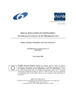
tuberculosis. On follow up, one submitted a sample
for PCR . Figure1 shows that within a period of six
months after being diagnosed as not having
tuberculosis, the PCR profile of the individual had
changed markedly, suggesting an increase in the
bacterial load in the sputum. Only seven of the 17
cases i.e. those who took ATT subsequently were
considered as true cases of tuberculosis, even though
the other six could be rated as highly probable cases
of tuberculosis (Table 1B). Table 2 summarizes the
final diagnosis of tuberculosis made in 71 cases
following a diagnostic review consisting of response
to ATT/ clinical follow up / information from X-
ray/ sputum investigations.
d. PCR data on controls with diseases other than
tuberculosis
PCR, culture examination and smear
microscopy were performed on 21 individuals with
Table 1B: Status of persons negative by smear
and culture but positive by PCR
Table 2: Summary : Final diagnosis of tuberculosis patients
Description Number Considered
true positives
(clinically)
Started ATT subsequently
Not started ATT, but highly
probable
Family contact
Deceased
+
Highly probable
!
Lost to follow up
7
3
1
2
4
7
Total 17 7
+
Immunocompromised with past history of tuberculosis
!
with symptom complex
Tuberculosis patients Diagnostic review
Description Number Culture positive &
response to ATT
Response to ATT and/or positive
on X ray/ microbiological data and
clinical follow up
Final
diagnosis
of TB
patients
Smear positive 36 32 4 36
Smear negative * 29 16 12 28
PCR positive 7 - 7 7
Total 72 48 (67.6%) 23 (32.4%) 71*
*1 smear negative case was false positive which was later determined on the basis of response to ATT and radiological profile after
3 months of treatment initiation and hence not considered in the final tally
COMPARISON OF PCR WITH ROUTINE DIAGNOSTICS72
Indian Journal of Tuberculosis
KAVITA MODI – PAREKH ET AL
respiratory diseases other than tuberculosis (Table
1A serial-c). These cases were as follows, two each
of asthma, pneumonia and cancer lung, one each of
post CABG, COPD and lung abscess and 12 with
cough and cold. All samples from these individuals
had tested negative for tuberculosis by all three
diagnostics. No PCR positivity being the expectation
for this group, the PCR results could be interpreted
as 100% efficient (Table 1A serial-c).
e. Comparative efficiency of PCR to routine
diagnostics like microscopy and culture
Table 3 presents the comparative efficiency
of PCR to diagnostics like microscopy and culture.
The sensitivity, specificity, positive and negative
predictive values for each of the diagnostics was
compared using the gold standards of smear
microscopy, culture, and combined microbiological
data along with chest radiographic findings and
information on clinical follow up. Of the 144 samples,
48 were confirmed on the basis of culture and
response to ATT, while 23 culture negative samples
were confirmed on the basis of response to ATT,
microbiological data and on clinical follow up.
PCR was the most sensitive diagnostic with
a sensitivity of 91.5% as against that of culture
(68%) and microscopy (51%). However, its
specificity was only 86% when compared to sputum
microscopy (100%) and culture (97%) (Table 3).
Comparison of PCR to conventional methods using
McNemars test (
χ
2
= 5.26, df=1, P<0.05) showed a
significant difference.
f. Report on utility of PCR amongst
inappropriate respiratory samples
Saliva mixed sputum specimens constituted
31% (45/144) of all respiratory samples. Thirty-one
of these were negative by all diagnostics while seven
of them were positive by routine diagnostics. Of the
latter, only one was positive by microscopy while
the remaining 6 belonged to individuals diagnosed
on the basis of radiological examination. Four of these
seven samples were also positive by culture and five
MW 2 3
100 bp
200 bp
300 bp
400 bp
500 bp
600 bp
700 bp
800 bp
900 bp
1000 bp
Figure 1: PCR profile of respiratory sample from
suspect at time of first reporting to the
microscopy centre (Lane 2) and after
six months (Lane 3). MW represents
DNA molecular weight marker.
Diagnostic Sensitivity
(%)
Specificity
(%)
Positive
predictive
value
Negative
predictive
value
Efficiency
Microscopy 51
(36/71)
100
(73/73)
100
(36/36)
67.5
(73/108)
76
(109/144)
Culture 68
(48/71)
97
(71/73)
96
(48/50)
75.5
(71/94)
83
(119/144)
PCR 91.5
(65/71)
86
(63/73)
87
(65/75)
91
(63/69)
89
(128/144)
Table 3:Comparative sensitivity, specificity, predictive value and efficiency of PCR to routine diagnostics.
73
Indian Journal of Tuberculosis
by PCR. In addition, seven saliva samples were
positive by PCR only (Table 1B). Thus microscopy,
culture, radiological examination and PCR could
diagnose one, four, six and eight cases respectively
of those individuals whose respiratory samples were
considered as inappropriate for microbiological
processing.
DISCUSSION
The need for an efficient tuberculosis
diagnostic becomes evident from the fact that for
every patient of tuberculosis who can be detected
using microscopy, nine have to be screened using
indirect methods due to the low sensitivity of
microscopy
17
.
This is the primary impetus for a world
wide effort for developing new tools to diagnose
tuberculosis
1
. The use of a molecular technique like
PCR for the laboratory detection of Mycobacteria in
respiratory and other tissue samples from
tuberculosis suspects has thus attracted enormous
attention. The present study demonstrates the utility
and limitations of PCR.
Among the total of 144 specimens studied,
the sensitivity of smear, culture and PCR was 51%,
68% and 91.5% respectively. Smear microscopy was
positive in only 67% of the culture positive samples.
In comparison, PCR was positive in 98% and it could
detect 83% of the smear negative cases that were
only radiologically positive. This aspect has great
potential in the laboratory diagnosis of tuberculosis,
particularly in paucibacillary cases. However, its
overall specificity was only 86% when compared to
smear and culture. PCR was negative in all negative
controls and did not show any cross reactivity with
the two MOTT isolates, which indicate good
specificity of the primers used.
This study has also indicated that PCR
can be a useful tool in those who are not able to
expectorate a proper sputum sample. Out of 45
such samples, PCR was able to detect 12 positives
while the routine diagnostic tests were positive
in only seven. Three of the additional seven
cases detected by PCR were considered as true
positives by the clinicians. In one of the subjects
who had persistent chest symptoms and whose
sample was available for PCR at the time of first
presentation and on follow up after 6 months, a
dramatic increase in bacillary load could be
detected by PCR.
The primary limitation of PCR arises from
the absence of a suitable gold standard to assess its
efficiency. When culture is used as a gold standard
in comparison studies, samples containing non-viable
Mycobacteria may lead to a false positive PCR,
thereby misleading clinicians. The primers MPB64
used in this study proved to be specific and should
hold promise for the future. However, studies with
larger numbers need to be taken up in order to validate
these results. In this context, a recent study examined
the cost-effectiveness of polymerase chain reaction
versus Ziehl-Neelsen smear microscopy for
diagnosis of tuberculosis in a high-burden, resource-
starved environment
18
. The study demonstrated that
costs per correctly diagnosed case were US dollar
41 and dollar 67 for smear microscopy and PCR,
respectively. When treatment costs were included,
including treatment of culture-negative cases, PCR
was found to be most cost-effective at dollar 382
versus dollar 412
18
.
ACKNOWLEDGEMENTS
We would like to thank Dr. A. K. Chakraborty
and the anonymous reviewers for comments and
suggestions on the manuscript, Dr N S Deodhar for
comments and suggestion on the work and Drs. Anil
Ravetkar, former Chief Medical Officer, Pune
Municipal Corporation, Dr. Nagkumar K, Chief
Medical Officer, Pimpri Chinchwad Municipal
Corporation, Dr. Dileep Jagtap, Tuberculosis Control
Program Officer, Dr. H V. Bahulkar and Dr. Rahul
Sakpal for various assistance. We acknowledge
the National Tuberculosis Institute, Bangalore for the
M.tuberculosis H37Rv strain. K.M-P acknowledges
the support from CSIR in the form of a Senior
Research Fellowship.
REFERENCES
1. Foulds J, and O’Brien J. New tools for the diagnosis of
tuberculosis, the perspective of developing countries. Int
J Tuberc Lung Dis 1998; 2: 778-783.
2. Kar A, Modi-Parekh K, and Chakroborty A.K. Advances
COMPARISON OF PCR WITH ROUTINE DIAGNOSTICS74
Indian Journal of Tuberculosis
in tuberculosis diagnostics. Health Administrator 2003;
15: 118-123.
3. Heifets L. Dilemmas and Realities of Rapid Diagnostic
Tests for Tuberculosis. Chest 2000; 118: 4-5.
4. Brisson-Noël A, Gicquel B, Lecossier D et al. Rapid
diagnosis of tuberculosis by amplification of
mycobacterial DNA in clinical samples. Lancet 1989;
4:1069 –71.
5. Claridge JE, Shawar R, Shinnick TM, Plikaytis BB. Large
scale use of polymerase chain reaction in a routine
mycobacteriology laboratory. J Clin Microbiol 1993; 31:
2049-56.
6. Noordhoek GT, Van Embden JD, and Kolk AH. Reliability
of nucleic acid amplification for detection of
Mycobacterium tuberculosis: an international
collaborative quality control study among 30 laboratories.
J Clin Microbiol 1996; 34: 2522-2525.
7. Della-Latta P, and Whittier S. Comprehensive evaluation
of performance, laboratory application, and clinical
usefulness of two direct amplification technologies for
the detection of Mycobacterium tuberculosis complex.
Am J Clin Pathol 1998; 110: 301-310.
8. Wang SX, and Tay L. Evaluation of three nucleic acid
amplification methods for direct detection of
Mycobacterium tuberculosis complex in respiratory
specimens. J Clin Microbiol 1999; 37: 1932-34.
9. Kambashi B, Mbulo G, McNerney R et al. Utility of
nucleic acid amplification techniques for the diagnosis of
pulmonary tuberculosis in sub-saharan Africa. Int J Tuberc
Lung Dis 2001; 5: 364-369.
10. Rattan A. PCR for diagnosis of tuberculosis: Where are
we now? Indian J Tuberc 2000; 47: 79-82.
11. Watterson SA, and Drobniewski FA. Modern laboratory
diagnosis of mycobacterial infections. J Clin Pathol 2000;
53: 727-32.
12. Noordhoek GT, Mulder S, Wallace P, Van Leon AM.
Multicentre Quality Control study for detection of
M.tuberculosis in clinical samples by nucleic amplification
methods. Clin Microbiol Infect 2004; 10: 295-301.
13. MMWR Centers for Disease Control and Prevention.
Update: nucleic acid amplification tests for tuberculosis.
Morb Mortal Wkly Rep 2000; 49: 593–594.
14. Kivihya-Ndugga L, van Cleeff M, Juma E, Kimwomi
J, et al. Comparison of PCR with the routine
procedure for diagnosis of tuberculosis in a population
with high prevalence of tuberculosis and human
immunodeficiency virus. J Clin Microbiol. 2004 Mar;
42(3): 1012-5.
15. Roberts GD. Mycobacteria and Norcardia. In: J. A.
Washington II, editor. Laboratory procedures in clinical
microbiology. New York : Springer Verlag, 1981 p. 365-
406.
16. Dar L, Sharma SK, Bhanu NV, Broor S, Chakraborty M,
Pande JN, Seth P. Diagnosis of pulmonary tuberculosis
by polymerase chain reaction for MPB64 gene: an
evaluation in a blind study. Indian J Chest Dis Allied Sci
1998 Jan-Mar; 40(1): 5-16.
17. Baily GVJ, Savic D, Gothi GD, Naidu VB, Nair SS. Potential
yield of pulmonary tuberculosis cases by direct microscopy
of sputum in a district of South India. Bull World Health
Organ 1967; 37(6): 875-892
18. van Cleeff M, Kivihya-Ndugga L, Githui W, et al.
Cost-effectiveness of polymerase chain reaction
versus Ziehl-Neelsen smear microscopy for diagnosis
of tuberculosis in Kenya. Int J Tuberc Lung Dis 2005
Aug; 9(8): 877-83.
KAVITA MODI – PAREKH ET AL
CHANCHAL SINGH MEMORIAL AWARD - 2006
The Tuberculosis Association of India awards every year a cash
prize of Rs. 1000/- to a medical graduate (non-medical scientists working as
bacteriologists, biochemists, etc, in the field of tuberculosis included) who is
below 45 years of age and is working in tuberculosis, for an original article
not exceeding 30 double spaced foolscap size pages (approximately 6,000
words, excluding charts and diagrams) on tuberculosis. Articles already
published or based on work of more than one author will not be considered.
Papers may be sent, in quadruplicate, to reach the Secretary-General,
Tuberculosis Association of India, 3, Red Cross Road, New Delhi-110001,
before 30th June, 2006.
75
Indian Journal of Tuberculosis
76
What To Do for Quitting Smoking