Báo cáo khoa học: The oleic acid complexes of proteolytic fragments of a-lactalbumin display apoptotic activity pdf
Bạn đang xem bản rút gọn của tài liệu. Xem và tải ngay bản đầy đủ của tài liệu tại đây (455.85 KB, 11 trang )
The oleic acid complexes of proteolytic fragments of
a-lactalbumin display apoptotic activity
Serena Tolin
1
, Giorgia De Franceschi
1
, Barbara Spolaore
1
, Erica Frare
1
, Marcella Canton
2
,
Patrizia Polverino de Laureto
1
and Angelo Fontana
1
1 CRIBI Biotechnology Centre, University of Padua, Italy
2 Department of Experimental Biomedical Sciences, University of Padua, Italy
Introduction
a-Lactalbumin (a-LA) is a small, acidic, Ca
2+
-bind-
ing protein involved in the biosynthesis of lactose,
being a component of the lactose synthase complex
[1]. For a few decades, a-LA has been the subject of
intensive structural investigations, and it has served
as a model system in many protein folding studies
[2,3]. The 123 residue chain of a-LA is organized into
a discontinuous a-helical domain composed of
residues 1–39 and 81–123, and a small b-domain
comprising the rest of the polypeptide chain [4,5]
(Fig. 1). A noteworthy property of a-LA is its ability
to adopt a partly folded or molten globule (MG)
state under a variety of conditions, including low pH.
This state, lacking the specific interactions of the ter-
tiary structure of the native protein, but maintaining
a high degree of secondary structure, has been exten-
sively analyzed with a variety of techniques and
approaches [6–9].
Keywords
apoptosis; HAMLET; oleic acid; protein
fragments; a-lactalbumin
Correspondence
P. Polverino de Laureto, CRIBI
Biotechnology Centre, University of Padua,
Viale G. Colombo 3, 35121 Padua, Italy
Fax: +39 049 827 6159
Tel: +39 049 827 6157
E-mail:
(Received 7 August 2009, revised 9 October
2009, accepted 27 October 2009)
doi:10.1111/j.1742-4658.2009.07466.x
The complexes formed by partially folded human and bovine
a-lactalbumin with oleic acid (OA) have been reported to display selective
apoptotic activity against tumor cells. These complexes were named human
(HAMLET) or bovine (BAMLET) alpha-lactalbumin made lethal to
tumor cells. Here, we analyzed the OA complexes formed by fragments of
bovine a-lactalbumin obtained by limited proteolysis of the protein. Speci-
fically, the fragments investigated were 53–103 and the two-chain fragment
species 1–40 ⁄ 53–123 and 1–40 ⁄ 104–123, these last being the N-terminal
fragment 1–40 covalently linked via disulfide bridges to the C-terminal
fragment 53–123 or 104–123. The OA complexes were obtained by mixing
the fatty acid and the fragments in solution (10-fold and 15-fold molar
excess of OA over protein fragment) or by chromatography of the frag-
ments loaded onto an OA-conditioned anion exchange column and salt-
induced elution of the OA complexes. Upon binding to OA, all fragments
acquire an enhanced content of a-helical secondary structure. All OA com-
plexes of the fragment species showed apoptotic activity for Jurkat tumor
cells comparable to that displayed by the OA complex of the intact pro-
tein. We conclude that the entire sequence of the protein is not required
to form an apoptotic OA complex, and we suggest that the apoptotic
activity of a protein–OA complex does not imply specific binding of the
protein.
Abbreviations
a-LA, a-lactalbumin; BAMLET, bovine a-lactalbumin made lethal to tumor cells; CAC, critical aggregate concentration; HAMLET, human
a-lactalbumin made lethal to tumor cells; MG, molten globule; OA, oleic acid; [h], mean residue ellipticity; TNS, 6-(p-toluidino)-2-
naphthalenesulfonate.
FEBS Journal 277 (2010) 163–173 ª 2009 The Authors Journal compilation ª 2009 FEBS 163
An interesting property of a-LA is its capacity to
interact with membranes and lipid bilayers [10–14], as
well as fatty acids [15]. In particular, a complex
formed by Ca
2+
-depleted a-LA in its partly folded
state with oleic acid (OA) has been extensively studied.
This OA complex, named human a-LA made lethal to
tumor cells (HAMLET), was initially isolated from
human milk, and was shown to selectively induce
apoptosis in tumor and immature cells, but not in
healthy cells [16–19]. It was proposed that the condi-
tions required to induce HAMLET formation in vivo
are those of the stomach of the breastfed child, where
low pH may partially unfold a-LA by releasing its
protein-bound Ca
2+
[19–21], and free OA can be
produced by lipases that hydrolyze milk triglycerides
[22,23]. HAMLET can also be prepared in vitro,by
application of the apo form of a-LA to an anion
exchange column equilibrated with OA and elution of
the OA complex with high salt concentration, followed
by dialysis and lyophilization [18]. It seems that the
formation of the OA complex relies on the fact that
the apo form of a-LA is more hydrophobic than the
holo form, and thus is prone to bind the hydrophobic
fatty acid [20]. The conformational features of a-LA in
the OA complex are those of a protein MG, and upon
binding to the protein, OA can probably stabilize this
altered protein conformation [21]. HAMLET-like com-
plexes with similar biological activity can be obtained
with a-LA from different species, including bovine,
equine, porcine, ovine and caprine a-LA [24]. The OA
complex of bovine a-LA was named bovine a-LA
made lethal to tumor cells (BAMLET) [21]. Moreover,
it was also shown that a-LA mutants with amino acid
replacements at the level of the Ca
2+
-binding loop
were capable of producing active OA complexes [21].
Despite the intensive research on HAMLET and
BAMLET, the molecular features of the active OA
complex in terms of protein ⁄ fatty acid stoichiometry
and monomeric ⁄ oligomeric state of the protein in the
complex are still not clarified, and are a matter of
debate in the current literature [20,25–27]. The molecu-
lar mechanism of interaction and physicochemical
properties of the OA complex are not understood, and
neither are the mechanisms and cellular events
involved in the toxicity of HAMLET. It was shown by
using labeled a-LA that the OA complex is able to
14053
104
1
40
53
103
1 123
H1
5–11
h1b
23–34
S1 S2 S3
H2
h2
86–98 105–110
h3c
H3 H4
123
123
(1−40/53−123)/OA
NaCl molarity ( )
0.0
0.5
1.0
BAMLET
(53−103)/OA
0.0
0.5
1.0
(1−40/104−123)/OA
Volume (mL)
0 20 40 60 80 0 20 40 60 80
020406080020406080
Relative absorbance (
___
)
α-LA
EDTA
EDTA
EDTA
EDTA
Aggregates
a
A
B
c
b
d
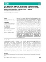
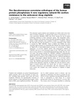
Fig. 1. (A) Top: scheme of the secondary
structure of the 123 residue chain of a-LA
[4]. The four a-helices (H1–H4) are indicated
by large boxes, and the corresponding chain
segments are given above them. The three
b-strands (S1, 41–44; S2, 47–50; S3, 55–56)
and the 3
10
helices (h1b, 18–20; h2, 77–80;
h3c, 115–118) are indicated by small boxes.
Bottom: schematic representation of the
three a-LA fragments investigated. The four
disulfide bridges (6–120, 28–111, 61–77,
and 73–91) are represented by thin lines,
and the gray box indicates the segment
encompassing the Ca
2+
-binding loop. (B)
Preparation of the OA complexes of a-LA (a)
and its fragments (b–d) by chromatography
on an OA-conditioned anion exchange col-
umn [18]. The protein material was applied
to a DEAE-Trisacryl M column conditioned
with OA, and the OA complexes were
eluted with a gradient of 1
M NaCl (dashed
lines). The solid bars indicate the fractions
of the effluent from the column that were
collected for further studies.
Oleic acid complexes of a-lactalbumin fragments S. Tolin et al.
164 FEBS Journal 277 (2010) 163–173 ª 2009 The Authors Journal compilation ª 2009 FEBS
bind to tumor cells and accumulate in the cell nuclei
[28]. These authors identified specific histone proteins
as nuclear targets for HAMLET. However, it was also
shown that a-LA in the absence of OA can interact
with histones and charged, disordered poly-a-amino
acids (i.e. poly-Lys and poly-Arg). This a-LA interac-
tion was shown to be driven by electrostatic forces
[29,30]. Therefore, the active species in the cell may be
the whole protein–fatty acid complex, the protein
alone, or even the OA by itself. In this last case, the
fatty acid aggregation state should be considered, as it
can be significantly influenced by the presence of a
protein in the same solution.
In this study, we analyzed the propensity of three
fragment species of bovine a-LA, obtained by limited
proteolysis of the protein [9,31,32], to bind OA and to
form biologically active OA complexes. As shown in
Fig. 1, the fragments have different structural charac-
teristics, with fragment 1–40 ⁄ 104–123 encompassing
three of the four a-helices of the native protein, frag-
ment 53–103 containing the chain segment that binds
Ca
2+
in the native protein, and fragment 1–40 ⁄ 53–123
being able to adopt, at neutral pH, an MG conforma-
tion resembling that of the MG conformation adopted
by intact a-LA at pH 2.0 [31,32]. The conformational
properties of the OA complexes of these fragments were
analyzed by far-UV CD measurements, and it was
shown that the fragments acquire an enhanced content
of a-helical secondary structure upon binding OA. The
physical and aggregation state of OA at physiological
pH was analyzed by fluorescence and turbidimetric
analyses. It was shown that the fragments, as well as
the entire protein, depress the critical concentration for
aggregate formation [critical aggregate concentration
(CAC)] of OA and induce the formation of small and
water-soluble OA aggregates. All OA complexes dis-
played apoptotic activity for tumor cells, and the extent
of their activity was comparable to that observed with
the OA complex of the intact protein, i.e. BAMLET.
Our results indicate that the entire 123 residue chain of
a-LA is not required for forming a cytotoxic OA com-
plex, and raise the possibility that the cell-damaging
effects of the various OA complexes could result from
an enhanced solubility of the otherwise poorly soluble
and inherently toxic fatty acid [33].
Results
Preparation of complexes of a-LA fragments
with OA
Two procedures were followed to prepare the
OA–fragment complexes, namely by simple mixing the
two components in solution, or by chromatography
using an OA-conditioned anion exchange column, as
described by Svensson et al. [18] for the preparation of
HAMLET. The two procedures were used here, as it is
not clear whether a mixing procedure results in a less
active or inactive complex [18,20,34]. Instead, we [35]
and others [26,36,37] have shown that it is indeed pos-
sible to prepare an OA complex displaying similar
structural properties to HAMLET or BAMLET, i.e.
to an OA complex prepared by chromatography. Nev-
ertheless, here we preferred to use and compare both
procedures in preparing the OA complexes.
Bovine a-LA and its fragments were loaded on an
anion exchange column conditioned with OA. The
chromatographic profile obtained with intact a-LA
was similar to that previously reported [18]. Salt and
EDTA were eluted first from the column. The free
protein was eluted from the column at low salt concen-
tration, whereas the OA complex was eluted at 1 m
salt. The three fragments strongly bound to the
OA-conditioned matrix, and their OA complexes could
be eluted by high salt (Fig. 1B). The amounts of pro-
tein fragment in the eluted OA complex, calculated on
the basis of the material loaded onto the column, were
50% for fragments 1–40 ⁄ 53–123 and 1–40 ⁄ 104–123,
and 25% for fragment 53–103, as estimated from
UV absorption measurements. This indicated that a
proportion of the protein fragment material remained
bound to the column. In the case of fragment 53–103,
aggregated species were eluted at a higher retention
time than that of the OA–fragment 53–103 complex.
Aggregation of the fragment was deduced from the
turbidity of the last eluted fraction (Fig. 1Bd). This
would account for the low recovery of soluble OA
complex of fragment 53–103. For the sake of compari-
son, the OA complexes were also prepared in solution
by direct mixing of the a-LA fragments with OA at a
molar ratio of 1 : 10 or 1 : 15 (see below).
Conformational properties of protein fragment
complexes with OA
The conformational properties of the OA complexes
formed by a-LA fragments prepared by chromatogra-
phy or by direct mixing in solution were analyzed by
far-UV CD spectroscopy in NaCl ⁄ P
i
(pH 7.4).
Figure 2A shows the far-UV CD spectra of fragment
1–40 ⁄ 104–123 in the presence of increasing concentra-
tions of OA. The CD spectrum of this fragment
appeared to be that of a largely disordered polypep-
tide, but upon addition of OA the spectrum acquired
the characteristics of a-helical secondary structure. It is
of interest that, in the presence of OA (protein ⁄ fatty
S. Tolin et al. Oleic acid complexes of a-lactalbumin fragments
FEBS Journal 277 (2010) 163–173 ª 2009 The Authors Journal compilation ª 2009 FEBS 165
acid molar ratio of 1 : 10), the CD spectrum of frag-
ment 1–40 ⁄ 104–123 was quite similar to that of the
corresponding OA complex prepared by chromato-
graphy, implying that the OA complexes prepared by
the two procedures displayed similar conformational
features. Analogous conformational effects of OA
binding were observed with fragment 53–103 in the
presence of 15 equivalents of OA (Fig. 2B). Thus, frag-
ment species 1–40 ⁄ 104–123 and 53–103 both appeared
to be rather disordered in solution at pH 7.4, but upon
binding OA they acquired a folded structure character-
ized by a significant content of a-helical structure, as
the OA complexes displayed far-UV CD spectra with
the typical minima of ellipticity at 208 and 222 nm of
the a-helical secondary structure [38].
The fragment species 1–40 ⁄ 53–123 comprises almost
all of the 123 residue chain of a-LA (Fig. 1), and
adopted a significantly folded structure in solution, as
shown by the characteristics of its far-UV CD spec-
trum (Fig. 2C). In this case, the addition of OA
induced a conformational change, but not as dramatic
as seen with the other two fragments. Here, we used
fragment solutions devoid of Ca
2+
, because we have
previously shown that the conformational features of
fragments 53–103 and 1–40 ⁄ 53–123, containing the
Ca
2+
-binding loop of the intact protein (Fig. 1), are
affected by Ca
2+
[31,32].
Determination of the aggregation state of OA
The phase behavior of OA is strongly dependent on
pH and fatty acid concentration [39]. In NaCl ⁄ P
i
(pH
7.4), OA forms oil droplets and vesicles of variable size
[40–42]. To understand the effect of protein fragments
on OA aggregation state, we measured the OA CAC.
We use this term because a complete morphological
characterization of the aggregate state of OA is not yet
available. The CAC of OA at pH 7.4 was estimated by
using the fluorescent dye 6-(p-toluidino)-2-napthalene-
sulfonate (TNS) [43]. In NaCl ⁄ P
i
(pH 7.4), the CAC of
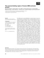
OA was calculated as 19.8 ± 0.3 lm (Fig. 3A, insert).
This value is similar to that previously determined for
OA [40]. The same measurements using TNS were con-
ducted in the presence of a-LA (Fig. 3A) or its frag-
ments (Fig. 3B). All protein species were able to
reduce by 20-fold the CAC value of OA. Estimated
values of the CAC of OA were 0.94 ± 0.24 lm in the
presence of a-LA, and 0.86 ± 0.29, 1.22 ± 0.24 and
0.93 ± 0.56 lm, respectively, in the presence of frag-
ment species 1–40 ⁄ 53–123, 1–40 ⁄ 104–123 and 53–103.
We also conducted turbidity analysis of OA solu-
tions and mixtures, as this method is often used for
measuring the critical vesicular concentration of lipids
[44]. Figure 3C shows the variation of absorbance (A)
at 400 nm of samples containing increasing amounts
of OA in the absence or presence of a-LA. The strik-
ing observation deriving from these measurements is
that the added protein was able to completely inhibit
the formation of large aggregates that caused light
scattering at 400 nm (Fig. 3C, open circles). Fragment
species 1–40 ⁄ 53–123 and 1–40 ⁄ 104–123 were also able
to similarly depress the OA aggregation. Fragment
53–103 also caused a reduction in the aggregation of
OA, but to a minor extent (Fig. 3D).
–15
–10
–5
0
5
[θ] x 10
–3
(deg·cm
2
·dmol
–1
)
–15
–10
–5
0
5
1 : 1
1 : 3
1 : 5
1 : 7
1 : 10
1 : 15
by column
1−40/104−123
190 210 230 250
190 210 230 250
190 210 230 250
–15
–10
–5
0
5
(53−103)/OA
(by mix 1 : 15)
(53−103)/OA
(by column)
53−103
B
A
C
(1−40/53−123)/OA
(by column)
1−40/53−123
Wavelength (nm)
(1−40/53−123)/OA
(by mix 1 : 15)
Fig. 2. Far-UV CD spectra of a-LA fragments in NaCl ⁄ P
i
(pH 7.4). (A) Far-UV CD spectra of fragment 1–40 ⁄ 104–123 in the absence (dotted
line) or presence (continuous line) of increasing amounts of OA. Numbers near the CD spectra refer to fragment ⁄ OA molar ratios of 1 : 1,
1 : 3, 1 : 5, 1 : 7, 1 : 10, and 1 : 15. The CD spectrum of the OA complex of the fragment obtained by chromatography is also shown
(dashed line). (B) CD spectra of the OA complex of fragment 53–103 obtained by chromatography (dashed line) or by mixing in solution
(continuous line). The spectrum of the OA free fragment is reported as a reference (dotted line). (C) CD spectra of fragment 1–40 ⁄ 53–123
(dotted line) and its OA complex obtained by chromatography (dashed line) and by mixing the fragment and OA in solution at a fragment ⁄
OA molar ratio of 1 : 15 (continuous line).
Oleic acid complexes of a-lactalbumin fragments S. Tolin et al.
166 FEBS Journal 277 (2010) 163–173 ª 2009 The Authors Journal compilation ª 2009 FEBS
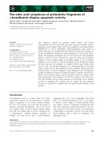
Cellular toxicity
The ability of the OA complexes of the a-LA frag-
ments to induce apoptosis-like cell death was exam-
ined. Assays were conducted on Jurkat cells, using the
OA complexes prepared by direct mixing or by chro-
matography using an OA-conditioned column (see
Experimental procedures). Cells treated with the
fragment–OA complexes suffered considerable loss of
viability through apoptosis-like death, whereas the
OA-free fragments displayed negligible toxicity
(Fig. 4). The OA complexes of the various fragment
species were also tested at a fragment ⁄ OA molar ratio
of 1 : 3, a condition that caused only a slight confor-
mational change in the fragment’s secondary structure,
as deduced from far-UV CD spectra. In this case, the
OA complexes did not display cellular toxicity (not
shown). The extent of apoptotic activity of the OA
complexes of the fragments was comparable to that
observed with the OA complex of the intact protein
prepared by chromatography, i.e. BAMLET, or by
mixing the intact protein with 15 equivalents of OA in
solution. In the absence of added protein species, OA
alone and at the concentration used for formation of
OA complexes displayed negligible toxicity, similarly
to NaCl ⁄ P
i
or the control sample (culture medium).
Conversely, as previously shown, OA can be toxic to
Jurkat cells via an apoptosis mechanism at higher con-
centrations and with a much longer duration of incu-
bation [33].
Fragment 1–40 ⁄ 104–123 is a two-chain species cross-
linked by the two disulfide bridges 6–120 and 28–123
of intact a-LA. Reduction of this fragment with
tris(2-carboxyethyl)phosphine, followed by S-alkylation
with iodoacetamide and RP-HPLC chromatography,
allowed us to prepare the single-chain, S-carboxami-
domethylated fragments 1–40 and 104–123. The inter-
action of OA with these fragments was monitored by
far-UV CD measurements. As shown in Fig. S1, OA
induced a-helical secondary structure in both frag-
ments, which were otherwise largely unfolded in the
absence of the fatty acid. It is of note that fragment
1–40 encompasses helix H1 (5–11) and helix H2
(23–34), and fragment 104–123 encompasses helix H4
(105–110), in native a-LA [4]. The OA complexes of
the two fragments, as obtained by mixing them with
15 equivalents of the fatty acid, displayed significant
apoptotic activity on Jurkat cells (Fig. S1, bottom). It
is of note that the OA–fragment 104–123 complex was
even more active than BAMLET, i.e. the OA–a-LA
complex prepared by column chromatography (see
Experimental procedures). Therefore, OA complexes of
Relative fluorescence
1.0
1.2
1.4
1.6
1.8
[OA] μM
0 20 40 60 80 100
Rel. fluorescence
0.8
1.0
1.2
1.4
1.6
1.8
[OA] (μM)
AB
D
02468100 2 46 810
0 100 200 300 400 500
0 100 200 300 400 500
A
400 nm
0.0
0.1
0.2
0.3
C
Fig. 3. Characterization of the physical state
of OA solutions by TNS fluorescence emis-
sion (A, B) and turbidity (C, D). All measure-
ments were conducted in NaCl ⁄ P
i
(pH 7.4),
in the absence (d) or presence of a-LA (s),
fragment 1–40 ⁄ 53–123 (
), fragment 1–
40 ⁄ 104–123 (n), or fragment 53–103 ()).
(A, B) Aliquots of OA (from 0 to 10 l
M)
were added to a solution of TNS (20 l
M),
and the intensity of fluorescence emission
at 460 nm was recorded, after excitation at
360 nm. The CAC is defined as the lipid
concentration at which the two linear por-
tions of the lines of fluorescence intensities
intersect [61]. The TNS fluorescence of OA
(up to 100 l
M) solutions was also measured
in the absence of intact a-LA (A, insert). (C,
D) Turbidimetric analysis of OA solutions in
the absence (filled circles) or presence of
10 l
M protein (open circles) or fragment
species (symbols as above). Measurements
of absorbance were conducted at 400 nm
on samples containing OA up to 500 l
M.
S. Tolin et al. Oleic acid complexes of a-lactalbumin fragments
FEBS Journal 277 (2010) 163–173 ª 2009 The Authors Journal compilation ª 2009 FEBS 167
peptide fragments even shorter than the three frag-
ments shown in Fig. 1 can display cellular toxicity.
Discussion
The data presented here indicate a strong mutual inter-
action between OA and a-LA fragments. Indeed, the
addition of OA induces enhancement of secondary
structure of the fragment species, and these signifi-
cantly modify the physical state of the fatty acid in
solution. The increase in the a-helical structure in the
fragment species upon addition of OA (Fig. 2) derives
from the fact that fatty acids, lipids and detergents can
provide a hydrophobic environment that is able to
induce and stabilize secondary structure of polypep-
tides [45–47]. The interaction of protein species with
OA is also simply shown by the fact that a turbid OA
suspension in water at neutral pH becomes clear after
the addition of protein ⁄ fragment species.
The OA complexes of the a-LA fragments have been
prepared by direct mixing in solution and by the chro-
matographic method of Svensson et al. [18], utilizing
an OA-conditioned anion exchange column, followed
by extensive dialysis of the OA complex eluted from
the column at high salt concentration, and then lyoph-
ilization. As the conformational and biological proper-
ties of the OA complexes prepared here by the two
methods are very similar, we consider the mixing
procedure in solution to be suitable, being easier,
reproducible and less cumbersome than the chromato-
graphic one. Furthermore, we have previously shown
that the mixing procedure can be effectively utilized
for the preparation of an a-LA–OA complex with
comparable structural features to BAMLET [35], and
other reports have more recently documented its
successful use [26,36,37].
The phase behavior of OA is critically dependent on
the ionization degree of the fatty acid, and is thus
affected by the pH and ionic strength of the aqueous
solution [40–42]. In NaCl ⁄ P
i
(pH 7.4), OA forms
aggregates of different sizes (diameter 25–250 nm), as
deduced by transmission electron microscopy (not
shown). The a-LA fragment species, as well as the
intact protein, strongly affect these structures. Both
turbidimetric and transmission electron microscopy
analyses show the disappearance of the large, aggre-
gated structures, and by means of fluorescence emis-
sion measurements, a 20-fold decrease in CAC was
found, indicating the formation of smaller aggregates
at a lower OA concentration in the presence of protein
species. As the formation of OA micelles requires the
complete ionization of the fatty acid molecules, and
micelles are formed at pH > 9 [39], it is likely that,
under the experimental conditions described here,
small vesicles or oil droplets are induced in OA in the
presence of the protein or its fragments.
A depression of the CAC of anionic detergents simi-
lar to that observed here with OA aggregates was
reported to occur also with other proteins, and this
effect was explained by considering both electrostatic
1−4
0
/53−123
(1
−
4
0
/104−123/OA) by column
(1−
4
0
/53
−1
2
3)/OA
by mix
(1
−
4
0
/53−123)/OA by column
(1−4
0
/104−123)/OA b
y
mix
1−4
0
/104−
1
2
3
(53−1
0
3)/OA
b
y
mix
α-L
A
/OA by
mix
5
3
−
1
0
3
(53
−1
0
3)/OA
b
y
column
BAMLET
α-L
A
OA
NaCl/P
i
CT
0
20
40
60
80
100
Late apoptosis
Early apoptosis
Cell death (%)
Fig. 4. Cytotoxicity of OA complexes. Jur-
kat cells (10
6
cells per mL) were incubated
at 37 °C with the OA complexes of a-LA or
its fragments prepared by column chroma-
tography or direct mixing in solution (see
Experimental procedures). All protein ⁄ frag-
ment samples were tested at 7 l
M.Asa
control, OA was tested at 100 l
M. After
incubation for 6 h, cell death by apoptosis
was evaluated by appropriate changes of
nuclei stained with Hoechst-33258 (early
apoptotic cells, gray bars) and propidium
iodide (late apoptotic ⁄ necrotic cells, open
bars). The test was also conducted on a-LA,
OA, NaCl ⁄ P
i
, and the medium (CT) as a con-
trol. Data are shown as percentage of BAM-
LET activity. Values are means ± standard
deviation of at least three experiments.
Oleic acid complexes of a-lactalbumin fragments S. Tolin et al.
168 FEBS Journal 277 (2010) 163–173 ª 2009 The Authors Journal compilation ª 2009 FEBS
and hydrophobic interactions [48–50]. A reduction of
the electrostatic repulsion between negative charges at
the surface of the detergent aggregates and positively
charged amino acid side chains of a protein allows the
formation of aggregates at a lower concentration.
However, considering that the apo form of a-LA is
negatively charged, it may well be that the interaction
with OA aggregates of this protein in its Ca
2+
-free
form is mostly mediated by hydrophobic interactions,
as the apo-a-LA is more hydrophobic than the holo
form [51,52]. Nonetheless, even the negatively charged
protein molecule may possess positively charged clus-
ters or areas that mediate the interaction with the neg-
atively charged head groups of OA aggregates, as, for
example, indicated by the fact that the negatively
charged a-synuclein contributes to CAC depression of
anionic surfactants [48]. In the 123 residue chain of
a-LA at neutral pH, Lys and Arg residues are clus-
tered at the level of helical segments A, C and D of
the native protein, whereas the central region of the
protein contains many negatively charged carboxylates
of Asp and Glu residues. Therefore, it could be that
fragment 53–103 interacts with the negatively charged
OA aggregates less effectively than the other fragments
investigated here, as shown by the results of turbidi-
metric analyses (Fig. 3D).
The mechanisms of biological activity of the OA
complexes of a-LA are not yet understood and, in fact,
a variety of diverse biological effects have been
described for HAMLET [20,25,27]. For this reason,
HAMLET was metaphorically named ‘Hydra’ [25].
Besides the cytotoxicity via an apoptotic mechanism,
an OA complex of human a-LA was shown to also
possess bactericidal activity against Streptococcus pneu-
moniae and Haemophilus influenzae [53]. It is of interest
that digestion of a-LA with trypsin and chymotrypsin
yields three peptides displaying bactericidal activity
against Gram-positive bacteria. These bactericidal spe-
cies are peptide 1–5 and the two-chain peptides linked
by a disulfide bridge, 17–31 ⁄ 109–114 and 61–68 ⁄ 75–80
[54]. However, the structural features responsible for
their bactericidal action were not clarified. Probably,
bactericidal action of the LA–OA complex requires a
different molecular mechanism than that occurring in
apoptosis. Hence, HAMLET-like complexes can be
detrimental by various cellular pathways, and exert
their actions by different molecular mechanisms.
The proteolytic fragments of a-LA investigated here
have widely differing chain lengths and amino acid
sequences (Fig. 1), and it therefore does not seem
possible to explain their cytotoxicity in terms of their
specific structural features. The variability in struc-
ture of the polypeptide chain in forming active OA
complexes seems to indicate instead that a generic poly-
peptide chain can eventually interact with OA, and thus
that the toxic action of an OA complex resides in the
fatty acid rather than in the protein moiety. The pres-
ent results show that OA displays new physicochemical
and aggregation properties in the presence of a-LA or
its fragments. With a decrease in the CAC of the fatty
acid in the presence of the protein or its fragments,
soluble and smaller aggregates of protein–OA or frag-
ment–OA complexes are easily formed and stabilized.
In previous studies, the tumor-selective cytotoxicity
of HAMLET or BAMLET was correlated with the
conformational properties of a-LA upon formation of
the OA complex [17,18]. In particular, it was proposed
that the fatty acid acts as a stabilizer of a partially
folded or MG conformation of the protein under phys-
iological conditions [21]. In a very recent paper, it was
reported that a recombinant mutant a-LA with all
eight Cys residues replaced by Ala residues (named all-
Ala mutant), and thus devoid of the four disulfide
bridges of the native protein, formed a cytotoxic OA
complex equivalent to HAMLET [55]. Even if the con-
formation of the all-Ala mutant at neutral pH is simi-
lar to the MG of a-LA at low pH [6–9], the addition
of OA to the all-Ala protein is required in order to
form a cytotoxic species, indicating that the fatty acid
is needed for the development of cytotoxicity [55].
Here, we show that a variety of a-LA fragments can
mimic the action of the entire 123 residue chain of the
protein in forming OA complexes displaying cytotoxic-
ity. A reasonable deduction from this and previous
studies is that the protein ⁄ peptide moiety can act as a
carrier of the inherently toxic fatty acid [33], and there-
fore that OA itself is the active species of a cytotoxic
protein ⁄ peptide complex. This view is in line with the
fact that all variants of a-LA of human, bovine,
equine, porcine and caprine origin, as well as recombi-
nant mutants of a-LA devoid of Ca
2+
-binding proper-
ties, were all able to form HAMLET-like complexes
with little difference in biological activity [21,24]. Inter-
estingly, it was recently reported that the OA complex
of lysozyme displays cellular toxicity similar to that of
HAMLET [56]. In our laboratory, we have performed
initial experiments indicating that even the 153 residue
chain of apomyoglobin can form cytotoxic complexes
when combined with OA [57].
In summary, the results of this study indicate that,
besides substantial variation in amino acid sequence of
the polypeptide chain of a-LA, severe truncation of
the polypeptide chain of the protein is also tolerated in
the formation of biologically active OA complexes.
Therefore, we are inclined to conclude that the poly-
peptide moiety can serve mainly as a carrier of the
S. Tolin et al. Oleic acid complexes of a-lactalbumin fragments
FEBS Journal 277 (2010) 163–173 ª 2009 The Authors Journal compilation ª 2009 FEBS 169
fatty acid. We have shown here that the addition of a
protein ⁄ fragment species strongly influences the aggre-
gation behavior of OA, in particular making it more
water-soluble and thus enhancing its intrinsic apoptotic
effects [33]. Nevertheless, we cannot exclude the possi-
bility that the protein itself can act in a synergic way
in the observed cytotoxicity of the OA complexes. This
could be particularly true in the case of OA complexes
of a-LA, considering that: (a) a-LA itself can display
an inherent apoptotic activity [58,59]; (b) a-LA alone
can interact with histones at the cellular level and thus
display cytotoxic effects [29,30]; and (c) even various
fragments of a-LA have been shown to have bacterici-
dal activity, and a-LA fragments can therefore be toxic
[54]. Despite these caveats regarding the specific role of
the protein moiety in HAMLET-like or BAMLET-like
species, it should be emphasized that the beneficial
effects of these OA complexes in selective killing a
variety of tumor cells appear to be remarkable and will
prompt additional studies on OA–protein ⁄ peptide
complexes as possible new anticancer agents.
Experimental procedures
Materials
Bovine a-LA and DEAE-Trisacryl M resin were purchased
from Sigma (St Louis, MO, USA); OA and the fluorescent
dye TNS were from Fluka (Buchs, Switzerland). All other
chemicals were of analytical reagent grade and were Sigma
or Fluka products.
Preparation of the OA complexes of a-LA
fragments
The a-LA fragments investigated, 1–40 ⁄ 53–123, 1–40 ⁄ 104–
123 and 53–103 (Fig. 1A), were produced by limited prote-
olysis of the protein with pepsin at pH 2.0 [9,31]. The OA
complexes of a-LA and its fragments were prepared follow-
ing two procedures, column chromatography and mixing in
solution.
Column chromatography
The protein material was loaded onto an OA-conditioned
anion exchange chromatographic column (1.0 · 7.0 cm),
following the procedure reported by Svensson et al. [18]. A
DEAE-Trisacryl M resin was employed, equilibrated with
10 mm Tris ⁄ HCl and 0.1 m NaCl (pH 8.5). An aliquot of
the protein or fragment material ( 2mgÆmL
)1
) was dis-
solved in 10 mm Tris ⁄ HCl (pH 8.5), containing 1 mm
EDTA, and then loaded onto the anion exchange column,
which was eluted with a gradient of 10 mm Tris ⁄ HCl and
1 m NaCl (pH 8.5). The absorbance of the effluent from
the column was monitored at 214 nm. The high-salt eluates
from the column containing the OA complexes were desalt-
ed by dialysis against water, using a membrane of 3.5 kDa
cut-off, and then lyophilized.
Mixing in solution
The OA complexes were prepared by direct mixing of pro-
tein species with 10 or 15 equivalents of OA dissolved
(20 mgÆmL
)1
), in ethanol and then diluted with NaCl ⁄ P
i
(8 mm Na
2
HPO
4
, 137 mm NaCl, 2 mm KH
2
PO
4
, 2.7 mm
KCl, pH 7.4) [35]. The fatty acid was added to the protein
solution, and the mixture was analyzed after 1 h of incuba-
tion in the dark.
CD spectroscopy
CD spectra were recorded on a Jasco J-710 spectropolarim-
eter (Tokyo, Japan). The spectra were recorded in NaCl ⁄ P
i
(pH 7.4), in the absence or presence of OA, at a pro-
tein ⁄ fragment concentration of 0.05–0.1 mgÆmL
)1
, using
1 mm quartz cells. The interaction of OA with protein spe-
cies was followed by far-UV CD measurements by adding
aliquots of an OA solution to the protein fragment samples
(10 lm) in NaCl ⁄ P
i
(pH 7.4). Mean residue ellipticity [h]is
reported as degÆcm
2
Ædmol
)1
. Protein fragment concentra-
tions were determined by absorption measurements at
280 nm on a double-beam Lambda-25 spectrophotometer
(Perkin-Elmer, Norwalk, CT, USA). The molar extinction
coefficients at 280 nm for a-LA fragments were
1.22 mg
)1
Æcm
)1
for fragment 53–103, 2.89 mg
)1
Æcm
)1
for
fragment 1–40 ⁄ 104–123, and 2.23 mg
)1
Æcm
)1
for fragment
1–40 ⁄ 53–123, as calculated according to Gill and von
Hippel [60].
Determination of the aggregation state of OA
The CAC of OA was determined by using the fluorescent
dye TNS [43]. The TNS (20 lm) fluorescence emission at
460 nm, after excitation at 360 nm, was measured at 25 °C
in the presence of increasing concentrations of OA in
NaCl ⁄ P
i
(pH 7.4). The analyses were conducted in the
absence or presence of a-LA or its fragments at 10 lm.
Three readings were taken, and the average fluorescence
intensities relative to blanks were plotted. The first and the
last five data points were joined separately by statistically
fitted straight lines. The CAC is defined as the lipid concen-
tration at which the two linear portions of the fluorescence
emission intensity lines intersect [61]. The aggregation state
of OA, in the absence or presence of a-LA or its fragments
(10 lm), was also analyzed by turbidity measurements at
400 nm of different samples containing increasing amounts
of OA (from 0 to 500 lm) in NaCl ⁄ P
i
(pH 7.4) [44].
Oleic acid complexes of a-lactalbumin fragments S. Tolin et al.
170 FEBS Journal 277 (2010) 163–173 ª 2009 The Authors Journal compilation ª 2009 FEBS
Apoptosis assays
Cell culture T-lymphoblastoid Jurkat cells were cultured in
RPMI-1640 medium supplemented with 10% heat-inacti-
vated fetal bovine serum, 2 mm glutamine, 100 IUÆmL
)1
penicillin and 100 lgÆmL
)1
streptomycin in 5% CO
2
⁄ 95%
air at 37 °C. The Jurkat cells (10
6
cells per mL) were incu-
bated with the protein ⁄ fragment samples in serum-free
medium for 6 h at 37 °C. These samples were tested at
7 lm, and the OA complexes were prepared by mixing 10
molar equivalents of OA for fragment 1–40 ⁄ 104–123 and
15 equivalents for fragments 1–40 ⁄ 53–123 and 53–103, as
well as intact a-LA. In order to assess cell viability, Jurkat
cells were stained with 10 lm Hoechst-33258 and 1 lm pro-
pidium iodide for 5 min, in order to allow visualization of
early and late apoptotic ⁄ necrotic cells, respectively. Cells
were then washed with Hanks’ balanced salt solution, and
visualized with an Olympus IMT-2 inverted microscope
equipped with a xenon lamp and a 12-bit digital, cooled,
charge-coupled device camera (Princeton Instruments,
Monmouth Junction, NJ, USA). Excitation ⁄ emission cubes
of 340 ⁄ 440 ± 25 nm and a 568 ⁄ 585 ± 25 nm long-pass fil-
ter were used for Hoechst-33258 and propidium iodide,
respectively. Three randomly selected fields were acquired
for each treatment. The corresponding bright field images
were also acquired, and the three channels were overlaid
using the appropriate function of metamorph software
(Universal Imaging, West Chester, PA, USA). The percent-
age of cell death and the standard deviation were calculated
from three acquisitions of each treatment. The data are
reported as percentage of BAMLET activity.
Acknowledgements
We gratefully acknowledge the financial support of the
Italian Ministry of University and Research (MIUR)
through PRIN-2004, PRIN-2006 and the FIRB Project
on Protein Misfolding and Aggregation (Project No.
RBNEOPX83). We thank M. Zambonin for his excel-
lent technical assistance. This work was presented at
the Symposium on HAMLET (12–14 May 2009, Lund,
Sweden) and at the XXI Symposium of the Protein
Society (21–25 July, 2007, Boston, MA, USA) [Protein
Sci 16 (Suppl. 1), Commun. 257].
References
1 Hill RL & Brew K (1975) Lactose synthetase. Adv
Enzymol 43, 411–490.
2 Kuwajima K (1996) The molten globule state of
a-lactalbumin. FASEB J 10, 102–109.
3 Fink AL (1995) Compact intermediate states in protein
folding. Annu Rev Biophys Biomol Struct 24, 495–522.
4 Pike AC, Brew K & Acharya KR (1996) Crystal struc-
tures of guinea-pig, goat and bovine alpha-lactalbumin
highlight the enhanced conformational flexibility of
regions that are significant for its action in lactose syn-
thase. Structure 4, 691–703.
5 Permyakov EA & Berliner LJ (2000) Alpha-lactalbu-
min: structure and function. FEBS Lett 473, 269–274.
6 Dolgikh DA, Gilmanshin RI, Brazhnikov EV, Bychk-
ova VE, Semisotnov GV, Venyaminov SY & Ptitsyn
OB (1981) a-Lactalbumin: compact state with fluctuat-
ing tertiary structure?. FEBS Lett 136, 311–315.
7 Alexandrescu AT, Evans PA, Pitkeathly M, Baum J &
Dobson CM (1993) Structure and dynamics of the acid-
denatured molten globule state of alpha-lactalbumin: a
two-dimensional NMR study. Biochemistry 32, 1707–
1718.
8 Ptitsyn OB (1995) Molten globule and protein folding.
Adv Protein Chem 47, 83–229.
9 Polverino de Laureto P, De Filippis V, Di Bello M,
Zambonin M & Fontana A (1995) Probing the molten
globule state of alpha-lactalbumin by limited proteoly-
sis. Biochemistry 34, 12596–12604.
10 Ban
˜
uelos S & Muga A (1995) Binding of molten glob-
ule-like conformations to lipid bilayers. Structure of
native and partially folded alpha-lactalbumin bound to
model membranes. J Biol Chem 270, 29910–29915.
11 Cawthern KM, Permyakov E & Berliner LJ (1996)
Membrane-bound states of alpha-lactalbumin: implica-
tions for the protein stability and conformation. Protein
Sci 5, 1394–1405.
12 Grishchenko VM, Kalinichenko LP, Deikus GY,
Veprintsev DB, Cawthern KM, Berliner LJ & Permya-
kov EA (1996) Interactions of alpha-lactalbumins with
lipid vesicles studied by tryptophan fluorescence.
Biochem Mol Biol Int 38, 453–466.
13 Halskau O, Froystein NA, Muga A & Martinez A
(2002) The membrane-bound conformation of a-lactal-
bumin studied by NMR-monitored
1
H exchange. J Mol
Biol 321, 99–110.
14 Agasøster AV, Halskau O, Fuglebakk E, Frøystein NA,
Muga A, Holmsen H & Martınez A (2003) The interac-
tion of peripheral proteins and membranes studied with
a-lactalbumin and phospholipid bilayers of various
compositions. J Biol Chem 278, 21790–21797.
15 Cawthern KM, Narayan M, Chaudhuri D, Permyakov
EA & Berliner LJ (1997) Interactions of alpha-lactalbu-
min with fatty acids and spin label analogs. J Biol Chem
272, 30812–30816.
16 Ha
¨
kansson A, Zhivotovsky B, Orrenius S, Sabharwal
HR & Svanborg C (1995) Apoptosis induced by a
human milk protein. Proc Natl Acad Sci USA 92, 8064–
8068.
17 Svensson M, Sabharwal H, Ha
¨
kansson A, Mossberg
AK, Lipniunas P, Leffler H, Svanborg C & Linse S
(1999) Molecular characterization of a-lactalbumin fold-
ing variants that induce apoptosis in tumor cells. J Biol
Chem 274, 6388–6396.
S. Tolin et al. Oleic acid complexes of a-lactalbumin fragments
FEBS Journal 277 (2010) 163–173 ª 2009 The Authors Journal compilation ª 2009 FEBS 171
18 Svensson M, Ha
¨
kansson A, Mossberg AK, Linse S &
Svanborg C (2000) Conversion of a-lactalbumin to a
protein inducing apoptosis. Proc Natl Acad Sci USA
97, 4221–4226.
19 Svensson M, Mossberg AK, Pettersson J, Linse S &
Svanborg C (2003) Lipids as cofactors in protein fold-
ing: stereo-specific lipid–protein interactions are
required to form HAMLET (human alpha-lactalbumin
made lethal to tumor cells). Protein Sci 12, 2805–2814.
20 Svanborg C, Agerstam H, Aronson A, Bjerkvig R,
Du
¨
ringer C, Fischer W, Gustafsson L, Hallgren O,
Leijonhuvud I, Linse S et al. (2003) HAMLET kills tumor
cells by an apoptosis-like mechanism: cellular, molecular
and therapeutic aspects. Adv Cancer Res 88, 1–29.
21 Svensson M, Fast J, Mossberg AK, Duringer C,
Gustafsson L, Hallgren O, Brooks CL, Berliner L,
Linse S & Svanborg C (2003) a-Lactalbumin unfolding
is not sufficient to cause apoptosis, but is required for
the conversion to HAMLET (human alpha-lactalbumin
made lethal to tumor cells). Protein Sci 12, 2794–2804.
22 Bernback S, Blackberg L & Hernell O (1990) The com-
plete digestion of human milk triacylglycerol in vitro
requires gastric lipase, pancreatic colipase-dependent
lipase and bile salt-stimulated lipase. J Clin Invest 85,
1221–1226.
23 Sarles J, Moreau H & Verger R (1992) Human gastric
lipase: ontogeny and variations in children. Acta Paedi-
atr 81, 511–513.
24 Pettersson J, Mossberg AK & Svanborg C (2006)
a-Lactalbumin species variation, HAMLET formation
and tumor cell death. Biochem Biophys Res Commun
345, 260–270.
25 Mok KH, Pettersson J, Orrenius S & Svanborg C
(2007) HAMLET, protein folding, and tumor cell
death. Biochem Biophys Res Commun 354, 1–7.
26 Knyazeva EL, Grishchenko VM, Fadeev RS, Akatov
VS, Permyakov SE & Permyakov EA (2008) Who is
Mr HAMLET? Interaction of human a-lactalbumin
with monomeric oleic acid. Biochemistry 47, 13127–
13137.
27 Pettersson–Kastberg J, Aits S, Gustafsson L, Mossberg
A, Storm P, Trulsson M, Persson F, Mok HK & Svan-
borg C (2008) Can misfolded proteins be beneficial?
The HAMLET case. Ann Med 41, 162–176.
28 Duringer C, Hamiche A, Gustafsson L, Kimura H &
Svanborg C (2003) HAMLET interacts with histones
and chromatin in tumor cell nuclei. J Biol Chem 278,
42131–42135.
29 Permyakov SE, Pershikova IV, Khokhlova TI, Uversky
VN & Permyakov EA. (2004) No need to be HAMLET
or BAMLET to interact with histones: binding of
monomeric alpha-lactalbumin to histones and basic
poly-amino acids. Biochemistry 43, 5575–5582.
30 Permyakov SE, Pershikova IV, Zhadan AP, Goers J,
Bakunts AG, Uversky VN, Berliner LJ & Permyakov
EA (2005) Conversion of human alpha-lactalbumin to
an apo-like state in the complexes with basic poly-
amino acids: toward understanding of the molecular
mechanism of antitumor action of HAMLET. J Prote-
ome Res 4, 564–569.
31 Polverino de Laureto P, Scaramella E, Frigo M, Gefter-
Wondrich F, De Filippis V, Zambonin M & Fontana A
(1999) Limited proteolysis of bovine a-lactalbumin: iso-
lation and characterization of protein domains. Protein
Sci 8, 2290–2303.
32 Polverino de Laureto P, Vinante D, Scaramella E, Frare
E & Fontana A (2001) Stepwise proteolytic removal of
the b-subdomain in a-lactalbumin: the protein remains
folded and can form the molten globule in acid solu-
tion. Eur J Biochem 286, 4324–4333.
33 Cury-Boaventura MF, Pompe
´
ia C & Curi R (2004)
Comparative toxicity of oleic acid and linoleic acid on
Jurkat cells. Clin Nutr 23, 721–732.
34 Fast J, Mossberg AK, Svanborg C & Linse S (2005)
Stability of HAMLET – a kinetically trapped alpha-
lactalbumin oleic acid complex. Protein Sci 14, 329–340.
35 Polverino de Laureto P, Frare E, Gottardo R & Fon-
tana A (2002) Molten globule of bovine a -lactalbumin at
neutral pH induced by heat, trifluoroethanol and oleic
acid: a comparative analysis by circular dichroism spec-
troscopy and limited proteolysis. Proteins 49, 385–397.
36 Yang F Jr, Zhang M, Chen J & Liang Y (2006) Struc-
tural changes of a-lactalbumin induced by low pH and
oleic acid. Biochim Biophys Acta 1764 , 1389–1396.
37 Zhang M, Yang F Jr, Yang F, Chen J, Zheng CY &
Liang Y (2009) Cytotoxic aggregates of alpha-lactalbumin
induced by unsaturated fatty acid induce apoptosis in
tumor cells. Chem Biol Interact 180, 131–142.
38 Woody RW (1995) Circular dichroism. Methods
Enzymol 246, 34–71.
39 Cistola DP, Hamilton J, Jackson D & Small D (1988) Ion-
ization and phase behavior of fatty acids in water: applica-
tion of Gibbs phase rule. Biochemistry 27, 1881–1888.
40 Edwards K, Silvander M & Karlsson G (1995) Aggre-
gate structure in dilute aqueous dispersions of oleic
acid ⁄ sodium oleate and oleic acid ⁄ sodium oleate ⁄ egg
phosphatidylcholine. Langmuir 11, 2429–2434.
41 Rogerson ML, Robinson BH, Bucak S & Walde P
(2006) Kinetic studies of the interaction of fatty acids
with phosphatidylcholine vesicles (liposomes). Colloids
Surf B Biointerfaces 48, 24–34.
42 Morigaki K & Walde P (2007) Fatty acid vesicles. Curr
Opin Colloid Interface Sci 12, 75–80.
43 Das AK & Hajra AK (1992) Critical micellar concen-
trations of palmitoyl dihydroxyacetone phosphate and
1-palmitoyl-rac-glycerol 3-phosphate. J Biol Chem 267,
9731–9737.
44 Namani T & Walde P (2005) From decanoate micelles
to decanoic acid ⁄ dodecylbenzenesulfonate vesicles.
Langmuir 21, 6210–6219.
Oleic acid complexes of a-lactalbumin fragments S. Tolin et al.
172 FEBS Journal 277 (2010) 163–173 ª 2009 The Authors Journal compilation ª 2009 FEBS
45 Johnson JE & Cornell RB (1999) Amphitropic proteins:
regulation by reversible membrane interactions. Mol
Membr Biol 16, 217–235.
46 Cornell RB & Taneva SG (2006) Amphipathic helices
as mediators of the membrane interaction of amphi-
tropic proteins, and as modulators of bilayer physical
properties. Curr Protein Pept Sci 7, 539–552.
47 Lee GA (2005) How lipids and proteins interact in a
membrane: a molecular approach. Mol Biosyst 1, 203–
212.
48 Necula M, Chirita CN & Kuret J (2003) Rapid anionic
micelle-mediated alpha-synuclein fibrillization in vitro.
J Biol Chem 278, 46674–46680.
49 Chirita C, Necula M & Kuret J (2003) Anionic micelles
and vesicles induce tau fibrillization in vitro. J Biol
Chem 278, 25644–25650.
50 Ruiz–Mirazo K, Stano P & Luisi PL (2006) Lysozyme
effect on oleic acid ⁄ oleate vesicles. J Liposome Res 16,
143–154.
51 Wijesinha-Bettoni R, Dobson CM & Redfield C (2001)
Comparison of the structural and dynamical properties
of holo and apo bovine alpha-lactalbumin by NMR
spectroscopy. J Mol Biol 307, 885–898.
52 Chrysina ED, Brew K & Acharya KR (2000) Crystal
structures of apo- and holo-bovine alpha-lactalbumin at
2.2-A
˚
resolution reveal an effect of calcium on inter-
lobe interactions. J Biol Chem 275, 37021–37029.
53 Ha
˚
kansson A, Svensson M, Mossberg AK, Sabharwal
H, Linse S, Lazou I, Lo
¨
nnerdal B & Svanborg C (2000)
A folding variant of alpha-lactalbumin with bactericidal
activity against Streptococcus pneumoniae. Mol Micro-
biol 35, 589–600.
54 Pellegrini A, Thomas U, Bramaz N, Hunziker P & von
Fellenberg R (1999) Isolation and identification of three
bactericidal domains in the bovine alpha-lactalbumin
molecule. Biochim Biophys Acta 1426, 439–448.
55 Pettersson-Kastberg J, Mossberg AK, Trulsson M,
Yong YJ, Min S, Lim Y, O’Brien JE, Svanborg C &
Mok KH (2009) Alpha-Lactalbumin, engineered to be
nonnative and inactive, kills tumor cells when in com-
plex with oleic acid: a new biological function resulting
from partial unfolding. J Mol Biol doi:10.1016/
j.jmb.2009.09.026
56 Wilhelm K, Darinskas A, Noppe W, Duchardt E, Mok
KH, Vukojevic
´
V, Schleucher J & Morozova-Roche LA
(2009) Protein oligomerization induced by oleic acid at
the solid–liquid interface – equine lysozyme cytotoxic
complexes. FEBS J 276, 3975–3989.
57 Pinato O, Spolaore B, Canton M, Polverino de Laureto
P & Fontana A (2008) The interaction of apomyoglobin
with oleic acid leads to a protein complex that displays
cellular toxicity. 53rd National Meeting of the Italian
Society of Biochemistry and Molecular Biology, Ricci-
one, Italy, 23–26 September 2008. Commun. 15 ⁄ 15
( />58 Sternhagen LG & Allen JC (2001) Growth rates of a
human colon adenocarcinoma cell line are regulated by
the milk protein alpha-lactalbumin. Adv Exp Med Biol
501, 115–120.
59 Xu M, Sugiura Y, Nagaoka S & Kanamaru Y (2005)
IEC-6 intestinal cell death induced by bovine milk
alpha-lactalbumin. Biosci Biotechnol Biochem 69
, 1082–
1089.
60 Gill SG & von Hippel PH (1989) Calculation of protein
extinction coefficients from amino acid sequence data.
Anal Biochem 182, 319–326.
61 Horowitz P (1977) A comparison between 8-anilino-
naphthalene-1-sulfonate and 2-p-toluidinylnaphthalene-
6-sulfonate as fluorescent indicators of the critical
micelle concentration of sodium dodecyl sulfate.
J Colloid Interface Sci 61, 197–198.
Supporting information
The following supporting information is available:
Fig. S1. Top: far-UV CD spectra of fragments 1–40
and 104–123 in the presence of increasing amounts of
OA. Bottom: cytotoxicity of the OA complexes of pep-
tides 1–40 and 104–123.
This supplementary material can be found in the
online version of this article.
Please note: As a service to our authors and readers,
this journal provides supporting information supplied
by the authors. Such materials are peer-reviewed and
may be re-organized for online delivery, but are not
copy-edited or typeset. Technical support issues arising
from supporting information (other than missing files)
should be addressed to the authors.
S. Tolin et al. Oleic acid complexes of a-lactalbumin fragments
FEBS Journal 277 (2010) 163–173 ª 2009 The Authors Journal compilation ª 2009 FEBS 173