Báo cáo khoa học: Comparing the substrate specificities of cytochrome c biogenesis Systems I and II docx
Bạn đang xem bản rút gọn của tài liệu. Xem và tải ngay bản đầy đủ của tài liệu tại đây (399.86 KB, 12 trang )
Comparing the substrate specificities of cytochrome c
biogenesis Systems I and II
Bioenergetics
Alan D. Goddard*, Julie M. Stevens*, Arnaud Rondelet, Elena Nomerotskaia, James W. A. Allen
and Stuart J. Ferguson
Department of Biochemistry, University of Oxford, UK
Introduction
Nature employs at least five distinct systems for the
biogenesis of c-type cytochromes [1–3]; this post-trans-
lational modification process covalently links the heme
cofactor to, normally, two cysteines in a CXXCH
motif. System I is found in many Gram-negative bacte-
ria and various mitochondria, including from plants
[4,5]; System II appears in Gram-positive and some
Gram-negative bacteria, and chloroplasts [6]; System
III occurs in many non-plant mitochondria [5]; System
IV is specific for the unusual cytochrome b
6
involved
in photosynthesis [7], and a fifth system, which remains
to be characterized, exists in trypanosomatids [8]. Very
unusually, some thermophilic cytochromes c are able
to form spontaneously in vitro or in the cytoplasm of
Escherichia coli [9], although it is believed that they are
naturally matured by one of the biogenesis systems
above.
The experimental amenability of E. coli has allowed
the heterologous replacement of its own cytochrome
c maturation (Ccm) machinery (encoded by the
ccmABCDEFGH operon, called System I, Fig. 1) with
systems from other organisms to facilitate their analy-
sis. The enzyme heme lyase (System III) has been
shown to function in E. coli cytoplasm [10] and to
Keywords
cytochrome c; cytochrome c maturation;
heme; heme provision; System II
Correspondence
S. J. Ferguson, Department of
Biochemistry, University of Oxford, South
Parks Road, Oxford OX1 3QU, UK
Fax: +44 1865 613201
Tel: +44 1865 613299
E-mail:
*These authors contributed equally to this
work
(Received 28 July 2009, revised 12 October
2009, accepted 25 November 2009)
doi:10.1111/j.1742-4658.2009.07517.x
c-Type cytochromes require specific post-translational protein systems,
which vary in different organisms, for the characteristic covalent attach-
ment of heme to the cytochrome polypeptide. Cytochrome c biogenesis
System II, found in chloroplasts and many bacteria, comprises four subun-
its, two of which (ResB and ResC) are the minimal functional unit. The
ycf5 gene from Helicobacter pylori encodes a fusion of ResB and ResC.
Heterologous expression of ResBC in Escherichia coli lacking its own bio-
genesis machinery allowed us to investigate the substrate specificity of Sys-
tem II. ResBC is able to attach heme to monoheme c-type cytochromes
c
550
from Paracoccus denitrificans and c
552
from Hydrogenobacter thermo-
philus, both normally matured by System I. The production of holocyto-
chrome is enhanced by the addition of exogenous reductant. Single-cysteine
variants of these cytochromes were not efficiently matured by System II,
but System I was able to produce detectable amounts of AXXCH variants;
this adds to evidence that there is no obligate requirement for a disulfide-
bonded intermediate for the latter c-type cytochrome biogenesis system. In
addition, System II was able to mature an AXXAH-containing variant into
a b-type cytochrome, with implications for both heme supply to the peri-
plasm and substrate recognition by System II.
Abbreviations
Ccm, cytochrome c maturation; IPTG, isopropyl thio-b-
D-galactoside; MESA, 2-mercaptoethane sulfonate.
726 FEBS Journal 277 (2010) 726–737 ª 2009 The Authors Journal compilation ª 2009 FEBS
produce mitochondrial holocytochrome c. System II
from Helicobacter pylori has been substituted for the
E. coli Ccm machinery, enabling a comparison of the
heme delivery activities of the two systems towards a
diheme cytochrome c [11]. In E. coli, natural cyto-
chrome c biogenesis requires at least 10 proteins, con-
trasting with the single protein constituting System III.
System II is of intermediate complexity and comprises
four proteins in, for example, Bacillus subtilis and
Bordetella pertussis, namely ResA, ResB, ResC and
CcdA [12,13] (Fig. 1). Notably, the genomes of some
Bordetella species, e.g. B. parapertussis, encode both
Systems I and II [1].
In common with System I, it is not clear whether
heme is transported across the membrane by System II
itself or by some other process. Heme must then be
attached to the CXXCH apocytochrome motif, the cys-
teine side-chains of which need to be in the reduced
state. CcdA (or, in some organisms, DsbD) is a mem-
brane protein that provides the required reducing power
to the thioredoxin-like protein ResA, which is thought
to reduce the apocytochrome [14]. ResB (also called
Ccs1 and CcsB) is a membrane protein of unknown
function with a large lumenal ⁄ extracytoplasmic domain
[15,16]. ResC (also called CcsA) is also membranous
with a soluble domain, and contains a tryptophan-rich
signature motif found in various cytochrome c biogene-
sis proteins (the System I proteins CcmC and CcmF),
which has been proposed to function in heme handling
[17–19]. The biogenesis machinery from H. pylori
appears to be a single protein that is a fusion of the pro-
teins ResB and ResC, making it a useful minimal
System II model. The expression of H. pylori ResBC
in E. coli allowed the heterologous maturation of
B. pertussis cytochrome c
4
, a cytochrome normally
matured by System II [11]. In addition, this approach
allowed the identification of essential histidine residues
within ResBC proposed to act as axial ligands to heme
iron [20]. However, little is known about the substrate
recognition or specificity of System II.
A particularly notable point for examination is the
ability of Systems I and II to mature single-cysteine-
containing c-type cytochromes (XXXCH or CXXXH
motifs). XXXCH cytochromes occur in nature in the
mitochondria of Euglenozoan organisms, such as Cri-
thidia fasciculata, and it has been demonstrated that
the overall structure of cytochrome c from this organ-
ism bears remarkable similarity to yeast cytochrome c
[21]. It is believed that organisms which possess such
single-cysteine c-type cytochromes exhibit an as yet
unidentified, novel biogenesis system. All fully
sequenced genomes of organisms expressing such sin-
gle-cysteine cytochromes lack identifiable homologues
of known c-type cytochrome biogenesis proteins. The
ability (or otherwise) of System I or II to mature such
cytochromes may shed light on the mechanism of
heme attachment in these systems. To date, System I
has not been clearly observed to attach heme to such
single-cysteine variants [22,23]. Contrastingly, System
II has been proposed to attach heme to an SXXCH
motif in NrfH [24].
In this work, we have cloned the ResBC-encoding
gene from H. pylori (ycf5) into the backbone plasmid
(pACYC184) of pEC86 which contains the E. coli ccm
operon [25] and which has been very widely used for
heterologous cytochrome c production. Heterologous
expression of H. pylori ResBC from this new plasmid
(pHP86) in E. coli allowed us to explore the substrate
Heme handling/ligation
System I
System II
CcmE
CcmF
ResA
ResB ResC CcdA
CcmH
CcmG
DsbD
D
CcmB
CcmA
CcmC
p-side
p-side
Disulfide isomerization
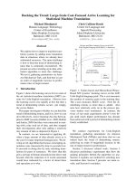
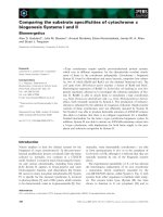
Fig. 1. Schematic representation of
cytochrome c biogenesis Systems I and II.
The systems illustrated are System I (the
Ccm system) from E. coli and System II
from B. subtilis. Note that ResBC is a fusion
protein in H. pylori. The two systems can
each be subdivided into proteins which
contribute to the handling of heme and
ligation of heme to the apocytochrome, and
those involved in the provision of reductant
to the apocytochrome in order to reduce a
potential disulfide bond in the CXXCH
heme-binding site. CcmH, which in some
organisms is two separate proteins CcmH
and CcmI [50], appears to be involved in
both heme handling ⁄ ligation and reductant
provision [2].
A. D. Goddard et al. Specificity of cytochrome c biogenesis System II
FEBS Journal 277 (2010) 726–737 ª 2009 The Authors Journal compilation ª 2009 FEBS 727
specificity of System II in direct comparison with
System I.
Results
Various c-type cytochrome proteins were used to probe
different aspects of the specificity of the expressed
cytochrome c biogenesis systems. Experiments were
conducted in a strain of E. coli lacking all known
endogenous cytochrome c biogenesis proteins, EC06.
In each case, control experiments were performed for
spontaneous heme attachment to exogenous cyto-
chromes [using the biogenesis system plasmid back-
bone containing no cytochrome c biogenesis genes
(AD377)]. In addition, correction was performed for
the formation of any endogenous E. coli cytochromes
catalyzed by the products of the different biogenesis
plasmids in the absence of a gene for an exogenous
cytochrome.
System II can mature monoheme c-type
cytochromes in E. coli
Cytochrome c
550
from Paracoccus denitrificans is well
characterized as a heterologous holocytochrome pro-
duced by the E. coli Ccm system [26]. The ability of
System II to attach heme to this monoheme bacterial
cytochrome in the periplasm of EC06 cells was tested.
Holocytochrome c
550
was detected in periplasmic
extracts of cells containing pHP86 (H. pylori ResBC)
and pKPD1 (cytochrome c
550
) and was quantified
spectroscopically (Fig. 2). The yield was approximately
0.6% of that with System I, which produces very large
amounts of the holocytochrome (Table 1). SDS-PAGE
analysis followed by heme staining (Fig. 2, right-hand
inset) shows a band of the expected mass ( 15 kDa
for P. denitrificans holocytochrome c
550
cleaved of its
signal peptide) for the cytochrome produced by System
II, the intensity of which is consistent with the amount
of cytochrome determined spectroscopically compared
with System I. The a-band of the pyridine hemo-
chrome spectrum, which is indicative of the saturation
of the heme vinyl groups to which the cysteine residues
attach, was found to be at 550 nm for the System
II-matured cytochrome c
550
, as expected for the forma-
tion of two thioether bonds (Fig. 2, left-hand inset).
Cytochrome c
550
made by System II (Fig. 2) was there-
fore indistinguishable from that made by System I, its
natural biogenesis machinery. This is the first demon-
stration that System II can mature a cytochrome nor-
mally matured by System I.
We also examined the biogenesis of Hydro-
genobacter thermophilus cytochrome c
552
. This System
0.035
0.030
0.025
Absorbance
14
550 560
570540530
0.020
MI II
6
Δ Absorbance
400 450 500 550 600 650
Wavelen
g
th (nm)
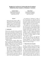
Fig. 2. Maturation of P. denitrificans cytochrome c
550
by System II.
Visible absorption spectra reflecting the formation of P. denitrificans
cytochrome c
550
and variants in periplasmic extracts of E. coli EC06
catalyzed by H. pylori ResBC: wild-type cytochrome c
550
(full line),
AXXCH-containing variant (broken ⁄ dotted line) and CXXAH-contain-
ing variant (broken line). The vertical scale bar represents 0.01
absorbance units. The spectra are vertically offset for clarity. Sam-
ples were reduced by the addition of a few grains of disodium dithi-
onite. The absorbance maxima for wild-type cytochrome c
550
are at
415, 521.5 and 550 nm. The inset spectrum shows the reduced
pyridine hemochrome spectrum of cytochrome c
550
produced by
H. pylori ResBC. The vertical line indicates 550 nm. The inset gel
illustrates the detection of c-type cytochromes via SDS-PAGE
analysis, and subsequent heme staining of the gel, of periplasmic
fractions from cells expressing P. denitrificans cytochrome c
550
and
the indicated biogenesis system (I or II). The periplasmic fraction
from cells expressing System I and cytochrome c
550
was diluted
20-fold before analysis (equating to 0.25–0.5 lg protein loaded,
compared with 5–10 lg for the undiluted System II-produced sam-
ple). The left-most lane (M) contains See-Blue Plus 2 protein mark-
ers of the indicated molecular weights (kDa).
Table 1. Levels of holocytochrome production for biogenesis
Systems I and II expressed in E. coli strain EC06. These values
have been corrected to account for any spontaneous formation of
the respective cytochromes and for background levels of endoge-
nous cytochrome production. The units are milligrams of holocyto-
chrome per gram of wet cell pellet. The values in parentheses are
standard deviations. ND, not detectable.
System I System II
Cytochrome c
550
CXXCH 4.07 (0.55) 0.024 (0.009)
AXXCH 0.045 (0.002) ND
CXXAH 0.023 (0.005) ND
Cytochrome c
552
CXXCH 0.99 (0.42) 0.16 (0.054)
AXXCH 0.030 (0.004) 0.009 (0.002)
CXXAH ND ND
Specificity of cytochrome c biogenesis System II A. D. Goddard et al.
728 FEBS Journal 277 (2010) 726–737 ª 2009 The Authors Journal compilation ª 2009 FEBS
I-matured thermophilic cytochrome has also been used
as a substrate to test the properties of the E. coli cyto-
chrome c biogenesis system [22]. System I is able to
produce large quantities of the c
552
holocytochrome
(Table 1 and Fig. 3). Co-expression of cytochrome c
552
with the System II plasmid resulted in approximately
16% of the level produced by System I, a much higher
proportion than that observed with the mesophilic
cytochrome c
550
. The spectroscopic features and mobil-
ity on SDS-PAGE of the System II-produced cyto-
chrome c
552
are identical to those of the same
cytochrome produced by System I (Fig. 3 and inset).
Spontaneous periplasmic assembly of cytochrome c
552
appears to occur, as some holocytochrome is detected
in the absence of any biogenesis system (Fig. 3). The
data presented in Table 1 have been corrected for the
mean level of spontaneous heme attachment. Uncata-
lyzed heme attachment to cytochrome c
552
is known to
occur in the E. coli cytoplasm [9], and a small amount
of cytoplasmic contamination of periplasmic extracts
can occur during preparation [22]. However, SDS-
PAGE analysis of the periplasmic fractions in this
study demonstrated that the spontaneous holocyto-
chrome formation detected was essentially all periplas-
mic, as the observed mass was consistent with that of
the cytochrome polypeptide cleaved of its periplasmic
targeting sequence. The mass of H. thermophilus holo-
cytochrome c
552
cleaved of its signal peptide is approx-
imately 9 kDa, whereas the uncleaved product has a
mass of approximately 11 kDa.
Maturation of single-cysteine holocytochromes
There are natural examples of cytochromes in which
heme is attached via a single thioether bond to a cys-
teine in the protein [8,27], which raises questions about
the purpose of covalent heme attachment via two
bonds [21,28]. The determination of whether the pres-
ence of two cysteine thiols is essential could also
address the requirement for an intramolecular disulfide
bond, known to occur within apocytochromes [29], in
the heme attachment reaction. It has been argued that
System II can attach heme to one SXXCH motif gen-
erated by site-directed mutagenesis in the tetraheme
cytochrome NrfH from Wolinella succinogenes [24].
However, this is an important point requiring further
investigation.
Cytochrome c
550
containing an AXXCH motif
(C35A) acquired approximately 1.1% of the level of
heme attachment observed for the wild-type CXXCH
protein when expressed with System I (Table 1). The
values in Table 1 are based on the absorption values
at single wavelengths. However, they are only taken to
indicate the presence of the specific holocytochrome
under investigation if the features of the spectrum, in
terms of wavelength maxima, and the position and
intensity of heme-staining bands on SDS-PAGE gels,
also appropriately demonstrate holocytochrome forma-
tion. The AXXCH variant produced by System I has
spectroscopic features indicative of single-cysteine holo-
cytochrome formation, and a band of the expected
mass is observed on heme-stained gels (data not
shown). The values in Table 1 imply that a small
amount of the C38A (CXXAH) variant may have
undergone heme attachment by System I. However,
using the criteria described above (spectra and heme
staining), we conclude that the single-wavelength
absorption intensity is not in fact indicative of C38A
holocytochrome. Effectively, therefore, the value in
Table 1 for the C38A variant of cytochrome c
550
matured by System I represents the lower limit of
detectability and the experimental error. Notably, the
production of the AXXCH variant of cytochrome c
550
was detected by western blotting using anti-cytochrome
6
14
Δ Absorbance
MIII
Wavelength (nm)
400 450 500 550 600
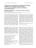
Fig. 3. Maturation of H. thermophilus cytochrome c
552
by Systems
I and II. Visible absorption spectra reflecting the formation of
H. thermophilus cytochrome c
552
in periplasmic extracts of E. coli
EC06 catalyzed by E. coli System I (full line), H. pylori System II
(broken line) and in the absence of any biogenesis system (AD377)
(broken ⁄ dotted line) (showing the level of spontaneous, i.e. uncata-
lyzed, holocytochrome formation). The vertical scale bar represents
0.2 absorbance units. The spectra are vertically offset for clarity
and normalized for wet cell weight. Samples were reduced by the
addition of a few grains of disodium dithionite. The absorbance
maxima are at 417, 521 and 552 nm. The inset gel illustrates the
detection of c-type cytochromes via SDS-PAGE analysis of periplas-
mic fractions from cells expressing H. thermophilus cytochrome
c
552
and the indicated biogenesis system (I or II), and subsequent
heme-staining of the gel. Loading was normalized for total protein
content. The left-most lane (M) contains See-Blue Plus 2 protein
markers of the indicated molecular weights (kDa).
A. D. Goddard et al. Specificity of cytochrome c biogenesis System II
FEBS Journal 277 (2010) 726–737 ª 2009 The Authors Journal compilation ª 2009 FEBS 729
c
550
serum, whereas the CXXAH variant was not
(Fig. S1, see Supporting Information).
Although it is possible that the CXXAH variant of
P. denitrificans cytochrome c
550
is unstable and cannot
be made, the two single-cysteine-containing (and
AXXAH) variants of H. thermophilus cytochrome c
552
can form stably in the E. coli cytoplasm [30,31]. Sys-
tem I was unable to produce the CXXAH variant
cytochrome c
552
, but some System I-dependent forma-
tion of the AXXCH variant (approximately 3% rela-
tive to CXXCH) was detected (this is the value after
subtraction to account for the level of spontaneous
holocytochrome formation). Figure 4 shows that the
heme-staining band corresponding to holo-c
552
AXXCH matured by System I is significantly more
intense than the band observed when no biogenesis
genes were co-expressed (i.e. with spontaneous holocy-
tochrome formation). This is a significant observation
regarding the substrate specificity of the Ccm system.
Neither single-cysteine holocytochrome c
550
was
detected with the coexpression of the System II plas-
mid, as shown in the spectra of the periplasmic
extracts in Fig. 2, which have no features indicative of
holocytochrome formation. It should be noted that
pEC86 (System I) complements the Ccm deletion of
E. coli strain EC06, whereas pHP86 (System II) does
not; thus our experimental errors as a result of back-
ground (endogenous) cytochrome production are much
larger with System I than with System II. Although
cultures grown in this work are considered to be aero-
bic, some microanaerobicity can occur, which causes
low-level expression of the endogenous E. coli c-type
cytochromes. System II appears to produce a very low
level of the AXXCH holocytochrome c
552
(Table 1),
compared with the CXXCH form. The analysis of 12
independent experiments revealed the production of
spectroscopically detectable AXXCH above the level
of spontaneous cytochrome formation in two of the
cultures. These data are responsible for the apparent
formation of AXXCH by System II when compared
with AD377 (reported as mean values in Table 1). It is
possible that in the majority of our observations
single-cysteine cytochromes were formed by System II
at such low levels that they were undetectable either by
spectroscopic analysis of periplasmic fractions or heme
staining of appropriate SDS-PAGE gels.
System II mediates the formation of a b-type
cytochrome
Unexpectedly, the spectra of periplasmic extracts of
cells containing the System II plasmid and the double-
alanine cytochrome c
550
(AXXAH motif, C35A ⁄ C38A)
indicated the presence of small amounts of a typical
low-spin, b-type cytochrome (Fig. 5). The Soret band
is red shifted by 4 nm and the a-band by 5 nm com-
pared with the wild-type cytochrome c
550
, as would be
expected for noncovalently bound heme (saturation of
each heme vinyl group on formation of a c-type cyto-
chrome causes a blue shift of 2–3 nm in the a-band of
the absorption spectrum). To confirm the presence of
variant cytochrome c
550
, we performed a western blot
of periplasmic extracts from this strain and a strain
containing only pKK223-3 (i.e. no cytochrome).
A band consistent with the molecular weight of
14
6
M I II -
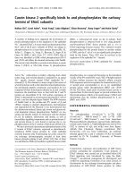
Fig. 4. Maturation of H. thermophilus cytochrome c
552
AXXCH vari-
ant. SDS-PAGE analysis and subsequent heme staining of periplas-
mic extracts from E. coli EC06 cells containing the H. thermophilus
cytochrome c
552
AXXCH variant and the indicated biogenesis sys-
tem [I or II (with the lane marked - being periplasm from cells
containing empty vector, AD377)]. Loading was normalized for total
protein content. The left-most lane (M) contains See-Blue Plus 2
protein markers of the indicated molecular weights (kDa).
Δ Absorbance
Wavelength (nm)
400 450 500 550 600 650
Fig. 5. Maturation of a b-type cytochrome AXXAH variant of
P. denitrificans cytochrome c
550
. Visible absorption spectra of peri-
plasmic extracts from E. coli EC06 cells expressing H. pylori ResBC
and P. denitrificans cytochrome c
550
(broken-dotted line), cyto-
chrome c
550
AXXAH variant (full line) and no cytochrome (pKK223-
3) (dotted line). The vertical scale bar represents 0.005 absorbance
units. Samples were reduced by the addition of a few grains of
disodium dithionite. The Soret and a-band absorbance maxima are
at 415 and 550 nm, respectively, for wild-type cytochrome c
550
,
and at around 419 and 555 nm for the AXXAH-containing variant.
The spectrum of the wild-type cytochrome c
550
has been reduced
by a factor of seven for clarity, and the spectra are vertically offset.
The vertical line indicates 550 nm.
Specificity of cytochrome c biogenesis System II A. D. Goddard et al.
730 FEBS Journal 277 (2010) 726–737 ª 2009 The Authors Journal compilation ª 2009 FEBS
cytochrome c
550
was evident in the strain expressing
AXXAH-containing cytochrome c
550
, but not in the
control strain containing pKK223-3 (Fig. S1, see Sup-
porting Information). It was not possible to detect low
levels of b-type complexes (if they exist) made with
System I because of the relatively high levels of endog-
enous cytochromes that are produced (see above)
would mask the spectroscopic features of the b-type
cytochrome. However, we were unable to detect the
formation of a b-type AXXAH variant by System I
using western blot analysis and anti-cytochrome c
550
serum (Fig. S1, see Supporting Information). In addi-
tion, no b-type AXXAH cytochrome was detected
when no System II biogenesis proteins were present.
These observations have implications for the provision
of heme to the periplasm by ResBC, and suggest that
it may facilitate heme delivery from the cytoplasm, in
agreement with a recent study by Frawley & Kranz
[20].
Provision of reductant increases significantly
System II-mediated c-type cytochrome formation
As the H. pylori biogenesis system expressed in this
study lacks the thiol-disulfide oxidoreductase compo-
nents that are thought to reduce the cysteine thiols in
the cytochrome heme-binding motif (neither ResA of
System II nor CcmG of System I are present), the
effect of the addition of a chemical reductant to the
growth medium was tested: 5 mm 2-mercaptoethane
sulfonate (MESA) was added to cells containing wild-
type (CXXCH) P. denitrificans cytochrome c
550
, and
the System II plasmid and holocytochrome contents
were determined. The added reductant caused a two-
to three-fold increase in holocytochrome formation
(data not shown). Exogenous chemical reductant has
been used to recover the phenotypes of strains lacking
other oxidoreductases [32,33]. The addition of 5 mm
MESA to cells expressing the single-cysteine c
550
C35A
variant did not result in the formation of detectable
holocytochrome, implying that the augmentation in
wild-type holocytochrome maturation with the addi-
tion of reductant is a result of the reduction of a disul-
fide in the apocytochrome.
Production of endogenous E. coli cytochromes
Escherichia coli contains a number of endogenous
c-type cytochromes that it expresses under different
anaerobic growth conditions. Some of these are
observed at low levels in periplasmic extracts when the
Ccm deletion strain EC06 is complemented with System
I (pEC86), but not with System II (pHP86), as shown in
Fig. 6. The two bands observed correspond to the
masses of the soluble cytochromes NapB (around
15 kDa) and NrfA (around 50 kDa). However, given the
relative maturation levels of exogenous cytochromes c
(see above), it may be that any endogenous cytochrome
matured by System II would be present below the lower
limit of detection in our experiments. We have
determined that the limit of detection for heme on a
heme-stained SDS-PAGE gel is 1 nmol per lane (A. D.
Goddard & S. J. Ferguson, unpublished observations).
E. coli NapB has two hemes and NrfA five. Therefore,
we would expect to detect 0.5 and 0.2 nmol of these
cytochromes, respectively.
Discussion
The complex and somewhat unpredictable natural
distribution of cytochrome c biogenesis systems does
not correlate specifically with the types of cyto-
chrome produced by the organisms concerned [5,34].
Cytochromes c vary widely in terms of overall fold,
heme iron ligands, number of hemes per polypeptide,
the presence of other cofactors, number of subunits,
being membrane-bound or soluble, as well as the way
in which the heme is linked to the protein (a few cyto-
chromes have single thioether bonds to heme). The
latter group includes the unusual cytochrome b
6
and
the trypanosomatid cytochromes c. The specificity of
E. coli System I has been studied extensively. It can
produce cytochromes c from a wide variety of organ-
isms, with many hemes per polypeptide, and even
attach heme to peptides as short as 12 residues in
length [35,36]. The specificity of some heme lyases
(System III) has also been determined; some organisms
62
49
38
14
28
17
MIII
6
Fig. 6. Analysis of endogenous cytochrome production. SDS-PAGE
analysis and subsequent heme staining of periplasmic extracts
from E. coli EC06 cells containing pKK223-3 (no exogenous cyto-
chrome) and the indicated biogenesis system (I or II). The left-most
lane (M) contains See-Blue Plus 2 protein markers of the indicated
molecular weights (kDa). Equal amounts of total protein were
loaded in each lane.
A. D. Goddard et al. Specificity of cytochrome c biogenesis System II
FEBS Journal 277 (2010) 726–737 ª 2009 The Authors Journal compilation ª 2009 FEBS 731
contain a heme lyase for each mitochondrial cyto-
chrome c (e.g. yeast cytochromes c and c
1
), whereas
others (e.g. animals) contain a single such enzyme that
apparently catalyzes heme attachment to both cyto-
chromes [37]. No study has examined the specificity of
System II, which in nature produces an array of
mono- and multiheme cytochromes.
System II produces monoheme bacterial
cytochromes
In this work, we have shown that coexpression of the
plasmid pHP86 (expressing System II) in E. coli with
the mesophilic cytochrome c
550
from P. denitrificans
and the thermophilic cytochrome c
552
from H. thermo-
philus, both naturally matured by System I, results in
heme attachment to these cytochromes, yielding prod-
ucts that are indistinguishable from those produced by
System I. This suggests that System II, in common
with System I and in contrast with System III [38], has
a broad substrate specificity and is able to mature c-
type cytochromes from a variety of sources, including
those that it does not naturally encounter. A relatively
high level of holocytochrome c
552
was produced (16%
relative to System I) considering that the System II
fusion protein is expressed heterologously and without
the remaining (disulfide oxidoreductase) components
of the biogenesis system. A previous study has inter-
preted a reduced level of cytochrome production by
System II compared with System I as the former hav-
ing a lower affinity for heme [11]; as no measurement
of the relative abundance of the biogenesis proteins
was shown, and there is no reason to believe that they
would be equally stable in E. coli, we have reservations
about this interpretation.
That higher levels of thermophilic cytochrome c
552
are produced by System II compared with a mesophilic
cytochrome (c
550
) is possibly a result of the higher sta-
bility of the apocytochrome c
552
when it is delivered
by the Sec system to the periplasm. Proteolytic degra-
dation of apocytochromes might compete with the
heme attachment machinery. In addition, apocyto-
chromes are susceptible to periplasmic oxidation of the
heme-binding cysteine residues. In our heterologous
System II, the oxidoreductase that would normally
reduce such a disulfide bond, ResA, is absent; the oxi-
dation would also slow heme attachment. Our observa-
tion that added reductant results in a substantial
increase in cytochrome c
550
production indicates that
oxidation of the heme-binding motif can reduce the
efficiency of heme attachment by System II. Neverthe-
less, it is becoming increasingly clear from this work
and others [20] that dithiol ⁄ disulfide oxidoreductases
are not strictly necessary for cytochrome c maturation
in the periplasm of E. coli.
Maturation of single-cysteine-attached
cytochromes c
We found no detectable heme attachment to the sin-
gle-cysteine-containing variants of P. denitrificans c
550
with coexpression of the System II plasmid. A very
low, variable level of heme attachment was observed
with the AXXCH variant of cytochrome c
552
, but none
with the CXXAH form. If there is a capability to
attach heme to a single-cysteine cytochrome then, in
common with System I, it is to a very low extent com-
pared with heme attachment to CXXCH, below the
level of detection of the analysis conducted in this
study. It is notable that, in the work of Simon et al.
[24], evidence was found for heme attachment to only
one SXXCH heme-binding motif of the four possible
(and investigated) in NrfH, and that no heme attach-
ment to CXXSH was reported [24]. It may be that the
observed heme attachment to one SXXCH motif was
not in fact catalyzed by System II, e.g. it was instead
spontaneous, perhaps facilitated by substantial folding
of the protein around the three other hemes attached
to CXXCH motifs by System II.
It has been reported previously that System I cannot
produce detectable levels of single-cysteine-containing
holocytochrome c
552
[22]. In that work, the lower level
of detectability was estimated as 2% of the wild-type
(CXXCH) holocytochrome yield. Control experiments
performed in the present work, to allow a direct com-
parison of System I and II plasmids (which are identi-
cal but for their encoded operons), permit a refinement
of the conclusion of the former work. We found here
low but detectable levels of the holo-forms of AXXCH
variants of both cytochromes c
550
and c
552
(1 and 3%
relative to the wild-type CXXCH cytochromes, respec-
tively) when expressed with pEC86. The difference is
presumably partly a result of the different E. coli
strains used. Here, we used EC06, a ccm deletion
strain, which had a significant effect on the amount of
background cytochrome c matured (producing no
detectable c-type cytochromes in the absence of a plas-
mid-borne biogenesis system). The sensitivity of the
analytical methods used (e.g. the former work did not
use heme-stained gels) may also contribute to the dif-
ferences. That System I can attach some heme to sin-
gle-cysteine-containing cytochromes is significant,
particularly in the context of a possible relationship
between heme attachment and a disulfide bond in the
CXXCH motif. The fact that two cysteines are not
absolutely essential for the heme attachment reaction
Specificity of cytochrome c biogenesis System II A. D. Goddard et al.
732 FEBS Journal 277 (2010) 726–737 ª 2009 The Authors Journal compilation ª 2009 FEBS
demonstrates that the chemistry of such a reaction is
not necessarily via an obligate disulfide-containing
intermediate. Breaking a disulfide bond could be envis-
aged as providing the driving force for formation of
the thioether bonds to heme. However, successful
in vitro thioether bond formation using a phosphine
(in the absence of thiol reagents) to reduce the apocy-
tochrome disulfide also indicated that disulfide-linked
chemistry is not involved in the heme attachment reac-
tion [39]. The present work implies that the formation of
the two thioether bonds is not thermodynamically neces-
sary to release heme from the covalent heme-binding
chaperone CcmE. We observe, as might be anticipated
[21,31,40], that the single-cysteine variant in which the
heme-binding cysteine is directly adjacent to the heme
iron-ligating histidine (i.e. XXXCH) is the more likely to
be recognized by the system and to undergo heme attach-
ment than is CXXXH. Nevertheless, it remains clear that
System I is far more effective and efficient at attaching
heme to apocytochrome c with two cysteines, rather than
one, in the heme-binding motif.
System II facilitates the formation of a b-type
cytochrome in the periplasm
In the absence of any biogenesis proteins, it was not
possible to detect the b-type forms (i.e. containing non-
covalently bound heme) of c-type cytochromes lacking
the heme-binding cysteine residues (i.e. the AXXAH
variants). Apocytochromes c appear to be proteolyti-
cally degraded when heme is not attached [41]. Because
of the clean background observed with the System II
plasmid (i.e. the lack of any endogenous c-type
cytochromes), it was possible to detect some b-type
cytochrome when cytochrome c
550
AXXAH was coex-
pressed with pHP86. It is possible that an equivalent
cytochrome is produced by System I, but that it is ren-
dered undetectable as a result of the production of
endogenous cytochromes c which mask the b-type
spectra [b-type cytochromes generally lose heme in
SDS-PAGE and therefore cannot be detected by the
heme staining of gels; western blotting with anti-cyto-
chrome c
550
serum failed to detect the presence of any
cytochrome (Fig. S1, see Supporting Information)].
Alternatively, it is possible that, as a result of a cova-
lent intermediate (CcmE–heme) [42], System I is
unable to pass heme to an AXXAH variant apocyto-
chrome; System II (ResBC) from Helicobacter hepati-
cus does not appear to covalently bind heme [20].
However, a recent study with Bacillus subtilis ResB
and ResC (unfused proteins in that organism) revealed
covalent binding of heme to the cytoplasmic side of
ResB [43]. It is therefore possible that an initial cova-
lent attachment of heme to ResB occurs, followed by
trafficking through ResC, before insertion of heme into
the periplasmic cytochrome. However, the residue
covalently bound to the heme of ResB was found to
be nonessential for cytochrome c biogenesis. The func-
tion, if any, of System II proteins covalently binding
heme therefore remains to be resolved.
That expression of the System II protein in E. coli
allows the formation of a b-type cytochrome suggests
that heme provision from the cytoplasm to the peri-
plasm might be performed by ResBC, concurrent with
recent observations [20]. The study by Frawley &
Kranz [20] also demonstrated the essentiality of H858
of H. hepaticus ResBC in holocytochrome formation,
and proposed that this residue, together with H77, is
involved in supplying heme to the periplasm. We note
that a H857E mutant in H. pylori ResBC (equivalent
to H858 in the H. hepaticus protein) is unable to
mature the b-type cytochrome described above (A. D.
Goddard & S. J. Ferguson, unpublished observations).
This is consistent with H857 playing a role in heme
transport. It is not known how heme is transported
across the inner membrane by System I, but it has
been shown conclusively that, contrary to earlier sug-
gestions, CcmA and CcmB are not involved in heme
transport in E. coli [44,45]. Notably, maturation of an
AXXAH-containing variant b -type cytochrome c
550
in
the present study indicates a nascent heme-binding site,
even in this mesophilic apocytochrome c (see also
[46]), as well as possible recognition features in the
apocytochrome, at least for cytochrome c biogenesis
System II, other than the CXXCH heme-binding
motif. These data also suggest that heme delivery to
apocytochrome and thioether bond formation by
System II are independent processes.
Materials and methods
Strains, plasmids and culture conditions
Escherichia coli strain EC06 [47] contains a chromosomal
deletion of the ccm operon and was used to examine holocy-
tochrome formation in the presence of the plasmid-encoded
biogenesis systems. E. coli strain DH5 a (Invitrogen, Paisley,
UK) was used for routine molecular biology. KOD poly-
merase (Merck Chemicals Ltd, Nottingham, UK) was used
for PCRs. All oligonucleotides used in this study are listed
in Table S1 (see Supporting Information).
Biogenesis plasmids
The plasmids used in this work are listed in Table S2. The
E. coli ccmABCDEFGH operon (System I) was expressed
A. D. Goddard et al. Specificity of cytochrome c biogenesis System II
FEBS Journal 277 (2010) 726–737 ª 2009 The Authors Journal compilation ª 2009 FEBS 733
from pEC86 [25]. To create a comparable plasmid lacking
any biogenesis system, inverse PCR was performed on
pEC86 using the primers AG234 and AG235, and the prod-
uct was self-ligated. This removed the entire ccm operon,
and the plasmid created is AD377 (no biogenesis system).
To create a suitable plasmid for the expression of other bio-
genesis systems, a XhoI site was introduced immediately
after the initiating ATG of ccmA in pEC86 via Quikchange
mutagenesis using the primers WC1 and WC2. The resul-
tant construct is pEC86x, in which the entire ccm operon
can be excised by digestion with XhoI and StuI. The ycf5
gene was amplified from H. pylori (strain 26695) genomic
DNA using oligonucleotides HelF and HelR. The PCR
product was cloned into XhoI ⁄ StuI-digested pEC86x. The
resultant plasmid for the expression of H. pylori ResBC is
pHP86 (System II).
Cytochrome c plasmids
P. denitrificans cytochrome c
550
was expressed from the iso-
propyl thio-b-d-galactoside (IPTG)-inducible promoter of
pKPD1 [26]. Mutations within the CXXCH heme-binding
motif of c
550
were created by Quikchange using the follow-
ing: c
550
C35A, C35AF and C35AR; c
550
C38A, C38AF
and C38AR; c
550
C35AC38A, C35AC38AF and
C35AC38AR. H. thermophilus cytochrome c
552
and its AX-
XCH, AXXAH and CXXAH variants were expressed from
the plasmids pEST210, pEST211, pEST212 and pEST213,
respectively [22].
In each case, the plasmid bearing the biogenesis system
confers resistance to chloramphenicol and the expression of
the proteins is constitutive. The plasmids bearing the cyto-
chromes are inducible with IPTG and confer resistance to
carbenicillin. All constructs were sequenced before use.
Routine cell growth was conducted using Luria–Bertani
medium supplemented with appropriate antibiotics.
Growth on solid medium used Luria–Bertani medium sup-
plemented with 1.5% bacteriological agar. For the prepara-
tion of periplasmic fractions, single colonies containing
appropriate plasmids were picked into 500 mL 2· TY
medium (16 gÆL
)1
peptone, 10 gÆL
)1
yeast extract, 5 gÆL
)1
NaCl), supplemented with 1 mm IPTG and appropriate
antibiotics, in 2 L flasks. Cultures were grown at 37 °C
with shaking at 200 r.p.m. for 16–20 h before harvesting.
Carbenicillin was used at 100 l g Æ mL
)1
and chlorampheni-
col at 34 lg Æ mL
)1
.
Analysis of cytochrome production
Periplasmic extractions were performed as described previ-
ously [22]. Extracts were analyzed by SDS-PAGE (Invitro-
gen pre-cast 10% Bis-Tris gels), followed by heme staining
[48], which stains proteins with covalently bound heme.
Samples were normalized for wet cell weight, and equal
amounts of protein were loaded per lane (5–10 lg).
Western blots to detect P. denitrificans cytochrome c
550
were performed according to the manufacturer’s instruc-
tions using anti-cytochrome c
550
rabbit serum and a
commercial alkaline-phosphatase-conjugated anti-rabbit
secondary IgG raised in goat. The marker used was See-
Blue Plus 2 (Invitrogen).
UV–visible spectroscopy was performed using a Perkin-
Elmer (Waltham, MA, USA) Lambda 2 spectrophotometer;
samples were reduced by the addition of a few grains of
disodium dithionite (Sigma-Aldrich Company Ltd, Poole,
UK). Pyridine hemochrome spectra were determined
according to the method described by Bartsch [49].
The normalized cytochrome content of each extract is pre-
sented as the number of milligrams of holocytochrome c
per gram of cell pellet. The data are averages of at least five
growths. The extinction coefficients used to calculate the
yields of holocytochromes were as follows: wild-type
H. thermophilus cytochrome c
552
, e = 182 mm
)1
Æcm
)1
at
417 nm; C11A c
552
, e = 179.5 mm
)1
Æcm
)1
at 422 nm; C14A
c
552
, e = 174.5 mm
)1
Æcm
)1
at 420 nm; C11A ⁄ C14A c
552
,
e = 145 mm
)1
Æcm
)1
at 425 nm; P. denitrificans cytochrome
c
550
wild-type and variants, 140 mm
)1
Æcm
)1
at 415 nm [22].
The extinction coefficients for the cytochrome c
550
variants
are unknown; the wild-type value was therefore used. Cor-
rections of the average normalized values for each dataset
were performed by subtracting the value observed when no
biogenesis genes were expressed (i.e. with plasmid AD377
and the relevant cytochrome plasmid, to correct for sponta-
neous holocytochrome production) and subtracting the
value observed when no heterologous cytochrome gene was
expressed (i.e. with plasmid pKK223-3 and the relevant bio-
genesis plasmid, to correct for the production of endoge-
nous E. coli cytochromes). Finally, the values for cells
expressing the two empty vectors AD377 and pKK223-3
were added back, so that any corrections were for endoge-
nous or spontaneous cytochrome c production only.
Cultures for the corrections were grown and analyzed at
least three times, and the mean values were used for the
corrections.
Acknowledgements
This work was supported by the Biotechnology and
Biological Sciences Research Council (BBSRC; grant
numbers BB ⁄ D523019 ⁄ 1, BB ⁄ E004865 ⁄ 1 and BB ⁄
D019753 ⁄ 1). J.W.A.A. is a BBSRC David Phillips
Fellow. A.D.G. gratefully acknowledges the E. P. Abra-
ham Cephalosporin Fund. We thank Professor David
Kelly (University of Sheffield) for kindly providing
H. pylori genomic DNA.
Since the submission of this manuscript Kern et al.
[50a] have also shown that System II cannot attach
heme to a single-cysteine motif in a cytochrome at
detectable levels [sentence added at proof stage].
Specificity of cytochrome c biogenesis System II A. D. Goddard et al.
734 FEBS Journal 277 (2010) 726–737 ª 2009 The Authors Journal compilation ª 2009 FEBS
References
1 Stevens JM, Daltrop O, Allen JW & Ferguson SJ
(2004) C-type cytochrome formation: chemical and
biological enigmas. Acc Chem Res 37, 999–1007.
2 Ferguson SJ, Stevens JM, Allen JW & Robertson IB
(2008) Cytochrome c assembly: a tale of ever increasing
variation and mystery? Biochim Biophys Acta 1777,
980–984.
3 Bowman SE & Bren KL (2008) The chemistry and
biochemistry of heme c: functional bases for covalent
attachment. Nat Prod Rep 25, 1118–1130.
4 Hamel P, Corvest V, Giege P & Bonnard G (2009)
Biochemical requirements for the maturation of
mitochondrial c-type cytochromes. Biochim Biophys
Acta 1793, 125–138.
5 Allen JW, Jackson AP, Rigden DJ, Willis AC, Fergu-
son SJ & Ginger ML (2008) Order within a mosaic dis-
tribution of mitochondrial c-type cytochrome
biogenesis systems? Febs J 275, 2385–2402.
6 Kranz RG, Beckett CS & Goldman BS (2002) Geno-
mic analyses of bacterial respiratory and cytochrome c
assembly systems: Bordetella as a model for the system
II cytochrome c biogenesis pathway. Res Microbiol
153, 1–6.
7 Kuras R, Saint-Marcoux D, Wollman FA & de Vitry
C (2007) A specific c-type cytochrome maturation sys-
tem is required for oxygenic photosynthesis. Proc Natl
Acad Sci USA 104, 9906–9910.
8 Allen JW, Ginger ML & Ferguson SJ (2004) Matura-
tion of the unusual single-cysteine (XXXCH) mito-
chondrial c-type cytochromes found in
trypanosomatids must occur through a novel biogene-
sis pathway. Biochem J 383, 537–542.
9 Sanbongi Y, Yang JH, Igarashi Y & Kodama T (1991)
Cloning, nucleotide sequence and expression of the
cytochrome c-552 gene from Hydrogenobacter thermo-
philus. Eur J Biochem 198, 7–12.
10 Pollock WB, Rosell FI, Twitchett MB, Dumont ME &
Mauk AG (1998) Bacterial expression of a mitochon-
drial cytochrome c. Trimethylation of lys72 in yeast
iso-1-cytochrome c and the alkaline conformational
transition. Biochemistry 37, 6124–6131.
11 Feissner RE, Richard-Fogal CL, Frawley ER, Lough-
man JA, Earley KW & Kranz RG (2006) Recombinant
cytochromes c biogenesis systems I and II and analysis
of haem delivery pathways in Escherichia coli. Mol
Microbiol 60, 563–577.
12 Le Brun NE, Bengtsson J & Hederstedt L (2000)
Genes required for cytochrome c synthesis in Bacillus
subtilis. Mol Microbiol 36, 638–650.
13 Hodson CT, Lewin A, Hederstedt L & Le Brun NE
(2008) The active-site cysteinyls and hydrophobic cav-
ity residues of ResA are important for cytochrome c
maturation in Bacillus subtilis. J Bacteriol 190, 4697–
4705.
14 Crow A, Le Brun NE & Oubrie A (2005) The role of
ResA in type II cytochrome c maturation. Biochem Soc
Trans 33, 149–151.
15 Dreyfuss BW, Hamel PP, Nakamoto SS & Merchant S
(2003) Functional analysis of a divergent system II pro-
tein, Ccs1, involved in c-type cytochrome biogenesis.
J Biol Chem 278, 2604–2613.
16 Beckett CS, Loughman JA, Karberg KA, Donato GM,
Goldman WE & Kranz RG (2000) Four genes are
required for the system II cytochrome c biogenesis
pathway in Bordetella pertussis, a unique bacterial
model. Mol Microbiol 38, 465–481.
17 Hamel PP, Dreyfuss BW, Xie Z, Gabilly ST &
Merchant S (2003) Essential histidine and tryptophan
residues in CcsA, a system II polytopic cytochrome c
biogenesis protein. J Biol Chem 278, 2593–2603.
18 Lee JH, Harvat EM, Stevens JM, Ferguson SJ & Saier
MH Jr (2007) Evolutionary origins of members of a
superfamily of integral membrane cytochrome c bio-
genesis proteins. Biochim Biophys Acta 1768, 2164–
2181.
19 Richard-Fogal CL, Frawley ER, Bonner ER, Zhu H,
San Francisco B & Kranz RG (2009) A conserved
haem redox and trafficking pathway for cofactor
attachment. EMBO J 28, 2349–2359.
20 Frawley ER & Kranz RG (2009) CcsBA is a cyto-
chrome c synthetase that also functions in heme trans-
port. Proc Natl Acad Sci USA 106, 10201–10206.
21 Fulop V, Sam KA, Ferguson SJ, Ginger ML & Allen
JW (2009) Structure of a trypanosomatid mitochon-
drial cytochrome c with heme attached via only one
thioether bond and implications for the substrate rec-
ognition requirements of heme lyase. Febs J 276, 2822–
2832.
22 Allen JWA, Tomlinson EJ, Hong L & Ferguson SJ
(2002) The Escherichia coli cytochrome c maturation
(Ccm) system does not detectably attach heme to single
cysteine variants of an apocytochrome c. J Biol Chem
277, 33559–33563.
23 Sambongi Y, Stoll R & Ferguson SJ (1996) Alteration
of haem-attachment and signal-cleavage sites for
Paracoccus denitrificans cytochrome c550 probes path-
way of c-type cytochrome biogenesis in Escherichia
coli. Mol Microbiol 19, 1193–1204.
24 Simon J, Eichler R, Pisa R, Biel S & Gross R (2002)
Modification of heme c binding motifs in the small
subunit (NrfH) of the Wolinella succinogenes cyto-
chrome c nitrite reductase complex. FEBS Lett 522,
83–87.
25 Arslan E, Schulz H, Zufferey R, Kunzler P & Tho
¨
ny-
Meyer L (1998) Overproduction of the Bradyrhizobium
japonicum c-type cytochrome subunits of the cbb3
A. D. Goddard et al. Specificity of cytochrome c biogenesis System II
FEBS Journal 277 (2010) 726–737 ª 2009 The Authors Journal compilation ª 2009 FEBS 735
oxidase in Escherichia coli. Biochem Biophys Res
Commun 251, 744–747.
26 Sambongi Y & Ferguson SJ (1994) Synthesis of holo
Paracoccus denitrificans cytochrome c550 requires
targeting to the periplasm whereas that of holo
Hydrogenobacter thermophilus cytochrome c552 does
not. Implications for c-type cytochrome biogenesis.
FEBS Lett 340, 65–70.
27 Stroebel D, Choquet Y, Popot JL & Picot D (2003)
An atypical haem in the cytochrome b(6)f complex.
Nature 426, 413–418.
28 Allen JW, Barker PD, Daltrop O, Stevens JM, Tomlin-
son EJ, Sinha N, Sambongi Y & Ferguson SJ (2005)
Why isn’t ‘standard’ heme good enough for c-type and
d1-type cytochromes? Dalton Trans 21, 3410–3418.
29 Daltrop O, Allen JW, Willis AC & Ferguson SJ (2002)
In vitro formation of a c-type cytochrome. Proc Natl
Acad Sci USA 99, 7872–7876.
30 Tomlinson EJ & Ferguson SJ (2000) Conversion of a c
type cytochrome to a b type that spontaneously forms
in vitro from apo protein and heme: implications for c
type cytochrome biogenesis and folding. Proc Natl
Acad Sci USA 97, 5156–5160.
31 Tomlinson EJ & Ferguson SJ (2000) Loss of either of
the two heme-binding cysteines from a class I c-type
cytochrome has a surprisingly small effect on physico-
chemical properties. J Biol Chem 275, 32530–32534.
32 Fabianek RA, Hofer T & Tho
¨
ny-Meyer L (1999)
Characterization of the Escherichia coli CcmH protein
reveals new insights into the redox pathway required
for cytochrome c maturation. Arch Microbiol 171,
92–100.
33 Sambongi Y & Ferguson SJ (1994) Specific thiol com-
pounds complement deficiency in c-type cytochrome
biogenesis in Escherichia coli carrying a mutation in a
membrane-bound disulphide isomerase-like protein.
FEBS Lett 353, 235–238.
34 Bertini I, Cavallaro G & Rosato A (2006) Cytochrome
c: occurrence and functions. Chem Rev 106, 90–115.
35 Braun M & Tho
¨
ny-Meyer L (2004) Biosynthesis of
artificial microperoxidases by exploiting the secretion
and cytochrome c maturation apparatuses of
Escherichia coli. Proc Natl Acad Sci USA 101,
12830–12835.
36 Allen JW & Ferguson SJ (2006) What is the substrate
specificity of the System I cytochrome c biogenesis
apparatus? Biochem Soc Trans 34, 150–151.
37 Bernard DG, Gabilly ST, Dujardin G, Merchant S &
Hamel PP (2003) Overlapping specificities of the mito-
chondrial cytochrome c and c1 heme lyases. J Biol
Chem 278, 49732–49742.
38 Sanders C & Lill H (2000) Expression of prokaryotic
and eukaryotic cytochromes c in Escherichia coli. Bio-
chim Biophys Acta 1459, 131–138.
39 Daltrop O & Ferguson SJ (2004) In vitro studies on
thioether bond formation between Hydrogenobacter
thermophilus apocytochrome c552 with metal-
loprotoporphyrin derivatives. J Biol Chem 279, 45347–
45353.
40 Rosell FI & Mauk AG (2002) Spectroscopic properties
of a mitochondrial cytochrome c with a single thioether
bond to the heme prosthetic group. Biochemistry 41,
7811–7818.
41 Gao T & O’Brian MR (2007) Control of DegP-depen-
dent degradation of c-type cytochromes by heme and
the cytochrome c maturation system in Escherichia coli .
J Bacteriol 189, 6253–6259.
42 Schulz H, Hennecke H & Tho
¨
ny-Meyer L (1998)
Prototype of a heme chaperone essential for cyto-
chrome c maturation. Science 281, 1197–1200.
43 Ahuja U, Kjelgaard P, Schulz BL, Tho
¨
ny-Meyer L &
Hederstedt L (2009) Haem-delivery proteins in cyto-
chrome c maturation System II. Mol Microbiol 73,
1058–1071.
44 Feissner RE, Richard-Fogal CL, Frawley ER & Kranz
RG (2006) ABC transporter-mediated release of a
haem chaperone allows cytochrome c biogenesis. Mol
Microbiol 61, 219–231.
45 Christensen O, Harvat EM, Tho
¨
ny-Meyer L, Ferguson
SJ & Stevens JM (2007) Loss of ATP hydrolysis activ-
ity by CcmAB results in loss of c-type cytochrome syn-
thesis and incomplete processing of CcmE. Febs J 274,
2322–2332.
46 Daltrop O & Ferguson SJ (2003) Cytochrome c matu-
ration. The in vitro reactions of horse heart apocyto-
chrome c and Paracoccus denitrificans apocytochrome
c550 with heme. J Biol Chem 278, 4404–4409.
47 Tho
¨
ny-Meyer L, Fischer F, Kunzler P, Ritz D &
Hennecke H (1995) Escherichia coli genes required
for cytochrome c maturation. J Bacteriol 177 , 4321–
4326.
48 Goodhew CF, Brown KR & Pettigrew GW (1986)
Haem staining in gels, a useful tool in the study of
bacterial c-type cytochromes. Biochim Biophys Acta
852, 288–294.
49 Bartsch RG (1971) Cytochromes: bacterial. Methods
Enzymol 23, 344–363.
50 Sanders C, Turkarslan S, Lee DW, Onder O, Kranz
RG & Daldal F (2008) The cytochrome c maturation
components CcmF, CcmH, and CcmI form a mem-
brane-integral multisubunit heme ligation complex.
J Biol Chem 283, 29715–29722.
50a Kern M, Eisel F, Scheithauer J, Kranz RG & Simon J
(2009) Substrate specificity of three cytochrome c haem
lyase isoenzymes from Wolinella succinogenes: uncon-
ventional haem c binding motifs are not sufficient for
haem c attachment by NrfI and CcsA1. Mol Microbiol,
doi:10.1111/j.1365-2958.2009.06965.x.
Specificity of cytochrome c biogenesis System II A. D. Goddard et al.
736 FEBS Journal 277 (2010) 726–737 ª 2009 The Authors Journal compilation ª 2009 FEBS
Supporting Information
The following supplementary material is available:
Fig. S1. Detection of P. denitrificans cytochrome c
550
and variants via western blot.
Table S1. Oligonucleotides used in this work.
Table S2. Plasmids used in this work.
This supplementary material can be found in the
online version of this article.
Please note: As a service to our authors and readers,
this journal provides supporting information supplied
by the authors. Such materials are peer-reviewed and
may be re-organized for online delivery, but are not
copy-edited or typeset. Technical support issues arising
from supporting information (other than missing files)
should be addressed to the authors.
A. D. Goddard et al. Specificity of cytochrome c biogenesis System II
FEBS Journal 277 (2010) 726–737 ª 2009 The Authors Journal compilation ª 2009 FEBS 737