Báo cáo khoa học: Alternative splicing: global insights potx
Bạn đang xem bản rút gọn của tài liệu. Xem và tải ngay bản đầy đủ của tài liệu tại đây (401.13 KB, 11 trang )
MINIREVIEW
Alternative splicing: global insights
Martina Hallegger*, Miriam Llorian* and Christopher W. J. Smith
Department of Biochemistry, University of Cambridge, UK
Introduction
Alternative splicing allows individual genes to produce
two or more variant mRNAs, which in many cases
encode functionally distinct proteins. With the progres-
sive generation of ever larger sequence datasets, the
proportion of multi-exon human genes that are known
to be alternatively spliced has expanded to 92–94%, of
which 85% have a minor isoform frequency of at least
15% [1,2]. Despite some debate about the extent to
which all of this alternative splicing is functionally
important [3], there is no disputing that alternative
splicing is a major contributor to the diverse repertoire
of transcriptomes and proteomes. Its importance is
underscored by the fact that misregulated alternative
splicing can lead to human disease [4,5]. As part of the
overarching effort to understand how the information
encrypted within genomes is used to generate fully
functional organisms, it is therefore necessary to deci-
pher the ‘RNA codes’ underlying regulated patterns of
alternative splicing.
Traditionally, research on alternative splicing regula-
tion focused on the study of minigene models in vitro
or in vivo. The picture that emerged is that regulation
of alternative splicing occurs via the action of numer-
ous RNA binding proteins expressed at variable levels
between tissues. These activators and repressors often
mediate their effects by binding to enhancer and silen-
cer elements within or surrounding alternatively spliced
exons (reviewed in [6]). Although much progress has
been made using model systems, a drawback is that
even when a model alternative splicing event has been
thoroughly characterized it is not immediately obvi-
ous which of its features are generally shared by
Keywords
alternative splicing; microarray; RNA-Seq
Correspondence
C. W. J. Smith, Department of
Biochemistry, University of Cambridge, 80
Tennis Court Road, Cambridge CB2 1GA,
UK
Fax: +44 1223 766002
Tel: +44 1223 333655
E-mail:
*These authors contributed equally to this
work
(Received 26 August 2009, accepted
22 October 2009)
doi:10.1111/j.1742-4658.2009.07521.x
Following the original reports of pre-mRNA splicing in 1977, it was
quickly realized that splicing together of different combinations of splice
sites – alternative splicing– allows individual genes to generate more than
one mRNA isoform. The full extent of alternative splicing only began to
be revealed once large-scale genome and transcriptome sequencing projects
began, rapidly revealing that alternative splicing is the rule rather than the
exception. Recent technical innovations have facilitated the investigation of
alternative splicing at a global scale. Splice-sensitive microarray platforms
and deep sequencing allow quantitative profiling of very large numbers of
alternative splicing events, whereas global analysis of the targets of RNA
binding proteins reveals the regulatory networks involved in post-transcrip-
tional gene control. Combined with sophisticated computational analysis,
these new approaches are beginning to reveal the so-called ‘RNA code’
that underlies tissue and developmentally regulated alternative splicing, and
that can be disrupted by disease-causing mutations.
Abbreviations
CLIP, UV cross-linking and immunoprecipitation; CELF, CUGBP and ETR3 like family (of RNA binding proteins); CUGBP, CUG binding
protein; miRNA, micro-RNA; RNP, ribonucleoprotein; MBNL, muscleblind like; PTB, polypyrimidine tract binding protein; SELEX, selective
evolution of ligands by exponential enrichment; SR protein, serine-arginine rich protein.
856 FEBS Journal 277 (2010) 856–866 ª 2010 The Authors Journal compilation ª 2010 FEBS
coregulated alternative splicing events as part of a
common regulatory programme, and which features
are oddities of the particular model system. Over
recent years new high-throughput methodologies have
allowed the analysis of thousands of alternative splic-
ing events in parallel. These tools – principally splice-
sensitive microarrays, but also medium-throughput
automated RT-PCR, and increasingly deep sequencing
– allow large-scale quantitative profiling of splice vari-
ants. This is important in allowing the generation of
large datasets of coregulated splicing events – a prere-
quisite for defining RNA codes. Biomedically, these
approaches can facilitate the identification of splicing
signatures that are associated with pathologies [7]. At
the same time, improved methods for defining the full
cellular complement of RNAs to which a particular
protein binds – for example, CLIP (UV cross-linking
and immunoprecipitation [8]) and its ‘next generation’
derivative HITS-CLIP [9] or CLIP-Seq [10] – as well
as a global analysis of alternative splicing changes pro-
duced as a result of splicing factor knockdown or
knockout, provide additional ‘factor-centric’ datasets
that can contribute to defining the codes.
Several recent reviews have covered different aspects
of these global analyses [11–15]. The aim of this mini-
review is to highlight some of the recently published
information that contributes towards breaking the
RNA code by the application of high-throughput
methodology, mainly focusing upon work in mamma-
lian systems. We start by providing a brief review of
the enabling technologies, and move on to discuss the
insights they have allowed and possible future develop-
ments.
Analogue and digital transcriptome
profiling
Early microarrays typically contained probes consisting
of full-length cDNAs or oligonucleotide probes located
towards the 3¢ end of transcripts, and were unable to
distinguish alternatively spliced isoforms. However, a
number of current array designs, in different ‘flavours’
depending on the location of the probes, can distin-
guish between splice variants (Fig. 1A, Table 1): (a) til-
ing arrays, with overlapping probes across a known
genomic sequence (a chromosome or an entire gen-
ome) [16]; (b) exon-body arrays, in which probes are
located within exons. For example, the Affymetrix
human ExonArray includes 1.4 million probe sets cor-
responding to all known human exons, ranging from
the well annotated to more speculative computational
predictions [17–20]; (c) splice-junction arrays, which
contain probes crossing spliced junctions [21]; or
(d) exon-junction arrays, which contain probes within
exons as well as across exon junctions. Among the
exon-junction arrays that have been used successfully
are human and mouse arrays interrogating 3100 and
3700 cassette exons, respectively [22,23]. A similar
design has been used to interrogate 8315 alternative
splicing events in Drosophila [24–26]. Finally, a ‘whole
transcript’ microarray monitoring 203 672 exons and
178 351 exon junctions has allowed the identification
of more than 24 000 human alternative splicing events
[27]. Such arrays have been applied successfully to
study changes in alternative splicing under different
conditions ranging from tissue-specific changes
[17,27,28], cancer-associated splicing [19,29], signal-
activated splicing [26,30], developmentally regulated
splicing [20,31], as well as to define functional targets
by splicing factor depletion [18,25,32–34] and alterna-
tive splicing events linked to nonsense-mediated decay
[35]. Although splice-sensitive microarrays have been
applied with great success (see Table 1), they have
some limitations, including cross-hybridization prob-
lems, limited dynamic range, as well as a low signal-to-
noise ratio due to background. In particular, many of
the normal rules for optimal probe design have to be
relaxed or ignored in the case of exon-junction probes.
Finally, arrays are not an ideal platform for discover-
ing new alternative splicing events, including, for
example, inclusion of pseudo-exons (see accompanying
review by Dhir and Buratti [36]), and they are limited
to organisms with sequenced genomes.
Sequence-based methods, including small tags, such
as expressed sequence tags, cap analysis of gene
expression [37], serial analysis of gene expression [38],
as well as full-length cDNAs [39,40], have been used to
obtain digital counts of transcript abundance, but they
have suffered from bias introduced in the sample prep-
aration, inability to detect lowly expressed genes and
low statistical power. The development of high-
throughput DNA sequencing technologies [10,41,42]
circumvents many of these previous barriers [1,43–48].
RNA-Seq has the capacity to generate millions of
short sequence reads (25–30 or 200–400 nucleotides
depending on the sequencing technology) of cDNAs
derived from polyA-enriched mRNA [45]. Reads are
then mapped on to unique locations on the genome
and annotated transcriptome (for splice-junction
reads), providing a digital count of expressed
sequences (exons). Differences in read densities across
genes in different conditions allow for quantification of
gene expression [2,43]. Comparison with microarray or
RT-PCR data shows that read counts give an accurate
estimate of relative gene expression levels across a very
broad dynamic range [1,2].
M. Hallegger et al. Alternative splicing: global insights
FEBS Journal 277 (2010) 856–866 ª 2010 The Authors Journal compilation ª 2010 FEBS 857
Because many sequence reads span exon–exon junc-
tions, RNA-Seq can identify novel splicing events. The
discovery of new alternative splicing events and
mRNA isoforms is an area where the new sequencing
technologies will have an immediate impact. However,
a greater challenge is to harness RNA-Seq for digital
quantitative profiling of alternative splicing (Fig. 1B).
In principle, changes in alternative splicing between
two conditions can be quantitated by comparing the
number of reads mapping to reciprocal events (e.g.
exon inclusion versus skipping) [2], or by normalizing
the number of reads mapping to a particular splice
junction or exon by the number of reads across the
gene. In practice, large-amplitude changes in alterna-
tive splicing events within genes that are themselves
highly expressed are readily detected (e.g. the ‘switch-
like’ events reported in [2]). Only in one-third of
105 000 annotated alternative splicing events were
reciprocal reads detected by Wang et al. [2], allowing
quantification of tissue-specific differential splicing
using a minimum threshold of 10% change in inclu-
sion ratio between tissues. However, more subtle
changes in alternative splicing within genes for which
few reads are available will evade detection [49].
Recent estimates suggest that 200 million reads would
be required to quantitate accurately the splicing levels
in 80% of genes [15]. In the future, the progressively
decreasing cost and increasing read lengths and volume
of high-throughput sequencing can only advance the
ability of RNA-Seq to profile alternative splicing quan-
titatively. Methods to ‘focus’ sequence reads on to
splice junctions, such as RNA-mediated annealing,
A
B
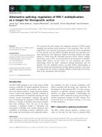
Fig. 1. High-throughput methods for global analyses of alternative splicing. (A) Schematic representation of different splice-sensitive micro-
arrays (adapted from [27]). Exon arrays, typically Affymetrix Exon Arrays, contain oligonucleotide probe sets for every known and predicted
exon. Junction arrays, typically used in [21], contain probes spanning exon junctions across annotated genes. Exon-junction arrays typically
contain both exon-body and exon-junction probes. The coverage of these arrays varies from a few thousand cassette exons [22,23] to all
annotated alternatively spliced genes in Drosophila [24–26] or every single annotated exon and exon junction in 18 000 human genes [27].
The bottom panel shows an example of differential exon usage for a typical cassette exon by means of the differential hybridization signals.
(B) RNA-Seq. The genomic structure for a typical cassette exon is depicted in the middle of the panel, where constitutive exons are shown
in purple and the alternative cassette exon in blue. Sequence reads obtained from the high-throughput method are represented in colour-
coded rectangles (see inset) and are mapped within the genomic sequence. The counting of reads corresponding to inclusion (upper) and
skipping (bottom) allows for the estimation of ‘inclusion ratios’ for the different alternatively spliced isoforms.
Alternative splicing: global insights M. Hallegger et al.
858 FEBS Journal 277 (2010) 856–866 ª 2010 The Authors Journal compilation ª 2010 FEBS
selection, extension and ligation [50] or preselection by
customized capture arrays [51], might enable more
cost-effective quantitative profiling of a large number
of alternative splicing events. In the meantime, some
of the splice-sensitive microarray platforms will remain
competitive.
Surveying splicing regulator targets
Cataloguing the targets of RNA binding proteins that
are known splicing regulators provides a complemen-
tary entry point for unravelling RNA codes. ‘Func-
tional targets’ can be classified as the set of alternative
splicing events that are affected by perturbing the
levels of a splicing regulator, by knockdown, knockout
or overexpression. These targets can be identified by
global transcriptome profiling tools, such as splice-
sensitive microarrays [18,25,32–34], medium-through-
put RT-PCR [52], RNA-Seq or even quantitative
proteomics [53]. However, apparent functional targets
can include indirect secondary targets.
A complementary approach is to identify direct
RNA ‘binding targets’. Selective evolution of ligands
by exponential enrichment (SELEX) is an initial fully
in vitro approach that defines the optimal binding site,
typically short variably degenerate motifs, for an RNA
binding protein by iterative selection from an ini-
tially fully degenerate sequence pool [54]. A variant
approach, genomic SELEX, uses RNA transcribed
from genomic DNA as the starting pool for selection
[55]. SELEX is a useful, although not obligatory,
precursor to methods that catalogue the actual RNA
species (mRNA or pre-mRNA) bound by a splicing
regulatory protein. Direct immunoprecipitation with-
out prior cross-linking (RNP immunoprecipitation)
followed by hybridization to arrays can be a useful
approach [25]. However, a more powerful approach
for identifying binding targets is CLIP (Fig. 2), which
was originally developed to identify targets of the
neuron-specific NOVA proteins [8,56]. RNA is first
cross-linked in vivo to bound protein by UV irradia-
tion, fragmented to 100 nucleotide tags, isolated by
immunoprecipitation, reverse transcribed and then
sequenced. A key feature of CLIP is that UV induces
‘zero-length’ cross-links only between RNA and
directly bound proteins, thereby allowing enrichment
Table 1. Summary of splice-sensitive microarray analyses.
Array design Experiment Species
Validation rate
(events tested) Reference
203 672 exons ⁄ 178 351 exon junctions 48 tissues and cell lines Human 74% (23 events tested) [27]
110 367 exons ⁄ 93 382 exon junctions Time course of heart development Mouse Not mentioned [31]
125 000 junction probes 52 tissues and cell lines Human 58% [21]
40 443 exon-junction probe sets Nova-2 knockout brains Mouse 100% (49 ⁄ 49) [72]
Affymetrix Exon Array Probe
sets for 1 million exons
Colon, bladder, prostate
cancer tissues
Human 66.67% (10 ⁄ 15) [29]
11 human tissues Human 86% [17]
Colon cancer Human 33% [19]
Lymphoblastoid cell lines Human 78% (25 ⁄ 32) [16]
hnRNPLL knockdown in T cells Human Not mentioned [18]
Mid-fetal brain Mouse 95% (65 ⁄ 68) [28]
hnRNPL knockdown Human 22% (11 ⁄ 50) [32]
PTB knockdown in N2A cells Mouse 27 ⁄ 30 [34]
Exon Array and array featuring
exon-body and exon-junction
probe sets
Erythropoiesis Human 6 events validated [20]
3126 cassette exons 10 adult mouse tissues Mouse [23]
3707 cassette exons 27 tissues and cell lines Mouse Not mentioned [22]
> 5000 cassette exons Activation of Jurkat cells Human 68% (17 ⁄ 25) [30]
3055 cassette exons Knockdown of UPF1, UPF2,
UPF3 in HeLa
Human 83% [35]
1300 exons Knockdown of Sam68 Mouse 68.5% (24 ⁄ 35) [33]
8315 mRNAs ⁄ 9868 alt
junction probes
Knockdown of SR and hnRNP
proteins in S2 cells
Drosophila 100% (6 ⁄ 6) [24]
Knockdown of hnRNP proteins
in S2 cells
Drosophila 70% [25]
Alternative splicing changes upon
insulin or Wingless stimulation
Drosophila 70% (11 ⁄ 15) [26]
M. Hallegger et al. Alternative splicing: global insights
FEBS Journal 277 (2010) 856–866 ª 2010 The Authors Journal compilation ª 2010 FEBS 859
of specifically bound sequences by immunoprecipita-
tion under stringent conditions. The original CLIP
procedure has now been modified, with direct high-
throughput sequencing of reverse transcribed tags
[9,10]. The so-called HITS-CLIP [9] or CLIP-Seq [10]
protocols allow saturated coverage of binding targets,
giving a truly global view of the RNP landscape of
individual proteins, and suggesting possible novel func-
tions. This ‘next generation’ CLIP approach has
already been applied to the splicing regulators NOVA
[57], FOX2 [58], SFRS1 (better known as SF2 ⁄ ASF)
[59,60], as well as the miRNA-associated protein,
argonaute [61]. The comprehensive view afforded by
this approach reveals additional, nonsplicing-related,
roles for these RNA binding proteins. For example,
a surprising new function for NOVA2 in alternative
poly(A)-site choice was discovered. Neuronal cells in
general tend to process at promoter-distal poly(A)-sites
and the NOVA2 targets follow this trend. Proliferating
cells produce shorter 3¢ UTRs and therefore reduce
the potential of miRNA regulation [62]. By the
same token, neuronal transcripts with long UTRs are
potentially more prone to regulatory inputs from both
miRNAs and 3¢ UTR binding proteins.
In practice, methods to define functional and
binding targets are complementary. A comprehensive
global analysis of the Drosophila homologues of the
mammalian hnRNPA ⁄ B proteins, hrp36, hrp38, hrp40,
hrp48, involved analysis by a splice-sensitive array of
alterations in alternative splicing upon knockdown,
determination of SELEX motifs in vitro and direct
immunoprecipitation without prior cross-linking
followed by hybridization to arrays using a whole
genome tiling array [25]. This provided many insights
into the functional redundancy and specialization of
this family, and provided hints about their probable
mechanism of action. Perhaps most surprisingly, in
view of popular models about antagonism between the
two families of proteins, very few alternative splicing
events were found to be regulated by both hnRNP and
SR proteins [24,25].
Tissue and individual variations in
alternative splicing
Over the last year, several reports have focussed on
the global analysis of transcript isoform differences
between human tissues [1,2,16,27,28,47,63,64], mouse
tissues [31,63], normal and cancer tissues [64], in
response to specific signalling pathways in Drosophila
[26], or developmental transitions in human brain [28],
mouse heart [31] and mouse stem cells [63]. The combi-
nation of these approaches has revealed extensive
transcript complexity.
Sequencing approaches show that many transcripts
extend beyond the previously annotated 5¢ and 3¢ gene
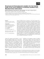
Fig. 2. HITS-CLIP. Intact tissue or tissue culture cells are UV irradiated to induce covalent cross-links between RNA and RNA binding pro-
teins. Cells are lysed under very stringent conditions and treated with DNAse and partially digested with RNAses. The RNA–RNP complex is
pulled-down by immunoprecipitation. The RNA is radioactively 5¢ labelled and ligated to a 5¢ RNA linker. The sample is run on SDS ⁄ PAGE
with neutral pH and blotted. Only RNA cross-linked to protein will be transferred on to the membrane. A small fragment of membrane is iso-
lated at a position that corresponds to the protein plus RNA between 50 and 100 nucleotides. After proteinase K digestion, the RNA is
recovered from the membrane and ligated on its 3¢ end to an RNA adapter with complementarity to the RT primer. The following PCR step
with primer complementary to ligated linkers also allows the addition of appropriate HITS-specific primer sequences (adapted from [76]).
Alternative splicing: global insights M. Hallegger et al.
860 FEBS Journal 277 (2010) 856–866 ª 2010 The Authors Journal compilation ª 2010 FEBS
boundaries [1,2,63]. Moreover, there has been a
substantial increase in the number of known alternative
splicing events, with the capacity of discovering new
splice junctions, ranging from 1400 in one study [63] to
between 4294 and 11 099 in another [1]. The majority of
detected alternative splicing events, including those
newly discovered, show clear tissue specificity, demon-
strating the importance of alternative splicing in tissue-
specific programmes of gene expression. In one study
alone, involving 400 million 32 base reads from 15
human tissues and cell lines, 22 000 tissue-specific alter-
native transcript events were identified [2]. A group of
alternative splicing events that shows extreme changes
between tissues – so-called ‘switch-like’ events – is asso-
ciated with the regulation of highly tissue-specific func-
tions by switching between distinct full-length isoforms
[2]. Perhaps unsurprisingly, some of these switch-like
alternative splicing events within highly expressed genes
(e.g. TPM1) have been used for many years as model
systems of regulated alternative splicing.
Interestingly, although in many cases alternative
splicing regulates functionally coherent groups of
genes, there is no significant overlap between those
genes that are differentially transcribed and those that
are differentially spliced within the same tissue or
within specific cell programmes [27,30,31,42,65]. For
example, upon T cell activation, genes related to the
immunological response are affected at the level of
transcription, whereas cell cycle genes are differentially
spliced [30]. These findings build upon the original
observations of Pan et al. [23] suggesting that overall
programmes of tissue-specific gene expression involve
independent subprogrammes operating on different
subsets of genes at the levels of transcription and splic-
ing [66]. On the other hand, in response to certain sig-
nalling pathways in Drosophila melanogaster cells, a
40% overlap was found between genes that undergo
both transcriptional and splicing changes, suggesting
that transcriptional and post-transcriptional co-ordina-
tion could be important to deploy quick responses
upon certain stimuli [26].
Sequencing and array studies have also provided
fascinating glimpses at the degree to which alternative
splicing varies between individuals. RNA-Seq of sam-
ples originating from seven cerebellar cortex samples
[2] and exon tiling array analysis of 57 lymphoblastoid
cell lines [16] both showed a significant association
between genomic variations (single nucleotide poly-
morphisms) and alternative splicing patterns. Happily
(for those working on mechanisms of tissue-specific
splicing), both studies indicated that although alterna-
tive splicing variation between individuals is common,
it is secondary to tissue-specific alternative splicing.
Motifs and maps
RNA-Seq and microarray analysis on tissues have
generated a genome-scale catalogue of isoform expres-
sion profiles [2,17,27,31]. These data provide a resource
to identify the RNA sequences involved in the regula-
tion of tissue-specific alternative splicing by motif
enrichment analysis. In some cases, the motifs associ-
ated with tissue-specific alternative splicing hint at the
involvement of ‘usual suspects’ – well-known splicing
regulators with defined binding sequences.
By microarray profiling 48 human tissues and sys-
tematically screening for 4-mer to 7-mer RNA ‘words’
associated with 24 426 alternatively spliced exons,
Castle et al. [27] identified 143 motifs enriched near
tissue-specific exons. Interestingly, the two most fre-
quent motifs, UCUCU and UGCAUG, coincide with
binding consensus sequences for PTB ⁄ nPTB and FOX
splicing factors, and show a distinct pattern of geno-
mic localization. Similar observations were made based
on RNA-Seq reads from 15 human tissues and cell
lines [2]. UCUCU motifs were enriched within a 200
nucleotide region upstream of cassette exons that are
upregulated in brain and striated muscle. The extent to
which these exons are spliced correlates inversely with
PTB expression levels [2,27], consistent with PTB’s
well-known role as a splicing repressor [67].
The Castle et al. [27] junction array was also used to
analyse alternative splicing during development of the
mouse heart, resulting in the identification of 63 devel-
opmentally regulated alternative splicing events, falling
into three temporal groups. More than half of these
events were regulated similarly during development of
the chicken heart [31]. Enriched motifs included bind-
ing sites for the CUGBP, MBNL, FOX, STAR and
PTB families of splicing factors. Forty-four of these
alternative splicing events were further investigated in
hearts from transgenic animals that overexpressed
CUGBP1 or were depleted of MBNL1. Of the 24 ex-
ons with altered inclusion levels, 13 were regulated by
CUGBP1, five by MBNL1 and six antagonistically by
both [31]. The switch in relative activities of CUGBP
and MBNL proteins during development appears to
explain a large subset of splicing transitions detected
during postnatal heart development.
Observation of enriched motifs in the cases above
allowed inferences to be drawn about the probable
cognate binding proteins, e.g. Fox, PTB, MBNL and
CELF proteins. However, there are more than 300
RNA binding proteins encoded in mammalian
genomes [68], which have the potential to act as splic-
ing regulators, but for most little or nothing is known
about their binding specificity. Traditional SELEX to
M. Hallegger et al. Alternative splicing: global insights
FEBS Journal 277 (2010) 856–866 ª 2010 The Authors Journal compilation ª 2010 FEBS 861
determine their binding specificity would be laborious.
However, a new array-based procedure may provide
the capability to rapidly derive the optimal binding
motifs for many of these proteins [69], which would
assist in future attempts to link factors with enriched
motifs.
NOVA and FOX maps
In the case of two families of mammalian proteins, the
FOX and NOVA proteins, a variety of techniques,
culminating in HITS-CLIP analysis, have converged
on very similar RNA maps, in which the precise
location of binding sites for the cognate proteins is
predictive of their action as either repressors or silenc-
ers of alternatively spliced exons.
The NOVA proteins are neuron-specific RNA bind-
ing proteins that are targets of a neuronal autoimmune
response associated with cancer. Analysis of these
proteins in the Darnell laboratory has led the way in
the global analysis of RNA binding protein function
[70]. SELEX analysis indicated that the optimal bind-
ing site for NOVA consisted of clusters of three
YCAY motifs [71], and importantly a cluster of such
motifs matched a cis element crucial for NOVA-regu-
lated alternative splicing of an exon in the GABA
A
gene. Analysis of alterations in alternative splicing in
the neocortex of wild-type and Nova2
) ⁄ )
mice using
an Affymetrix prototype junction array with 40 000
probe sets allowed the identification of 50 alterna-
tive splicing events that were NOVA regulated [72].
The genes affected by NOVA-dependent alternative
splicing were highly enriched for proteins involved in
synaptic function, emphasizing the fact that alternative
splicing targets functionally coherent groups of genes.
The CLIP method was originally developed to analyse
in vivo NOVA binding RNAs by conventional cloning
and sequencing of purified RNA tags. Of the moderate
number of sequence tags identified, only 20% con-
tained clusters of YCAY motifs, but in these cases the
tags were often associated with NOVA-regulated
alternative splicing events [56]. On the basis of the
accumulated group of validated NOVA targets, a bio-
informatic exercise was carried out to identify clusters
of YCAY motifs within 200 nucleotides of alternative
exons or their flanking constitutive exons, and more-
over to predict whether these clusters would act as
enhancers or silencers [73]. The resulting NOVA RNA
map contained various intronic and exonic silencers, as
well as intronic enhancers. NOVA clusters within the
downstream intron were invariably enhancers, whereas
within the exon and most positions in the upstream
intron they were silencers. Most recently, the NOVA
RNA map has been refined by high-throughput
sequencing (using the Roche 454 platform) of NOVA2
CLIP tags from mouse neocortex, with confirmation
of splicing outcomes by splice-junction array compari-
son of wild-type and Nova2
) ⁄ )
mice [57]. As expected,
the comprehensive HITS-CLIP approach rediscovered
many of the previously known NOVA targets, as well
as many new ones. The refined NOVA map showed
that NOVA binding clusters within 500 nucleotides of
the alternative 5¢ splice site or constitutive 3¢ splice site
acted as enhancers, whereas NOVA binding within 500
nucleotides of the constitutive 5¢ splice site or sur-
rounding the NOVA-regulated exon was inhibitory.
The FOX1 and -2 proteins are alternative splicing
regulators that have a single RNA binding domain
with an unusual degree of specificity for the cognate
UGCAUG binding site [74]. In a number of recent
global transcriptome profiling studies, FOX binding
motifs were found to be associated with exons regu-
lated in striated muscle and neurons [2,27,75], consis-
tent with the expression patterns of FOX1 and -2.
Analysis of breast and ovarian cancer using an RT-
PCR panel of alternative splicing events indicated that
one-third of cases of increased exon skipping in cancer
were associated with downstream FOX sites. More-
over, FOX2 expression is lower in breast cancer and
its own alternative splicing is altered in ovarian cancer
[64]. Closer analysis of the various FOX datasets
showed an interesting position-dependent effect, remi-
niscent of the NOVA map [57,73]. When located
downstream of alternative splicing exons, FOX binding
sites act as enhancers, whereas on the upstream side
they act as repressors (Fig. 3). The FOX ‘RNA map’
was also converged upon by two additional
approaches that used FOX binding sites and mRNA
targets as the starting point. The long and nondegener-
ate nature of the FOX binding site allowed Zhang
et al. [75] to conduct a computational search for posi-
tionally conserved UGCAUG motifs within 200 nucle-
otides of internal exons across 28 vertebrate genomes.
Comparing the bioinformatics with data collected from
the Castle et al. [27] custom exon-junction array for
alternative splicing in 47 different tissues and cell lines,
they identified the position dependency of FOX bind-
ing sites. Finally, CLIP-Seq analysis was carried out
for FOX2 binding sites in human embryonic stem cells
[58]. Of 5.3 million 36 nucleotide reads, 4.4 million
mapped to unique genomic locations leading to the
identification of > 3500 clusters representing genuine
FOX2 binding events. Surprisingly, although the
UGCAUG motif was highly enriched, an exact
match was only found in 22% of clusters, and even
the core GCAUG pentamer was present in only 33%,
Alternative splicing: global insights M. Hallegger et al.
862 FEBS Journal 277 (2010) 856–866 ª 2010 The Authors Journal compilation ª 2010 FEBS
indicating that FOX2 can bind to other sites, perhaps
in co-operation with other proteins. FOX2 sites were
highly enriched around alternative splicing exons and a
similar position-dependent FOX2 activity map was
deduced. Interestingly, it appears that FOX2 is a key
player in a splicing regulatory network in human
embryonic stem cells. The alternative splicing events
regulated by FOX2 were highly enriched for splicing
regulatory proteins, including numerous hnRNP and
SR proteins and an autoregulatory splicing event in
the FOX2 gene itself. [58]. In contrast, different sets of
FOX2 targets were identified in neural progenitors,
with the major functional enrichment being for cyto-
skeletal proteins, consistent with other reports
[27,58,64,75].
Towards a predictive splicing map
Global alternative splicing profiling points towards
the association of some sequence motifs and their
cognate binding proteins with some tissue-specific
splicing programmes, whereas the NOVA and FOX
splicing maps indicate the position-dependent activity
of some splicing regulators. But even the activity of
FOX and NOVA when bound at particular locations
is dependent upon the binding and activity of other
factors. There is still some way to go before a full
tissue-specific splicing code, with the ability to predict
the consequences of mutations, is deciphered. A
recent study highlighted one of the important future
directions. The Frey and Blencowe groups have
developed a machine-learning approach in which the
tissue-specific splicing profiles of 3707 mouse cassette
exons, gathered using a custom junction-array plat-
form [22], have been combined with over a thousand
separate ‘RNA features’ in order to generate a ‘splic-
ing code’ that predicts changes in exon inclusion
between tissues. The features include known protein
binding sequences (including FOX, NOVA and
PTB ⁄ nPTB), motifs with predicted silencer or enhan-
cer activity, secondary structures, conservation, exon
and intron size, and whether exon inclusion or skip-
ping introduces a premature termination codon.
Using this approach, distinct combinations of fea-
tures are found to be predictive of five different tis-
sue categories of alternative splicing: central nervous
system, muscle, embryo, ‘digestive organs’ (including
liver, kidney, gut) and tissue independent (B. Frey,
personal communication). This pioneering study is
based upon a moderate number of cassette exons and
27 tissue-specific datasets, but it provides a clear
direction for future endeavours. Further refinement
of the splicing code will be readily achieved by a
combination of additional tissue datasets and analysis
of transcriptomes of defined cell types (most tissues
contain a variety of differentiated cell types), together
with larger numbers and different categories of alter-
native splicing events. The ability to sequence the
transcriptomes of single cells [63] will also be enor-
mously helpful as improved methods for sequencing-
based quantitative profiling of alternative splicing are
developed. Of course, defining the logic of the code
will pose many questions about the underlying mech-
anisms. For example, why do FOX and NOVA pro-
teins inhibit from an upstream position, but activate
from downstream of an alternative exon? As the
details of the splicing codes are revealed, there will
be scope for a great deal of further mechanistic dis-
section at the molecular level. However, in contrast
to earlier work on alternative splicing mechanisms,
experimentalists will know in advance that they are
revealing the mechanisms of generally applicable pro-
grammes.
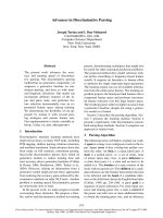
A
B
Fig. 3. Position-dependent activity of FOX proteins. (A) Enrichment
of UGCAUG motifs in the downstream intron is associated with
increased exon inclusion in heart, skeletal muscle, brain and cere-
bellar cortex. Higher motif frequency in the upstream intron is asso-
ciated with reduced inclusion in skeletal muscle. Adapted from [2].
(B) Enrichment of FOX binding sites on the upstream side of alter-
natively spliced exons, indicated by the blue line, is associated with
FOX-dependent exon skipping, whereas enrichment on the down-
stream side (red line) is associated with FOX-dependent inclusion.
Adapted from [2,58,64,75].
M. Hallegger et al. Alternative splicing: global insights
FEBS Journal 277 (2010) 856–866 ª 2010 The Authors Journal compilation ª 2010 FEBS 863
Acknowledgements
We thank Brendan Frey for comments on the manu-
script and for communicating unpublished data. Work
in the CWJS laboratory is funded by the Wellcome
Trust (programme grant 077877) and by EC grant
EURASNET-LSHG-CT-2005-518238.
References
1 Pan Q, Shai O, Lee LJ, Frey BJ & Blencowe BJ (2008)
Deep surveying of alternative splicing complexity in the
human transcriptome by high-throughput sequencing.
Nat Genet 40, 1413–1415.
2 Wang ET, Sandberg R, Luo S, Khrebtukova I, Zhang
L, Mayr C, Kingsmore SF, Schroth GP & Burge CB
(2008) Alternative isoform regulation in human tissue
transcriptomes. Nature 456, 470–476.
3 Melamud E & Moult J (2009) Stochastic noise in splic-
ing machinery. Nucleic Acids Res 37, 4873–4886.
4 Raponi M & Baralle D (2009) Alternative splicing: good
and bad effects of translationally silent substitutions.
FEBS J 277, doi:10.1111/j.1742-4658.2009.07519.x.
5 Faustino NA & Cooper TA (2003) Pre-mRNA splicing
and human disease. Genes Dev 17, 419–437.
6 Matlin AJ, Clark F & Smith CW (2005) Understanding
alternative splicing: towards a cellular code. Nat Rev
Mol Cell Biol 6, 386–398.
7 Soreq L, Gilboa-Geffen A, Berrih-Aknin S, Lacoste P,
Darvasi A, Soreq E, Bergman H & Soreq H (2008)
Identifying alternative hyper-splicing signatures in
MG-thymoma by exon arrays. PLoS ONE 3, e2392.
8 Ule J, Jensen K, Mele A & Darnell RB (2005) CLIP:
a method for identifying protein-RNA interaction sites
in living cells. Methods 37, 376–386.
9 Jensen KB & Darnell RB (2008) CLIP: crosslinking and
immunoprecipitation of in vivo RNA targets of
RNA-binding proteins. Methods Mol Biol 488 , 85–98.
10 Wang Z, Gerstein M & Snyder M (2009) RNA-Seq: a
revolutionary tool for transcriptomics. Nat Rev Genet
10, 57–63.
11 Ben-Dov C, Hartmann B, Lundgren J & Valcarcel J
(2008) Genome-wide analysis of alternative pre-mRNA
splicing. J Biol Chem 283, 1229–1233.
12 Hartmann B & Valcarcel J (2009) Decrypting the
genome’s alternative messages. Curr Opin Cell Biol 21,
377–386.
13 Moore MJ & Silver PA (2008) Global analysis of
mRNA splicing. RNA 14, 197–203.
14 Wang Z & Burge CB (2008) Splicing regulation: from a
parts list of regulatory elements to an integrated splicing
code. RNA 14, 802–813.
15 Blencowe BJ, Ahmad S & Lee LJ (2009) Current-gener-
ation high-throughput sequencing: deepening insights
into mammalian transcriptomes. Genes Dev 23,
1379–1386.
16 Kwan T, Benovoy D, Dias C, Gurd S, Provencher C,
Beaulieu P, Hudson TJ, Sladek R & Majewski J (2008)
Genome-wide analysis of transcript isoform variation in
humans. Nat Genet 40, 225–231.
17 Clark TA, Schweitzer AC, Chen TX, Staples MK, Lu
G, Wang H, Williams A & Blume JE (2007) Discovery
of tissue-specific exons using comprehensive human
exon microarrays. Genome Biol 8, R64.
18 Oberdoerffer S, Moita LF, Neems D, Freitas RP,
Hacohen N & Rao A (2008) Regulation of CD45
alternative splicing by heterogeneous ribonucleoprotein,
hnRNPLL. Science 321, 686–691.
19 Gardina PJ, Clark TA, Shimada B, Staples MK, Yang
Q, Veitch J, Schweitzer A, Awad T, Sugnet C, Dee S
et al. (2006) Alternative splicing and differential gene
expression in colon cancer detected by a whole genome
exon array. BMC Genomics 7, 325.
20 Yamamoto ML, Clark TA, Gee SL, Kang JA,
Schweitzer AC, Wickrema A & Conboy JG (2009)
Alternative pre-mRNA splicing switches modulate gene
expression in late erythropoiesis. Blood 113, 3363–3370.
21 Johnson JM, Castle J, Garrett-Engele P, Kan Z, Loerch
PM, Armour CD, Santos R, Schadt EE, Stoughton R
& Shoemaker DD (2003) Genome-wide survey of
human alternative pre-mRNA splicing with exon junc-
tion microarrays. Science 302, 2141–2144.
22 Fagnani M, Barash Y, Ip JY, Misquitta C, Pan Q,
Saltzman AL, Shai O, Lee L, Rozenhek A, Mohammad
N et al. (2007) Functional coordination of alternative
splicing in the mammalian central nervous system.
Genome Biol 8, R108.
23 Pan Q, Shai O, Misquitta C, Zhang W, Saltzman AL,
Mohammad N, Babak T, Siu H, Hughes TR, Morris
QD et al. (2004) Revealing global regulatory features of
mammalian alternative splicing using a quantitative
microarray platform. Mol Cell 16, 929–941.
24 Blanchette M, Green RE, Brenner SE & Rio DC (2005)
Global analysis of positive and negative pre-mRNA
splicing regulators in Drosophila . Genes Dev 19,
1306–1314.
25 Blanchette M, Green RE, MacArthur S, Brooks AN,
Brenner SE, Eisen MB & Rio DC (2009) Genome-wide
analysis of alternative pre-mRNA splicing and RNA-
binding specificities of the Drosophila hnRNP A ⁄ B
family members. Mol Cell 33, 438–449.
26 Hartmann B, Castelo R, Blanchette M, Boue S, Rio
DC & Valcarcel J (2009) Global analysis of alternative
splicing regulation by insulin and wingless signaling in
Drosophila cells. Genome Biol 10, R11.
27 Castle JC, Zhang C, Shah JK, Kulkarni AV, Kalsotra
A, Cooper TA & Johnson JM (2008) Expression of
24,426 human alternative splicing events and predicted
Alternative splicing: global insights M. Hallegger et al.
864 FEBS Journal 277 (2010) 856–866 ª 2010 The Authors Journal compilation ª 2010 FEBS
cis regulation in 48 tissues and cell lines. Nat Genet 40,
1416–1425.
28 Johnson MB, Kawasawa YI, Mason CE, Krsnik Z,
Coppola G, Bogdanovic D, Geschwind DH, Mane
SM, State MW & Sestan N (2009) Functional and
evolutionary insights into human brain development
through global transcriptome analysis. Neuron 62, 494–
509.
29 Thorsen K, Sorensen KD, Brems-Eskildsen AS,
Modin C, Gaustadnes M, Hein AM, Kruhoffer M,
Laurberg S, Borre M, Wang K et al. (2008) Alterna-
tive splicing in colon, bladder, and prostate cancer
identified by exon array analysis. Mol Cell Proteomics
7, 1214–1224.
30 Ip JY, Tong A, Pan Q, Topp JD, Blencowe BJ &
Lynch KW (2007) Global analysis of alternative splicing
during T-cell activation. RNA 13, 563–572.
31 Kalsotra A, Xiao X, Ward AJ, Castle JC, Johnson JM,
Burge CB & Cooper TA (2008) A postnatal switch of
CELF and MBNL proteins reprograms alternative
splicing in the developing heart. Proc Natl Acad Sci
USA 105, 20333–20338.
32 Hung LH, Heiner M, Hui J, Schreiner S, Benes V &
Bindereif A (2008) Diverse roles of hnRNP L in mam-
malian mRNA processing: a combined microarray and
RNAi analysis. RNA 14, 284–296.
33 Chawla G, Lin CH, Han A, Shiue L, Ares M Jr &
Black DL (2009) Sam68 regulates a set of alternatively
spliced exons during neurogenesis. Mol Cell Biol 29,
201–213.
34 Xing Y, Stoilov P, Kapur K, Han A, Jiang H, Shen S,
Black DL & Wong WH (2008) MADS: a new and
improved method for analysis of differential alternative
splicing by exon-tiling microarrays. RNA 14,
1470–1479.
35 Saltzman AL, Kim YK, Pan Q, Fagnani MM, Maquat
LE & Blencowe BJ (2008) Regulation of multiple core
spliceosomal proteins by alternative splicing-coupled
nonsense-mediated mRNA decay. Mol Cell Biol 28,
4320–4330.
36 Dhir A & Buratti E (2009) Alternative splicing: role of
pseudoexons in human disease and potential therapeutic
strategies. FEBS J 277, doi:10.1111/j.1742-4658.2009.
07520.x.
37 Shiraki T, Kondo S, Katayama S, Waki K, Kasukawa
T, Kawaji H, Kodzius R, Watahiki A, Nakamura M,
Arakawa T et al. (2003) Cap analysis gene expression
for high-throughput analysis of transcriptional starting
point and identification of promoter usage. Proc Natl
Acad Sci USA 100, 15776–15781.
38 Velculescu VE, Zhang L, Vogelstein B & Kinzler KW
(1995) Serial analysis of gene expression. Science 270,
484–487.
39 Iida K, Fukami-Kobayashi K, Toyoda A, Sakaki Y,
Kobayashi M, Seki M & Shinozaki K (2009) Analysis
of multiple occurrences of alternative splicing events in
Arabidopsis thaliana using novel sequenced full-length
cDNAs. DNA Res 15, 155–164.
40 Kim YC, Wu Q, Chen J, Xuan Z, Jung YC, Zhang
MQ, Rowley JD & Wang SM (2009) The transcriptome
of human CD34+ hematopoietic stem-progenitor cells.
Proc Natl Acad Sci USA 106, 8278–8283.
41 Ansorge WJ (2009) Next-generation DNA sequencing
techniques. N Biotechnol 25, 195–203.
42 Calarco JA, Saltzman AL, Ip JY & Blencowe BJ (2007)
Technologies for the global discovery and analysis of
alternative splicing. Adv Exp Med Biol
623, 64–84.
43 Mortazavi A, Williams BA, McCue K, Schaeffer L &
Wold B (2008) Mapping and quantifying mammalian
transcriptomes by RNA-Seq. Nat Meth 5, 621–628.
44 Nagalakshmi U, Wang Z, Waern K, Shou C, Raha D,
Gerstein M & Snyder M (2008) The transcriptional
landscape of the yeast genome defined by RNA
sequencing. Science 320, 1344–1349.
45 Wilhelm BT, Marguerat S, Watt S, Schubert F, Wood
V, Goodhead I, Penkett CJ, Rogers J & Bahler J (2008)
Dynamic repertoire of a eukaryotic transcriptome
surveyed at single-nucleotide resolution. Nature 453,
1239–1243.
46 Lister R, O’Malley RC, Tonti-Filippini J, Gregory BD,
Berry CC, Millar AH & Ecker JR (2008) Highly inte-
grated single-base resolution maps of the epigenome in
Arabidopsis. Cell 133, 523–536.
47 Sultan M, Schulz MH, Richard H, Magen A, Klingen-
hoff A, Scherf M, Seifert M, Borodina T, Soldatov A,
Parkhomchuk D et al. (2008) A global view of gene
activity and alternative splicing by deep sequencing of
the human transcriptome. Science 321, 956–960.
48 Cloonan N, Forrest AR, Kolle G, Gardiner BB, Faulk-
ner GJ, Brown MK, Taylor DF, Steptoe AL, Wani S,
Bethel G et al. (2008) Stem cell transcriptome profiling
via massive-scale mRNA sequencing. Nat Meth 5,
613–619.
49 Li H, Lovci MT, Kwon YS, Rosenfeld MG, Fu XD &
Yeo GW (2008) Determination of tag density required
for digital transcriptome analysis: application to an
androgen-sensitive prostate cancer model. Proc Natl
Acad Sci USA 105, 20179–20184.
50 Yeakley JM, Fan JB, Doucet D, Luo L, Wickham E, Ye
Z, Chee MS & Fu XD (2002) Profiling alternative splic-
ing on fiber-optic arrays. Nat Biotechnol 20, 353–358.
51 Hodges E, Xuan Z, Balija V, Kramer M, Molla MN,
Smith SW, Middle CM, Rodesch MJ, Albert TJ, Hannon
GJ et al. (2007) Genome-wide in situ exon capture for
selective resequencing. Nat Genet 39, 1522–1527.
52 Venables JP, Koh CS, Froehlich U, Lapointe E,
Couture S, Inkel L, Bramard A, Paquet ER, Watier V,
Durand M et al. (2008) Multiple and specific mRNA
processing targets for the major human hnRNP
proteins. Mol Cell Biol 28, 6033–6043.
M. Hallegger et al. Alternative splicing: global insights
FEBS Journal 277 (2010) 856–866 ª 2010 The Authors Journal compilation ª 2010 FEBS 865
53 Spellman R, Llorian M & Smith CW (2007) Crossregu-
lation and functional redundancy between the splicing
regulator PTB and its paralogs nPTB and ROD1. Mol
Cell 27, 420–434.
54 Tuerk C & Gold L (1990) Systematic evolution of
ligands by exponential enrichment: RNA ligands to
bacteriophage T4 DNA polymerase. Science 249,
505–510.
55 Kim S, Shi H, Lee DK & Lis JT (2003) Specific SR
protein-dependent splicing substrates identified through
genomic SELEX. Nucleic Acids Res 31, 1955–1961.
56 Ule J, Jensen KB, Ruggiu M, Mele A, Ule A & Darnell
RB (2003) CLIP identifies Nova-regulated RNA
networks in the brain. Science 302, 1212–1215.
57 Licatalosi DD, Mele A, Fak JJ, Ule J, Kayikci M, Chi
SW, Clark TA, Schweitzer AC, Blume JE, Wang X et al.
(2008) HITS-CLIP yields genome-wide insights into
brain alternative RNA processing. Nature 456, 464–469.
58 Yeo GW, Coufal NG, Liang TY, Peng GE, Fu XD &
Gage FH (2009) An RNA code for the FOX2 splicing
regulator revealed by mapping RNA-protein interac-
tions in stem cells. Nat Struct Mol Biol 16, 130–137.
59 Sanford JR, Coutinho P, Hackett JA, Wang X,
Ranahan W & Caceres JF (2008) Identification of
nuclear and cytoplasmic mRNA targets for the
shuttling protein SF2 ⁄ ASF. PLoS ONE 3, e3369.
60 Sanford JR, Wang X, Mort M, Vanduyn N, Cooper
DN, Mooney SD, Edenberg HJ & Liu Y (2009) Splic-
ing factor SFRS1 recognizes a functionally diverse land-
scape of RNA transcripts. Genome Res 19, 381–394.
61 Chi SW, Zang JB, Mele A & Darnell RB (2009)
Argonaute HITS-CLIP decodes microRNA-mRNA
interaction maps. Nature 460, 479–486.
62 Sandberg R, Neilson JR, Sarma A, Sharp PA & Burge
CB (2008) Proliferating cells express mRNAs with
shortened 3¢ untranslated regions and fewer microRNA
target sites. Science 320, 1643–1647.
63 Tang F, Barbacioru C, Wang Y, Nordman E, Lee C,
Xu N, Wang X, Bodeau J, Tuch BB, Siddiqui A et al.
(2009) mRNA-Seq whole-transcriptome analysis of a
single cell. Nat Meth 6, 377–382.
64 Venables JP, Klinck R, Koh C, Gervais-Bird J, Bram-
ard A, Inkel L, Durand M, Couture S, Froehlich U,
Lapointe E et al. (2009) Cancer-associated regulation of
alternative splicing. Nat Struct Mol Biol 16, 670–676.
65 Calarco JA, Xing Y, Caceres M, Calarco JP, Xiao X,
Pan Q, Lee C, Preuss TM & Blencowe BJ (2007) Global
analysis of alternative splicing differences between
humans and chimpanzees. Genes Dev 21, 2963–2975.
66 Blencowe BJ (2006) Alternative splicing: new insights
from global analyses. Cell 126, 37–47.
67 Spellman R & Smith CW (2006) Novel modes of splic-
ing repression by PTB. Trends Biochem Sci 31, 73–76.
68 McKee AE, Minet E, Stern C, Riahi S, Stiles CD &
Silver PA (2005) A genome-wide in situ hybridization
map of RNA-binding proteins reveals anatomically
restricted expression in the developing mouse brain.
BMC Dev Biol 5, 14.
69 Ray D, Kazan H, Chan ET, Castillo LP, Chaudhry S,
Talukder S, Blencowe BJ, Morris Q & Hughes TR
(2009) Rapid and systematic analysis of the RNA rec-
ognition specificities of RNA-binding proteins. Nat
Biotechnol 7, 667–670.
70 Ule J & Darnell RB (2007) Functional and mechanistic
insights from genome-wide studies of splicing regulation
in the brain. Adv Exp Med Biol 623, 148–160.
71 Buckanovich RJ & Darnell RB (1997) The neuronal
RNA binding protein Nova-1 recognizes specific RNA
targets in vitro and in vivo. Mol Cell Biol 17,
3194–3201.
72 Ule J, Ule A, Spencer J, Williams A, Hu JS, Cline M,
Wang H, Clark T, Fraser C, Ruggiu M et al. (2005)
Nova regulates brain-specific splicing to shape the syn-
apse. Nat Genet 37, 844–852.
73 Ule J, Stefani G, Mele A, Ruggiu M, Wang X, Taneri
B, Gaasterland T, Blencowe BJ & Darnell RB (2006)
An RNA map predicting Nova-dependent splicing regu-
lation. Nature 444, 580–586.
74 Auweter SD, Fasan R, Reymond L, Underwood JG,
Black DL, Pitsch S & Allain FH (2006) Molecular basis
of RNA recognition by the human alternative splicing
factor Fox-1. EMBO J 25, 163–173.
75 Zhang C, Zhang Z, Castle J, Sun S, Johnson J, Krainer
AR & Zhang MQ (2008) Defining the regulatory
network of the tissue-specific splicing factors Fox-1 and
Fox-2. Genes Dev 22, 2550–2563.
76 Wang Z, Tollervey J, Briese M, Turner D & Ule J
(2009) CLIP: construction of cDNA libraries for high-
throughput sequencing from RNAs cross-linked to pro-
teins in vivo. Methods 48, 287–293.
Alternative splicing: global insights M. Hallegger et al.
866 FEBS Journal 277 (2010) 856–866 ª 2010 The Authors Journal compilation ª 2010 FEBS