Báo cáo khoa học: Amyloid oligomers: dynamics and toxicity in the cytosol and nucleus ppt
Bạn đang xem bản rút gọn của tài liệu. Xem và tải ngay bản đầy đủ của tài liệu tại đây (258.73 KB, 11 trang )
MINIREVIEW
Amyloid oligomers: dynamics and toxicity in the cytosol
and nucleus
Akira Kitamura
1
and Hiroshi Kubota
2
1 Department of Molecular Cell Dynamics, Faculty of Advanced Life Science, Hokkaido University, Kita-ku, Sapporo, Japan
2 Department of Life Science, Faculty of Engineering and Resource Science, Akita University, Akita, Japan
Introduction
The accumulation of misfolded proteins in the cytosol
and nucleus causes neurodegenerative disease [1–3].
For example, proteins harboring expanded polygluta-
mine (polyQ) tracts cause polyQ diseases, which
include Huntington’s disease and several spinocerebel-
lar ataxias [4,5], and mutations in superoxide dismu-
tase 1 (SOD1) lead to familial amyotrophic lateral
sclerosis (ALS) [6,7]. In these diseases, inclusions of
the mutant proteins are found in the neuronal cells of
patients and the accumulation of misfolded proteins is
considered to be a primary cause of neuronal dysfunc-
tion and death. The aggregation-prone nature of the
mutant proteins suggests that misfolded proteins dis-
turb neuronal cell functions via unnecessary interac-
tions with normal proteins. However, the mechanism
by which mutant proteins exert their cytotoxicity is lar-
gely unknown. Although these diseases have a late
onset, as symptoms appear in adulthood, the molecu-
lar mechanisms underlying the age-dependent onset are
poorly understood. Moreover, little is known about
Keywords
live cell imaging; misfolded protein;
molecular chaperone; neurodegenerative
disease; neuronal cell death; oligomer;
protein aggregation; protein degradation;
protein interaction; spectroscopic analysis
Correspondence
H. Kubota, Department of Life Science,
Faculty of Engineering and Resource
Science, Akita University, 1-1 Tegatagakuen-
cho, Akita 010-8502, Japan
Fax: +81 18 75 3053
Tel: +81 18 75 3053
E-mail:
(Received 4 September 2009, revised 29
November 2009, accepted 1 December
2010)
doi:10.1111/j.1742-4658.2010.07570.x
The accumulation of misfolded proteins in the cytosol and nucleus of
neuronal cells leads to neurodegenerative disorders. Polyglutamine diseases
are caused by polyglutamine-expanded proteins, whereas mutations in
superoxide dismutase 1 lead to amyotrophic lateral sclerosis. These struc-
turally unstable mutant species perturb essential interactions between nor-
mal proteins and tend to aggregate because of the presence of exposed
hydrophobic surfaces. Accumulating evidence suggests that soluble species,
including misfolded monomers and oligomers, are more toxic than large
insoluble aggregates or inclusions. Spectroscopic analysis, including fluores-
cence recovery after photobleaching and fluorescence loss in photobleach-
ing, in living cells revealed that protein aggregates of misfolded proteins
are dynamic structures that interact with other proteins, such as molecular
chaperones. Fluorescence correlation spectroscopy analysis detected soluble
oligomers ⁄ aggregates of misfolded proteins in cell extracts. Fluorescence
resonance energy transfer analysis supported the notion that soluble oligo-
mers ⁄ aggregates are formed before the formation of inclusions in vivo.
Here, we reviewed the characteristics of oligomers and aggregates of
misfolded proteins, with a particular focus on those revealed by spectro-
scopic analysis, and discussed how these oligomers may be toxic to cells.
Abbreviations
ALS, amyotrophic lateral sclerosis; AR, androgen receptor; CCT, chaperonin containing t-complex polypeptide 1; CFP, cyan fluorescent
protein; FCCS, fluorescence cross-correlation spectroscopy; FCS, fluorescence correlation spectroscopy; FRAP, fluorescence recovery after
photobleaching; FRET, fluorescence resonance energy transfer; GFP, green fluorescent protein; HDAC6, histone deacetylase 6; HSP, heat
shock protein; polyQ, polyglutamine; RFP, red fluorescent protein; SCA, spinocerebellar ataxia; SOD1, superoxide dismutase 1; YFP, yellow
fluorescent protein.
FEBS Journal 277 (2010) 1369–1379 ª 2010 The Authors Journal compilation ª 2010 FEBS 1369
how the mutant proteins specifically damage particular
neuronal cells.
Recent progress in spectroscopic imaging analysis,
using proteins tagged with green fluorescent protein
(GFP) and related cyan, yellow and red fluorescent
proteins (e.g. CFP, YFP, RFP respectively) allowed us
to trace the tagged proteins in living cells [8]. Mutant
proteins that cause polyQ disease and ALS have been
tagged with these fluorescent proteins and analyzed by
fluorescence microscopy-based spectroscopic analysis,
as well as by conventional biochemical experiments. The
spectroscopic techniques used for living cells include flu-
orescence recovery after photobleaching (FRAP), fluo-
rescence loss in photobleaching (FLIP) and
fluorescence ⁄ Fo
¨
rster resonance energy transfer (FRET)
(Fig. 1). These techniques reveal real-time movements
and interactions of misfolded proteins in living cells.
The recent application of fluorescence correlation
spectroscopy (FCS), which is a microscopy-based
technique used for the analysis of fluorescent molecules
at the single-molecule sensitivity [9,10], to misfolded
mutant proteins succeeded in detecting their soluble
oligomers ⁄ aggregates in cell extracts. Together with
evidence from other cell biological and biochemical
analyses, we discussed the role of soluble oligomers of
toxic species in protein-misfolding diseases, including
polyQ disease and ALS.
Soluble oligomers of misfolded
proteins as potentially toxic species
PolyQ-expanded proteins and ALS-linked mutant
SOD1 are structurally unstable [5,7]. These proteins
thus tend to aggregate and interact with other proteins
via exposed hydrophobic surfaces, leading to the pertur-
bation of cellular activities (Fig. 2). The hydrophobic
surfaces of misfolded proteins can be masked by molec-
ular chaperones, and the aggregation of misfolded pro-
teins is inhibited by chaperones through this activity
[11,12]. However, the concentration of chaperones is
limited in living cells, and these chaperones are required
for the folding of newly synthesized normal proteins.
Thus, the overloading of cellular chaperoning capacity
by misfolded mutant proteins results in increased mis-
folding of normal proteins and further enhancement of
co-aggregation. In this state, the degradation of mis-
folded proteins is diminished by their insolubility, and
cellular functions are severely damaged by a negative
chain reaction. This situation can be explained by
escape from (or collapse of) the protein homeostasis net-
work [13,14], and the accumulating incapacitation of
protein homeostasis may explain, in part, the late onset
of neurodegenerative disorders associated with protein
misfolding. It should be noted that chaperone functions
and substrate proteins differ among chaperones, to a
certain extent, and their expression levels vary according
to cell type. These differences may affect the protein
species whose functions are inhibited and the cell types
that are damaged under disease conditions.
The decrease in the amount of functional proteins
(e.g. transcription factors) as a result of becoming
trapped in aggregates may explain the toxicity of mis-
folded proteins. However, a number of studies suggest
that sequestration of misfolded proteins into inclusions
is protective [15]. The total amount of exposed hydro-
phobic surfaces in misfolded proteins is much greater
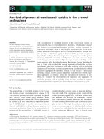
Detection of
conformational
change
Dynamics of
correctly folded
monomer
Detection of
small oligomer
Detection of
soluble aggregate
or amyloid fibril
FRAP/FLIP
FCS
FCCS
FRET
Unsuitable
Good
Not applicable
Not applicable
Unsuitable
Not sensitive*
Less sensitive****
Good****
Unsuitable
Less sensitive**
Good****
Less sensitive***
Unsuitable
Good
Good****
Less quantitative
Dynamics of
inclusion body
Good
Not applicable
Not applicable
Less quantitative
Fig. 1. Suitability of spectroscopic methods for the analysis of protein aggregation. Asterisks indicate the following: *, FCS can be applied
only when the size of molecule is greatly altered by conformational change; **, FCS can detect most oligomers but this method is less sen-
sitive for the detection of very small oliomers such as dimers or trimers; ***, FRET detects dimers and larger oligomers ⁄ aggregates as com-
plexes but cannot determine their sizes; and ****, these methods have not been used in in vivo studies for the indicated purposes despite
the availability. FCCS, fluorescence cross-correlation spectroscopy; FLIP, fluorescence loss in photobleaching; FRAP, fluorescence recovery
after photobleaching.
Dynamics and toxicity of cytosolic amyloid oligomers A. Kitamura and H. Kubota
1370 FEBS Journal 277 (2010) 1369–1379 ª 2010 The Authors Journal compilation ª 2010 FEBS
in the monomeric and oligomeric states than in the
inclusion state under conditions where the number of
misfolded proteins per cell is identical. Although
misfolded monomers can be trapped and refolded by
molecular chaperones and degraded by the ubiquitin–
proteasome system [16], oligomers are more resistant
to refolding by chaperones and to degradation by the
proteasome. Thus, soluble oligomers are considered as
highly toxic to cells. Inhibition of oligomer formation
is probably useful to protect cells against the toxicity
of misfolded proteins.
Sequestration of soluble oligomers ⁄ aggregates into
inclusions or aggresomes by microtubule-dependent
transport is considered to play a role in the removal of
the potentially toxic soluble species from the cytosol
[17]. Indeed, cells harboring polyQ-expanded Hunting-
tin inclusions were reported to be more resistant to the
toxicity of misfolded proteins than cells exhibiting dif-
fusible patterns [18]. However, an opposing effect was
reported using an ALS-linked mutant SOD1 [19], sug-
gesting that the cell-protection activity exerted by
inclusion ⁄ aggresome formation can be affected by dif-
ferences in protein characters and other factors (e.g.
expression level, time course and cell type). Structural
differences between inclusions ⁄ aggresomes may also
affect the cell-protection activity; polyQ-expanded
Huntingtin proteins are tightly associated and immo-
bile in the inclusion [20], whereas mutant SOD1 pro-
teins are loosely packed in the aggresome and partly
exchangeable with cytosolic SOD1 proteins [19]. In a
Drosophila model of spinobulbar muscular atrophy,
which is a neurodegenerative disease caused by expan-
sion of a polyglutamine repeat in the androgen recep-
tor (AR), histone deacetylase 6 (HDAC6) was shown
to play an essential role in preventing polyglutamine
toxicity [21]. HDAC6 is a microtuble-associated pro-
tein that interacts with polyubiquitinated misfolded
proteins and dynein motors [22]. Through these inter-
actions, HDAC6 mediates inclusion ⁄ aggresome forma-
tion of misfolded proteins in a microtubule-dependent
manner. In this sequestration system, however, the
details of transported species (e.g. monomer, oligomer
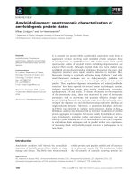
Non-toxic
conformation
Toxic
monomer
Toxic small
oligomer
Degradation
by proteasome
Degradation
resistant
Soluble
aggregate
Inclusion
Degradation
by autophagy
Chaperone
Newly-synthesized polypeptide
Aberrant interaction
with other proteins
Conformational change
and further binding
Inactivation of
other proteins
A
B
C
Depletion of chaperones
b
y
oli
g
omers
Increased misfolding
b
y
impaired chaperonin
g
activit
y
Further stimulation of
a
gg
re
g
ation
Fig. 2. Possible mechanisms of oligomer
toxicity in neurodegenerative disease.
(A) Small oligomers are hardly degraded
by the proteasome and autophagy.
(B) Oligomers inhibit protein functions by
aberrant interaction and co-aggregation.
(C) Depletion of molecular chaperones by
oligomers leads to further stimulation of
co-aggregation.
A. Kitamura and H. Kubota Dynamics and toxicity of cytosolic amyloid oligomers
FEBS Journal 277 (2010) 1369–1379 ª 2010 The Authors Journal compilation ª 2010 FEBS 1371
or soluble aggregate) remain unknown. Thus, these
observations are not contradictory with the notion that
soluble oligomers are highly toxic to cells.
The autophagy–lysosome pathway plays a role in
the clearance of misfolded protein aggregates [23,24]
and this pathway is a candidate system for the removal
of soluble oligomers ⁄ aggregates. For example, beclin 1,
an essential component of the autophagy system, is
required for the effective removal of polyQ aggregates
[25]. In the study by Pandey et al. [21] the amount of
polyQ-expanded AR aggregates were significantly
decreased by the over-expression of HDAC6, but
increased by the knockdown of autophagy com-
ponents. Interestingly, in the fly that expresses polyQ-
expanded AR, the rescue of eye degeneration by
HDAC6 was dependent on autophagic activity. A sim-
ilar role of HDAC6 was shown in mammalian cells
[26], and an essential role of autophagy in preventing
neurodegeneration was reported using knockout mice
[27]. These observations suggest a link between the
microtubule-dependent formation of aggresome and
the autophagy-dependent clearance of misfolded pro-
teins. Recently, the ubiquitin-binding protein p62 (also
known as sequestosome 1) was shown to stimulate the
aggregation of ubiquitinated proteins and to interact
with LC3, an essential component of autophagy [28].
The p62 protein is required for the prevention of pol-
yQ toxicity [29] and interacts with ALS-linked mutant
SOD1 [30]. Thus, p62-bound misfolded proteins in a
soluble state (e.g. soluble aggregates) may be removed
by an autophagy-mediated degradation system. How-
ever, the autophagy-mediated pathway is probably
inefficient for the removal of small oligomers (e.g.
trimer, tetramer, etc.), even though proteins such as
p62 assist specific recognition, because this system uses
bulk sequestration of cytosolic regions via the forma-
tion of double membranes. Further investigations are
required to understand what size of aggregated species
is effectively removed by the autophagy-mediated deg-
radation system in a selective manner in living cells.
Two types of neurodegenerative
diseases caused by misfolded proteins
in the cytosol and nucleus
PolyQ diseases
Expansion of the polyQ tract in at least nine proteins
causes neurodegenerative disorders [4,5,31,32]. PolyQ
expansion in the Huntingtin protein causes Hunting-
ton’s disease, and polyQ-expanded ataxin-1 leads to
spinocerebellar ataxia type 1 (SCA1; also known as
olivopontocerebellar atrophy type 1). PolyQ expansion
in ataxin-3 is the cause of the most common dominant
ataxia, spinocerebellar ataxia type 3 (SCA3, also
known as Machado–Joseph disease). SCA2, SCA6,
SCA7 and SCA17 are also caused by polyQ expansion,
and expansion of the polyQ repeat in the AR is
responsible for spinal and bulbar muscular atrophy.
The expanded polyQ tract is encoded by a CAG repeat
in the causative genes, and polyQ diseases are inherited
dominantly. PolyQ repeats contain approximately
10–30 glutamine residues in healthy individuals,
whereas they are expanded to more than 40 repeats in
patients.
In the polyQ diseases, inclusions containing polyQ-
expanded proteins are found in the nucleus and ⁄ or
cytoplasm of neuronal cells in the brain of patients.
The function of neuronal cells is progressively dis-
turbed, leading to cell death. PolyQ-expanded proteins
aggregate easily in vivo and in vitro, and these aggre-
gates are very difficult to dissolve, even in the presence
of strong detergents, such as SDS [33]. PolyQ-
expanded proteins trap normal functional proteins in
the aggregation process, which suggests a possible
mechanism of toxicity. PolyQ aggregates have been
shown to be rich in b-sheets [34], and Nagai et al. [35]
demonstrated that b-sheet-containing monomers and
oligomers produced in vitro are toxic to cultured neu-
ronal cells when introduced by microinjection.
A recent study of the conformation and toxicity of
polyQ-expanded Huntingtin indicated that fragile amy-
loid, which is rich in exposed and flexible regions, is
significantly more toxic than rigid amyloid, which
comprises buried and fixed regions [36]. As the former
is considered to break into oligomers and to be
accessible to other proteins more easily than the latter,
these observations are consistent with the notion that
oligomers with specific conformations are toxic to
cells.
Molecular chaperones, including heat shock protein
(HSP)70, cognate of HSP70 (HSC70), chaperonin con-
taining t-complex polypeptide 1 (CCT) (also called
TRiC), HSP40 (DnaJ) and small HSPs (e.g. HSP27
and crystalins), play crucial roles in the protection of
cells against the toxicity of polyQ-expanded proteins
[11,12]. For example, HSP70, which is a cytosolic
molecular chaperone that interacts with the hydropho-
bic surfaces of denatured and misfolded proteins, pre-
vents aggregation of polyQ-expanded proteins and
inhibits their toxicity [37–39]. The cytosolic chaperonin
CCT is a molecular chaperone that prevents b-sheet
aggregation by recognizing hydrophobic b-strands [40];
this chaperone prevents aggregate formation and toxic-
ity of polyQ-expanded proteins [41–44]. CCT weakly
recognizes monomeric and oligomeric forms (2–5 mers)
Dynamics and toxicity of cytosolic amyloid oligomers A. Kitamura and H. Kubota
1372 FEBS Journal 277 (2010) 1369–1379 ª 2010 The Authors Journal compilation ª 2010 FEBS
of Huntingin-Q53, but not fibril forms of the protein
[43]. Depletion of CCT activity by RNA interference
in polyQ-expressing cells results in increased amounts
of soluble aggregates and cell death [42], which sug-
gests that CCT interferes with polyQ aggregation by
trapping monomers or small oligomers, thus inhibiting
their toxicity. These observations support the notion
that b-sheet-rich oligomers of polyQ-expanded proteins
are toxic to cells and that inhibition of oligomer for-
mation is an effective strategy for the inhibition of
polyQ toxicity.
Familial ALS caused by mutant SOD1
ALS is a neurodegenerative disorder characterized by
the progressive loss of motor neurons. Although 90%
of ALS cases are sporadic, the remaining 10% are
caused by genetic mutations that are inherited domi-
nantly. Dominant inheritance of familial ALS suggests
a toxic gain of function, similarly to other protein-
misfolding diseases, such as the polyQ diseases.
However, the molecular mechanisms of ALS toxicity
are largely unknown. Moreover, little is known about
how the mutant gene products specifically damage and
kill motor neurons. Several causative genes have been
identified for familial ALS, including SOD1, TDP-43
and FUS ⁄ TLS [45]. Mutations in SOD1 are the most
common cause of familial ALS, and SOD1 mutants
aggregate in the cytosol of neuronal cells [6,46,47].
More than 100 ALS-linked mutations have been iden-
tified in SOD1 and these mutant proteins are structur-
ally unstable [7,48]. Because of structural instability,
the SOD1 mutants are thought to expose hydrophobic
surfaces more easily than the wild-type protein. Thus,
these proteins tend to aggregate and potentially exert
their toxicity via aberrant interactions with other
normal proteins.
Recently, Wang et al. [49] used a mouse model of
ALS-linked mutant SOD1 (G85R) to show that soluble
oligomers of mutant SOD1 are detectable biochemically
in spinal cord extracts before the onset of visible motor
neuron dysfunction. Similar oligomers were also
detected biochemically in Caenorhabditis elegans
expressing the G85R mutant [50]. In the mouse model,
insoluble aggregates were detected at the onset of symp-
toms, which suggests that soluble oligomers are further
aggregated into inclusions. These observations suggest
that soluble oligomers of mutant SOD1 appear when
cellular chaperoning and other quality-control pathways
are overwhelmed by the accumulation of misfolded
proteins. Although the molecular chaperone HSC70 was
associated with soluble species of mutant SOD1 at any
stage, HSP110, which is a nucleotide exchange factor of
HSP70 ⁄ HSC70, was associated with the mutant protein
after the initiation of motor neuron dysfunction. The
structure and toxicity of soluble oligomers may differ
according to the stage of disease progression.
Extracellular oligomers have been suggested to be a
pathogenic factor of neurodegenerative diseases,
including Alzheimer’s disease and prion diseases [51–
53]. For example, amyloid-b peptide (Ab) oligomers
induce synaptic disfunction, probably by interfering
with receptor-dependent signaling pathways via bind-
ing to synaptic plasma membranes. In the case of the
ALS-linked mutant, SOD1, Urushitani et al. [54] indi-
cated (using cultured cells) that these mutants are
secreted from neuronal cells through a chromogranin-
mediated pathway and that extracellular mutant SOD1
triggers microgliosis and neuronal cell death. In a
mouse model of ALS, a conditional knockout of
mutant SOD1 in astrocytes revealed that these cells
affect the disease progression, but not the onset, of
ALS in a noncell autonomous manner [55]. In this
report, extracellular mutant SOD1 was suggested as a
candidate for the mediator. These observations suggest
that extracellular mutant SOD1 may play an addi-
tional role in the pathogenesis of mutant SOD1-medi-
ated ALS. As in vitro studies for mutant SOD1
indicate that post-translational events (including metal
binding and disulfide formation) affect oligomer and
fibril formation [56,57], the aggregation state may be
altered by extracellular environmental conditions.
Thus, like other neurodegenerative diseases, extracellu-
lar oligomers of mutant SOD1 might act as a
toxic species, although this possibility remains to be
investigated.
In vivo dynamics of misfolded proteins
revealed by spectroscopic imaging
analyses
FRAP and FLIP analyses of aggregates and
interacting proteins
Time-lapse observation of fluorescently labeled mole-
cules is often used to trace the movement of cellular
structures. However, this method cannot analyze the
mobility of molecules distributed uniformly and is
unsuitable for the determination of molecular-exchange
rates from one structure to another. FRAP is a method
that measures the mobility of rapidly moving fluores-
cent molecules in a living cell [8]. Molecules labeled with
a fluorescent protein (i.e. GFP and related proteins) are
bleached in a region of interest for a short time-period
and the subsequent movement of fluorescent molecules
from the unbleached area is quantitatively analyzed by
A. Kitamura and H. Kubota Dynamics and toxicity of cytosolic amyloid oligomers
FEBS Journal 277 (2010) 1369–1379 ª 2010 The Authors Journal compilation ª 2010 FEBS 1373
the recovery of fluorescence intensity. This method is
useful for the quantitative analysis of the mobility of
aggregation-prone proteins in a living cell (Fig. 1). In
FLIP analysis, fluorescently labeled molecules are con-
tinuously bleached in a region and the fluorescence of
unbleached areas is measured. FLIP can be used for the
analysis of molecular transfers between two or more
regions, regardless of the speed of movement, even if
this method is less quantitative than FRAP. Thus,
FRAP is very useful for determining the locoregional
mobility of proteins, whereas FLIP can comprehen-
sively analyze protein trafficking.
The mobility of polyQ-expanded proteins in inclu-
sions has been analyzed by FRAP and FLIP. FRAP
analysis of polyQ-expanded ataxin-3 tagged with GFP
(GFP-ataxin-3-Q82) revealed that polyQ-expanded
ataxin-3 is immobile in the nuclear inclusion [58]. In
addition, FLIP analysis indicated that polyQ-expanded
ataxin-3 is unable to shuttle between the inclusions and
the nucleoplasm. These results demonstrate that the
inclusion body formed by polyQ-expanded ataxin-3 is a
structure that is immobilized in the nucleus. By contrast,
FRAP analysis of GFP-ataxin-1-Q84 demonstrated that
ataxin-1 is mobile in nucleoplasmic inclusions [59].
Interestingly, there are two types of ataxin-1 inclusions:
one undergoes fast and complete exchange with a nucle-
oplasmic pool and the other exhibits slow exchange
rates. The slowly exchanging inclusions contain high
levels of ubiquitin and low levels of proteasome, which
suggests a role that is distinct from that of the rapidly
exchanging inclusions. Inverse FRAP analysis of ataxin-
1 indicated that wild-type ataxin-1 shuttles between the
nucleus and the cytosol, whereas polyQ-expanded
ataxin-1 is not exported from the nucleus [60]. These
observations suggest that the ataxin-1 accumulated in
the nucleus becomes a species that is unable to pass
through nuclear pores. FRAP was also used to analyze
the dynamics of ALS-linked mutant SOD1 in cytosolic
inclusions [19]. Mutant SOD1 shuttles dynamically
between the inclusion body and the cytosol in neuronal
cells, which suggests that the inclusion body of mutant
SOD1 is not an immobile structure. By contrast, polyQ
and polyQ-expanded Huntingtin formed immobile
inclusions in the cytosol and in the nucleus [20]. Thus,
there are at least two types of inclusions – mobile inclu-
sions and immobile inclusions – which is consistent with
a recent study proposing two distinct inclusion-like
compartments for protein quality control [61].
FRAP and FLIP are also useful for analyzing the
transient association of other proteins with the inclu-
sions. FRAP analysis of HSP70–YFP in Huntingtin-
150Q–CFP inclusions revealed that HSP70 is mobile
within the inclusion [20]. As the movement of HSP70 is
significantly slower in the inclusion than in the cytosol,
HSP70 appears to interact transiently with aggregated
mutant proteins in the inclusion. These observations
indicate that HSP70 localized in inclusions is not co-
aggregated in inclusions and thus may play a role in the
modulation of the potentially toxic hydrophobic sur-
faces of polyQ aggregates. Interaction of HSP70 with
the ALS-linked mutant SOD1 was analyzed using FLIP
[19]. By continuous photobleaching of YFP–SOD1 in a
small cytosolic region, the fluorescence intensity of the
nonbleached area was decreased more slowly in aggre-
gate-containing cells than in aggregate-free cells. These
observations suggest a dynamic interaction between
HSP70 and mobile inclusions of mutant SOD1 in living
cells. HSP70 might shuttle with misfolded mutant
SOD1 between the inclusions and the cytosol.
FCS analysis of oligomers and soluble
aggregates
The mobility or exchange rate of aggregate-prone
proteins in inclusions has been estimated using FRAP,
as described above. However, this method is unsuitable
for determining the diffusion coefficients of rapidly
moving molecules (or particles), because the diffusion
rates are faster than the image capture rate on the
detector. By contrast, FCS is appropriate for this pur-
pose [8–10]. For FCS analysis, a very small fluorescence
detection volume (the so-called confocal volume) is cre-
ated using optics similar to that of a confocal micro-
scope. In the FCS optics, fluorescent molecules are
excited by a diffracted narrow laser beam and detected
in a pinhole aperture-regulated thin layer. When fluo-
rescent molecules pass through the confocal volume,
fluorescence fluctuation is detected using a highly sensi-
tive photodetector. The fluctuation is analyzed as a cal-
culated autocorrelation function, which provides the
residence time of diffusing molecules in the confocal
volume. As diffusion coefficients correlate with the fric-
tion between the molecule and the solvent, the molecu-
lar mass of the molecules can be calculated by
assuming the molecular shape (e.g. sphere or rod). FCS
analysis allows determination of the concentration of
fluorescent molecules and of fluorescence intensity per
molecule (or counts per particle) thus, the distribution
of differently sized oligomeric species can be estimated.
As FCS analysis can be performed in living cells as well
as in solution [10,62], this technique is becoming a pow-
erful tool for the quantitative analysis of protein com-
plexes, including soluble oligomers ⁄ aggregates of
misfolded proteins, as described below.
The presence of soluble oligomers (or soluble aggre-
gates) has been demonstrated by FCS analysis using
Dynamics and toxicity of cytosolic amyloid oligomers A. Kitamura and H. Kubota
1374 FEBS Journal 277 (2010) 1369–1379 ª 2010 The Authors Journal compilation ª 2010 FEBS
extracts of cultured cells expressing long polyQ repeats
or polyQ-expanded Huntingtin tagged with GFP or
YFP [42,63]. The amount of soluble oligomers ⁄ aggre-
gates was significantly increased by the RNA interfer-
ence-mediated knockdown of the cytosolic chaperonin
CCT, which suggests that CCT prevents oligomer for-
mation of polyQ-expanded proteins in an early step of
aggregation, under normal conditions [42]. In this
study, records of count rate indicated that bright parti-
cles of Q82–GFP and Huntingtin-Q143–YFP passed
through the confocal volume in the CCT-depleted cell
extract. In another study using FCS, fluorescence
intensity per particle increased for Q45–GFP and
Q81–GFP in a time-dependent manner [63]. Further-
more, a polyQ-binding polypeptide (QBP1) signifi-
cantly inhibited the increase of fluorescence intensity
per particle for Q45–GFP. These observations suggest
that the soluble oligomers ⁄ aggregates detected by FCS
contain multiple polyQ-expanded proteins, which are
probably homo-oligomeric, at least in part.
Fluorescence cross-correlation spectroscopy (FCCS)
detects the direct interaction between two fluorescent
molecules at a near single-molecule sensitivity [10,64].
For FCCS measurements, two molecules are labeled
with different fluorophores that are distant in wave-
length, and a solution of these labeled molecules is
analyzed using FCS equipment. An interaction
between the denatured proteins and small HSPs was
determined using FCCS in vitro [65]. Although this
study was carried out in vitro, FCCS can be performed
in living cells or in cell lysates. Thus, this method has
the potential to analyze the interaction between aggre-
gation-prone protein and binding protein (e.g. chaper-
ones) in living cells.
FRET measurements to analyze molecular
interactions in aggregates
FRET provides a useful tool with which to detect
interactions between proteins labeled with a fluorescent
tag. FRET analysis is a method that measures energy
transfer from a donor fluorophore to a nearby acceptor
chromophore. FRET efficiency is highly dependent on
the distance between the donor and the acceptor. Sev-
eral methods have been used to detect FRET signals,
including spectral scanning, ratio imaging, the recovery
of donor fluorescence after acceptor photobleaching
and fluorescence lifetime measurement of the donor.
Molecular interactions between neurodegenerative
disease-associated proteins in aggregates have been ana-
lyzed by FRET using the ratio imaging method. For
example, polyQ-expanded proteins show strong FRET
in the inclusions when they are tagged with enhanced
(E)CFP as a donor and EYFP as an acceptor [20,66].
PolyQ-expanded proteins (Q82–CFP) co-aggregate with
normal-length polyQ (Q19–YFP), as detected by FRET
[20]. Because FRAP analysis in Q82–FLAG-based
inclusions indicates that the exchange rate of Q19–GFP
is faster than that of Q82–GFP, Q19 interacts weakly
with Q82. Although TATA-box binding protein, which
contains a short polyQ tract, is also sequestered in the
Q82 inclusion, this protein exhibited more rapid
exchange than Q19–GFP. Thus, protein mobility in
inclusions appears to depend on the external polypep-
tide sequence as well as on the length of the polyQ
repeat. In this report, the FRET efficiency was variable
among cells, which suggests the presence of cell-to-cell
heterogeneity in the molecular interactions within
Q82–CFP ⁄ Q19–YFP aggregates. As FRET analysis via
fluorescence recovery of the donor after acceptor photo-
bleaching for Q40 in C. elegans also indicates the pres-
ence of heterogeneity in living neurons [67], the
molecular interactions of polyQ proteins may be affected
by unknown cellular conditions. FRET was also used to
screen for inhibitors of polyQ aggregation in cultured
cells, which indicates that this method has high-through-
put potential [68]. Recently, Takahashi et al. [69]
reported that soluble FRET-positive species of polyQ-
expanded proteins were detected before inclusion-body
formation, and FRET signals were significantly
decreased by inhibitors of aggregation. These observa-
tions suggest that soluble oligomers ⁄ aggregates of pol-
yQ-expanded proteins are formed before the formation
of inclusion bodies, which is consistent with the results
of FCS analysis described above [42,63] and with studies
reporting 4-50 nm particles of immunopurified polyQ-
expansion proteins by atomic force microscopy [70,71].
Fluorescence lifetime measurement of a donor
fluorophore is available for quantitative FRET analy-
sis [72]. Fluorescence lifetime imaging microscopy can
determine the distribution of fluorescence lifetime and
is appropriate for cell-based assays. Fluorescence life-
time imaging microscopy analysis led to the detection
of FRET signals for an ubiquitination substrate pro-
tein tagged with EGFP as a donor and ubiquitin
tagged with REACh (a non-fluorescent variant of
yellow fluorescent protein) as an acceptor in cultured
cells, which indicates that this method can be used to
analyze the distribution of ubiquitin conjugates of spe-
cific proteins [73]. The ubiquitination of ALS-linked
SOD1 in cultured cells was analyzed using this system,
which revealed that the distribution of fluorescence
lifetime was different for the G85R and G93A mutants
[74]. These observations are consistent with the fact
that mutant SOD1 is polyubiquitinated for rapid
degradation by the proteasome [75,76] and that the
A. Kitamura and H. Kubota Dynamics and toxicity of cytosolic amyloid oligomers
FEBS Journal 277 (2010) 1369–1379 ª 2010 The Authors Journal compilation ª 2010 FEBS 1375
G85R mutant is more structurally unstable than the
G93A mutant [6,7]. Interestingly, the FRET efficiency
of the polyubiquitinated mutant, SOD1–G85R, was
stronger in a region near the plasma membrane than
in inclusions, which suggests a role for the juxtamem-
brane region in protein quality control. As proteaso-
mal function is important for eliminating the potential
toxicity of mutant proteins, such as SOD1, detailed
spatiotemporal examination of the polyubiquitination
of mutant SOD1 using FRET may be useful for inves-
tigating the details of the role of the ubiquitin–protea-
some system in neurodegenerative diseases in vivo.By
contrast, polyubiquitinated SOD1–G93A emitted
strong FRET signals in perinuclear inclusions. The
localization of the polyubiquitinated mutant SOD1
may be affected by the structure of the mutant pro-
teins, because the G93A mutant is more structurally
stable than the G85R mutant. The FRET efficiency of
SOD1–ubiquitin conjugates in cultured cells was highly
correlated with that of SOD1–HSP70 complexes,
which suggests that HSP70 plays a role in the poly-
ubiquitination of mutant SOD1, perhaps by maintain-
ing mutant SOD1 in a ubiquitination-competent state.
Although FRET signals indicate steady-state molec-
ular interactions, the combination of FRET analysis
with other spectroscopic methods, including FRAP
and FCS, provides important information on the
dynamic biophysical properties of protein aggregates.
The optical system of FCS can be applied to single-
molecule FRET analysis. This method was used to
analyze aggregate formation and protein interactions
of polyQ-expanded proteins in vitro [43]. In this study,
the fluorescence intensity of FRET and non-FRET sig-
nals was measured at the single-molecule sensitivity
level using a dual-color system, and direct interaction
between polyQ-expanded Huntingtin (Huntingtin-Q53)
and the molecular chaperone CCT was detected using
single-molecule FRET. Similar methods may be appli-
cable to the analysis of the molecular structure of
polyQ-expanded proteins or ALS-linked SOD1 in
oligomers and aggregates formed in living cells. Inter-
molecular FRET analysis has been applied to yeast
Sup35 prion proteins and revealed that Sup35 exists as
a monomer at low concentrations in vitro and adopts a
compact state in yeast [77]. Intermolecular FRET may
also be useful for the analysis of the aggregation of
polyQ-expanded proteins or ALS-linked SOD1 in vivo.
Conclusions and perspectives
We reviewed the dynamics and toxicity of misfolded
proteins (including polyQ-expanded proteins and ALS-
linked SOD1) in living cells, with a particular focus on
soluble oligomers ⁄ aggregates. Accumulating evidence
strongly suggests that soluble oligomers of misfolded
proteins are toxic to cells. However, the exact molecu-
lar mechanisms that underlie this cytotoxicity remain
unknown. Real-time spatiotemporal observation of
misfolded proteins in vivo is essential for the full
understanding of these mechanisms, and spectroscopic
analyses in living cells will greatly aid the detailed
analysis of the processes involved in these diseases. In
addition to the techniques described in this review,
single-molecule observations in living cells may be
required to elucidate how misfolded proteins produce
toxic oligomers and interact with other proteins. The
improvement of microscopic techniques will promote
the understanding of the dynamics and toxicity of mis-
folded proteins in living cells in the near future.
Acknowledgements
AK was supported by a fellowship of the Japan
Society for the Promotion of Science (JSPS). HK was
supported by Grant-in-Aid for Scientific Research
Programs from the Ministry of Education, Culture,
Sports, Science and Technology of Japan, and the
Japanese Society for the Promotion of Science.
References
1 Taylor JP, Hardy J & Fischbeck KH (2002) Toxic pro-
teins in neurodegenerative disease. Science 296, 1991–
1995.
2 Ross CA & Poirier MA (2004) Protein aggregation
and neurodegenerative disease. Nat Med Suppl 10,
S10–S17.
3 Chiti F & Dobson CM (2006) Protein misfolding, func-
tional amyloid, and human disease. Annu Rev Biochem
75, 333–366.
4 Gatchel JR & Zoghbi HY (2005) Diseases of unstable
repeat expansion: mechanisms and common principles.
Nat Rev Genet 6, 743–755.
5 Williams AJ & Paulson HL (2008) Polyglutamine
neurodegeneration: protein misfolding revisited. Trends
Neurosci 31, 521–528.
6 Boillee S, Vande Velde C & Cleveland DW (2006) ALS:
a disease of motor neurons and their nonneuronal
neighbors. Neuron 52, 39–59.
7 Shaw BF & Valentine JS (2007) How do ALS-associated
mutations in superoxide dismutase 1 promote aggrega-
tion of the protein? Trends Biochem Sci 32, 78–85.
8 Lippincott-Schwartz J, Snapp E & Kenworthy A (2001)
Studying protein dynamics in living cells. Nat Rev Mol
Cell Biol 2, 444–456.
9 Rigler R, Mets U, Widengren J & Kask P (1993) Fluo-
rescence correlation spectroscopy with high count rate
Dynamics and toxicity of cytosolic amyloid oligomers A. Kitamura and H. Kubota
1376 FEBS Journal 277 (2010) 1369–1379 ª 2010 The Authors Journal compilation ª 2010 FEBS
and low background: analysis of translational diffusion.
Eur Biophys J 22, 169–175.
10 Kim SA & Schwille P (2003) Intracellular applications
of fluorescence correlation spectroscopy: prospects for
neuroscience. Curr Opin Neurobiol 13, 583–590.
11 Sakahira H, Breuer P, Hayer-Hartl MK & Hartl FU
(2002) Molecular chaperones as modulators of polyglu-
tamine protein aggregation and toxicity. Proc Natl Acad
Sci U S A 99 Suppl 4, 16412–16418.
12 Muchowski PJ & Wacker JL (2005) Modulation of
neurodegeneration by molecular chaperones. Nat Rev
Neurosci 6, 11–22.
13 Morimoto RI (2008) Proteotoxic stress and inducible
chaperone networks in neurodegenerative disease and
aging. Genes Dev 22, 1427–1438.
14 Powers ET, Morimoto RI, Dillin A, Kelly JW & Balch
WE (2009) Biological and chemical approaches to dis-
eases of proteostasis deficiency. Annu Rev Biochem 78,
959–991.
15 Ross CA & Poirier MA (2005) Opinion: what is the role
of protein aggregation in neurodegeneration? Nat Rev
Mol Cell Biol 6, 891–898.
16 Ciechanover A & Brundin P (2003) The ubiquitin
proteasome system in neurodegenerative diseases: some-
times the chicken, sometimes the egg. Neuron 40, 427–
446.
17 Kopito RR (2000) Aggresomes, inclusion bodies and
protein aggregation. Trends Cell Biol 10, 524–530.
18 Arrasate M, Mitra S, Schweitzer ES, Segal MR &
Finkbeiner S (2004) Inclusion body formation reduces
levels of mutant huntingtin and the risk of neuronal
death. Nature 431, 805–810.
19 Matsumoto G, Stojanovic A, Holmberg CI, Kim S &
Morimoto RI (2005) Structural properties and neuronal
toxicity of amyotrophic lateral sclerosis-associated
Cu ⁄ Zn superoxide dismutase 1 aggregates. J Cell Biol
171, 75–85.
20 Kim S, Nollen EA, Kitagawa K, Bindokas VP & Mor-
imoto RI (2002) Polyglutamine protein aggregates are
dynamic. Nat Cell Biol 4, 826–831.
21 Pandey UB, Nie Z, Batlevi Y, McCray BA, Ritson GP,
Nedelsky NB, Schwartz SL, DiProspero NA, Knight
MA, Schuldiner O et al. (2007) HDAC6 rescues neu-
rodegeneration and provides an essential link between
autophagy and the UPS. Nature 447, 859–863.
22 Kawaguchi Y, Kovacs JJ, McLaurin A, Vance JM, Ito
A & Yao TP (2003) The deacetylase HDAC6 regulates
aggresome formation and cell viability in response to
misfolded protein stress. Cell 115, 727–738.
23 Rubinsztein DC (2006) The roles of intracellular
protein-degradation pathways in neurodegeneration.
Nature 443, 780–786.
24 Mizushima N, Levine B, Cuervo AM & Klionsky DJ
(2008) Autophagy fights disease through cellular self-
digestion. Nature 451, 1069–1075.
25 Shibata M, Lu T, Furuya T, Degterev A, Mizushima N,
Yoshimori T, MacDonald M, Yankner B & Yuan J
(2006) Regulation of intracellular accumulation of mutant
Huntingtin by Beclin 1. J Biol Chem 281, 14474–14485.
26 Iwata A, Riley BE, Johnston JA & Kopito RR (2005)
HDAC6 and microtubules are required for autophagic
degradation of aggregated huntingtin. J Biol Chem 280
,
40282–40292.
27 Hara T, Nakamura K, Matsui M, Yamamoto A,
Nakahara Y, Suzuki-Migishima R, Yokoyama M,
Mishima K, Saito I, Okano H et al. (2006) Suppression
of basal autophagy in neural cells causes neurodegener-
ative disease in mice. Nature 441, 885–889.
28 Komatsu M, Waguri S, Koike M, Sou YS, Ueno T,
Hara T, Mizushima N, Iwata J, Ezaki J, Murata S
et al. (2007) Homeostatic levels of p62 control cytoplas-
mic inclusion body formation in autophagy-deficient
mice. Cell 131, 1149–1163.
29 Bjorkoy G, Lamark T, Brech A, Outzen H, Perander
M, Overvatn A, Stenmark H & Johansen T (2005)
p62 ⁄ SQSTM1 forms protein aggregates degraded by
autophagy and has a protective effect on huntingtin-
induced cell death. J Cell Biol 171, 603–614.
30 Gal J, Strom AL, Kwinter DM, Kilty R, Zhang J,
Shi P, Fu W, Wooten MW & Zhu H (2009) Sequesto-
some 1 ⁄ p62 links familial ALS mutant SOD1 to LC3
via an ubiquitin-independent mechanism. J Neurochem
111, 1062–1073.
31 Ross CA (2002) Polyglutamine pathogenesis: emergence
of unifying mechanisms for Huntington’s disease and
related disorders. Neuron 35, 819–822.
32 Bates G (2003) Huntingtin aggregation and toxicity in
Huntington’s disease. Lancet 361, 1642–1644.
33 Kazantsev A, Preisinger E, Dranovsky A, Goldgaber D
& Housman D (1999) Insoluble detergent-resistant
aggregates form between pathological and nonpatholog-
ical lengths of polyglutamine in mammalian cells. Proc
Natl Acad Sci U S A 96, 11404–11409.
34 Perutz MF, Finch JT, Berriman J & Lesk A (2002)
Amyloid fibers are water-filled nanotubes. Proc Natl
Acad Sci U S A 99, 5591–5595.
35 Nagai Y, Inui T, Popiel HA, Fujikake N, Hasegawa K,
Urade Y, Goto Y, Naiki H & Toda T (2007) A toxic
monomeric conformer of the polyglutamine protein.
Nat Struct Mol Biol 14, 332–340.
36 Nekooki-Machida Y, Kurosawa M, Nukina N, Ito K,
Oda T & Tanaka M (2009) Distinct conformations of
in vitro and in vivo amyloids of huntingtin-exon1 show
different cytotoxicity. Proc Natl Acad Sci U S A 106,
9679–9684.
37 Muchowski PJ, Schaffar G, Sittler A, Wanker EE,
Hayer-Hartl MK & Hartl FU (2000) Hsp70 and hsp40
chaperones can inhibit self-assembly of polyglutamine
proteins into amyloid-like fibrils. Proc Natl Acad Sci
USA97, 7841–7846.
A. Kitamura and H. Kubota Dynamics and toxicity of cytosolic amyloid oligomers
FEBS Journal 277 (2010) 1369–1379 ª 2010 The Authors Journal compilation ª 2010 FEBS 1377
38 Cummings CJ, Sun Y, Opal P, Antalffy B, Mestril R,
Orr HT, Dillmann WH & Zoghbi HY (2001)
Over-expression of inducible HSP70 chaperone
suppresses neuropathology and improves motor func-
tion in SCA1 mice. Hum Mol Genet 10, 1511–1518.
39 Schaffar G, Breuer P, Boteva R, Behrends C, Tzvetkov
N, Strippel N, Sakahira H, Siegers K, Hayer-Hartl M
& Hartl FU (2004) Cellular toxicity of polyglutamine
expansion proteins: mechanism of transcription factor
deactivation. Mol Cell 15, 95–105.
40 Kubota S, Kubota H & Nagata K (2006) Cytosolic
chaperonin protects folding intermediates of Gbeta
from aggregation by recognizing hydrophobic beta-
strands. Proc Natl Acad Sci U S A 103, 8360–8365.
41 Nollen EA, Garcia SM, van Haaften G, Kim S, Chavez
A, Morimoto RI & Plasterk RH (2004) Genome-wide
RNA interference screen identifies previously unde-
scribed regulators of polyglutamine aggregation. Proc
Natl Acad Sci U S A 101, 6403–6408.
42 Kitamura A, Kubota H, Pack CG, Matsumoto G,
Hirayama S, Takahashi Y, Kimura H, Kinjo M,
Morimoto RI & Nagata K (2006) Cytosolic chaperonin
prevents polyglutamine toxicity with altering the aggre-
gation state. Nat Cell Biol 8, 1163–1170.
43 Behrends C, Langer CA, Boteva R, Bottcher UM,
Stemp MJ, Schaffar G, Rao BV, Giese A, Kretzschmar
H, Siegers K et al. (2006) Chaperonin TRiC Promotes
the Assembly of polyQ Expansion Proteins into
Nontoxic Oligomers. Mol Cell 23, 887–897.
44 Tam S, Geller R, Spiess C & Frydman J (2006) The
chaperonin TRiC controls polyglutamine aggregation
and toxicity through subunit-specific interactions. Nat
Cell Biol 8, 1155–1162.
45 Lagier-Tourenne C & Cleveland DW (2009) Rethink-
ing ALS: the FUS about TDP-43. Cell 136, 1001–
1004.
46 Rosen DR, Siddique T, Patterson D, Figlewicz DA,
Sapp P, Hentati A, Donaldson D, Goto J, O’Regan JP,
Deng HX et al. (1993) Mutations in Cu ⁄ Zn superoxide
dismutase gene are associated with familial amyotrophic
lateral sclerosis. Nature 362, 59–62.
47 Cleveland DW & Rothstein JD (2001) From Charcot to
Lou Gehrig: deciphering selective motor neuron death
in ALS. Nat Rev Neurosci 2, 806–819.
48 Hart PJ (2006) Pathogenic superoxide dismutase struc-
ture, folding, aggregation and turnover. Curr Opin
Chem Biol 10, 131–138.
49 Wang J, Farr GW, Zeiss CJ, Rodriguez-Gil DJ, Wilson
JH, Furtak K, Rutkowski DT, Kaufman RJ, Ruse CI,
Yates JR 3rd et al. (2009) Progressive aggregation
despite chaperone associations of a mutant SOD1-YFP
in transgenic mice that develop ALS. Proc Natl Acad
Sci U S A 106, 1392–1397.
50 Wang J, Farr GW, Hall DH, Li F, Furtak K, Dreier L
& Horwich AL (2009) An ALS-linked mutant SOD1
produces a locomotor defect associated with aggrega-
tion and synaptic dysfunction when expressed in neu-
rons of Caenorhabditis elegans. PLoS Genet 5,
e1000350.
51 Klein WL (2006) Synaptic targeting by Abeta oligomers
(ADDLS) as a basis for memory loss in early Alzhei-
mer’s disease. Alzheimers Dement 2, 43–55.
52 Haass C & Selkoe DJ (2007) Soluble protein oligomers
in neurodegeneration: lessons from the Alzheimer’s
amyloid beta-peptide. Nat Rev Mol Cell Biol 8,
101–112.
53 Ferreira ST, Vieira MN & De Felice FG (2007) Soluble
protein oligomers as emerging toxins in Alzheimer’s and
other amyloid diseases. IUBMB Life 59, 332–345.
54 Urushitani M, Sik A, Sakurai T, Nukina N, Takahashi
R & Julien JP (2006) Chromogranin-mediated secretion
of mutant superoxide dismutase proteins linked to
amyotrophic lateral sclerosis. Nat Neurosci 9, 108–118.
55 Yamanaka K, Chun SJ, Boillee S, Fujimori-Tonou N,
Yamashita H, Gutmann DH, Takahashi R, Misawa H
& Cleveland DW (2008) Astrocytes as determinants of
disease progression in inherited amyotrophic lateral
sclerosis. Nat Neurosci 11, 251–253.
56 Furukawa Y, Kaneko K, Yamanaka K, O’Halloran TV
& Nukina N (2008) Complete loss of post-translational
modifications triggers fibrillar aggregation of SOD1 in
the familial form of amyotrophic lateral sclerosis. J Biol
Chem 283, 24167–24176.
57 Teilum K, Smith MH, Schulz E, Christensen LC,
Solomentsev G, Oliveberg M & Akke M (2009)
Transient structural distortion of metal-free Cu ⁄ Zn
superoxide dismutase triggers aberrant oligomerization.
Proc Natl Acad Sci U S A 106, 18273–18278.
58 Chai Y, Shao J, Miller VM, Williams A & Paulson HL
(2002) Live-cell imaging reveals divergent intracellular
dynamics of polyglutamine disease proteins and sup-
ports a sequestration model of pathogenesis. Proc Natl
Acad Sci U S A 99, 9310–9315.
59 Stenoien DL, Mielke M & Mancini MA (2002) Intranu-
clear ataxin1 inclusions contain both fast- and slow-
exchanging components. Nat Cell Biol 4, 806–810.
60 Irwin S, Vandelft M, Pinchev D, Howell JL, Graczyk J,
Orr HT & Truant R (2005) RNA association and nucle-
ocytoplasmic shuttling by ataxin-1. J Cell Sci 118, 233–
242.
61 Kaganovich D, Kopito R & Frydman J (2008) Misfold-
ed proteins partition between two distinct quality con-
trol compartments. Nature 454, 1088–1095.
62 Pack C, Saito K, Tamura M & Kinjo M (2006) Micro-
environment and effect of energy depletion in the
nucleus analyzed by mobility of multiple oligomeric
EGFPs. Biophys J 91, 3921–3936.
63 Takahashi Y, Okamoto Y, Popiel HA, Fujikake N,
Toda T, Kinjo M & Nagai Y (2007) Detection of
polyglutamine protein oligomers in cells by fluores-
Dynamics and toxicity of cytosolic amyloid oligomers A. Kitamura and H. Kubota
1378 FEBS Journal 277 (2010) 1369–1379 ª 2010 The Authors Journal compilation ª 2010 FEBS
cence correlation spectroscopy. J Biol Chem 282,
24039–24048.
64 Bacia K, Kim SA & Schwille P (2006) Fluorescence
cross-correlation spectroscopy in living cells. Nat
Methods 3, 83–89.
65 Hirose M, Tohda H, Giga-Hama Y, Tsushima R, Zako
T, Iizuka R, Pack C, Kinjo M, Ishii N & Yohda M
(2005) Interaction of a small heat shock protein of the
fission yeast, Schizosaccharomyces pombe, with a dena-
tured protein at elevated temperature. J Biol Chem 280,
32586–32593.
66 Rajan RS, Illing ME, Bence NF & Kopito RR (2001)
Specificity in intracellular protein aggregation and inclu-
sion body formation. Proc Natl Acad Sci U S A 98,
13060–13065.
67 Brignull HR, Moore FE, Tang SJ & Morimoto RI
(2006) Polyglutamine proteins at the pathogenic thresh-
old display neuron-specific aggregation in a pan-neuro-
nal Caenorhabditis elegans model. J Neurosci 26,
7597–7606.
68 Pollitt SK, Pallos J, Shao J, Desai UA, Ma AA,
Thompson LM, Marsh JL & Diamond MI (2003)
A rapid cellular FRET assay of polyglutamine aggrega-
tion identifies a novel inhibitor. Neuron 40, 685–694.
69 Takahashi T, Kikuchi S, Katada S, Nagai Y, Nishizawa
M & Onodera O (2008) Soluble polyglutamine oligo-
mers formed prior to inclusion body formation are
cytotoxic. Hum Mol Genet 17, 345–356.
70 Mukai H, Isagawa T, Goyama E, Tanaka S, Bence NF,
Tamura A, Ono Y & Kopito RR (2005) Formation of
morphologically similar globular aggregates from
diverse aggregation-prone proteins in mammalian cells.
Proc Natl Acad Sci U S A 102, 10887–10892.
71 Wong SL, Chan WM & Chan HY (2008) Sodium
dodecyl sulfate-insoluble oligomers are involved in
polyglutamine degeneration. FASEB J 22, 3348–3357.
72 Yasuda R (2006) Imaging spatiotemporal dynamics of
neuronal signaling using fluorescence resonance energy
transfer and fluorescence lifetime imaging microscopy.
Curr Opin Neurobiol 16, 551–561.
73 Ganesan S, Ameer-Beg SM, Ng TT, Vojnovic B &
Wouters FS (2006) A dark yellow fluorescent protein
(YFP)-based Resonance Energy-Accepting Chromo-
protein (REACh) for Forster resonance energy
transfer with GFP. Proc Natl Acad Sci U S A 103,
4089–4094.
74 Ganesan S, Rohde G, Eckermann K, Sroka K, Schaefer
MK, Dohm CP, Kermer P, Haase G, Wouters F, Bahr
M et al. (2008) Mutant SOD1 detoxification mechanisms
in intact single cells. Cell Death Differ 15, 312–321.
75 Johnston JA, Dalton MJ, Gurney ME & Kopito RR
(2000) Formation of high molecular weight complexes
of mutant Cu, Zn-superoxide dismutase in a mouse
model for familial amyotrophic lateral sclerosis. Proc
Natl Acad Sci U S A 97, 12571–12576.
76 Niwa J, Yamada S, Ishigaki S, Sone J, Takahashi M,
Katsuno M, Tanaka F, Doyu M & Sobue G (2007)
Disulfide bond mediates aggregation, toxicity, and ubiq-
uitylation of familial amyotrophic lateral sclerosis-
linked mutant SOD1. J Biol Chem 282, 28087–28095.
77 Mukhopadhyay S, Krishnan R, Lemke EA, Lindquist S
& Deniz AA (2007) A natively unfolded yeast prion
monomer adopts an ensemble of collapsed and rapidly
fluctuating structures. Proc Natl Acad Sci U S A 104,
2649–2654.
A. Kitamura and H. Kubota Dynamics and toxicity of cytosolic amyloid oligomers
FEBS Journal 277 (2010) 1369–1379 ª 2010 The Authors Journal compilation ª 2010 FEBS 1379